

The DOI matches its Crossref record; 194 of the 194 references it cites were found in a registry or an index.
The Role of APOE4 in Disrupting the Homeostatic Functions of Astrocytes and Microglia in Aging and Alzheimer’s Disease
Open accessAbstract
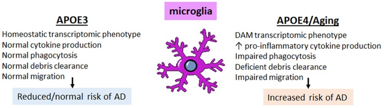
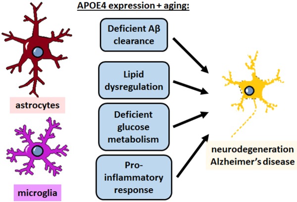
APOE4 is the greatest genetic risk factor for late-onset Alzheimer's disease (AD), increasing the risk of developing the disease by 3-fold in the 14% of the population that are carriers. Despite 25 years of research, the exact mechanisms underlying how APOE4 contributes to AD pathogenesis remain incompletely defined. APOE in the brain is primarily expressed by astrocytes and microglia, cell types that are now widely appreciated to play key roles in the pathogenesis of AD; thus, a picture is emerging wherein APOE4 disrupts normal glial cell biology, intersecting with changes that occur during normal aging to ultimately cause neurodegeneration and cognitive dysfunction. This review article will summarize how APOE4 alters specific pathways in astrocytes and microglia in the context of AD and the aging brain. APOE itself, as a secreted lipoprotein without enzymatic activity, may prove challenging to directly target therapeutically in the classical sense. Therefore, a deeper understanding of the underlying pathways responsible for APOE4 toxicity is needed so that more tractable pathways and drug targets can be identified to reduce APOE4-mediated disease risk.
Overview
The claims and statistics of this paper haven’t been checked yet.
Run the check — free, no account needed. It reads the open-access text and checks the sources it cites.
Integrity signals
Registry
Cites
Statistics
Figures
Verification
Claims the paper makes
The claims and statistics of this paper haven’t been checked yet.
Run the check — free, no account needed. It reads the open-access text and checks the sources it cites.
Claims other papers make about this one
Each is checked against this paper’s own text: does it say what the citing paper says it does?
The source backs part of the claim, not all of it — or backs it for a different population, condition or comparison.
In the citing paper
“ApoE is the most abundant apolipoprotein in the brain, where it is primarily expressed by astrocytes and reactive microglia7, regulating cholesterol metabolism8,9.”
From Decreased lipidated ApoE‐receptor interactions confer protection against pathogenicity of ApoE and its lipid cargoes in lysosomes (2025) · cites this paper as [7]
Experimental — accuracy not yet measuredThe source backs part of the claim, not all of it — or backs it for a different population, condition or comparison.
In the citing paper
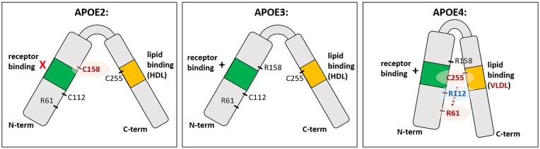
“The Cys112 residue in APOE2 reduces its receptor binding capacity, congruent with the increased levels of lipoproteins in plasma, leading to hyperlipidemia III (Fernandez et al., 2019).”
From APOE2, E3, and E4 differentially modulate cellular homeostasis, cholesterol metabolism, and inflammatory response in isogenic iPSC-derived astrocytes (2022) · cites this paper as Fernandez et al., 2019
Experimental — accuracy not yet measured
Self-check
Percentages, sums and reported differences re-computed from the paper’s own numbers.
The self-check needs the paper’s full text.
Citations
Cites
Corder, E. H., Saunders, A. M., Strittmatter, W. J., Schmechel, D. E., Gaskell, P. C., Small, G. W., Roses, A. D., Haines, J. L., & Pericak-Vance, M. A. (1993) · Science · doi:10.1126/science.8346443 · Cited by 9,429
Neurotoxic reactive astrocytes are induced by activated microglia
Liddelow, S. A., Guttenplan, K. A., Clarke, L. E., Bennett, F. C., Bohlen, C. J., Schirmer, L., Bennett, M. L., Münch, A. E., Chung, W.-S., Peterson, T. C., et al. (2017) · Nature · doi:10.1038/nature21029 · Cited by 7,971
A Unique Microglia Type Associated with Restricting Development of Alzheimer’s Disease
Keren-Shaul, H., Spinrad, A., Weiner, A., Matcovitch-Natan, O., Dvir-Szternfeld, R., Ulland, T. K., David, E., Baruch, K., Lara-Astaiso, D., Toth, B., et al. (2017) · Cell · doi:10.1016/j.cell.2017.05.018 · Cited by 5,493
Meta-Analysis of 74,046 Individuals Identifies 11 New Susceptibility Loci for Alzheimer’s Disease
(EADI), E. A. D. I., Lambert, J.-C., (GERAD), G. A. E. R. I. A. D., Alzheimer's Disease Genetic Consortium (ADGC), (CHARGE), C. F. H. A. A. R. I. G. E., Ibrahim-Verbaas, C. A., Harold, D., Naj, A. C., Sims, R., Bellenguez, C., et al. (2013) · Nature Genetics · doi:10.1038/ng.2802 · Cited by 4,336
Strittmatter, W. J., Saunders, A. M., Schmechel, D., Pericak-Vance, M., Enghild, J., Salvesen, G. S., & Roses, A. D. (1993) · Proceedings of the National Academy of Sciences · doi:10.1073/pnas.90.5.1977 · Cited by 4,268
Synaptic Pruning by Microglia Is Necessary for Normal Brain Development
Paolicelli, R. C., Bolasco, G., Pagani, F., Maggi, L., Scianni, M., Panzanelli, P., Giustetto, M., Ferreira, T. A., Guiducci, E., Dumas, L., et al. (2011) · Science · doi:10.1126/science.1202529 · Cited by 4,244
Microglia Sculpt Postnatal Neural Circuits in an Activity and Complement-Dependent Manner
Schafer, D. P., Lehrman, E. K., Kautzman, A. G., Koyama, R., Mardinly, A. R., Yamasaki, R., Ransohoff, R. M., Greenberg, M. E., Barres, B. A., & Stevens, B. (2012) · Neuron · doi:10.1016/j.neuron.2012.03.026 · Cited by 4,089
Apolipoprotein E: Cholesterol Transport Protein with Expanding Role in Cell Biology
Mahley, R. W. (1988) · Science · doi:10.1126/science.3283935 · Cited by 4,028
Association of apolipoprotein E allele ϵ4 with late‐onset familial and sporadic Alzheimer's disease
Saunders, A. M., Strittmatter, W. J., Schmechel, D., St. George-Hyslop, P. H., Pericak-Vance, M. A., Joo, S. H., Rosi, B. L., Gusella, J. F., Crapper-MacLachlan, D. R., Alberts, M. J., et al. (1993) · Neurology · doi:10.1212/wnl.43.8.1467 · Cited by 3,786
Oakley, H., Cole, S. L., Logan, S., Maus, E., Shao, P., Craft, J., Guillozet-Bongaarts, A., Ohno, M., Disterhoft, J., Van Eldik, L., et al. (2006) · The Journal of Neuroscience · doi:10.1523/jneurosci.1202-06.2006 · Cited by 3,530
Cited by
Sorted by citations
Molecular and cellular mechanisms underlying the pathogenesis of Alzheimer’s disease
Guo, T., Zhang, D., Zeng, Y., Huang, T. Y., Xu, H., & Zhao, Y. (2020) · Molecular Neurodegeneration · doi:10.1186/s13024-020-00391-7 · Cited by 994
ApoE in Alzheimer’s disease: pathophysiology and therapeutic strategies
Raulin, A.-C., Doss, S. V., Trottier, Z. A., Ikezu, T. C., Bu, G., & Liu, C.-C. (2022) · Molecular Neurodegeneration · doi:10.1186/s13024-022-00574-4 · Cited by 756
Astrocytic and microglial cells as the modulators of neuroinflammation in Alzheimer’s disease
Singh, D. (2022) · Journal of Neuroinflammation · doi:10.1186/s12974-022-02565-0 · Cited by 569
APOE and Alzheimer’s Disease: From Lipid Transport to Physiopathology and Therapeutics
Husain, M. A., Laurent, B., & Plourde, M. (2021) · Frontiers in Neuroscience · doi:10.3389/fnins.2021.630502 · Cited by 353
Fernández-Calle, R., Konings, S. C., Frontiñán-Rubio, J., García-Revilla, J., Camprubí-Ferrer, L., Svensson, M., Martinson, I., Boza-Serrano, A., Venero, J. L., Nielsen, H. M., et al. (2022) · Molecular Neurodegeneration · doi:10.1186/s13024-022-00566-4 · Cited by 278
Andersen, J. V., Schousboe, A., & Verkhratsky, A. (2022) · Progress in Neurobiology · doi:10.1016/j.pneurobio.2022.102331 · Cited by 263
Alzheimer’s Risk Factors Age, APOE Genotype, and Sex Drive Distinct Molecular Pathways
Zhao, N., Ren, Y., Yamazaki, Y., Qiao, W., Li, F., Felton, L. M., Mahmoudiandehkordi, S., Kueider-Paisley, A., Sonoustoun, B., Arnold, M., et al. (2020) · Neuron · doi:10.1016/j.neuron.2020.02.034 · Cited by 225
Emerging Microglia Biology Defines Novel Therapeutic Approaches for Alzheimer’s Disease
Lewcock, J. W., Schlepckow, K., Di Paolo, G., Tahirovic, S., Monroe, K. M., & Haass, C. (2020) · Neuron · doi:10.1016/j.neuron.2020.09.029 · Cited by 221
Cell type‐specific roles of APOE4 in Alzheimer disease
Blumenfeld, J., Yip, O., Kim, M. J., & Huang, Y. (2024) · Nature Reviews Neuroscience · doi:10.1038/s41583-023-00776-9 · Cited by 202
Lee, S., Devanney, N. A., Golden, L. R., Smith, C. T., Schwartz, J. L., Walsh, A. E., Clarke, H. A., Goulding, D. S., Allenger, E. J., Morillo-Segovia, G., et al. (2023) · Cell Reports · doi:10.1016/j.celrep.2023.112196 · Cited by 203
Li, R.-Y., Qin, Q., Yang, H.-C., Wang, Y.-Y., Mi, Y.-X., Yin, Y.-S., Wang, M., Yu, C.-J., & Tang, Y. (2022) · Molecular Neurodegeneration · doi:10.1186/s13024-022-00542-y · Cited by 204
Apolipoprotein E4, inhibitory network dysfunction, and Alzheimer’s disease
Najm, R., Jones, E. A., & Huang, Y. (2019) · Molecular Neurodegeneration · doi:10.1186/s13024-019-0324-6 · Cited by 183
The duality of amyloid-β: its role in normal and Alzheimer’s disease states
Azargoonjahromi, A. (2024) · Molecular Brain · doi:10.1186/s13041-024-01118-1 · Cited by 214
ApoE Lipidation as a therapeutic target in Alzheimer’s disease
Lanfranco, M. F., Ng, C. A., & Rebeck, G. W. (2020) · International Journal of Molecular Sciences · doi:10.3390/ijms21176336 · Cited by 189
Expression and secretion of apoE isoforms in astrocytes and microglia during inflammation
Lanfranco, M. F., Sepulveda, J., Kopetsky, G., & Rebeck, G. W. (2021) · Glia · doi:10.1002/glia.23974 · Cited by 164
Pires, M., & Rego, A. C. (2023) · International Journal of Molecular Sciences · doi:10.3390/ijms24010778 · Cited by 157
Neuroinflammation as a Factor of Neurodegenerative Disease: Thalidomide Analogs as Treatments
Jung, Y. J., Tweedie, D., Scerba, M. T., & Greig, N. H. (2019) · Frontiers in Cell and Developmental Biology · doi:10.3389/fcell.2019.00313 · Cited by 156
Beyond the CNS: The many peripheral roles of APOE
Martínez-Martínez, A. B., Torres-Perez, E., Devanney, N., Del Moral, R., Johnson, L. A., & Arbones-Mainar, J. M. (2020) · Neurobiology of Disease · doi:10.1016/j.nbd.2020.104809 · Cited by 149
APOE alleles and diet in brain aging and Alzheimer's disease
Yassine, H. N., & Finch, C. E. (2020) · Frontiers in Aging Neuroscience · doi:10.3389/fnagi.2020.00150 · Cited by 145
Mahan, T. E., Wang, C., Bao, X., Choudhury, A., Ulrich, J. D., & Holtzman, D. M. (2022) · Molecular Neurodegeneration · doi:10.1186/s13024-022-00516-0 · Cited by 134
Zhang, Q., Ma, C., Chin, L., & Li, L. (2020) · Science Advances · doi:10.1126/sciadv.abc5802 · Cited by 130
Ren, Y. L., Savadlou, A., Park, S., Siska, P., Epp, J. R., & Sargin, D. (2023) · Frontiers in Neuroendocrinology · doi:10.1016/j.yfrne.2023.101061 · Cited by 137
Contribution of astrocytes to neuropathology of neurodegenerative diseases
Acıoğlu, Ç., Li, L., & Elkabes, S. (2021) · Brain Research · doi:10.1016/j.brainres.2021.147291 · Cited by 119
APOE genotype and sex affect microglial interactions with plaques in Alzheimer’s disease mice
Stephen, T. L., Cacciottolo, M., Balu, D., Morgan, T. E., LaDu, M. J., Finch, C. E., & Pike, C. J. (2019) · Acta Neuropathologica Communications · doi:10.1186/s40478-019-0729-z · Cited by 115
The Microbiota-Gut-Immune-Glia (MGIG) Axis in Major Depression
Rudzki, L., & Maes, M. H. J. (2020) · Molecular Neurobiology · doi:10.1007/s12035-020-01961-y · Cited by 115
Vascular Dementia and Underlying Sex Differences
Akhter, F., Persaud, A., Zaokari, Y., Zhao, Z., & Zhu, D. (2021) · Frontiers in Aging Neuroscience · doi:10.3389/fnagi.2021.720715 · Cited by 119
Duong, M. T., Nasrallah, I. M., Wolk, D. A., Chang, C. C. Y., & Chang, T. (2021) · Frontiers in Aging Neuroscience · doi:10.3389/fnagi.2021.647990 · Cited by 110
de Leeuw, S. M., Kirschner, A. W. T., Lindner, K., Rust, R., Budny, V., Wolski, W. E., Gavin, A.-C., Nitsch, R. M., & Tackenberg, C. (2022) · Stem Cell Reports · doi:10.1016/j.stemcr.2021.11.007 · Cited by 105
Lee, S.-I., Jeong, W., Lim, H., Cho, S., Lee, H., Jang, Y., Cho, J., Bae, S., Lin, Y.-T., Tsai, L.-H., et al. (2021) · Stem Cell Reports · doi:10.1016/j.stemcr.2021.07.017 · Cited by 105
Destabilizing heterochromatin by APOE mediates senescence
Zhao, H., Ji, Q., Wu, Z., Wang, S., Ren, J., Yan, K., Wang, Z., Hu, J., Chu, Q., Hu, H., et al. (2022) · Nature Aging · doi:10.1038/s43587-022-00186-z · Cited by 104
How Can Insulin Resistance Cause Alzheimer’s Disease?
Yoon, J. H., Hwang, J., Son, S. U., Choi, J., You, S.-W., Park, H., Cha, S.-Y., & Maeng, S. (2023) · International Journal of Molecular Sciences · doi:10.3390/ijms24043506 · Cited by 106
Glia in neurodegeneration: Drivers of disease or along for the ride?
Gleichman, A. J., & Carmichael, S. T. (2020) · Neurobiology of Disease · doi:10.1016/j.nbd.2020.104957 · Cited by 90
Fatty acid oxidation and photoreceptor metabolic needs
Fu, Z., Kern, T. S., Hellström, A., & Smith, L. E. H. (2020) · Journal of Lipid Research · doi:10.1194/jlr.tr120000618 · Cited by 98
Oeckl, P., Anderl-Straub, S., Von Arnim, C. A. F., Baldeiras, I., Diehl-Schmid, J., Grimmer, T., Halbgebauer, S., Kort, A. M., Lima, M., Marques, T. M., et al. (2022) · Journal of Neurology, Neurosurgery & Psychiatry · doi:10.1136/jnnp-2021-328547 · Cited by 94
Cholesterol as a key player in amyloid β-mediated toxicity in Alzheimer’s disease
Rudajev, V., & Novotny, J. (2022) · Frontiers in Molecular Neuroscience · doi:10.3389/fnmol.2022.937056 · Cited by 94
Guo, J. L., Braun, D., Fitzgerald, G. A., Hsieh, Y.-T., Rougé, L., Litvinchuk, A., Steffek, M., Propson, N. E., Heffner, C. M., Discenza, C., et al. (2025) · Cell · doi:10.1016/j.cell.2024.10.027 · Cited by 91
“6 ApoE is the most abundant apolipoprotein in the brain, where it is primarily expressed by astrocytes and reactive microglia, 7 regulating cholesterol metabolism.”
Ritzel, R. M., Li, Y., Jiao, Y., Lei, Z., Doran, S. J., He, J., Shahror, R. A., Henry, R. J., Khan, R., Tan, C., et al. (2023) · Science Advances · doi:10.1126/sciadv.add1101 · Cited by 90
Shifting paradigms: The central role of microglia in Alzheimer’s disease
Schwabe, T., Srinivasan, K., & Rhinn, H. (2020) · Neurobiology of Disease · doi:10.1016/j.nbd.2020.104962 · Cited by 84
Xue, T., Ji, J., Sun, Y., Huang, X., Cai, Z., Yang, J., Guo, W., Guo, R., Cheng, H., & Sun, X. (2021) · Acta Pharmaceutica Sinica B · doi:10.1016/j.apsb.2021.10.012 · Cited by 84
Avraham, O., Chamessian, A. G., Feng, R., Yang, L., Halevi, A. E., Moore, A. M., Gereau, R. W., & Cavalli, V. (2022) · Pain · doi:10.1097/j.pain.0000000000002628 · Cited by 83
Cholesterol Metabolism in Aging and Age-Related Disorders
Saher, G. (2023) · Annual Review of Neuroscience · doi:10.1146/annurev-neuro-091922-034237 · Cited by 85
APOE4 confers transcriptomic and functional alterations to primary mouse microglia
Machlovi, S. I., Neuner, S. M., Hemmer, B. M., Khan, R., Liu, Y., Huang, M., Zhu, J. D., Castellano, J. M., Cai, D., Marcora, E., et al. (2022) · Neurobiology of Disease · doi:10.1016/j.nbd.2022.105615 · Cited by 73
The CD33 short isoform is a gain-of-function variant that enhances Aβ1–42 phagocytosis in microglia
Bhattacherjee, A., Jung, J., Zia, S., Ho, M. F. S., Eskandari‐Sedighi, G., Laurent, C. D. S., McCord, K. A., Bains, A., Sidhu, G., Sarkar, S., et al. (2021) · Molecular Neurodegeneration · doi:10.1186/s13024-021-00443-6 · Cited by 77
Neuroimmune contributions to Alzheimer’s disease: a focus on human data
Haage, V., & De Jager, P. L. (2022) · Molecular Psychiatry · doi:10.1038/s41380-022-01637-0 · Cited by 74
Neuron-astrocyte omnidirectional signaling in neurological health and disease
Pathak, D., & Sriram, K. (2023) · Frontiers in Molecular Neuroscience · doi:10.3389/fnmol.2023.1169320 · Cited by 72
Zhang, M., He, P., & Bian, Z. (2021) · Frontiers in Molecular Neuroscience · doi:10.3389/fnmol.2021.685143 · Cited by 69
Wang, Y., Mishra, A., & Brinton, R. D. (2020) · F1000Research · doi:10.12688/f1000research.21599.1 · Cited by 65
Jasiecki, J., Targońska, M., & Wasąg, B. (2021) · International Journal of Molecular Sciences · doi:10.3390/ijms22042033 · Cited by 67
Role of Neuron and Glia in Alzheimer’s Disease and Associated Vascular Dysfunction
Bandyopadhyay, S. (2021) · Frontiers in Aging Neuroscience · doi:10.3389/fnagi.2021.653334 · Cited by 64
Larramona-Arcas, R., González-Arias, C., Perea, G., Gutiérrez, A., Vitorica, J., García-Barrera, T., Gómez-Ariza, J. L., Pascua-Maestro, R., Ganfornina, M. D., Kara, E., et al. (2020) · Molecular Neurodegeneration · doi:10.1186/s13024-020-00382-8 · Cited by 56
Bai, X., Mai, M., Yao, K., Zhang, M., Huang, Y., Zhang, W., Guo, X., Xu, Y., Zhang, Y., Qurban, A., et al. (2022) · Acta Neuropathologica Communications · doi:10.1186/s40478-022-01338-3 · Cited by 51
Iannucci, J., Sen, A., & Grammas, P. (2021) · Current Issues in Molecular Biology · doi:10.3390/cimb43010018 · Cited by 57
ApoE4 attenuates autophagy via FoxO3a repression in the brain
Sohn, H.-Y., Kim, S.-I., Park, J.-Y., Park, S.-H., Koh, Y. H., Kim, J., & Jo, C. (2021) · Scientific Reports · doi:10.1038/s41598-021-97117-6 · Cited by 54
Lizama, B. N., Kahle, J., Catalano, S. M., Caggiano, A. O., Grundman, M., & Hamby, M. E. (2023) · International Journal of Molecular Sciences · doi:10.3390/ijms24076251 · Cited by 54
Self-citationMolecular Insight into the Therapeutic Promise of TargetingAPOE4for Alzheimer’s Disease
Mamun, A. A., Uddin, M. S., Bashar, M. F. B., Zaman, S., Begum, Y., Bulbul, I. J., Islam, M. S., Sarwar, M. S., Mathew, B., Amran, M. S., et al. (2020) · Oxidative Medicine and Cellular Longevity · doi:10.1155/2020/5086250 · Cited by 54
Jiang, Y., Zhou, X., Wong, H. Y., Ouyang, L., Ip, F. C. F., Chau, V. M. N., Lau, S., Wu, W., Wong, D. Y. K., Seo, H., et al. (2022) · Nature Aging · doi:10.1038/s43587-022-00241-9 · Cited by 56
Park, J., Kim, H., Kim, J., & Cheon, M. (2020) · PLoS Computational Biology · doi:10.1371/journal.pcbi.1008099 · Cited by 51
Astrocytic Calcium and cAMP in Neurodegenerative Diseases
Sobolczyk, M., & Boczek, T. (2022) · Frontiers in Cellular Neuroscience · doi:10.3389/fncel.2022.889939 · Cited by 53
Wang, R., Oh, J. M., Motovylyak, A., Ma, Y., Sager, M. A., Rowley, H. A., Johnson, K. M., Gallagher, C. L., Carlsson, C. M., Bendlin, B. B., et al. (2021) · Journal of Cerebral Blood Flow & Metabolism · doi:10.1177/0271678x211021313 · Cited by 49
Nutritional metabolism and cerebral bioenergetics in Alzheimer's disease and related dementias
Yassine, H. N., Self, W. K., Kerman, B. E., Santoni, G., Shanmugam, N. K. N., Abdullah, L. I., Golden, L. R., Fonteh, A. N., Harrington, M. G., Gräff, J., et al. (2022) · Alzheimer s & Dementia · doi:10.1002/alz.12845 · Cited by 52
Macaron, T., Giudici, K. V., Bowman, G. L., Sinclair, A. J., Stephan, E., Vellas, B. J., & Barreto, P. D. S. (2021) · Ageing Research Reviews · doi:10.1016/j.arr.2021.101300 · Cited by 52
Redefining transcriptional regulation of the APOE gene and its association with Alzheimer’s disease
Lee, E.-G., Tulloch, J., Chen, S., Leong, L., Saxton, A. D., Kraemer, B. C., Darvas, M., Keene, C. D., Shutes‐David, A., Todd, K. W., et al. (2020) · PLoS ONE · doi:10.1371/journal.pone.0227667 · Cited by 49
Wang, Z.-H., Xia, Y., Liu, P., Liu, X., Edgington‐Mitchell, L. E., Lei, K., Yu, S. P., Wang, X., & Ye, K. (2021) · Progress in Neurobiology · doi:10.1016/j.pneurobio.2021.102032 · Cited by 48
Wang, Z.-H., Xia, Y., Wu, Z., Kang, S. S., Zhang, J.-C., Liu, P., Liu, X., Song, W., Huin, V., Dhaenens, C., et al. (2021) · Progress in Neurobiology · doi:10.1016/j.pneurobio.2021.102212 · Cited by 48
Oxidative Stress: Pathological Driver in Chronic Neurodegenerative Diseases
Chong, Z. Z., & Souayah, N. (2025) · Antioxidants · doi:10.3390/antiox14060696 · Cited by 50
Foo, H., Thalamuthu, A., Jiang, J., Koch, F. C., Mather, K. A., Wen, W. X., & Sachdev, P. S. (2020) · Neurobiology of Aging · doi:10.1016/j.neurobiolaging.2020.11.002 · Cited by 46
Rudajev, V., & Novotný, J. (2023) · Cell & Bioscience · doi:10.1186/s13578-023-01127-y · Cited by 52
Mercury and Alzheimer’s disease: a look at the links and evidence
Azar, J., Yousef, M. H., El‐Fawal, H. A. N., & Abdelnaser, A. (2021) · Metabolic Brain Disease · doi:10.1007/s11011-020-00649-5 · Cited by 46
The effect of aged microglia on synaptic impairment and its relevance in neurodegenerative diseases
Triviño, J. J., & von Bernhardi, R. (2021) · Neurochemistry International · doi:10.1016/j.neuint.2021.104982 · Cited by 43
Borràs, C., Mercer, A., Sirisi, S., Alcolea, D., Escolà-Gil, J. C., Blanco-Vaca, F., & Tondo, M. (2022) · International Journal of Molecular Sciences · doi:10.3390/ijms23169356 · Cited by 39
Cellular senescence and the blood–brain barrier: Implications for aging and age-related diseases
Knopp, R. C., Erickson, M. A., Rhea, E. M., Reed, M. J., & Banks, W. A. (2023) · Experimental Biology and Medicine · doi:10.1177/15353702231157917 · Cited by 47
Roles of TREM2 in the Pathological Mechanism and the Therapeutic Strategies of Alzheimer's Disease
Lin, M., Yu, J., Zhang, W., Lao, F., & Huang, H. (2024) · The Journal of Prevention of Alzheimer s Disease · doi:10.14283/jpad.2024.164 · Cited by 46
Multi-functional role of apolipoprotein E in neurodegenerative diseases
Islam, S., Noorani, A. A., Sun, Y., Michikawa, M., & Zou, K. (2025) · Frontiers in Aging Neuroscience · doi:10.3389/fnagi.2025.1535280 · Cited by 43
Chen, Y., Hong, T., Chen, F., Sun, Y., Wang, Y., & Cui, L. (2021) · Frontiers in Aging Neuroscience · doi:10.3389/fnagi.2021.631827 · Cited by 40
Dehghan, A., Pinto, R. C., Karaman, İ., Huang, J. V., Durainayagam, B. R., Ghanbari, M., Nazeer, A., Zhong, Q., Liggi, S., Whiley, L., et al. (2022) · Proceedings of the National Academy of Sciences · doi:10.1073/pnas.2206083119 · Cited by 38
Synapses, Microglia, and Lipids in Alzheimer’s Disease
Paasila, P. J., Aramideh, J. A., Sutherland, G. T., & Graeber, M. B. (2022) · Frontiers in Neuroscience · doi:10.3389/fnins.2021.778822 · Cited by 40
Comprehensive Overview of Alzheimer’s Disease: Etiological Insights and Degradation Strategies
Singh, M. K., Shin, Y., Ju, S., Han, S., Kim, S. S., & Kang, I. (2024) · International Journal of Molecular Sciences · doi:10.3390/ijms25136901 · Cited by 43
Walker, C. D., Risher, W. C., & Risher, M. (2020) · Cells · doi:10.3390/cells9020297 · Cited by 36
Marottoli, F. M., Trevino, T. N., Geng, X. H., Arbieva, Z. H., Kanabar, P. N., Maienschein‐Cline, M., Lee, J. C., Lutz, S. E., & Tai, L. M. (2021) · Frontiers in Cell and Developmental Biology · doi:10.3389/fcell.2021.668296 · Cited by 38
Repurposing Immunomodulatory Imide Drugs (IMiDs) in Neuropsychiatric and Neurodegenerative Disorders
Jung, Y. J., Tweedie, D., Scerba, M. T., Kim, D. S., Palmas, M. F., Pisanu, A., Carta, A. R., & Greig, N. H. (2021) · Frontiers in Neuroscience · doi:10.3389/fnins.2021.656921 · Cited by 37
Proal, A. D., & VanElzakker, M. B. (2020) · Immunometabolism · doi:10.20900/immunometab20210003 · Cited by 36
NOX-induced oxidative stress is a primary trigger of major neurodegenerative disorders
Zilberter, Y., Tabuena, D. R., & Zilberter, M. Y. (2023) · Progress in Neurobiology · doi:10.1016/j.pneurobio.2023.102539 · Cited by 35
Giarratana, A. O., Zheng, C., Reddi, S., Teng, S. L., Berger, D. S., Adler, D., Sullivan, P. M., Thakker‐Varia, S., & Alder, J. (2020) · Scientific Reports · doi:10.1038/s41598-020-76849-x · Cited by 39
Fote, G. M., Geller, N. R., Efstathiou, N. E., Hendricks, N., Vavvas, D. G., Reidling, J. C., Thompson, L. M., & Steffan, J. S. (2022) · Journal of Cell Science · doi:10.1242/jcs.258687 · Cited by 36
Essers, E., Binter, A.-C., Neumann, A., White, T. J. H., Alemany, S., & Guxens, M. (2022) · Environmental Research · doi:10.1016/j.envres.2022.114595 · Cited by 32
Sergi, D., Zauli, E., Tisato, V., Secchiero, P., Zauli, G., & Cervellati, C. (2023) · International Journal of Molecular Sciences · doi:10.3390/ijms24054403 · Cited by 32
Villa, C., Rivellini, E., Lavitrano, M., & Combi, R. (2022) · Journal of Personalized Medicine · doi:10.3390/jpm12010029 · Cited by 34
Alzheimer’s disease: a tale of two diseases?
Nardini, E., Hogan, R., Flamier, A., & Bernier, G. (2021) · Neural Regeneration Research · doi:10.4103/1673-5374.308070 · Cited by 33
Eid, A., Mhatre‐Winters, I., Sammoura, F. M., Edler, M. K., von Stein, R., Hossain, M. M., Han, Y., Lisci, M., Carney, K., Konsolaki, M., et al. (2022) · Environmental Health Perspectives · doi:10.1289/ehp10576 · Cited by 31
Nanomedicine based strategies for oligonucleotide traversion across the blood–brain barrier
Amulya, E., Sikder, A., Vambhurkar, G. B., Shah, S., Khatri, D. K., Raghuvanshi, R. S., Singh, S. B., & Srivastava, S. (2023) · Journal of Controlled Release · doi:10.1016/j.jconrel.2023.01.031 · Cited by 30
Role of TET1-mediated epigenetic modulation in Alzheimer's disease
Armstrong, M. J., Jin, Y., Vattathil, S., Huang, Y., Schroeder, J. P., Bennet, D. A., Qin, Z. S., Wingo, T. S., & Jin, P. (2023) · Neurobiology of Disease · doi:10.1016/j.nbd.2023.106257 · Cited by 32
CDH6 and HAGH protein levels in plasma associate with Alzheimer’s disease in APOE ε4 carriers
Ahmad, S., Milan, M. D. C., Hansson, O., Demirkan, A., Agustin, R., Sáez, M. E., Giagtzoglou, N., Cabrera-Socorro, A., Bakker, M. H. M., Ramirez, A., et al. (2020) · Scientific Reports · doi:10.1038/s41598-020-65038-5 · Cited by 31
Emerging hiPSC Models for Drug Discovery in Neurodegenerative Diseases
Trudler, D., Ghatak, S., & Lipton, S. A. (2021) · International Journal of Molecular Sciences · doi:10.3390/ijms22158196 · Cited by 30
Riviere, M., Langbaum, J. B., Turner, R. S., Rinne, J. O., Sui, Y., Cazorla, P., Ricart, J., Meneses, K., Caputo, A., Tariot, P. N., et al. (2024) · Alzheimer's & Dementia · doi:10.1002/alz.13532 · Cited by 28
Xu, D. C., Sas‐Nowosielska, H., Donahue, G., Huang, H., Pourshafie, N., Good, C. R., & Berger, S. L. (2024) · Acta Neuropathologica Communications · doi:10.1186/s40478-023-01696-6 · Cited by 35
de Frutos‐Lucas, J., Sewell, K. R., García‐Colomo, A., Markovic, S. J., Erickson, K. I., & Brown, B. M. (2023) · Alzheimer s Research & Therapy · doi:10.1186/s13195-023-01170-4 · Cited by 26
Mhatre‐Winters, I., Eid, A., Han, Y., Tieu, K., & Richardson, J. R. (2022) · International Journal of Molecular Sciences · doi:10.3390/ijms23179829 · Cited by 26
Trares, K., Gao, X. Y., Perna, L., Rujescu, D., Stocker, H. R., Möllers, T., Beyreuther, K. T., Brenner, H., & Schöttker, B. (2020) · Alzheimer s & Dementia · doi:10.1002/alz.12081 · Cited by 25
A systems biology approach for studying neurodegenerative diseases
Lam, S. C. C., Bayraktar, A., Zhang, C., Türkez, H., Nielsen, J., Borén, J., Shoaie, S., Uhlén, M., & Mardinoğlu, A. (2020) · Drug Discovery Today · doi:10.1016/j.drudis.2020.05.010 · Cited by 25
Microfluidic Platforms to Unravel Mysteries of Alzheimer’s Disease: How Far Have We Come?
Prasanna, P., Rathee, S., Rahul, V., Mandal, D., Goud, M. S. C., Yadav, P., Hawthorne, S. J., Sharma, A., Gupta, P. K., Ojha, S. K., et al. (2021) · Life · doi:10.3390/life11101022 · Cited by 25
Metadata
- Authors
- Fernandez, C. G., Hamby, M. E., McReynolds, M. L., & Ray, W. J.
- Affiliations
- The University of Texas MD Anderson Cancer Center (US)
- Corresponding author
- Fernandez, Celia G.
- Venue
- Frontiers in Aging Neuroscience
- Publisher
- Frontiers Media SA
- Published
- 2019-02-11
- Volume
- 11
- Pages
- 14-14
- ISSN
- 1663-4365 (ISSN-L)
- PMC
- PMC6378415
- OpenAlex
- W2911484089
- Cited by
- 277
- References
- 194
- Citations by year
- 2026: 25 · 2025: 32 · 2024: 31 · 2023: 54 · 2022: 50 · 2021: 36 · 2020: 43 · 2019: 6
- FWCI
- 3.97
- Type
- journal article
- Language
- en
- Open access
- gold
- doi.org
- Licence
- Locations
- Frontiers in Aging Neuroscience (publishedVersion, cc-by)
- Europe PMC (PubMed Central) (publishedVersion, cc-by)
- PubMed Central (publishedVersion, cc-by)
- PubMed (publishedVersion)
- DOAJ (DOAJ: Directory of Open Access Journals) (submittedVersion, cc-by-sa)
- Funders
- Robert A. and Renee E. Belfer Family Foundation
- Topics
- Life Sciences › Neuroscience › Cellular and Molecular Neuroscience › Alzheimer's disease research and treatments
- Life Sciences › Neuroscience › Neurology › Neuroinflammation and Neurodegeneration Mechanisms
- Health Sciences › Medicine › Psychiatry and Mental health › Dementia and Cognitive Impairment Research
- Keywords
- APOE ε4, Alzheimer's disease, astrocytes, microglia, brain aging, neurodegeneration, cognitive impairment, therapeutic target, genetic risk factors
About these checks
Registry, retraction, Cites and statistics results are computed, not judged, with one exception: a printed reference that no index lists is read once by a model before it counts as a citation error. Claims are extracted by a model and kept only when their quote appears verbatim in the text; each cited source is then checked, with a model’s help, for whether it says what the claim relies on. Those verdicts can be wrong — read the claim’s quote and the cited source before relying on one.
Statistics and arithmetic are recomputed, not judged.
Confidence bands are shown only for fields calibrated on our annotated test set. None is calibrated yet, so no band is shown.
What counts as a claim ›How we measure confidence ›How we check this ›
Are you an author, or do you see a problem with this page? Request a review
