

The DOI matches its Crossref record; 76 of the 76 references it cites were found in a registry or an index.
Microglia, seen from the CX3CR1 angle
Open accessAbstract
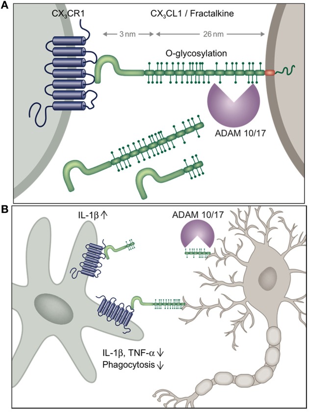
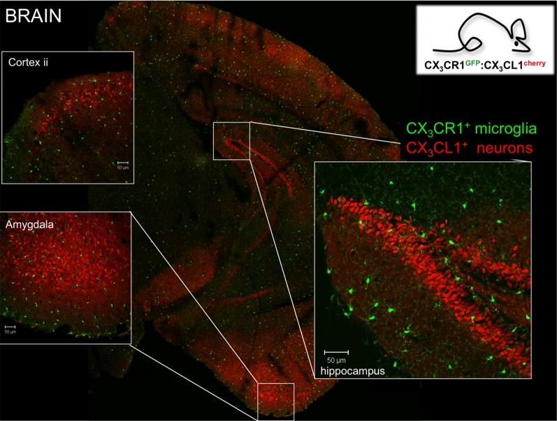
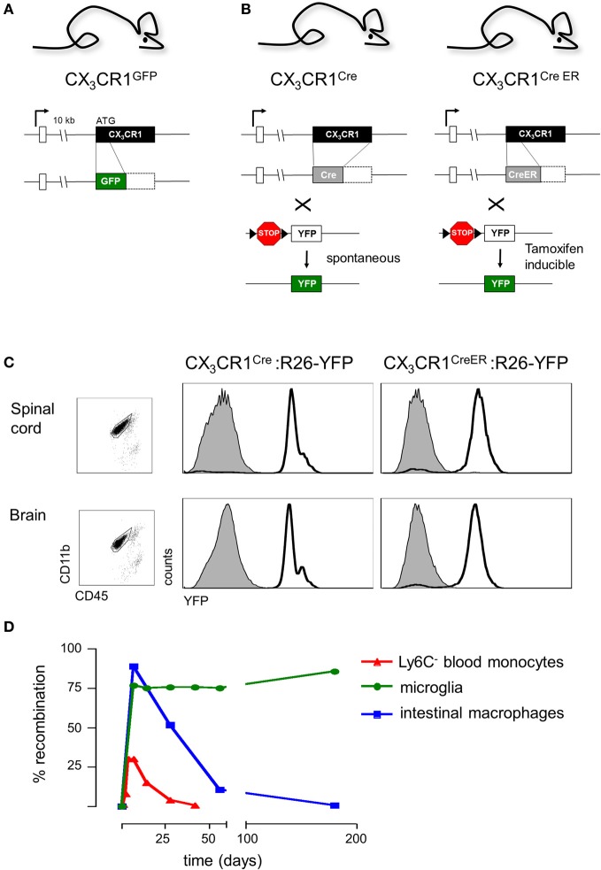
Microglial cells in brain and spinal cord are characterized by high expression of the chemokine receptor CX3CR1. Expression of the sole CX3CR1 ligand, the membrane-tethered and sheddable chemokine CX3CL1/fractalkine, is restricted in the brain parenchyma to selected neurons. Here we summarize our current understanding of the physiological role of CX3CR1 for microglia function and the CX3C axis in microglial/neuronal crosstalk in homeostasis and under challenge. Moreover, we will discuss the efforts of our laboratory and others to exploit CX3CR1 promoter activity for the visualization and genetic manipulation of microglia to probe their functional contributions in the central nerve system (CNS) context.
Overview
The claims and statistics of this paper haven’t been checked yet.
Run the check — free, no account needed. It reads the open-access text and checks the sources it cites.
Integrity signals
Registry
Cites
Statistics
Figures
Verification
Claims the paper makes
The claims and statistics of this paper haven’t been checked yet.
Run the check — free, no account needed. It reads the open-access text and checks the sources it cites.
Claims other papers make about this one
Each is checked against this paper’s own text: does it say what the citing paper says it does?
In the citing paper
“Activated microglia also support neuronal survival and function by engulfing pathogens, cellular debris, or other neurotoxic entities and can synthesize various neurotrophic factors to promote tissue maintenance and repair (63, 64). However, the excessive or sustained activation of microglial cells (such as in chronic neurodegenerative diseases) usually produces significant inflammatory collateral”
From The HPA – Immune Axis and the Immunomodulatory Actions of Glucocorticoids in the Brain (2014) · cites this paper as [64]
Self-check
Percentages, sums and reported differences re-computed from the paper’s own numbers.
The self-check needs the paper’s full text.
Citations
Cites
Resting microglial cells are highly dynamic surveillants of brain parenchyma in vivo
Nimmerjahn, A., Kirchhoff, F., & Helmchen, F. (2005) · Science · doi:10.1126/science.1110647 · Cited by 5,827
Fate Mapping Analysis Reveals That Adult Microglia Derive from Primitive Macrophages
Ginhoux, F., Greter, M., Leboeuf, M., Nandi, S., See, P., Gokhan, S., Mehler, M. F., Conway, S. J., Ng, L. G., Stanley, E. R., et al. (2010) · Science · doi:10.1126/science.1194637 · Cited by 5,175
Synaptic Pruning by Microglia Is Necessary for Normal Brain Development
Paolicelli, R. C., Bolasco, G., Pagani, F., Maggi, L., Scianni, M., Panzanelli, P., Giustetto, M., Ferreira, T. A., Guiducci, E., Dumas, L., et al. (2011) · Science · doi:10.1126/science.1202529 · Cited by 4,244
Microglia Sculpt Postnatal Neural Circuits in an Activity and Complement-Dependent Manner
Schafer, D. P., Lehrman, E. K., Kautzman, A. G., Koyama, R., Mardinly, A. R., Yamasaki, R., Ransohoff, R. M., Greenberg, M. E., Barres, B. A., & Stevens, B. (2012) · Neuron · doi:10.1016/j.neuron.2012.03.026 · Cited by 4,089
ATP mediates rapid microglial response to local brain injury in vivo
Davalos, D., Grutzendler, J., Yang, G., Kim, J. V., Zuo, Y., Jung, S., Littman, D. R., Dustin, M. L., & Gan, W.-B. (2005) · Nature Neuroscience · doi:10.1038/nn1472 · Cited by 3,913
Self-citationMicroglia: Active sensor and versatile effector cells in the normal and pathologic brain
Hanisch, U.-K., & Kettenmann, H. (2007) · Nature Neuroscience · doi:10.1038/nn1997 · Cited by 3,676
Fate Mapping Reveals Origins and Dynamics of Monocytes and Tissue Macrophages under Homeostasis
Yona, S., Kim, K.-W., Wolf, Y., Mildner, A., Varol, D., Breker, M., Strauss-Ayali, D., Viukov, S., Guilliams, M., Misharin, A., et al. (2013) · Immunity · doi:10.1016/j.immuni.2012.12.001 · Cited by 3,245
Self-citationCre reporter strains produced by targeted insertion of EYFP and ECFP into the ROSA26 locus
Srinivas, S., Watanabe, T., Lin, C.-S., William, C. M., Tanabe, Y., Jessell, T. M., & Costantini, F. (2001) · BMC Developmental Biology · doi:10.1186/1471-213x-1-4 · Cited by 3,121
Development of Monocytes, Macrophages, and Dendritic Cells
Geissmann, F., Manz, M. G., Jung, S., Sieweke, M. H., Merad, M., & Ley, K. (2010) · Science · doi:10.1126/science.1178331 · Cited by 3,049
Jung, S., Aliberti, J., Graemmel, P., Sunshine, M. J., Kreutzberg, G. W., Sher, A., & Littman, D. R. (2000) · Molecular and Cellular Biology · doi:10.1128/mcb.20.11.4106-4114.2000 · Cited by 2,632
Cited by
Sorted by citations
Items 101–200 of 316
Scianni, M., Antonilli, L., Chece, G., Cristalli, G., Castro, M. A. D., Limatola, C., & Maggi, L. (2013) · Journal of Neuroinflammation · doi:10.1186/1742-2094-10-108 · Cited by 70
New Insights Into Tissue Macrophages: From Their Origin to the Development of Memory
Italiani, P., & Boraschi, D. (2015) · Immune Network · doi:10.4110/in.2015.15.4.167 · Cited by 68
Reiss, D. J., Maduna, T., Maurin, H., Audouard, E., & Gavériaux‐Ruff, C. (2020) · Journal of Neuroscience Research · doi:10.1002/jnr.24626 · Cited by 62
Schwarz, J. M., Smith, S. H., & Bilbo, S. D. (2013) · Psychopharmacology · doi:10.1007/s00213-013-3180-z · Cited by 61
Neuroinflammation in retinitis pigmentosa: Therapies targeting the innate immune system
Zhao, L., Hou, C., & Yan, N. (2022) · Frontiers in Immunology · doi:10.3389/fimmu.2022.1059947 · Cited by 65
Lynch, M. A. (2013) · Immunology · doi:10.1111/imm.12156 · Cited by 64
Villar, J., Cros, A., de Juan, A., Alaoui, L., Bonté, P., Lau, C. M., Tiniakou, I., Reizis, B., & Segura, É. (2022) · Nature Immunology · doi:10.1038/s41590-022-01374-0 · Cited by 63
Puccini, J. M., Marker, D. F., Fitzgerald, T., Barbieri, J., Kim, C. S., Miller‐Rhodes, P., Lu, S., Dewhurst, S., & Gelbard, H. A. (2015) · Journal of Neuroscience · doi:10.1523/jneurosci.0650-14.2015 · Cited by 59
Genetic manipulation of microglia during brain development and disease
Wieghofer, P., & Prinz, M. (2015) · Biochimica et Biophysica Acta (BBA) - Molecular Basis of Disease · doi:10.1016/j.bbadis.2015.09.019 · Cited by 60
Neuronal apoptosis drives remodeling states of microglia and shifts in survival pathway dependence
Anderson, S. R., Roberts, J. M., Ghena, N., Irvin, E., Schwakopf, J., Cooperstein, I. B., Bosco, A., & Vetter, M. L. (2022) · eLife · doi:10.7554/elife.76564 · Cited by 57
Yazdankhah, M., Shang, P., Ghosh, S., Hose, S., Liu, H., Weiss, J., Fitting, C. S., Bhutto, I. A., Zigler, J. S., Qian, J. X., et al. (2020) · Progress in Retinal and Eye Research · doi:10.1016/j.preteyeres.2020.100886 · Cited by 57
Activation of the human chemokine receptor CX3CR1 regulated by cholesterol
Lu, M., Zhao, W., Han, S., Lin, X., Xu, T., Tan, Q., Wang, M., Yi, C., Chu, X., Yang, W., et al. (2022) · Science Advances · doi:10.1126/sciadv.abn8048 · Cited by 56
Redefining our vision: an updated guide to the ocular immune system
Wu, M., Fletcher, E. L., Chinnery, H. R., Downie, L. E., & Mueller, S. N. (2024) · Nature reviews. Immunology · doi:10.1038/s41577-024-01064-y · Cited by 60
Çalışkan, G., French, T., Lacalle, S. E., del Ángel, M., Steffen, J., Heimesaat, M. M., Dunay, I. R., & Stork, O. (2021) · Brain Behavior and Immunity · doi:10.1016/j.bbi.2021.10.007 · Cited by 58
Glucocorticoids as Regulators of Macrophage-Mediated Tissue Homeostasis
Díaz-Jiménez, D., Kolb, J. P., & Cidlowski, J. A. (2021) · Frontiers in Immunology · doi:10.3389/fimmu.2021.669891 · Cited by 60
Alboni, S., Benatti, C., Montanari, C., Tascedda, F., & Brunello, N. (2013) · European Journal of Pharmacology · doi:10.1016/j.ejphar.2013.08.046 · Cited by 54
CX3CR1 deficiency accelerates the development of retinopathy in a rodent model of type 1 diabetes
Beli, E., Dominguez, J. M., Hu, P., Thinschmidt, J. S., Caballero, S., Calzi, S. L., Luo, D., Shanmugam, S., Salazar, T. E., Duan, Y., et al. (2016) · Journal of Molecular Medicine · doi:10.1007/s00109-016-1433-0 · Cited by 52
Ueno, M., & Yamashita, T. (2014) · Current Opinion in Neurobiology · doi:10.1016/j.conb.2014.02.004 · Cited by 56
Sun, S., Yu, H., Yu, H., Mei, H., Ni, W., Zhang, Y., Guo, L., He, Y., Xue, Z., Ni, Y., et al. (2014) · Molecular Neurobiology · doi:10.1007/s12035-014-8712-y · Cited by 53
Spinal microglial activation in a murine surgical model of knee osteoarthritis
Tran, P. B., Miller, R. E., Ishihara, S., Miller, R. E., & Malfait, A. (2016) · Osteoarthritis and Cartilage · doi:10.1016/j.joca.2016.09.007 · Cited by 50
Pimentel‐Coelho, P. M., Michaud, J.-P., & Rivest, S. (2013) · Journal of Neuroscience · doi:10.1523/jneurosci.0535-13.2013 · Cited by 51
CCL17 exerts a neuroimmune modulatory function and is expressed in hippocampal neurons
Fülle, L., Offermann, N., Hansen, J. N., Breithausen, B., Erazo, A. B., Schanz, O., Radau, L., Gondorf, F., Knöpper, K., Alferink, J., et al. (2018) · Glia · doi:10.1002/glia.23507 · Cited by 52
Early Sex Differences in the Immune-Inflammatory Responses to Neonatal Ischemic Stroke
Villapol, S., Faivre, V., Joshi, P., Moretti, R., Besson, V. C., & Charriaut‐Marlangue, C. (2019) · International Journal of Molecular Sciences · doi:10.3390/ijms20153809 · Cited by 50
Microglia Signatures: A Cause or Consequence of Microglia-Related Brain Disorders?
Mirarchi, A., Albi, E., & Arcuri, C. (2024) · International Journal of Molecular Sciences · doi:10.3390/ijms252010951 · Cited by 52
Microglial activation during epileptogenesis in a mouse model of tuberous sclerosis complex
Zhang, B., Zou, J., Han, L., Rensing, N. R., & Wong, M. (2016) · Epilepsia · doi:10.1111/epi.13429 · Cited by 50
Zhang, B., Zou, J., Han, L., Beeler, B., Friedman, J. L., Griffin, E., Piao, Y., Rensing, N. R., & Wong, M. (2018) · Epilepsia · doi:10.1111/epi.14526 · Cited by 50
CX3CL1 (Fractalkine) Protein Expression in Normal and Degenerating Mouse Retina: In Vivo Studies
Zieger, M. V., Ahnelt, P. K., & Uhrín, P. (2014) · PLoS ONE · doi:10.1371/journal.pone.0106562 · Cited by 46
Cysteine cathepsins as therapeutic targets in inflammatory diseases
Vizovišek, M., Vidak, E., Javoršek, U., Mikhaylov, G., Bratovš, A., & Turk, B. (2020) · Expert opinion on therapeutic targets · doi:10.1080/14728222.2020.1746765 · Cited by 39
Microglial-specific depletion of TAK1 is neuroprotective in the acute phase after ischemic stroke
Zeyen, T., Noristani, R., Habib, S., Heinisch, O., Slowik, A., Huber, M., Schulz, J., Reich, A., & Habib, P. (2020) · Journal of molecular medicine · doi:10.1007/s00109-020-01916-9 · Cited by 41
Brandi, E., Torres-Garcia, L., Svanbergsson, A., Haikal, C., Liu, D., Li, W., & Li, J. (2022) · Frontiers in Aging Neuroscience · doi:10.3389/fnagi.2022.910988 · Cited by 48
Microglial cGAS–STING signaling underlies glaucoma pathogenesis
Liu, Y., Wang, A., Chen, C., Zhang, Q., Shen, Q., Zhang, D., Xiao, X., Chen, S., Lian, L., Le, Z., et al. (2024) · Proceedings of the National Academy of Sciences · doi:10.1073/pnas.2409493121 · Cited by 47
Meireles, M., Marques, C., Norberto, S., Fernandes, I., Mateus, N. F. D. C. B., Rendeiro, C., Spencer, J. P. E., Faria, A., & Calhau, C. (2015) · The Journal of Nutritional Biochemistry · doi:10.1016/j.jnutbio.2015.05.008 · Cited by 45
de Cossío, L. F., Lacabanne, C., Bordeleau, M., Castino, G., Kyriakakis, P., & Tremblay, M. (2021) · Brain Behavior and Immunity · doi:10.1016/j.bbi.2021.07.025 · Cited by 43
Vázquez, C., Tolón, R. M., Pazos, M. R., Moreno, M., Koester, E. C., Cravatt, B. F., Hillard, C. J., & Romero, J. (2015) · Neurobiology of Disease · doi:10.1016/j.nbd.2015.04.005 · Cited by 42
Sommer, D., Corstjens, I., Sanchez, S., Dooley, D., Lemmens, S., Broeckhoven, J. V., Bogie, J. F. J., Vanmierlo, T., Vidal, P. M., Rose‐John, S., et al. (2019) · Brain Behavior and Immunity · doi:10.1016/j.bbi.2019.02.032 · Cited by 42
Mouhieddine, T. H., Wang, K., Kobeissy, F. H., Itani, M. M., & Nokkari, A. (2014) · Neural Regeneration Research · doi:10.4103/1673-5374.133129 · Cited by 42
Microglial contribution to the pathology of neurodevelopmental disorders in humans
Matulevičiūtė, R., Akinluyi, E. T., Muntslag, T. A. O., Dewing, J. M., Long, K. R., Vernon, A. C., Tremblay, M., & Menassa, D. A. (2023) · Acta Neuropathologica · doi:10.1007/s00401-023-02629-2 · Cited by 41
Leaw, B., Zhu, D., Tan, J. L., Muljadi, R. C. M., Saad, M. I., Mockler, J. C., Wallace, E. M., Lim, R., & Tolcos, M. (2017) · Stem Cell Research & Therapy · doi:10.1186/s13287-017-0496-3 · Cited by 40
Ślusarczyk, J., Trojan, E., Wydra, K., Głombik, K., Chamera, K., Kucharczyk, M. W., Budziszewska, B., Kubera, M., Lasoń, W., Filip, M., et al. (2016) · Biochemical Pharmacology · doi:10.1016/j.bcp.2016.05.008 · Cited by 41
De Felice, E., Gonçalves de Andrade, E., Golia, M. T., González Ibáñez, F., Khakpour, M., Di Castro, M. A., Garofalo, S., Di Pietro, E., Benatti, C., Brunello, N., et al. (2022) · Journal of Neuroinflammation · doi:10.1186/s12974-022-02655-z · Cited by 39
Microglia and Brain Disorders: The Role of Vitamin D and Its Receptor
Mirarchi, A., Albi, E., Beccari, T., & Arcuri, C. (2023) · International Journal of Molecular Sciences · doi:10.3390/ijms241511892 · Cited by 47
Resident immune responses to spinal cord injury: role of astrocytes and microglia
Brockie, S. T., Zhou, C., & Fehlings, M. G. (2023) · Neural Regeneration Research · doi:10.4103/1673-5374.389630 · Cited by 41
Wilhelmsson, U., Andersson, D., de Pablo, Y., Pekny, R., Ståhlberg, A., Mulder, J., Mitsios, N., Hortobágyi, T., Pekny, M., & Pekna, M. (2017) · Cerebral Cortex · doi:10.1093/cercor/bhx069 · Cited by 38
Differential Neuroinflammatory Response in Male and Female Mice: A Role for BDNF
Rossetti, A. C., Paladini, M. S., Trepci, A., Mallien, A. S., Riva, M. A., Gass, P., & Molteni, R. (2019) · Frontiers in Molecular Neuroscience · doi:10.3389/fnmol.2019.00166 · Cited by 41
McDonough, A., Noor, S., Lee, R. V., Dodge, R. E., Strosnider, J. S., Shen, J., Davidson, S., Möller, T., Garden, G. A., & Weinstein, J. R. (2019) · Glia · doi:10.1002/glia.23701 · Cited by 40
Pandur, E., Tamási, K., Pap, R., Varga, E., Miseta, A., & Sipos, K. (2019) · Cellular and Molecular Neurobiology · doi:10.1007/s10571-019-00694-4 · Cited by 37
Zahaf, A., Kassoussi, A., Hutteau-Hamel, T., Mellouk, A., Marie, C., Zoupi, L., Tsouki, F., Mattern, C., Bobé, P., Schumacher, M., et al. (2023) · Nature Communications · doi:10.1038/s41467-023-36846-w · Cited by 37
Yasueda, A., Kayama, H., Murohashi, M., Nishimura, J., Wakame, K., Komatsu, K., Ogino, T., Miyoshi, N., Takahashi, H., Uemura, M., et al. (2020) · Scientific Reports · doi:10.1038/s41598-020-65306-4 · Cited by 37
López-López, A., Gelpí, E., Lopategui, D. M., & Vidal-Taboada, J. M. (2017) · Molecular Neurobiology · doi:10.1007/s12035-017-0489-3 · Cited by 35
Ito, N., Hirose, E., Ishida, T., Hori, A., Nagai, T., Kobayashi, Y., Kiyohara, H., Oikawa, T., Hanawa, T., & Odaguchi, H. (2017) · Journal of Neuroinflammation · doi:10.1186/s12974-017-0876-8 · Cited by 36
Regulation of microglia phagocytosis and potential involvement of exercise
Li, C., Wang, Y., Xing, Y., Han, J., Zhang, Y., Zhang, A., Hu, J., Hua, Y., & Bai, Y. (2022) · Frontiers in Cellular Neuroscience · doi:10.3389/fncel.2022.953534 · Cited by 37
Béchade, C., D’Andrea, I., Etienne, F., Verdonk, F., Moutkine, I., Banas, S. M., Kolodziejczak, M., Diaz, S. L., Parkhurst, C. N., Gan, W., et al. (2020) · Glia · doi:10.1002/glia.23918 · Cited by 37
Delivery Platforms for CRISPR/Cas9 Genome Editing of Glial Cells in the Central Nervous System
Meneghini, V., Peviani, M., Luciani, M., Zambonini, G., & Gritti, A. (2021) · Frontiers in Genome Editing · doi:10.3389/fgeed.2021.644319 · Cited by 36
Petrella, C., Strimpakos, G., Torcinaro, A., Middei, S., Ricci, V., Gargari, G., Mora, D., Santa, F. D., & Vecchioli, S. F. (2021) · Pharmacological Research · doi:10.1016/j.phrs.2021.105795 · Cited by 39
Loss of Fractalkine Signaling Exacerbates Axon Transport Dysfunction in a Chronic Model of Glaucoma
Breen, K., Anderson, S., Steele, M. R., Calkins, D. J., Bosco, A., & Vetter, M. L. (2016) · Frontiers in Neuroscience · doi:10.3389/fnins.2016.00526 · Cited by 35
Monitoring in vivo function of cortical microglia
Brawek, B., & Garaschuk, O. (2017) · Cell Calcium · doi:10.1016/j.ceca.2017.02.011 · Cited by 35
Sarma, J. D., Burrows, A. C., Rayman, P. A., Hwang, M., Kundu, S., Sharma, N., Bergmann, C. C., & Sen, G. C. (2020) · PLoS Pathogens · doi:10.1371/journal.ppat.1009034 · Cited by 34
Defining Microglial States and Nomenclature: A Roadmap to 2030Preprint
Paolicelli, R. C., Sierra, A., Stevens, B., Tremblay, M., Aguzzi, A., Ajami, B., Amit, I., Audinat, É., Bechmann, I., Bennett, M. L., et al. (2022) · SSRN Electronic Journal · doi:10.2139/ssrn.4065080 · Cited by 33
Deplete and repeat: microglial CSF1R inhibition and traumatic brain injury
Boland, R., & Kokiko‐Cochran, O. N. (2024) · Frontiers in Cellular Neuroscience · doi:10.3389/fncel.2024.1352790 · Cited by 32
Laouarem, Y., Kassoussi, A., Zahaf, A., Hutteau-Hamel, T., Mellouk, A., Bobé, P., Mattern, C., Schumacher, M., & Traiffort, É. (2021) · Glia · doi:10.1002/glia.23967 · Cited by 31
Albertini, G., D’Andrea, I., Druart, M., Béchade, C., Nieves-Rivera, N., Etienne, F., Magueresse, C. L., Rebsam, A., Heck, N., Maroteaux, L., et al. (2023) · Molecular Psychiatry · doi:10.1038/s41380-023-02048-5 · Cited by 37
A novel molecular class that recruits HDAC/MECP2 complexes to PU.1 motifs reduces neuroinflammation
Ralvenius, W. T., Mungenast, A. E., Woolf, H., Huston, M. M., Gillingham, T. Z., Godin, S. K., Penney, J., Cam, H. P., Gao, F., Fernandez, C. G., et al. (2023) · Journal of Experimental Medicine · doi:10.1084/jem.20222105 · Cited by 29
“Thus, we crossed CK-p25 mice with CX 3 CR 1 :CreERT2:PU.1 fl/fl mice to enable conditional ablation of the PU.1 gene in a majority of myeloid cells, including most microglia (Auffray et al., 2009; Wolf et al., 2013), through induction of Cre expression.”
Blon, D. L., Hoornaert, C., Daans, J., Santermans, E., Hens, N., Goossens, H., Berneman, Z. N., & Ponsaerts, P. (2014) · Immunology and Cell Biology · doi:10.1038/icb.2014.49 · Cited by 31
Voluntary exercise ameliorates synaptic pruning deficits in sleep-deprived adolescent mice
Tuan, L., Tsao, C.-Y., Lee, L. J., & Lee, L.-J. (2020) · Brain Behavior and Immunity · doi:10.1016/j.bbi.2020.12.017 · Cited by 34
Beckers, L., Geric, I., Stroobants, S., Beel, S., Van Damme, P., D’Hooge, R., & Baes, M. (2019) · Journal of Neuroinflammation · doi:10.1186/s12974-019-1442-3 · Cited by 30
Tichauer, J. E., Arellano, G., Acuña, E., González, L. F., Kannaiyan, N. R., Murgas, P., Panadero-Medianero, C., Ibañez-Vega, J., Burgos, P. I., Loda, E., et al. (2023) · Frontiers in Immunology · doi:10.3389/fimmu.2023.1191838 · Cited by 33
Modulation of Microglial Function by ATP-Gated P2X7 Receptors: Studies in Rat, Mice and Human
Tewari, M., Michalski, S. A., & Egan, T. M. (2024) · Cells · doi:10.3390/cells13020161 · Cited by 36
Microglial Cx3cr1knockout reduces prion disease incubation time in mice
Grizenkova, J., Akhtar, S., Brandner, S., Collinge, J., & Lloyd, S. E. (2014) · BMC Neuroscience · doi:10.1186/1471-2202-15-44 · Cited by 29
Microglial cells: Sensors for neuronal activity and microbiota-derived molecules
D’Alessandro, G., Marrocco, F., & Limatola, C. (2022) · Frontiers in Immunology · doi:10.3389/fimmu.2022.1011129 · Cited by 31
Zhang, Y., Park, Y. S., & Kim, I. (2023) · International Journal of Molecular Sciences · doi:10.3390/ijms241814236 · Cited by 31
Interaction of microglia with the microenvironment in spinal cord injury
Timofeeva, A. V., Akhmetzyanova, E. R., Rizvanov, A. A., & Mukhamedshina, Y. O. (2024) · Neuroscience · doi:10.1016/j.neuroscience.2024.11.074 · Cited by 29
Bedoui, Y., Neal, J. W., & Gasque, P. (2018) · Journal of Neuroimmune Pharmacology · doi:10.1007/s11481-018-9793-6 · Cited by 28
Modulators of microglia: a patent review
Papa, S., Caron, I., Rossi, F., & Veglianese, P. (2016) · Expert Opinion on Therapeutic Patents · doi:10.1517/13543776.2016.1135901 · Cited by 26
The Shh receptor Boc is important for myelin formation and repair
Zakaria, M., Ferent, J., Hristovska, I., Laouarem, Y., Zahaf, A., Kassoussi, A., Mayeur, M.-E., Pascual, O., Charron, F., & Traiffort, É. (2019) · Development · doi:10.1242/dev.172502 · Cited by 28
Social defeat-induced increase in the conditioned rewarding effects of cocaine: Role of CX3CL1
Montagud‐Romero, S., Montesinos, J., Pavón, F. J., Blanco-Gandía, M. C., Ballestín, R., de Fonseca, F. R., Miñarro, J., Guerri, C., & Rodríguez‐Arias, M. (2019) · Progress in Neuro-Psychopharmacology and Biological Psychiatry · doi:10.1016/j.pnpbp.2019.109753 · Cited by 26
Prydz, A., Stahl, K., Zahl, S., Skauli, N., Skare, Ø., Ottersen, O. P., & Amiry-Moghaddam, M. R. (2020) · Cells · doi:10.3390/cells9112418 · Cited by 26
Banerjee, J., Dorfman, M. D., Fasnacht, R. D., Douglass, J. D., Wyse‐Jackson, A. C., Barría, A., & Thaler, J. P. (2022) · International Journal of Molecular Sciences · doi:10.3390/ijms23126380 · Cited by 26
Pilipović, I., Stojić-Vukanić, Z., Prijić, I., Jasnic, N. I., & Leposavić, G. (2019) · Neurobiology of Disease · doi:10.1016/j.nbd.2019.104665 · Cited by 27
Contributions of Microglia to Structural Synaptic Plasticity
Kim, K. H., Son, S. M., & Mook‐Jung, I. (2013) · Journal of Experimental Neuroscience · doi:10.4137/jen.s11269 · Cited by 25
Neiva, I., Malva, J. O., & Valero, J. (2014) · Frontiers in Cellular Neuroscience · doi:10.3389/fncel.2014.00202 · Cited by 24
Yang, X., Wang, J., Jia, X.-T., Yang, Y., Fang, Y., Ying, X., Li, H., Zhang, M., Wei, J., & Pan, Y. (2025) · Journal of Alzheimer s Disease · doi:10.1177/13872877241313223 · Cited by 22
The Role of Microglial CX3CR1 in Schizophrenia-Related Behaviors Induced by Social Isolation
Zhou, H., Wang, J., Zhang, Y., Shao, F., & Wang, W. (2020) · Frontiers in Integrative Neuroscience · doi:10.3389/fnint.2020.551676 · Cited by 21
Zhang, W., Karschnia, P., von Mücke-Heim, I.-A., Mulazzani, M., Zhou, X., Blobner, J., Mueller, N., Teske, N., Dede, S., Xu, T., et al. (2021) · Neoplasia · doi:10.1016/j.neo.2021.09.001 · Cited by 21
Ge, Y., Yang, J., Chen, J., Dai, M., Dou, X., Yao, S., Yao, C., & Lin, Y. (2024) · CNS Neuroscience & Therapeutics · doi:10.1111/cns.14551 · Cited by 20
Zhou, K., Han, J., Lund, H., Boggavarapu, N. R., Lauschke, V. M., Goto, S., Cheng, H., Wang, Y., Tachi, A., Xie, C., et al. (2022) · Journal of Neuroinflammation · doi:10.1186/s12974-022-02381-6 · Cited by 20
The innate immune response in tauopathies
Johnson, A. M., & Lukens, J. R. (2023) · European Journal of Immunology · doi:10.1002/eji.202250266 · Cited by 22
Noristani, H. N., Saint-Martin, G., Cardoso, M., Sidi‐Boulenouar, R., Catteau, M., Coillot, C., Goze‐Bac, C., & Perrin, F. E. (2018) · Journal of Neurotrauma · doi:10.1089/neu.2017.5613 · Cited by 20
Effect of High Glucose on Human Alveolar Macrophage Phenotype and Phagocytosis of Mycobacteria
Vance, J., Santos, A., Sadofsky, L. R., Morice, A. H., & Cervantes, J. (2018) · Lung · doi:10.1007/s00408-018-0181-z · Cited by 19
Blood brain barrier as an interface for alcohol induced neurotoxicity during development
Siqueira, M., & Stipursky, J. (2022) · NeuroToxicology · doi:10.1016/j.neuro.2022.03.009 · Cited by 19
Bobotis, B. C., Halvorson, T., Carrier, M., & Tremblay, M. (2024) · Frontiers in Cellular Neuroscience · doi:10.3389/fncel.2024.1317125 · Cited by 22
Microglia pack a toolbox for life
Zengeler, K. E., & Lukens, J. R. (2024) · Trends in Immunology · doi:10.1016/j.it.2024.03.010 · Cited by 19
Striebel, J. F., Race, B., Carroll, J. A., Phillips, K. L., & Chesebro, B. W. (2016) · Journal of General Virology · doi:10.1099/jgv.0.000442 · Cited by 19
Immunology of Retinitis Pigmentosa and Gene Therapy–Associated UveitisPreprint
Yang, P. G., Mustafi, D., & Pepple, K. L. (2023) · Cold Spring Harbor Perspectives in Medicine · doi:10.1101/cshperspect.a041305 · Cited by 20
Ayilara, G. O., & Owoyele, B. V. (2023) · IBRO Neuroscience Reports · doi:10.1016/j.ibneur.2023.05.010 · Cited by 18
Fetal CCL2 signaling mediates offspring social behavior and recapitulates effects of prenatal stress
Chen, H. J., Galley, J. D., Verosky, B. G., Yang, F. T., Rajasekera, T. A., Bailey, M. T., & Gur, T. (2023) · Brain Behavior and Immunity · doi:10.1016/j.bbi.2023.10.032 · Cited by 18
Microglia in Prion Diseases: Angels or Demons?
Peggion, C., Stella, R., Lorenzon, P., Spisni, E., Bertoli, A., & Massimino, M. L. (2020) · International Journal of Molecular Sciences · doi:10.3390/ijms21207765 · Cited by 19
Fractalkine Attenuates Microglial Cell Activation Induced by Prenatal Stress
Ślusarczyk, J., Trojan, E., Głombik, K., Chamera, K., Roman, A., Budziszewska, B., & Basta‐Kaim, A. (2016) · Neural Plasticity · doi:10.1155/2016/7258201 · Cited by 17
RetractedBeckers, L., Stroobants, S., D’Hooge, R., & Baes, M. I. (2018) · Frontiers in Cellular Neuroscience · doi:10.3389/fncel.2018.00136 · Cited by 17
Choi, J., Kim, J. H., Hossain, F. M. A., Uyangaa, E., Park, S. O., Kim, B., Kim, K., & Eo, S. K. (2019) · Frontiers in Immunology · doi:10.3389/fimmu.2019.01467 · Cited by 17
Hemonnot-Girard, A.-L., Valverde, A., Hua, J., Delaygue, C., Linck, N., Maurice, T., Rassendren, F., & Hirbec, H. (2020) · Brain Behavior and Immunity · doi:10.1016/j.bbi.2020.10.021 · Cited by 17
Metadata
- Authors
- Wolf, Y., Yona, S., Kim, K.-W., & Jung, S.
- Affiliations
- Weizmann Institute of Science (IL)
- University College London (GB)
- Yale University (US)
- Corresponding author
- Jung, Steffen
- Venue
- Frontiers in Cellular Neuroscience
- Publisher
- Frontiers Media SA
- Published
- 2013
- Volume
- 7
- Pages
- 26-26
- ISSN
- 1662-5102 (ISSN-L)
- PMC
- PMC3600435
- OpenAlex
- W1963664876
- Cited by
- 316
- References
- 76
- Citations by year
- 2026: 16 · 2025: 13 · 2024: 22 · 2023: 29 · 2022: 19 · 2021: 33 · 2020: 27 · 2019: 27 · 2018: 25 · 2017: 27 · 2016: 20 · 2015: 20 · 2014: 28 · 2013: 10
- FWCI
- 14.59
- Type
- journal article
- Language
- en
- Open access
- gold
- doi.org
- Licence
- cc-by
- Locations
- Frontiers in Cellular Neuroscience (publishedVersion, cc-by)
- Europe PMC (PubMed Central) (publishedVersion, cc-by)
- PubMed Central (publishedVersion, cc-by)
- PubMed (publishedVersion)
- DOAJ (DOAJ: Directory of Open Access Journals) (submittedVersion, cc-by-sa)
- UCL Discovery (University College London) (submittedVersion)
- Funders
- Deutsche Forschungsgemeinschaft
- Israel Science Foundation
- Topics
- Life Sciences › Neuroscience › Neurology › Neuroinflammation and Neurodegeneration Mechanisms
- Health Sciences › Medicine › Oncology › Chemokine receptors and signaling
- Life Sciences › Neuroscience › Cellular and Molecular Neuroscience › Neuroscience and Neuropharmacology Research
- Keywords
- microglia, CX3CR1, chemokine receptor signaling, CNS homeostasis
About these checks
Registry, retraction, Cites and statistics results are computed, not judged, with one exception: a printed reference that no index lists is read once by a model before it counts as a citation error. Claims are extracted by a model and kept only when their quote appears verbatim in the text; each cited source is then checked, with a model’s help, for whether it says what the claim relies on. Those verdicts can be wrong — read the claim’s quote and the cited source before relying on one.
Statistics and arithmetic are recomputed, not judged.
Confidence bands are shown only for fields calibrated on our annotated test set. None is calibrated yet, so no band is shown.
What counts as a claim ›How we measure confidence ›How we check this ›
Are you an author, or do you see a problem with this page? Request a review
