

The DOI matches its Crossref record; 78 of the 78 references it cites were found in a registry or an index.
Microglial diversity by responses and responders
Open accessAbstract
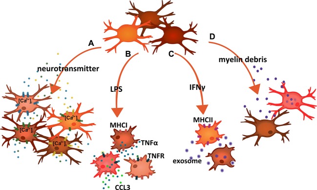
Microglia are the principal resident innate immune cells of the CNS. Their contributions to the normal development of the CNS, the maintenance and plasticity of neuronal networks and the safeguarding of proper functionality are becoming more and more evident. Microglia also survey the tissue homeostasis to respond rapidly to exogenous and endogenous threats, primarily with a protective outcome. However, excessive acute activation, chronic activity or an improper adaptation of their functional performance can foster neuropathologies. A key to the versatile response behavior of these cells is their ability to commit to reactive phenotypes, which reveal enormous complexity. Yet the respective profiles of induced genes and installed functions may build up on heterogeneous contributions of cellular subsets. Here, we discuss findings and concepts that consider the variety of microglial activities and response options as being based-at least in part-on a diversity of the engaged cells. Whether it is the production of proinflammatory cytokines, clearance of tissue debris, antigen presentation or the ability to sense neurotransmitters, microglial cells present with an unanticipated heterogeneity of their constitutive and inducible features. While the organizational principles of this heterogeneity are still largely unknown, functional implications are already perceptible.
Overview
The claims and statistics of this paper haven’t been checked yet.
Run the check — free, no account needed. It reads the open-access text and checks the sources it cites.
Integrity signals
Registry
Cites
Statistics
Figures
Verification
Claims the paper makes
The claims and statistics of this paper haven’t been checked yet.
Run the check — free, no account needed. It reads the open-access text and checks the sources it cites.
Claims other papers make about this one
Each is checked against this paper’s own text: does it say what the citing paper says it does?
The source backs part of the claim, not all of it — or backs it for a different population, condition or comparison.
In the citing paper
“In AD, brain microglial cells are activated in response to Aβ and other neuropathological changes and undergo complex neuroinflammation processes,54 playing either a protective or detrimental role in the disease.55, 56”
From Acoustofluidic assembly of 3D neurospheroids to model Alzheimer's disease (2020) · cites this paper as [54]
Experimental — accuracy not yet measured
Self-check
Percentages, sums and reported differences re-computed from the paper’s own numbers.
The self-check needs the paper’s full text.
Citations
Cites
Exploring the full spectrum of macrophage activation
Mosser, D. M., & Edwards, J. P. (2008) · Nature Reviews Immunology · doi:10.1038/nri2448 · Cited by 9,144
Resting microglial cells are highly dynamic surveillants of brain parenchyma in vivo
Nimmerjahn, A., Kirchhoff, F., & Helmchen, F. (2005) · Science · doi:10.1126/science.1110647 · Cited by 5,827
Fate Mapping Analysis Reveals That Adult Microglia Derive from Primitive Macrophages
Ginhoux, F., Greter, M., Leboeuf, M., Nandi, S., See, P., Gokhan, S., Mehler, M. F., Conway, S. J., Ng, L. G., Stanley, E. R., et al. (2010) · Science · doi:10.1126/science.1194637 · Cited by 5,175
Macrophage biology in development, homeostasis and disease
Wynn, T. A., Chawla, A., & Pollard, J. W. (2013) · Nature · doi:10.1038/nature12034 · Cited by 4,684 · cited 2×
“mononuclear phagocytic system, as to their self-maintenance as well as properties acquired and exhibited within the context of their tissue environment (Ginhoux et al., 2010; Gautier et al., 2012; Schulz et al., 2012; Davies et al., 2013; Greter and Merad, 2013; Kierdorf et al., 2013; Wynn et al., 2013).”
BackgroundSynaptic Pruning by Microglia Is Necessary for Normal Brain Development
Paolicelli, R. C., Bolasco, G., Pagani, F., Maggi, L., Scianni, M., Panzanelli, P., Giustetto, M., Ferreira, T. A., Guiducci, E., Dumas, L., et al. (2011) · Science · doi:10.1126/science.1202529 · Cited by 4,244
Microglia Sculpt Postnatal Neural Circuits in an Activity and Complement-Dependent Manner
Schafer, D. P., Lehrman, E. K., Kautzman, A. G., Koyama, R., Mardinly, A. R., Yamasaki, R., Ransohoff, R. M., Greenberg, M. E., Barres, B. A., & Stevens, B. (2012) · Neuron · doi:10.1016/j.neuron.2012.03.026 · Cited by 4,089
ATP mediates rapid microglial response to local brain injury in vivo
Davalos, D., Grutzendler, J., Yang, G., Kim, J. V., Zuo, Y., Jung, S., Littman, D. R., Dustin, M. L., & Gan, W.-B. (2005) · Nature Neuroscience · doi:10.1038/nn1472 · Cited by 3,913
Microglia: Active sensor and versatile effector cells in the normal and pathologic brain
Hanisch, U.-K., & Kettenmann, H. (2007) · Nature Neuroscience · doi:10.1038/nn1997 · Cited by 3,676
Self-citationKettenmann, H., Hanisch, U.-K., Noda, M., & Verkhratsky, A. (2011) · Physiological Reviews · doi:10.1152/physrev.00011.2010 · Cited by 3,547
Self-citationDaneman, R., & Prat, A. (2015) · Cold Spring Harbor Perspectives in Biology · doi:10.1101/cshperspect.a020412 · Cited by 3,437
Cited by
Sorted by citations
Microglia in physiology and disease
Wolf, S. A., Boddeke, H. W. G. M., & Kettenmann, H. (2017) · Annual Review of Physiology · doi:10.1146/annurev-physiol-022516-034406 · Cited by 1,426
Glia–neuron interactions in the mammalian retina
Vecino, E., Rodriguez, F. D., Ruzafa, N., Pereiro, X., & Sharma, S. C. (2015) · Progress in Retinal and Eye Research · doi:10.1016/j.preteyeres.2015.06.003 · Cited by 863
Inflammatory Response in the CNS: Friend or Foe?
Sochocka, M., Diniz, B. S., & Leszek, J. (2017) · Molecular Neurobiology · doi:10.1007/s12035-016-0297-1 · Cited by 573
Microglial subtypes: diversity within the microglial community
Stratoulias, V., Venero, J. L., Tremblay, M., & Joseph, B. (2019) · The EMBO Journal · doi:10.15252/embj.2019101997 · Cited by 564
Neuroinflammation in the central nervous system: Symphony of glial cells
Yang, Q., & Zhou, J. (2018) · Glia · doi:10.1002/glia.23571 · Cited by 539
Immunoneuropsychiatry — novel perspectives on brain disorders
Pape, K., Tamouza, R., Leboyer, M., & Zipp, F. (2019) · Nature Reviews Neurology · doi:10.1038/s41582-019-0174-4 · Cited by 461
Bidirectional Microglia–Neuron Communication in Health and Disease
Szepesi, Z., Manouchehrian, O., Bachiller, S., & Deierborg, T. (2018) · Frontiers in Cellular Neuroscience · doi:10.3389/fncel.2018.00323 · Cited by 459
Streit, W. J., Xue, Q.-S., Tischer, J., & Bechmann, I. (2014) · Acta Neuropathologica Communications · doi:10.1186/s40478-014-0142-6 · Cited by 317
Microglia: Housekeeper of the Central Nervous System
Kabba, J. A., Xu, Y., Christian, H., Ruan, W., Kitchen, C., Xiang, Y., Zhang, L., Saavedra, J. M., & Pang, T. (2017) · Cellular and Molecular Neurobiology · doi:10.1007/s10571-017-0504-2 · Cited by 265
The Ischemic Environment Drives Microglia and Macrophage Function
Fumagalli, S., Perego, C., Pischiutta, F., Zanier, E. R., & Simoni, M. G. D. (2015) · Frontiers in Neurology · doi:10.3389/fneur.2015.00081 · Cited by 261
Transcriptional control of microglia phenotypes in health and disease
Holtman, I. R., Skola, D., & Glass, C. K. (2017) · Journal of Clinical Investigation · doi:10.1172/jci90604 · Cited by 218
Microglia Mediated Neuroinflammation: Focus on PI3K Modulation
Cianciulli, A., Porro, C., Calvello, R., Trotta, T., Lofrumento, D. D., & Panaro, M. A. (2020) · Biomolecules · doi:10.3390/biom10010137 · Cited by 210
Targeting microglia for the treatment of Alzheimer’s Disease
Wes, P. D., Sayed, F. A., Bard, F., & Gan, L. (2016) · Glia · doi:10.1002/glia.22988 · Cited by 185
Gundersen, V., Storm‐Mathisen, J., & Bergersen, L. H. (2015) · Physiological Reviews · doi:10.1152/physrev.00024.2014 · Cited by 179
Wes, P. D., Holtman, I. R., Boddeke, E. W. G. M., Möller, T., & Eggen, B. J. L. (2016) · Glia · doi:10.1002/glia.22866 · Cited by 167
Microglia change from a reactive to an age-like phenotype with the time in culture
Caldeira, C., Oliveira, A. F., Cunha, C., Vaz, A. R., Falcão, A. S., Fernandes, A., & Brites, D. (2014) · Frontiers in Cellular Neuroscience · doi:10.3389/fncel.2014.00152 · Cited by 156
Reactive astrogliosis in stroke: Contributions of astrocytes to recovery of neurological function
Sims, N. R., & Yew, W. P. (2017) · Neurochemistry International · doi:10.1016/j.neuint.2016.12.016 · Cited by 149
Axon Initial Segment–Associated Microglia
Baalman, K., Marin, M. A., Ho, T., Godoy, M. I., Cherian, L., Robertson, C., & Rasband, M. (2015) · Journal of Neuroscience · doi:10.1523/jneurosci.3751-14.2015 · Cited by 129
Siman, R., Cocca, R., & Dong, Y. (2015) · PLoS ONE · doi:10.1371/journal.pone.0142340 · Cited by 122
Vascular and Inflammatory Factors in the Pathophysiology of Blast-Induced Brain Injury
Elder, G. A., Sosa, M. A. G., Gasperi, R. D., Stone, J. R., Dickstein, D. L., Haghighi, F. G., Hof, P. R., & Ahlers, S. T. (2015) · Frontiers in Neurology · doi:10.3389/fneur.2015.00048 · Cited by 115
Microglial Priming as Trained Immunity in the Brain
Haley, M. J., Brough, D., Quintin, J., & Allan, S. M. (2019) · Neuroscience · doi:10.1016/j.neuroscience.2017.12.039 · Cited by 104
Cellular and molecular pathophysiology in the progression of Parkinson’s disease
Zaman, V., Shields, D. C., Shams, R., Drasites, K. P., Matzelle, D. D., Haque, A., & Banik, N. L. (2021) · Metabolic Brain Disease · doi:10.1007/s11011-021-00689-5 · Cited by 119
Lewis, N. D., Hill, J. D., Juchem, K. W., Stefanopoulos, D. E., & Modis, L. K. (2014) · Journal of Neuroimmunology · doi:10.1016/j.jneuroim.2014.09.014 · Cited by 110
Benfotiamine Attenuates Inflammatory Response in LPS Stimulated BV-2 Microglia
Bozic, I., Savic, D., Laketa, D., Bjelobaba, I., Milenkovic, I., Pekovic, S., Nedeljkovic, N., & Lavrnja, I. (2015) · PLOS ONE · doi:10.1371/journal.pone.0118372 · Cited by 101
PDE4B as a microglia target to reduce neuroinflammation
Pearse, D. D., & Hughes, Z. A. (2016) · Glia · doi:10.1002/glia.22986 · Cited by 98
A neurodegenerative cellular stress response linked to dark microglia and toxic lipid secretion
Flury, A. G., Aljayousi, L., Park, H.-J., Khakpour, M., Mechler, J., Aziz, S., McGrath, J. D., Deme, P., Sandberg, C., Ibáñez, F. G., et al. (2024) · Neuron · doi:10.1016/j.neuron.2024.11.018 · Cited by 102
Wlodarczyk, A., Cédile, O., Jensen, K. N., Jasson, A., Mony, J. T., Khorooshi, R., & Owens, T. (2015) · Frontiers in Immunology · doi:10.3389/fimmu.2015.00463 · Cited by 88
Dendritic cells in brain diseases
Ludewig, P., Gallizioli, M., Urra, X., Behr, S., Brait, V. H., Gelderblom, M., Magnus, T. U., & Planas, A. M. (2015) · Biochimica et Biophysica Acta (BBA) - Molecular Basis of Disease · doi:10.1016/j.bbadis.2015.11.003 · Cited by 86
The Evolving Biology of Microglia in Alzheimer's Disease
Malm, T. M., Jay, T. R., & Landreth, G. E. (2015) · Neurotherapeutics · doi:10.1007/s13311-014-0316-8 · Cited by 85
Weng, L., Zhang, H., Li, X., Zhan, H., Chen, F., Han, L., Xu, Y., & Cao, X. (2016) · International Immunopharmacology · doi:10.1016/j.intimp.2016.12.018 · Cited by 81
Yew, W. P., Djukic, N. D., Jayaseelan, J. S. P., Walker, F. R., Roos, K. A. A., Chataway, T., Muyderman, H., & Sims, N. R. (2019) · Journal of Neuroinflammation · doi:10.1186/s12974-018-1379-y · Cited by 82
Innate immune response is differentially dysregulated between bipolar disease and schizophrenia
de Baumont, A., Maschietto, M. C., Lima, L., Carraro, D. M., Olivieri, E. H. R., Fiorini, A., Barreta, L. A. N., Palha, J. A., Belmonte-de-Abreu, P. S., Moreira‐Filho, C. A., et al. (2014) · Schizophrenia Research · doi:10.1016/j.schres.2014.10.055 · Cited by 75
Microglia are continuously activated in the circumventricular organs of mouse brain
Takagi, S., Furube, E., Nakano, Y., Morita, M., & Miyata, S. (2017) · Journal of Neuroimmunology · doi:10.1016/j.jneuroim.2017.10.008 · Cited by 70
Acoustofluidic assembly of 3D neurospheroids to model Alzheimer's disease
Cai, H., Ao, Z., Hu, L., Moon, Y., Wu, Z., Lu, H.-C., Kim, J., & Guo, F. (2020) · The Analyst · doi:10.1039/d0an01373k · Cited by 70
The estrogen–macrophage interplay in the homeostasis of the female reproductive tract
Pepe, G., Locati, M., Torre, S. D., Mornata, F., Cignarella, A., Maggi, A. C., & Vegeto, E. (2018) · Human Reproduction Update · doi:10.1093/humupd/dmy026 · Cited by 78
Microglia and HPA axis in depression: An overview of participation and relationship
Pereira, G. C., Piton, E., dos Santos, B. M., Ramanzini, L. G., Camargo, L. F. M., da Silva, R. M., & Bochi, G. V. (2021) · The World Journal of Biological Psychiatry · doi:10.1080/15622975.2021.1939154 · Cited by 72
Role of Non-Neuronal Cells in Body Weight and Appetite Control
Argente-Arizón, P., Freire‐Regatillo, A., Argente, J., & Chowen, J. A. (2015) · Frontiers in Endocrinology · doi:10.3389/fendo.2015.00042 · Cited by 70
Llorens-Martín, M., Jurado‐Arjona, J., Fuster-Matanzo, A., Hernández, F., Rábano, A., & Ávila, J. (2014) · Translational Psychiatry · doi:10.1038/tp.2014.92 · Cited by 69
Joost, E., Jordão, M. J. C., Mages, B., Prinz, M., Bechmann, I., & Krueger, M. (2019) · Brain Structure and Function · doi:10.1007/s00429-019-01834-8 · Cited by 66
Brendecke, S. M., & Prinz, M. (2015) · Seminars in Immunopathology · doi:10.1007/s00281-015-0520-6 · Cited by 68
Shao, R., Sun, D., Hu, Y., & Cui, D. (2021) · Journal of Neuroscience Research · doi:10.1002/jnr.24761 · Cited by 64
Song, J., Choi, S., & Kim, B. C. (2017) · Frontiers in Cellular Neuroscience · doi:10.3389/fncel.2017.00064 · Cited by 63
Microglial Interferon Signaling and White Matter
McDonough, A., Lee, R. V., & Weinstein, J. R. (2017) · Neurochemical Research · doi:10.1007/s11064-017-2307-8 · Cited by 63
Inhibitory Effects of Myricetin on Lipopolysaccharide-Induced Neuroinflammation
Jang, J.-H., Lee, S. H., Jung, K., Yoo, H.-R., & Park, G. (2020) · Brain Sciences · doi:10.3390/brainsci10010032 · Cited by 59
Role of metabolic programming in the modulation of microglia phagocytosis by lipids
Nadjar, A. (2018) · Prostaglandins, Leukotrienes and Essential Fatty Acids · doi:10.1016/j.plefa.2018.07.006 · Cited by 49
Fernandes, B. S., Dai, Y., Jia, P., & Zhao, Z. (2022) · European Neuropsychopharmacology · doi:10.1016/j.euroneuro.2022.06.001 · Cited by 49
Geng, Y., Zhu, S., Cheng, P., Lu, Z., Xu, H., Shi, J., & Xu, Z. (2017) · Phytomedicine · doi:10.1016/j.phymed.2017.01.005 · Cited by 46
Mestre, L., Carrillo-Salinas, F. J., Mecha, M., Feliú, A., Espejo, C., Alvarez-Cermeño, J. C., Villar, L. M., & Guaza, C. (2019) · Frontiers in Immunology · doi:10.3389/fimmu.2019.01374 · Cited by 48
Dando, S. J., Golborne, C. N., Chinnery, H. R., Ruitenberg, M. J., & McMenamin, P. G. (2016) · Glia · doi:10.1002/glia.23005 · Cited by 46
Non-Neuronal Cells in the Hypothalamic Adaptation to Metabolic Signals
Freire‐Regatillo, A., Argente-Arizón, P., Argente, J., García‐Segura, L. M., & Chowen, J. A. (2017) · Frontiers in Endocrinology · doi:10.3389/fendo.2017.00051 · Cited by 48
Benfotiamine upregulates antioxidative system in activated BV-2 microglia cells
Bozic, I. D., Savić, D. Z., Stevanović, I. D., Peković, S., Nedeljković, N., & Lavrnja, I. C. (2015) · Frontiers in Cellular Neuroscience · doi:10.3389/fncel.2015.00351 · Cited by 44
Memory-Like Responses of Brain Microglia Are Controlled by Developmental State and Pathogen Dose
Lajqi, T., Stojiljkovic, M. R., Williams, D. L., Hudalla, H., Bauer, M. W., Witte, O. W., Wetzker, R., Bauer, R., & Schmeer, C. W. (2020) · Frontiers in Immunology · doi:10.3389/fimmu.2020.546415 · Cited by 41
Synapses, Microglia, and Lipids in Alzheimer’s Disease
Paasila, P. J., Aramideh, J. A., Sutherland, G. T., & Graeber, M. B. (2022) · Frontiers in Neuroscience · doi:10.3389/fnins.2021.778822 · Cited by 40
Salminen, A., Kaarniranta, K., & Kauppinen, A. (2018) · Cellular and Molecular Life Sciences · doi:10.1007/s00018-018-2844-6 · Cited by 39
Wogram, E., Wendt, S., Matyash, M., Pivneva, T. A., Draguhn, A., & Kettenmann, H. (2016) · European Journal of Neuroscience · doi:10.1111/ejn.13256 · Cited by 33
Grace, P. M., Shimizu, K., Strand, K. A., Rice, K. C., Deng, G., Watkins, L. R., & Herson, P. S. (2015) · Brain Behavior and Immunity · doi:10.1016/j.bbi.2015.03.005 · Cited by 37
Interferon‐β regulates the production of IL‐10 by toll‐like receptor‐activated microglia
Lobo‐Silva, D., Carriche, G. M., Castro, A. G., Roque, S., & Saraiva, M. (2017) · Glia · doi:10.1002/glia.23172 · Cited by 35
Fukushima, S., Furube, E., Itoh, M., Nakashima, T., & Miyata, S. (2015) · Journal of Neuroimmunology · doi:10.1016/j.jneuroim.2015.05.014 · Cited by 35
The use and limitations of single‐cell mass cytometry for studying human microglia function
Fernández‐Zapata, C., Leman, J. K. H., Priller, J., & Böttcher, C. (2020) · Brain Pathology · doi:10.1111/bpa.12909 · Cited by 38
Amos, P. J., Fung, S., Case, A., Kifelew, J., Osnis, L., Smith, C. S., Green, K., Naydenov, A. V., Aloi, M. S., Hubbard, J. J., et al. (2017) · ASN NEURO · doi:10.1177/1759091417716610 · Cited by 34
Zhao, C., Lei, H., Zhang, Y., Lin, L., Xu, S., Cai, J., Li, P., Wang, L., Wang, X., & Peng, Y. (2015) · Journal of Asian Natural Products Research · doi:10.1080/10286020.2015.1099524 · Cited by 32
Zhao, C., Hou, W.-Z., Lei, H., Huang, L., Wang, S., Cui, D., Xing, C., Wang, X., & Peng, Y. (2017) · Acta Pharmaceutica Sinica B · doi:10.1016/j.apsb.2017.04.007 · Cited by 29
Extracellular acidosis impairs P2Y receptor-mediated Ca2+ signalling and migration of microglia
Langfelder, A., Okonji, P. E., Deca, D., Wei, W., & Glitsch, M. D. (2015) · Cell Calcium · doi:10.1016/j.ceca.2015.01.004 · Cited by 28
Karczewski, J., Zielińska, A., Staszewski, R., Eder, P., Dobrowolska, A., & Souto, E. B. (2022) · International Journal of Molecular Sciences · doi:10.3390/ijms23116145 · Cited by 27
Alterations in CD200-CD200R1 System during EAE Already Manifest at Presymptomatic Stages
Valente, T., Serratosa, J., Perpiñá, U., Saura, J., & Solà, C. (2017) · Frontiers in Cellular Neuroscience · doi:10.3389/fncel.2017.00129 · Cited by 27
A presumed antagonistic LPS identifies distinct functional organization of TLR4 in mouse microglia
Döring, C., Regen, T., Gertig, U., van Rossum, D., Winkler, A., Saiepour, N., Brück, W., Hanisch, U., & Jáňová, H. (2017) · Glia · doi:10.1002/glia.23151 · Cited by 24
Self-citationWhat has single‐cell RNA sequencing revealed about microglial neuroimmunology?
Kubick, N., Flournoy, P. C. H., Klimovich, P., Manda, G., & Mickael, M. (2020) · Immunity Inflammation and Disease · doi:10.1002/iid3.362 · Cited by 27
Zhang, Q., Liu, X., Yan, L., Zhao, R., An, J., Liu, C., & Yang, H. (2019) · Phytomedicine · doi:10.1016/j.phymed.2019.152966 · Cited by 25
Gullo, F., Amadeo, A., Donvito, G., Lecchi, M., Costa, B., Constanti, A., & Wanke, E. (2014) · Frontiers in Cellular Neuroscience · doi:10.3389/fncel.2014.00361 · Cited by 26
Neuroinflammation and Alzheimer's disease: Unravelling the molecular mechanisms
Kakkar, A., Singh, H., Singh, B. K., Kumar, A., Mishra, A. K., & Chopra, H. (2025) · Journal of Alzheimer s Disease · doi:10.1177/13872877251374353 · Cited by 26
Infections in the Developing Brain: The Role of the Neuro-Immune Axis
Kim, J., Erice, C., Rohlwink, U. K., & Tucker, E. W. (2022) · Frontiers in Neurology · doi:10.3389/fneur.2022.805786 · Cited by 28
Astrocyte phenotypes: Emphasis on potential markers in neuroinflammation.
Bozic, I. D., Savić, D. Z., & Lavrnja, I. C. (2021) · PubMed · doi:10.14670/hh-18-284 · Cited by 22
Targeting Kv1.3 channels to reduce white matter pathology after traumatic brain injury
Reeves, T. M., Trimmer, P., Colley, B. S., & Phillips, L. (2016) · Experimental Neurology · doi:10.1016/j.expneurol.2016.06.011 · Cited by 17
Hypoperfusion is a potential inducer of immunosuppressive network in Alzheimer's disease
Salminen, A. (2020) · Neurochemistry International · doi:10.1016/j.neuint.2020.104919 · Cited by 20
Ravenhill, S. M., Evans, A. H., & Crewther, S. G. (2023) · Antioxidants · doi:10.3390/antiox12051117 · Cited by 22
Molecular mechanisms underlying resting-state brain functional correlates of behavioral inhibition
Cui, S., Jiang, P., Cheng, Y., Cai, H., Zhu, J., & Yu, Y. (2023) · NeuroImage · doi:10.1016/j.neuroimage.2023.120415 · Cited by 20
Zhu, H., Liu, J., Zhou, J., Jin, Y., Zhao, Q., Jiang, X., & Gao, H. (2024) · Journal of Ethnopharmacology · doi:10.1016/j.jep.2024.118651 · Cited by 22
Dual role of microglia in health and disease: pushing the balance toward repair
Ferreira, R., & Bernardino, L. I. (2015) · Frontiers in Cellular Neuroscience · doi:10.3389/fncel.2015.00051 · Cited by 20
Sorrentino, S., Ascari, R., Maderna, E., Catania, M., Ghetti, B. F., Tagliavini, F., Giaccone, G., & Fede, G. D. (2021) · International Journal of Molecular Sciences · doi:10.3390/ijms22052780 · Cited by 23
Nagayach, A., Bhaskar, R., & Patro, I. K. (2022) · Journal of Chemical Neuroanatomy · doi:10.1016/j.jchemneu.2022.102160 · Cited by 18
Kamigaki, M., Hide, I., Yanase, Y., Shiraki, H., Harada, K., Tanaka, Y., Seki, T., Shirafuji, T., Tanaka, S., Hide, M., et al. (2016) · Neurochemistry International · doi:10.1016/j.neuint.2016.01.003 · Cited by 19
Adenosine A1 Receptor mRNA Expression by Neurons and Glia in the Auditory Forebrain
Hackett, T. A. (2018) · The Anatomical Record · doi:10.1002/ar.23907 · Cited by 17
Copper disrupts S-nitrosothiol signaling in activated BV2 microglia
Rossi‐George, A., & Guo, C. (2016) · Neurochemistry International · doi:10.1016/j.neuint.2016.05.011 · Cited by 16
Yang, L. L., Zhang, Y., Yu, X., Li, D., Liu, N., Xue, X., & Fu, J. (2023) · Journal of Immunology Research · doi:10.1155/2023/5149306 · Cited by 15
Coelho, V. R., Viau, C. M., Staub, R. B., de Souza, M. S., Pflüger, P., Regner, G. G., Pereira, P., & Saffi, J. (2017) · NeuroImmunoModulation · doi:10.1159/000481095 · Cited by 15
Hispolon Suppresses LPS- or LTA-Induced iNOS/NO Production and Apoptosis in BV-2 Microglial Cells
Wu, M., Chien, C.-C., Cheng, K.-T., Subbaraju, G. V., & Chen, Y. (2017) · The American Journal of Chinese Medicine · doi:10.1142/s0192415x17500896 · Cited by 15
Lang, G.-P., Ndongson‐Dongmo, B., Lajqi, T., Brodhun, M., Han, Y., Wetzker, R., Frasch, M. G., & Bauer, R. (2020) · Journal of Neuroinflammation · doi:10.1186/s12974-020-01954-7 · Cited by 15
Velez-Perez, A., Holder, M. K., Fountain, S., & Blaustein, J. D. (2020) · eNeuro · doi:10.1523/eneuro.0505-19.2020 · Cited by 14
Akingbade, G. T., Ijomone, O. M., Imam, A., Aschner, M., & Ajao, M. S. (2022) · Environmental Toxicology and Pharmacology · doi:10.1016/j.etap.2022.103872 · Cited by 13
Extracellular Vesicles Cargo in Modulating Microglia Functional Responses
Torre, M. E. L., Panaro, M. A., Ruggiero, M., Polito, R., Cianciulli, A., Filannino, F. M., Lofrumento, D. D., Antonucci, L., Benameur, T., Monda, V., et al. (2022) · Biology · doi:10.3390/biology11101426 · Cited by 15
Nanomaterials for neurodegenerative diseases: Molecular mechanisms guided design and applications
Li, T., Hou, X., Yu, Q., Duan, X., Yan, P., Zhu, H., Xie, Z., & Zhang, H. (2021) · Nano Research · doi:10.1007/s12274-021-3865-2 · Cited by 12
Disturbed sensitive equilibrium led by stress‐induced inflammation in psychiatric illness
Zhao, H., Jin, R., & Hu, J. (2022) · General Psychiatry · doi:10.1136/gpsych-2022-100910 · Cited by 12
Shao, J., Yin, X. L., Lang, Y., Ding, M., Zhang, B., Sun, Q., Jiang, X., Song, J., & Cui, L. (2022) · Molecular Neurobiology · doi:10.1007/s12035-022-03099-5 · Cited by 12
Poised for action: USP18 restrains microglial activation in the white matter
Takata, K., & Ginhoux, F. (2015) · The EMBO Journal · doi:10.15252/embj.201591899 · Cited by 10
Thomaty, S., Pézard, L., Xerri, C., & Brezun, J. (2017) · Neuroscience · doi:10.1016/j.neuroscience.2017.02.035 · Cited by 10
Back to the Future: The Role of Infections in Psychopathology. Focus on OCD.
Vecchia, A. D., & Marazziti, D. (2022) · PubMed Central · doi:10.36131/cnfioritieditore20220407 · Cited by 10
Asaba, T., Hamano, S., Nanmo, A., Seo, J., Kageyama, T., & Fukuda, J. (2025) · Regenerative Therapy · doi:10.1016/j.reth.2025.06.013 · Cited by 8
Ruzafa, N., Pereiro, X., Fonollosa, A., Araiz, J., Acera, A., & Vecino, E. (2021) · Frontiers in Pharmacology · doi:10.3389/fphar.2021.606232 · Cited by 8
General Physiology and Pathophysiology of Microglia During Neuroinflammation
Patro, I. K., Nagayach, A., Sinha, S., & Patro, N. (2016) · Springer eBooks · doi:10.1007/978-981-10-1711-7_2 · Cited by 8
Cummings, J. L., Wu, Y. L., Dixon, C. E., Henchir, J. J., Simard, J. M., Panigrahy, A., Kochanek, P. M., Jha, R. M., & Aneja, R. K. (2023) · Journal of Neuroinflammation · doi:10.1186/s12974-023-02692-2 · Cited by 7
Metadata
- Authors
- Gertig, U., & Hanisch, U.-K.
- Affiliations
- University of Göttingen (DE)
- Corresponding author
- Hanisch, Uwe-Karsten
- Venue
- Frontiers in Cellular Neuroscience
- Publisher
- Frontiers Media SA
- Published
- 2014-04-01
- Volume
- 8
- Pages
- 101-101
- ISSN
- 1662-5102 (ISSN-L)
- PMC
- PMC3978327
- OpenAlex
- W1968597551
- Cited by
- 123
- References
- 78
- Citations by year
- 2026: 2 · 2025: 2 · 2024: 4 · 2023: 5 · 2022: 10 · 2021: 9 · 2020: 11 · 2019: 12 · 2018: 6 · 2017: 18 · 2016: 13 · 2015: 23 · 2014: 8
- FWCI
- 1.67
- Type
- journal article
- Language
- en
- Open access
- gold
- doi.org
- Licence
- cc-by
- Locations
- Frontiers in Cellular Neuroscience (publishedVersion, cc-by)
- Europe PMC (PubMed Central) (publishedVersion, cc-by)
- PubMed Central (publishedVersion, cc-by)
- PubMed (publishedVersion)
- DOAJ (DOAJ: Directory of Open Access Journals) (submittedVersion, cc-by-sa)
- GoeScholar The Publication Server of the Georg-August-Universität Göttingen (Georg-August-Universität Göttingen) (submittedVersion, cc-by)
- Funders
- Deutsche Forschungsgemeinschaft (SFB TRR 43, TRR-43)
- Topics
- Life Sciences › Neuroscience › Neurology › Neuroinflammation and Neurodegeneration Mechanisms
- Life Sciences › Neuroscience › Cellular and Molecular Neuroscience › Neuroscience and Neuropharmacology Research
- Life Sciences › Biochemistry, Genetics and Molecular Biology › Molecular Biology › Inflammasome and immune disorders
- Keywords
- microglia, microglial heterogeneity, microglial activation, inflammatory cytokines, debris removal, antigen presentation, neurotransmitter detection, CNS homeostasis, neuroinflammation, neuropathology
About these checks
Registry, retraction, Cites and statistics results are computed, not judged, with one exception: a printed reference that no index lists is read once by a model before it counts as a citation error. Claims are extracted by a model and kept only when their quote appears verbatim in the text; each cited source is then checked, with a model’s help, for whether it says what the claim relies on. Those verdicts can be wrong — read the claim’s quote and the cited source before relying on one.
Statistics and arithmetic are recomputed, not judged.
Confidence bands are shown only for fields calibrated on our annotated test set. None is calibrated yet, so no band is shown.
What counts as a claim ›How we measure confidence ›How we check this ›
Are you an author, or do you see a problem with this page? Request a review
