

The DOI matches its Crossref record; 98 of the 100 references it cites were found in a registry or an index, and 2 are not checked yet.
Dipy, a library for the analysis of diffusion MRI data
Open accessAbstract
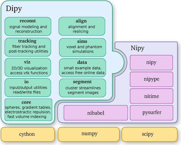
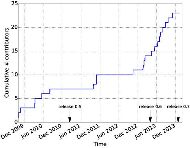
Diffusion Imaging in Python (Dipy) is a free and open source software project for the analysis of data from diffusion magnetic resonance imaging (dMRI) experiments. dMRI is an application of MRI that can be used to measure structural features of brain white matter. Many methods have been developed to use dMRI data to model the local configuration of white matter nerve fiber bundles and infer the trajectory of bundles connecting different parts of the brain. Dipy gathers implementations of many different methods in dMRI, including: diffusion signal pre-processing; reconstruction of diffusion distributions in individual voxels; fiber tractography and fiber track post-processing, analysis and visualization. Dipy aims to provide transparent implementations for all the different steps of dMRI analysis with a uniform programming interface. We have implemented classical signal reconstruction techniques, such as the diffusion tensor model and deterministic fiber tractography. In addition, cutting edge novel reconstruction techniques are implemented, such as constrained spherical deconvolution and diffusion spectrum imaging (DSI) with deconvolution, as well as methods for probabilistic tracking and original methods for tractography clustering. Many additional utility functions are provided to calculate various statistics, informative visualizations, as well as file-handling routines to assist in the development and use of novel techniques. In contrast to many other scientific software projects, Dipy is not being developed by a single research group. Rather, it is an open project that encourages contributions from any scientist/developer through GitHub and open discussions on the project mailing list. Consequently, Dipy today has an international team of contributors, spanning seven different academic institutions in five countries and three continents, which is still growing.
Overview
The claims and statistics of this paper haven’t been checked yet.
Run the check — free, no account needed. It reads the open-access text and checks the sources it cites.
Integrity signals
Registry
Cites
Statistics
Figures
Verification
Claims the paper makes
The claims and statistics of this paper haven’t been checked yet.
Run the check — free, no account needed. It reads the open-access text and checks the sources it cites.
Claims other papers make about this one
Each is checked against this paper’s own text: does it say what the citing paper says it does?
The cited source says what the claim attributes to it.
In the citing paper
“In contrast to projects such as DiPy (Garyfallidis et al., 2014) , this library does not provide functionality to manipulate raw image data within Python itself, but provides functionality to invoke and manage external high-performance executables to perform the actual computations.”
From MRtrix3: A fast, flexible and open software framework for medical image processing and visualisation (2019) · cites this paper as Garyfallidis et al., 2014
Self-check
Percentages, sums and reported differences re-computed from the paper’s own numbers.
The self-check needs the paper’s full text.
Citations
Cites
A Threshold Selection Method from Gray-Level Histograms
Otsu, N. (1979) · IEEE Transactions on Systems, Man and Cybernetics · doi:10.1109/tsmc.1979.4310076 · Cited by 44,708 · cited 1×
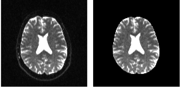
“…median_otsu (see Figure 3 ). from dipy.segment.mask import median_otsu mask, S0_mask = median_otsu(data [:, :, :, 0]) median_otsu uses first a median filter to smooth the S0 and then Otsu’s method, an automated histogram method (Otsu, 1979) to separate the brain (foreground) from its background.”
MethodAdvances in functional and structural MR image analysis and implementation as FSL
Smith, S. M., Jenkinson, M., Woolrich, M. W., Beckmann, C. F., Behrens, T. E. J., Johansen-Berg, H., Bannister, P. R., De Luca, M., Drobnjak, I., Flitney, D. E., et al. (2004) · NeuroImage · doi:10.1016/j.neuroimage.2004.07.051 · Cited by 13,630
Fischl, B. (2012) · NeuroImage · doi:10.1016/j.neuroimage.2012.01.021 · Cited by 9,526 · cited 4×
“…al., 2006), AFNI (Cox, 2012), MITK (Fritzsche et al., 2012), BrainVoyager QX (Goebel, 2012), DTI-Query/Quench (Sherbondy et al., 2005), FreeSurfer (Fischl, 2012), FSL-FDT (Smith et al., 2004), MedInria (Toussaint et al., 2007), MRtrix (Tournier et al., 2012), Diffusion Toolkit/Trackvis (Wang et…”
MethodInfluentialThe NumPy Array: A Structure for Efficient Numerical Computation
van der Walt, S., Colbert, S. C., & Varoquaux, G. (2011) · Computing in Science & Engineering · doi:10.1109/mcse.2011.37 · Cited by 9,381
MR diffusion tensor spectroscopy and imaging
Basser, P. J., Mattiello, J., & LeBihan, D. (1994) · Biophysical Journal · doi:10.1016/s0006-3495(94)80775-1 · Cited by 5,970 · cited 2×
“Another alternative is to make assumptions about the distribution of the PDF such as a Gaussian assumption, which leads to the popular diffusion tensor imaging (DTI) technique (Basser et al., 1994).”
BackgroundMethodThe WU-Minn Human Connectome Project: An overview
Van Essen, D. C., Smith, S. M., Barch, D. M., Behrens, T. E. J., Yacoub, E., & Ugurbil, K. (2013) · NeuroImage · doi:10.1016/j.neuroimage.2013.05.041 · Cited by 5,651
The basis of anisotropic water diffusion in the nervous system—A technical review.
Beaulieu, C. (2002) · NMR in Biomedicine · doi:10.1002/nbm.782 · Cited by 4,628
Basser, P. J., & Pierpaoli, C. (1996) · Journal of Magnetic Resonance, Series B · doi:10.1006/jmrb.1996.0086 · Cited by 4,306 · cited 3×
“…of the myelin wrapping the axons in the voxel may affect both MD and FA, changes in other factors, such as the distribution of directions of crossing of fiber populations through the voxel may also affect these measures (Basser and Pierpaoli, 1996; Jones et al., 2013; Wandell and Yeatman, 2013).”
BackgroundMethodMedical image analysis: progress over two decades and the challenges ahead
Duncan, J. S., & Ayache, N. (2000) · IEEE Transactions on Pattern Analysis and Machine Intelligence · doi:10.1109/34.824822 · Cited by 4,167
IPython: A System for Interactive Scientific Computing
Pérez, F., & Granger, B. (2007) · Computing in Science & Engineering · doi:10.1109/mcse.2007.53 · Cited by 4,129 · cited 1×
“There has been large growth in the number of Python users, there are many Python tools for scientific computing (Pérez and Granger, 2007; Pérez et al., 2011; McKinney, 2012), and there are complementary neuroimaging packages in Python1.”
BackgroundPrinciples of Nuclear Magnetic Resonance Microscopy
Callaghan, P. T. (1991) · doi:10.1093/oso/9780198539445.001.0001 · Cited by 3,614
Three-dimensional tracking of axonal projections in the brain by magnetic resonance imaging
Mori, S., Crain, B. J., Chacko, V. P., & van Zijl, P. C. M. (1999) · Annals of Neurology · doi:10.1002/1531-8249(199902)45:2 · Cited by 3,513
The Human Connectome: A Structural Description of the Human Brain
Sporns, O., Tononi, G., & Kötter, R. (2005) · PLoS Computational Biology · doi:10.1371/journal.pcbi.0010042 · Cited by 3,260 · cited 1×
“The connection matrix that results from an exhaustive connectivity analysis of all parts of the cerebral cortex is sometimes referred to as a connectome (Sporns et al., 2005) and understanding the structure and function of the connectome is a central goal of contemporary neuroscience.”
BackgroundDiffusion tensor MR imaging of the human brain
Pierpaoli, C., Jezzard, P., Basser, P. J., Barnett, A., & Di Chiro, G. (1996) · Radiology · doi:10.1148/radiology.201.3.8939209 · Cited by 2,664 · cited 4×
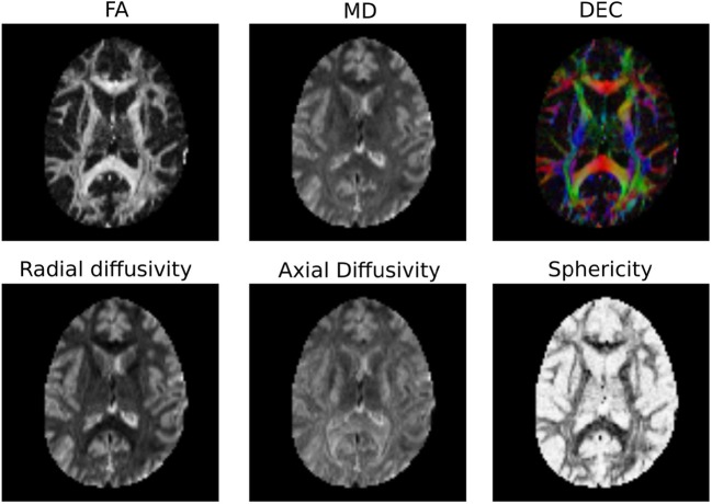
“…common to represent the primary diffusion direction using a red-green-blue (RGB) representation and create a DEC (Directionally Encoded Color) map (Pierpaoli et al., 1996; Pajevic and Pierpaoli, 1999) which is also known as color FA (see Figure 4 ): from dipy.reconst.dti import color_fa cfa =…”
BackgroundMethodInfluentialDysmyelination revealed through MRI as increased radial (but unchanged axial) diffusion of water
Song, S.-K., Sun, S.-W., Ramsbottom, M. J., Chang, C., Russell, J., & Cross, A. H. (2002) · NeuroImage · doi:10.1006/nimg.2002.1267 · Cited by 2,582
White matter integrity, fiber count, and other fallacies: The do's and don'ts of diffusion MRI
Jones, D. K., Knösche, T. R., & Turner, R. (2013) · NeuroImage · doi:10.1016/j.neuroimage.2012.06.081 · Cited by 2,402 · cited 1×
“…of the myelin wrapping the axons in the voxel may affect both MD and FA, changes in other factors, such as the distribution of directions of crossing of fiber populations through the voxel may also affect these measures (Basser and Pierpaoli, 1996; Jones et al., 2013; Wandell and Yeatman, 2013).”
BackgroundTournier, J., Calamante, F., & Connelly, A. (2007) · NeuroImage · doi:10.1016/j.neuroimage.2007.02.016 · Cited by 2,191 · cited 6×
“The method is inspired by CSD (Tournier et al., 2007) with the main difference that the CSD is applied directly to the initial signal and the SDT directly to the ODF (Descoteaux, 2008; Descoteaux et al., 2009).”
BackgroundMethodInfluentialNipype: A Flexible, Lightweight and Extensible Neuroimaging Data Processing Framework in Python
Gorgolewski, K. J., Burns, C. D., Madison, C. M., Clark, D., Halchenko, Y. O., Waskom, M., & Ghosh, S. (2011) · Frontiers in Neuroinformatics · doi:10.3389/fninf.2011.00013 · Cited by 2,162
Tuch, D. S. (2004) · Magnetic Resonance in Medicine · doi:10.1002/mrm.20279 · Cited by 1,880
Tracking neuronal fiber pathways in the living human brain
Conturo, T. E., Lori, N. F., Cull, T. S., Akbudak, E., Snyder, A. Z., Shimony, J. S., McKinstry, R. C., Burton, H., & Raichle, M. E. (1999) · Proceedings of the National Academy of Sciences · doi:10.1073/pnas.96.18.10422 · Cited by 1,774 · cited 3×
“We created an algorithm that has many similarities with the classical deterministic methods (Conturo et al., 1999; Mori et al., 1999; Basser et al., 2000) and with more recent ones such as those described in Descoteaux et al. (2009) and Yeh et al. (2010).”
MethodTournier, J.-D., Calamante, F., Gadian, D. G., & Connelly, A. (2004) · NeuroImage · doi:10.1016/j.neuroimage.2004.07.037 · Cited by 1,635 · cited 2×
“However, the spherical deconvolution problem is ill-posed and thus severely affected by noise (Tournier et al., 2004).”
BackgroundHigh angular resolution diffusion imaging reveals intravoxel white matter fiber heterogeneity
Tuch, D. S., Reese, T. G., Wiegell, M. R., Makris, N., Belliveau, J. W., & Wedeen, V. J. (2002) · Magnetic Resonance in Medicine · doi:10.1002/mrm.10268 · Cited by 1,548 · cited 4×
“Due to their practical mathematical properties, there have been several proposals using spherical harmonics to describe the apparent diffusion coefficient (ADC) computed from HARDI, starting from the work of Frank (2001, 2002) and Alexander et al. (2002) and Tuch et al. (2002).”
BackgroundMethodInfluentialMRtrix: Diffusion tractography in crossing fiber regions
Tournier, J., Calamante, F., & Connelly, A. (2012) · International Journal of Imaging Systems and Technology · doi:10.1002/ima.22005 · Cited by 1,370
Mapping complex tissue architecture with diffusion spectrum magnetic resonance imaging
Wedeen, V. J., Hagmann, P., Tseng, W. I., Reese, T. G., & Weisskoff, R. M. (2005) · Magnetic Resonance in Medicine · doi:10.1002/mrm.20642 · Cited by 1,339 · cited 10×
“This can be used in exactly the same way as the standard DSI method: the unit vector ˆ u where r = r ˆ u . ψ DSI is a function on a sphere.”
BackgroundMethodInfluentialEdelman, R. R., & Warach, S. J. (1993) · New England Journal of Medicine · doi:10.1056/nejm199303183281109 · Cited by 1,207
Log-Euclidean metrics for fast and simple calculus on diffusion tensors
Arsigny, V., Fillard, P., Pennec, X., & Ayache, N. (2006) · Magnetic Resonance in Medicine · doi:10.1002/mrm.20965 · Cited by 1,113
MRI Atlas of Human White Matter
Mori, S. (2005) · Cited by 1,103
Generalized ${ q}$-Sampling Imaging
Yeh, F.-C. F., Wedeen, V. J., & Tseng, W. I. (2010) · IEEE Transactions on Medical Imaging · doi:10.1109/tmi.2010.2045126 · Cited by 1,030 · cited 4×
“We created an algorithm that has many similarities with the classical deterministic methods (Conturo et al., 1999; Mori et al., 1999; Basser et al., 2000) and with more recent ones such as those described in Descoteaux et al. (2009) and Yeh et al. (2010).”
BackgroundMethodInfluentialMRI atlas of human white matter
Ling, C. L., & Rumpel, H. (2006) · Concepts in Magnetic Resonance Part A · doi:10.1002/cmr.a.20052 · Cited by 1,021
ExploreDTI: a graphical toolbox for processing, analyzing, and visualizing diffusion MR data
Leemans, A., Jeurissen, B., Sijbers, J., & Jones, D. K. (2009) · Cited by 874
Regularized, fast, and robust analytical Q-ball imaging
Descoteaux, M., Angelino, E., Fitzgibbons, S., & Deriche, R. (2007) · Magnetic Resonance in Medicine · doi:10.1002/mrm.21277 · Cited by 850
Self-citationAxcaliber: A method for measuring axon diameter distribution from diffusion MRI
Assaf, Y., Blumenfeld‐Katzir, T., Yovel, Y., & Basser, P. J. (2008) · Magnetic Resonance in Medicine · doi:10.1002/mrm.21577 · Cited by 849 · cited 1×
“We have also started looking into tissue characterization in microstructure (Assaf et al., 2008).”
Tract profiles of white matter properties: automating fiber-tract quantification
Yeatman, J. D., Dougherty, R. F., Myall, N. J., Wandell, B. A., & Feldman, H. M. (2012) · PLoS ONE · doi:10.1371/journal.pone.0049790 · Cited by 840
Deterministic and probabilistic tractography based on complex fibre orientation distributions
Descoteaux, M., Deriche, R., Knosche, T. R., & Anwander, A. (2009) · IEEE Transactions on Medical Imaging · doi:10.1109/tmi.2008.2004424 · Cited by 698 · cited 6×
“You can use the SDT in the following way: from dipy.reconst.csdeconv import ConstrainedSDTModel as SdtModel sdt_model = SdtModel(gtab, ratio) Here the response function is provided as a scalar parameter ratio , which is the ratio of the smallest eigenvalue to the largest eigenvalue.”
BackgroundMethodInfluentialSelf-citationRESTORE: Robust estimation of tensors by outlier rejection
Chang, L., Jones, D. K., & Pierpaoli, C. M. (2005) · Magnetic Resonance in Medicine · doi:10.1002/mrm.20426 · Cited by 611 · cited 1×
“In addition, there is ongoing research on methods to robustly fit the tensor model, in the face of noisy data (Chang et al., 2005, 2012).”
MethodBasser, P. J., & Pierpaoli, C. M. (2011) · Journal of Magnetic Resonance · doi:10.1016/j.jmr.2011.09.022 · Cited by 551 · cited 3×
“…of the myelin wrapping the axons in the voxel may affect both MD and FA, changes in other factors, such as the distribution of directions of crossing of fiber populations through the voxel may also affect these measures (Basser and Pierpaoli, 1996; Jones et al., 2013; Wandell and Yeatman, 2013).”
BackgroundMethodDetection and modeling of non‐Gaussian apparent diffusion coefficient profiles in human brain data
Alexander, D. C., Barker, G. J., & Arridge, S. (2002) · Magnetic Resonance in Medicine · doi:10.1002/mrm.10209 · Cited by 538
Characterization of anisotropy in high angular resolution diffusion‐weighted MRI
Frank, L. R. (2002) · Magnetic Resonance in Medicine · doi:10.1002/mrm.10156 · Cited by 520 · cited 1×
“Due to their practical mathematical properties, there have been several proposals using spherical harmonics to describe the apparent diffusion coefficient (ADC) computed from HARDI, starting from the work of Frank (2001, 2002) and Alexander et al. (2002) and Tuch et al. (2002).”
MethodProceedings of the 36th International Conference on Software Engineering
(2014) · doi:10.1145/2568225 · Cited by 477
Measurement of fiber orientation distributions using high angular resolution diffusion imaging
Anderson, A. W. (2005) · Magnetic Resonance in Medicine · doi:10.1002/mrm.20667 · Cited by 461 · cited 1×
“This q-ball ODF ψ QBI can be obtained analytically from a SH estimation of the diffusion signal (Anderson, 2005; Hess et al., 2006; Descoteaux et al., 2007): where P l ( j ) is the Legendre polynomial of order l corresponding to coefficient j and the coefficients have been normalized by the S0…”
MethodCamino: Open-Source Diffusion-MRI Reconstruction and Processing
Cook, P. A., Bai, Y., Nedjati-Gilani, S., Seunarine, K. K., Hall, M. G., Parker, G. J. M., & Alexander, D. C. (2006) · Cited by 453
Diffusion MRI: Theory, methods, and applications
Jones, D. K. (2011) · Cited by 450
Özarslan, E., Koay, C. G., Shepherd, T. M., Komlosh, M. E., Irfanoglu, M. O., Pierpaoli, C. M., & Basser, P. J. (2013) · NeuroImage · doi:10.1016/j.neuroimage.2013.04.016 · Cited by 394
Anisotropy in high angular resolution diffusion-weighted MRI
Frank, L. R. (2001) · Magnetic Resonance in Medicine · doi:10.1002/mrm.1125 · Cited by 389
Q‐ball reconstruction of multimodal fiber orientations using the spherical harmonic basis
Hess, C. P., Mukherjee, P., Han, E. T., Xu, D., & Vigneron, D. B. (2006) · Magnetic Resonance in Medicine · doi:10.1002/mrm.20931 · Cited by 373
Aganj, I., Lenglet, C., Sapiro, G., Yacoub, E. S., Uǧurbil, K., & Harel, N. (2010) · Magnetic Resonance in Medicine · doi:10.1002/mrm.22365 · Cited by 369
Probabilistic fiber tracking using the residual bootstrap with constrained spherical deconvolution
Jeurissen, B., Leemans, A., Jones, D. K., Tournier, J., & Sijbers, J. (2010) · Human Brain Mapping · doi:10.1002/hbm.21032 · Cited by 369 · cited 1×
“…example the fiber orientation distribution function (fODF) obtained by fitting the CSD (constrained spherical deconvolution) model to diffusion data can be used as an estimate of the PDF, because it represents a fiber distribution associated with the measured diffusion signal (Jeurissen et al., 2011).”
MethodImproving diffusion MRI using simultaneous multi-slice echo planar imaging
Setsompop, K., Cohen‐Adad, J., Gagoski, B. A., Raij, T., Yendiki, A., Keil, B. R., Wedeen, V. J., & Wald, L. L. (2012) · NeuroImage · doi:10.1016/j.neuroimage.2012.06.033 · Cited by 357
AFNI: What a long strange trip it's been
Cox, R. W. (2011) · NeuroImage · doi:10.1016/j.neuroimage.2011.08.056 · Cited by 344 · cited 2×
“Many of these tools are written in C/C ++ : 3D Slicer (Pieper et al., 2006), AFNI (Cox, 2012), MITK (Fritzsche et al., 2012), BrainVoyager QX (Goebel, 2012), DTI-Query/Quench (Sherbondy et al., 2005), FreeSurfer (Fischl, 2012), FSL-FDT (Smith et al., 2004), MedInria (Toussaint et al., 2007), MRtrix…”
MethodThe spatial mapping of translational diffusion coefficients by the NMR imaging technique
Taylor, D. G., & Bushell, M. C. (1985) · Physics in Medicine and Biology · doi:10.1088/0031-9155/30/4/009 · Cited by 344 · cited 1×
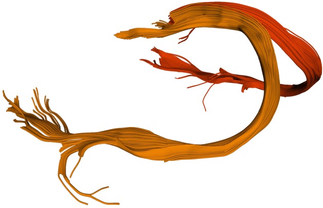
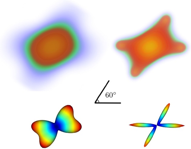
“INTRODUCTION Diffusion MRI (dMRI) (LeBihan and Breton, 1985; Merboldt et al., 1985; Taylor and Bushell, 1985) is an MRI technique (Callaghan, 1991) that provides information about the structure of neuronal pathways found in the white matter and other body tissue with fiber-like structure (see Figure 1).”
BackgroundDiffusion Imaging, White Matter, and Psychopathology
Thomason, M. E., & Thompson, P. M. (2011) · Annual Review of Clinical Psychology · doi:10.1146/annurev-clinpsy-032210-104507 · Cited by 316 · cited 1×
“Variance in a variety of behavioral (Ben-Shachar et al., 2007) and clinical (Thomason and Thompson, 2011) measures can be predicted based on these statistics, suggesting that they are reporting on meaningful variability in brain structure.”
BackgroundQuickBundles, a Method for Tractography Simplification
Garyfallidis, E., Brett, M., Correia, M., Williams, G. B., & Nimmo‐Smith, I. (2012) · Frontiers in Neuroscience · doi:10.3389/fnins.2012.00175 · Cited by 315
Self-citationSelf-diffusion NMR imaging using stimulated echoes
Merboldt, K., Hänicke, W., & Frahm, J. (1985) · Journal of Magnetic Resonance (1969) · doi:10.1016/0022-2364(85)90111-8 · Cited by 307
Design of multishell sampling schemes with uniform coverage in diffusion MRI
Caruyer, E., Lenglet, C., Sapiro, G., & Rachid Deriche, B. (2013) · Magnetic Resonance in Medicine · doi:10.1002/mrm.24736 · Cited by 304 · cited 3×
“…Sherbrooke, QC, Canada 2 MRC Cognition and Brain Sciences Unit, University of Cambridge, Cambridge, UK 3 Henry H. Wheeler, Jr. Brain Imaging Center, University of California, Berkeley, CA, USA 4 Department of Neurology and Graduate Group in Bioengineering, University of California, San Francisco,…”
BackgroundInfluentialNMR microscopy of dynamic displacements: k-space and q-space imaging
Callaghan, P. T., Eccles, C. D., & Xia, Y. (1988) · Journal of Physics E Scientific Instruments · doi:10.1088/0022-3735/21/8/017 · Cited by 303 · cited 1×
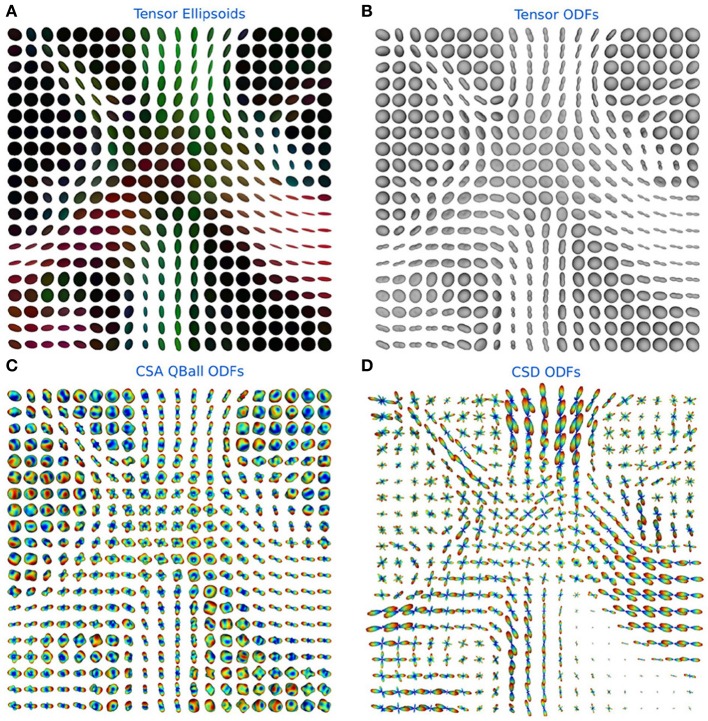
“In practice, the first methods to calculate the PDF were q-space imaging (QSI) (Callaghan et al., 1988) and the most recent diffusion spectrum imaging (DSI) (Wedeen et al., 2005) (see section 6.2).”
MethodInfluentialPieper, S. D., Lorensen, B., Schroeder, W. J., & Kikinis, R. (2006) · IEEE International Symposium on Biomedical Imaging (ISBI) · doi:10.1109/isbi.2006.1625012 · Cited by 293
Orthogonal tensor invariants and the analysis of diffusion tensor magnetic resonance images
Ennis, D. B., & Kindlmann, G. (2006) · Magnetic Resonance in Medicine · doi:10.1002/mrm.20741 · Cited by 276 · cited 1×
“Not only can users compute FA, MD, DEC, but also other statistics that have been proposed in the literature, such as the diffusion linearity, planarity and sphericity (Westin et al., 1997), as well as the tensor mode, tensor norm (Ennis and Kindlmann, 2006), radial and axial diffusivity.”
BackgroundThe Connectome Mapper: An Open-Source Processing Pipeline to Map Connectomes with MRI
Daducci, A., Gerhard, S., Griffa, A., Lemkaddem, A., Cammoun, L., Gigandet, X., Meuli, R. A., Hagmann, P., & Thiran, J. (2012) · PLoS ONE · doi:10.1371/journal.pone.0048121 · Cited by 275
Changes in water diffusion due to Wallerian degeneration in peripheral nerve
Beaulieu, C., Does, M. D., Snyder, R. E., & Allen, P. S. (1996) · Magnetic Resonance in Medicine · doi:10.1002/mrm.1910360419 · Cited by 263
Lenglet, C., Rousson, M., Rachid Deriche, B., & Faugeras, O. D. (2006) · Journal of Mathematical Imaging and Vision · doi:10.1007/s10851-006-6897-z · Cited by 248 · cited 1×
“…meth-ods have been implemented, including non-linear least-squares, ordinary least-squares, and weighted least-squares fitting (Chung et al., 2006) as well as Riemannian modeling-based techniques that assure that the estimated tensor is positive definite (Arsigny et al., 2006; Lenglet et al., 2006).”
MethodKoay, C. G., Chang, L., Carew, J. D., Pierpaoli, C. M., & Basser, P. J. (2006) · Journal of Magnetic Resonance · doi:10.1016/j.jmr.2006.06.020 · Cited by 244 · cited 1×
“The first is that there is still ongoing research on fitting methods for the tensor model (Koay et al., 2006).”
MethodPython: An Ecosystem for Scientific Computing
Pérez, F., Granger, B., & Hunter, J. D. (2010) · Computing in Science & Engineering · doi:10.1109/mcse.2010.119 · Cited by 243
Imagerie de diffusion in-vivo par résonance magnétique nucléaire
Bihan, D. L., & Breton, E. (1985) · HAL · Cited by 238
Descoteaux, M., Angelino, E. L., Fitzgibbons, S., & Rachid Deriche, B. (2006) · Magnetic Resonance in Medicine · doi:10.1002/mrm.20948 · Cited by 235
Self-citationTractometer: Towards validation of tractography pipelines
Côté, M.-A., Girard, G., Boré, A., Garyfallidis, E., Houde, J.-C., & Descoteaux, M. (2013) · Medical Image Analysis · doi:10.1016/j.media.2013.03.009 · Cited by 228 · cited 6×
“It is often useful in dMRI to filter, group or count streamlines based on their interactions with one or several ROIs (Côté et al., 2013).”
BackgroundMethodInfluentialSelf-citationWhite matter pathways in reading
Ben‐Shachar, M., Dougherty, R. F., & Wandell, B. A. (2007) · Current Opinion in Neurobiology · doi:10.1016/j.conb.2007.03.006 · Cited by 198 · cited 1×
“Variance in a variety of behavioral (Ben-Shachar et al., 2007) and clinical (Thomason & Thompson, 2011) measures can be predicted based on these statistics, suggesting that they are reporting on meaningful variability in brain structure.”
BackgroundInternational Society for Magnetic Resonance in Medicine
(1997) · Journal of Cerebral Blood Flow & Metabolism · doi:10.1097/00004647-199701000-00016 · Cited by 181
Assessing Test-Driven Development at IBM
Maximilien, E. M. (2003) · Cited by 178
Multiple q-shell diffusion propagator imaging
Descoteaux, M., Rachid Deriche, B., Bihan, D. L., Mangin, J., & Poupon, C. (2010) · Medical Image Analysis · doi:10.1016/j.media.2010.07.001 · Cited by 177 · cited 1×
“To reduce the acquisition requirements of DSI, several techniques have been proposed to compute the ODF from Equation (5) using only a single b -value dMRI acquisition, often called single-shell high angular resolution diffusion imaging (HARDI) (Tuch et al., 2002; Descoteaux et al., 2011).”
MethodSelf-citationBrainVoyager — Past, present, future
Goebel, R. (2012) · NeuroImage · doi:10.1016/j.neuroimage.2012.01.083 · Cited by 175 · cited 1×
“Many of these tools are written in C/C ++ : 3D Slicer (Pieper et al., 2006), AFNI (Cox, 2012), MITK (Fritzsche et al., 2012), BrainVoyager QX (Goebel, 2012), DTI-Query/Quench (Sherbondy et al., 2005), FreeSurfer (Fischl, 2012), FSL-FDT (Smith et al., 2004), MedInria (Toussaint et al., 2007), MRtrix…”
MethodAn invitation to reproducible computational research
Donoho, D. L. (2010) · Biostatistics · doi:10.1093/biostatistics/kxq028 · Cited by 167 · cited 1×
“In addition, since the code is entirely free and open, Dipy is ideally suited to facilitate reproducible research (Donoho, 2010).”
BackgroundProbabilistic streamline q-ball tractography using the residual bootstrap
Berman, J., Chung, S., Mukherjee, P., Hess, C. P., Han, E. T., & Henry, R. G. (2007) · NeuroImage · doi:10.1016/j.neuroimage.2007.08.021 · Cited by 167 · cited 1×
“For example, residual bootstrap fiber tracking methods fall into this category (Berman et al., 2008).”
BackgroundDiffusion MRI of complex tissue structure
Tuch, D. S. (2002) · Cited by 156
Assessing test-driven development at IBM
Maximilien, E. M., & Williams, L. A. (2003) · Proceedings/Proceedings - International Conference on Software Engineering · doi:10.1109/icse.2003.1201238 · Cited by 138 · cited 1×
“Dipy also follows a test-driven development philosophy, whereby all code should be accompanied by a suite of tests to exercise all corner cases (Maximilien and Williams, 2013).”
BackgroundMedINRIA: Medical Image Navigation and Research Tool by INRIA
Toussaint, N., Souplet, J.-C., & Fillard, P. (2007) · Cited by 131
Comparison of bootstrap approaches for estimation of uncertainties of DTI parameters
Chung, S., Lü, Y., & Henry, R. G. (2006) · NeuroImage · doi:10.1016/j.neuroimage.2006.07.001 · Cited by 131
Biological development of reading circuits
Wandell, B. A., & Yeatman, J. D. (2013) · Current Opinion in Neurobiology · doi:10.1016/j.conb.2012.12.005 · Cited by 128 · cited 1×
“…of the myelin wrapping the axons in the voxel may affect both MD and FA, changes in other factors, such as the distribution of directions of crossing of fiber populations through the voxel may also affect these measures (Basser and Pierpaoli, 1996; Jones et al., 2013; Wandell and Yeatman, 2013).”
BackgroundAccelerated diffusion spectrum imaging in the human brain using compressed sensing
Menzel, M. I., Tan, E. T., Khare, K., Sperl, J. I., King, K. F., Tao, X., Hardy, C. J., & Marinelli, L. (2011) · Magnetic Resonance in Medicine · doi:10.1002/mrm.23064 · Cited by 123 · cited 1×
“DSI needs long acquisition time although recent techniques based on Compressed Sensing ideas (Menzel et al., 2011; Bilgic et al., 2012; Gramfort et al., 2013), and multi-slice imaging (Setsompop et al., 2012) have considerably accelerated the DSI acquisition.”
MethodExploring Connectivity of the Brain's White Matter with Dynamic Queries
Sherbondy, A. J., Akers, D. L., Mackenzie, R., Dougherty, R. F., & Wandell, B. A. (2005) · IEEE Transactions on Visualization and Computer Graphics · doi:10.1109/tvcg.2005.59 · Cited by 116
Probabilistic fibre tracking: Differentiation of connections from chance events
Morris, D. M., Embleton, K. V., & Parker, G. J. M. (2008) · NeuroImage · doi:10.1016/j.neuroimage.2008.06.012 · Cited by 113 · cited 2×
“The uncertainty in fiber direction can be estimated as an fODF, this distribution can be used along with a Monte Carlo sampling to generate streamlines (Morris et al., 2008).”
MethodChang, L., Walker, L., & Pierpaoli, C. M. (2012) · Magnetic Resonance in Medicine · doi:10.1002/mrm.24173 · Cited by 112 · cited 1×
“In addition, there is ongoing research on methods to robustly fit the tensor model, in the face of noisy data (Chang et al., 2005, 2012).”
MethodHalchenko, Y. O., & Hanke, M. (2012) · Frontiers in Neuroinformatics · doi:10.3389/fninf.2012.00022 · Cited by 108
Tristán‐Vega, A., Westin, C., & Aja‐Fernández, S. (2009) · NeuroImage · doi:10.1016/j.neuroimage.2009.04.049 · Cited by 106
McKinney, W. (2012) · Cited by 106
TractoR: Magnetic Resonance Imaging and Tractography withR
Clayden, J. D., Maniega, S. M., Storkey, A., King, M. D., Bastin, M. E., & Clark, C. A. (2011) · Journal of Statistical Software · doi:10.18637/jss.v044.i08 · Cited by 98
Neher, P., Reicht, I., van Bruggen, T., Goch, C. J., Reisert, M., Nolden, M., Zelzer, S., Meinzer, H.-P., Stieltjes, B., & Fritzsche, K. H. (2012) · Methods of Information in Medicine · doi:10.3414/me11-02-0031 · Cited by 83 · cited 2×
“Many of these tools are written in C/C ++ : 3D Slicer (Pieper et al., 2006), AFNI (Cox, 2012), MITK (Fritzsche et al., 2012), BrainVoyager QX (Goebel, 2012), DTI-Query/Quench (Sherbondy et al., 2005), FreeSurfer (Fischl, 2012), FSL-FDT (Smith et al., 2004), MedInria (Toussaint et al., 2007), MRtrix…”
Method18th Annual Meeting of the Organization for Human Brain Mapping
Gorgolewski, K. J., Storkey, A., Bastin, M. E., & Pernet, C. R. (2012) · Cited by 80
High angular resolution diffusion MRI (from local estimation to segmentation and tractography)
Descoteaux, M., & DERICHE, R. (2008) · OpenGrey (Institut de l'Information Scientifique et Technique) · Cited by 70
Self-citationAccelerated diffusion spectrum imaging with compressed sensing using adaptive dictionaries
Bilgiç, B., Setsompop, K., Cohen‐Adad, J., Yendiki, A., Wald, L. L., & Elfar Adalsteinsson, A. (2012) · Magnetic Resonance in Medicine · doi:10.1002/mrm.24505 · Cited by 69 · cited 1×
“DSI needs long acquisition time although recent techniques based on Compressed Sensing ideas (Menzel et al., 2011; Bilgic et al., 2012; Gramfort et al., 2013), and multi-slice imaging (Setsompop et al., 2012) have considerably accelerated the DSI acquisition.”
Method(2012) · Oxford University Press eBooks · doi:10.1093/med/9780195369779.001.0001 · Cited by 65
Does, M. D., & Gore, J. C. (2000) · Magnetic Resonance in Medicine · doi:10.1002/1522-2594(200006)43:6 · Cited by 62
Denoising and fast diffusion imaging with physically constrained sparse dictionary learning
Gramfort, A., Poupon, C., & Descoteaux, M. (2013) · Medical Image Analysis · doi:10.1016/j.media.2013.08.006 · Cited by 41 · cited 1×
“DSI needs long acquisition time although recent techniques based on Compressed Sensing ideas (Menzel et al., 2011; Bilgic et al., 2012; Gramfort et al., 2013), and multi-slice imaging (Setsompop et al.”
MethodSelf-citationDeconvolution in diffusion spectrum imaging
Canales-Rodríguez, E. J., Iturria‐Medina, Y., Alemán‐Gómez, Y., & Melie‐García, L. (2009) · NeuroImage · doi:10.1016/j.neuroimage.2009.11.066 · Cited by 40 · cited 9×
“The main choice to consider is the estimation of the single fiber response function. We assume that R is derived from a prolate tensor, where the single-tensor model is accurate. The eigenvalues of this tensor are estimated from the voxels with FA > 0.7. The input parameter response is a tuple with two parameters: (a)…”
BackgroundMethodInfluentialTractography in the Study of the Human Brain: A Neurosurgical Perspective
Fortin, D., Aubin-Lemay, C., Boré, A., Girard, G., Houde, J., Whittingstall, K., & Descoteaux, M. (2012) · Canadian Journal of Neurological Sciences / Journal Canadien des Sciences Neurologiques · doi:10.1017/s0317167100015560 · Cited by 30 · cited 1×
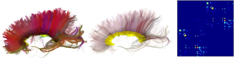
“Figure 7 shows an example of QuickBundles applied on the human brain dataset described in Fortin et al. (2012), using the same parameters shown in the code listing above.”
MethodSelf-citationSeo, Y., Shinar, H., Morita, Y., & Navon, G. (1999) · Magnetic Resonance in Medicine · doi:10.1002/(sici)1522-2594(199909)42:3 · Cited by 19
Diffusion MRI: Overview and clinical applications in neuroradiology
Maas, L. C., & Mukherjee, P. (2005) · Applied Radiology · doi:10.37549/ar1383 · Cited by 18 · cited 1×
“The reduction in extracellular water fraction results in a decrease in MD in the affected regions (Maas and Mukherjee, 2005).”
BackgroundNumano, T., Homma, K., Iwasaki, N., Hyodo, K., Nitta, N., & Hirose, T. (2006) · Magnetic Resonance Imaging · doi:10.1016/j.mri.2005.12.011 · Cited by 17 · cited 1×
“Hence, fiber tracking is a general term that can be used in any field that reconstructs fibrous structures, such as white matter axon fibers, muscle fibers, prostate fibers and even celery fibers (Numano et al., 2006).”
BackgroundOrganization for Human Brain Mapping
(2009) · NeuroImage · doi:10.1016/j.neuroimage.2009.04.012 · Cited by 16
Constrained spherical deconvolution on signal and ODF valuesPreprint
Garyfallidis, E., St‐Jean, S., Paquette, M., Pierrick Coupé, A., & Descoteaux, M. (2013) · doi:10.13140/2.1.1130.4323 · Cited by 1
Self-citationBasser (2000) · doi:10.1002/1522-2594(200010)44:4
Research works printed but not linked: 9
- Chamberland, 2013, Explore the brain white matter networks in real-time: multi-sticks fiber tracking
- Garyfallidis, 2012, Towards an Accurate Brain Tractography
- Garyfallidis, 2011, Dipy - a novel software library for diffusion MR and tractography
- LeBihan, 1985, Imagerie de diffusion in vivo par résonance magnétique nucléaire, C. R. Acad. Sci. Paris, 301, 1109
- Pajevic, 1999, Color schemes to represent the orientation of anisotropic tissues from diffusion tensor data: application to white matter fiber tract mapping in the human brain, Magn. Reson. Med, 42, 526, 10.1002/(SICI)1522-2594(199909)42:3526::AID-MRM153.3.CO;2-A · DOI
- Schroeder, 2001, The Visualization Toolkit-User's Guide
- Vaillancourt, 2011, A fiber navigator for neurosurgical planning
- Wang, 2007, Diffusion toolkit: a software package for diffusion imaging data processing and tractography, Proc. Int. Soc. Magn. Reson. Med, 15, 3720
- Westin, 1997, Geometrical diffusion measures for MRI from tensor basis analysis, Proc. ISMRM, 97, 1742
Cited by
Sorted by citations
MRtrix3: A fast, flexible and open software framework for medical image processing and visualisation
Tournier, J.-D., Smith, R., Raffelt, D., Tabbara, R., Dhollander, T., Pietsch, M., Christiaens, D., Jeurissen, B., Yeh, C.-H., & Connelly, A. (2019) · NeuroImage · doi:10.1016/j.neuroimage.2019.116137 · Cited by 2,974
Tractography-based connectomes are dominated by false-positive connections
Maier-Hein, K. H., Neher, P., Houde, J.-C., Côté, M.-A., Garyfallidis, E., Zhong, J., Chamberland, M., Yeh, F.-C., Lin, Y.-C., Ji, Q., et al. (2016) · doi:10.1101/084137 · Cited by 1,376
“The full script used to run this bundle recognition implementation was based on the DIPY library 78 (www.dipy.org) and is available online (Supplementary Software 1).”
Self-citationHarmonization of multi-site diffusion tensor imaging data
Fortin, J.-P., Parker, D., Tunç, B., Watanabe, T., Elliott, M. A., Ruparel, K., Roalf, D. R., Satterthwaite, T. D., Gur, R. C., Gur, R. E., et al. (2017) · NeuroImage · doi:10.1016/j.neuroimage.2017.08.047 · Cited by 1,261
“Subsequently, the FA and MD maps were calculated from the resultant tensor image using the python package dipy [Garyfallidis et al., 2014].”
MethodDiffusion models in medical imaging: A comprehensive survey
Kazerouni, A., Aghdam, E. K., Heidari, M., Azad, R., Fayyaz, M., Hacihaliloglu, I., & Merhof, D. (2023) · Medical Image Analysis · doi:10.1016/j.media.2023.102846 · Cited by 1,006
“They elaborated their findings on one private and three public datasets (Sherbrooke 3-Shell [140], Stanford HARDI [141], and Parkinson’s Progression Markers Initiative (PPMI) [142]) and reported superior denoising performances.”
BackgroundTractSeg - Fast and accurate white matter tract segmentation
Wasserthal, J., Neher, P., & Maier-Hein, K. H. (2018) · NeuroImage · doi:10.1016/j.neuroimage.2018.07.070 · Cited by 664
Metadata
- Authors
- Garyfallidis, E., Brett, M., Amirbekian, B., Rokem, A., van der Walt, S., Descoteaux, M., Nimmo-Smith, I., & Contributors, D.
- Affiliations
- Université de Sherbrooke (CA)
- University of California, Berkeley (US)
- University of California, San Francisco (US)
- Stanford University (US)
- Corresponding author
- Garyfallidis, Eleftherios
- Venue
- Frontiers in Neuroinformatics
- Publisher
- Frontiers Media SA
- Published
- 2014-02-21
- Volume
- 8
- Pages
- 8-8
- ISSN
- 1662-5196 (ISSN-L)
- PMC
- PMC3931231
- OpenAlex
- W2062791478
- Cited by
- 1,371
- References
- 98
- Citations by year
- 2026: 147 · 2025: 192 · 2024: 177 · 2023: 162 · 2022: 151 · 2021: 147 · 2020: 100 · 2019: 89 · 2018: 54 · 2017: 79 · 2016: 34 · 2015: 26 · 2014: 12
- FWCI
- 39.38
- Type
- journal article
- Language
- en
- Open access
- gold
- doi.org
- Licence
- cc-by
- Locations
- Frontiers in Neuroinformatics (publishedVersion, cc-by)
- Europe PMC (PubMed Central) (publishedVersion, cc-by)
- PubMed Central (publishedVersion, cc-by)
- PubMed (publishedVersion)
- DOAJ (DOAJ: Directory of Open Access Journals) (submittedVersion)
- University of Birmingham Research Portal (University of Birmingham) (submittedVersion, cc-by)
- Funders
- Fonds Québécois de la Recherche sur la Nature et les Technologies
- Natural Sciences and Engineering Research Council of Canada
- National Eye Institute (F32EY022294)
- Topics
- Health Sciences › Medicine › Radiology, Nuclear Medicine and Imaging › Advanced Neuroimaging Techniques and Applications
- Life Sciences › Neuroscience › Cognitive Neuroscience › Functional Brain Connectivity Studies
- Health Sciences › Medicine › Radiology, Nuclear Medicine and Imaging › Advanced MRI Techniques and Applications
- Keywords
- diffusion MRI, DIPY, white matter, fiber tractography, diffusion tensor model, constrained spherical deconvolution, diffusion spectrum imaging, probabilistic tracking, open source software, Python library
About these checks
Registry, retraction, Cites and statistics results are computed, not judged, with one exception: a printed reference that no index lists is read once by a model before it counts as a citation error. Claims are extracted by a model and kept only when their quote appears verbatim in the text; each cited source is then checked, with a model’s help, for whether it says what the claim relies on. Those verdicts can be wrong — read the claim’s quote and the cited source before relying on one.
Statistics and arithmetic are recomputed, not judged.
Confidence bands are shown only for fields calibrated on our annotated test set. None is calibrated yet, so no band is shown.
What counts as a claim ›How we measure confidence ›How we check this ›
Are you an author, or do you see a problem with this page? Request a review
