

The DOI matches its Crossref record; 98 of the 100 references it cites were found in a registry or an index, and 2 are not checked yet.
Dipy, a library for the analysis of diffusion MRI data
Open accessAbstract
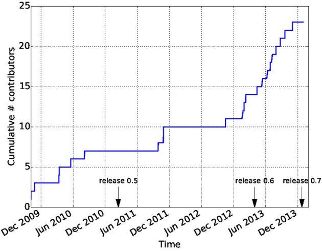
Diffusion Imaging in Python (Dipy) is a free and open source software project for the analysis of data from diffusion magnetic resonance imaging (dMRI) experiments. dMRI is an application of MRI that can be used to measure structural features of brain white matter. Many methods have been developed to use dMRI data to model the local configuration of white matter nerve fiber bundles and infer the trajectory of bundles connecting different parts of the brain. Dipy gathers implementations of many different methods in dMRI, including: diffusion signal pre-processing; reconstruction of diffusion distributions in individual voxels; fiber tractography and fiber track post-processing, analysis and visualization. Dipy aims to provide transparent implementations for all the different steps of dMRI analysis with a uniform programming interface. We have implemented classical signal reconstruction techniques, such as the diffusion tensor model and deterministic fiber tractography. In addition, cutting edge novel reconstruction techniques are implemented, such as constrained spherical deconvolution and diffusion spectrum imaging (DSI) with deconvolution, as well as methods for probabilistic tracking and original methods for tractography clustering. Many additional utility functions are provided to calculate various statistics, informative visualizations, as well as file-handling routines to assist in the development and use of novel techniques. In contrast to many other scientific software projects, Dipy is not being developed by a single research group. Rather, it is an open project that encourages contributions from any scientist/developer through GitHub and open discussions on the project mailing list. Consequently, Dipy today has an international team of contributors, spanning seven different academic institutions in five countries and three continents, which is still growing.
Overview
The claims and statistics of this paper haven’t been checked yet.
Run the check — free, no account needed. It reads the open-access text and checks the sources it cites.
Integrity signals
Registry
Cites
Statistics
Figures
Verification
Claims the paper makes
The claims and statistics of this paper haven’t been checked yet.
Run the check — free, no account needed. It reads the open-access text and checks the sources it cites.
Claims other papers make about this one
Each is checked against this paper’s own text: does it say what the citing paper says it does?
The cited source says what the claim attributes to it.
In the citing paper
“In contrast to projects such as DiPy (Garyfallidis et al., 2014) , this library does not provide functionality to manipulate raw image data within Python itself, but provides functionality to invoke and manage external high-performance executables to perform the actual computations.”
From MRtrix3: A fast, flexible and open software framework for medical image processing and visualisation (2019) · cites this paper as Garyfallidis et al., 2014
Self-check
Percentages, sums and reported differences re-computed from the paper’s own numbers.
The self-check needs the paper’s full text.
Citations
Cites
A Threshold Selection Method from Gray-Level Histograms
Otsu, N. (1979) · IEEE Transactions on Systems, Man and Cybernetics · doi:10.1109/tsmc.1979.4310076 · Cited by 44,708 · cited 1×
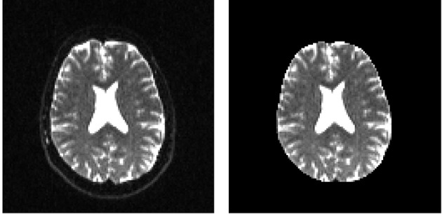
“…median_otsu (see Figure 3 ). from dipy.segment.mask import median_otsu mask, S0_mask = median_otsu(data [:, :, :, 0]) median_otsu uses first a median filter to smooth the S0 and then Otsu’s method, an automated histogram method (Otsu, 1979) to separate the brain (foreground) from its background.”
MethodAdvances in functional and structural MR image analysis and implementation as FSL
Smith, S. M., Jenkinson, M., Woolrich, M. W., Beckmann, C. F., Behrens, T. E. J., Johansen-Berg, H., Bannister, P. R., De Luca, M., Drobnjak, I., Flitney, D. E., et al. (2004) · NeuroImage · doi:10.1016/j.neuroimage.2004.07.051 · Cited by 13,630
Fischl, B. (2012) · NeuroImage · doi:10.1016/j.neuroimage.2012.01.021 · Cited by 9,526 · cited 4×
“…al., 2006), AFNI (Cox, 2012), MITK (Fritzsche et al., 2012), BrainVoyager QX (Goebel, 2012), DTI-Query/Quench (Sherbondy et al., 2005), FreeSurfer (Fischl, 2012), FSL-FDT (Smith et al., 2004), MedInria (Toussaint et al., 2007), MRtrix (Tournier et al., 2012), Diffusion Toolkit/Trackvis (Wang et…”
MethodInfluentialThe NumPy Array: A Structure for Efficient Numerical Computation
van der Walt, S., Colbert, S. C., & Varoquaux, G. (2011) · Computing in Science & Engineering · doi:10.1109/mcse.2011.37 · Cited by 9,381
MR diffusion tensor spectroscopy and imaging
Basser, P. J., Mattiello, J., & LeBihan, D. (1994) · Biophysical Journal · doi:10.1016/s0006-3495(94)80775-1 · Cited by 5,970 · cited 2×
“Another alternative is to make assumptions about the distribution of the PDF such as a Gaussian assumption, which leads to the popular diffusion tensor imaging (DTI) technique (Basser et al., 1994).”
BackgroundMethodThe WU-Minn Human Connectome Project: An overview
Van Essen, D. C., Smith, S. M., Barch, D. M., Behrens, T. E. J., Yacoub, E., & Ugurbil, K. (2013) · NeuroImage · doi:10.1016/j.neuroimage.2013.05.041 · Cited by 5,651
The basis of anisotropic water diffusion in the nervous system—A technical review.
Beaulieu, C. (2002) · NMR in Biomedicine · doi:10.1002/nbm.782 · Cited by 4,628
Basser, P. J., & Pierpaoli, C. (1996) · Journal of Magnetic Resonance, Series B · doi:10.1006/jmrb.1996.0086 · Cited by 4,306 · cited 3×
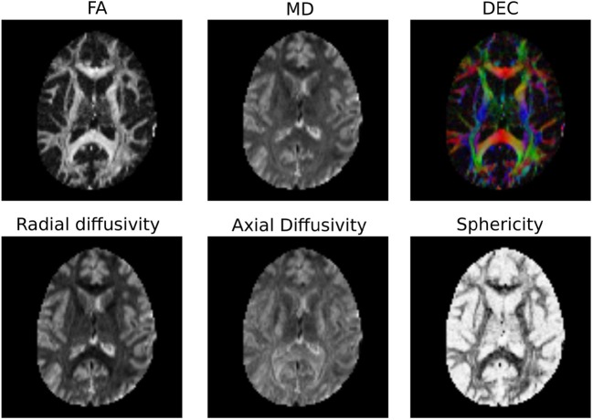
“…of the myelin wrapping the axons in the voxel may affect both MD and FA, changes in other factors, such as the distribution of directions of crossing of fiber populations through the voxel may also affect these measures (Basser and Pierpaoli, 1996; Jones et al., 2013; Wandell and Yeatman, 2013).”
BackgroundMethodMedical image analysis: progress over two decades and the challenges ahead
Duncan, J. S., & Ayache, N. (2000) · IEEE Transactions on Pattern Analysis and Machine Intelligence · doi:10.1109/34.824822 · Cited by 4,167
IPython: A System for Interactive Scientific Computing
Pérez, F., & Granger, B. (2007) · Computing in Science & Engineering · doi:10.1109/mcse.2007.53 · Cited by 4,129 · cited 1×
“There has been large growth in the number of Python users, there are many Python tools for scientific computing (Pérez and Granger, 2007; Pérez et al., 2011; McKinney, 2012), and there are complementary neuroimaging packages in Python1.”
Background
Research works printed but not linked: 9
- Chamberland, 2013, Explore the brain white matter networks in real-time: multi-sticks fiber tracking
- Garyfallidis, 2012, Towards an Accurate Brain Tractography
- Garyfallidis, 2011, Dipy - a novel software library for diffusion MR and tractography
- LeBihan, 1985, Imagerie de diffusion in vivo par résonance magnétique nucléaire, C. R. Acad. Sci. Paris, 301, 1109
- Pajevic, 1999, Color schemes to represent the orientation of anisotropic tissues from diffusion tensor data: application to white matter fiber tract mapping in the human brain, Magn. Reson. Med, 42, 526, 10.1002/(SICI)1522-2594(199909)42:3526::AID-MRM153.3.CO;2-A · DOI
- Schroeder, 2001, The Visualization Toolkit-User's Guide
- Vaillancourt, 2011, A fiber navigator for neurosurgical planning
- Wang, 2007, Diffusion toolkit: a software package for diffusion imaging data processing and tractography, Proc. Int. Soc. Magn. Reson. Med, 15, 3720
- Westin, 1997, Geometrical diffusion measures for MRI from tensor basis analysis, Proc. ISMRM, 97, 1742
Cited by
Sorted by citations
MRtrix3: A fast, flexible and open software framework for medical image processing and visualisation
Tournier, J.-D., Smith, R., Raffelt, D., Tabbara, R., Dhollander, T., Pietsch, M., Christiaens, D., Jeurissen, B., Yeh, C.-H., & Connelly, A. (2019) · NeuroImage · doi:10.1016/j.neuroimage.2019.116137 · Cited by 2,974
Tractography-based connectomes are dominated by false-positive connections
Maier-Hein, K. H., Neher, P., Houde, J.-C., Côté, M.-A., Garyfallidis, E., Zhong, J., Chamberland, M., Yeh, F.-C., Lin, Y.-C., Ji, Q., et al. (2016) · doi:10.1101/084137 · Cited by 1,376
“The full script used to run this bundle recognition implementation was based on the DIPY library 78 (www.dipy.org) and is available online (Supplementary Software 1).”
Self-citationHarmonization of multi-site diffusion tensor imaging data
Fortin, J.-P., Parker, D., Tunç, B., Watanabe, T., Elliott, M. A., Ruparel, K., Roalf, D. R., Satterthwaite, T. D., Gur, R. C., Gur, R. E., et al. (2017) · NeuroImage · doi:10.1016/j.neuroimage.2017.08.047 · Cited by 1,261
“Subsequently, the FA and MD maps were calculated from the resultant tensor image using the python package dipy [Garyfallidis et al., 2014].”
MethodDiffusion models in medical imaging: A comprehensive survey
Kazerouni, A., Aghdam, E. K., Heidari, M., Azad, R., Fayyaz, M., Hacihaliloglu, I., & Merhof, D. (2023) · Medical Image Analysis · doi:10.1016/j.media.2023.102846 · Cited by 1,006
“They elaborated their findings on one private and three public datasets (Sherbrooke 3-Shell [140], Stanford HARDI [141], and Parkinson’s Progression Markers Initiative (PPMI) [142]) and reported superior denoising performances.”
BackgroundTractSeg - Fast and accurate white matter tract segmentation
Wasserthal, J., Neher, P., & Maier-Hein, K. H. (2018) · NeuroImage · doi:10.1016/j.neuroimage.2018.07.070 · Cited by 664
A massive 7T fMRI dataset to bridge cognitive neuroscience and artificial intelligence
Allen, E., St-Yves, G., Wu, Y., Breedlove, J. L., Prince, J. S., Dowdle, L. T., Nau, M., Caron, B. A., Pestilli, F., Charest, I., et al. (2021) · Nature Neuroscience · doi:10.1038/s41593-021-00962-x · Cited by 679
Kamagata, K., Andica, C., Takabayashi, K., Saito, Y., Taoka, T., Nozaki, H., Kikuta, J., Fujita, S., Hagiwara, A., Kamiya, K., et al. (2022) · Neurology · doi:10.1212/wnl.0000000000201300 · Cited by 397
“org/).(28) Mean cerebral FW-WM was extracted using the T1w imaging–based WM mask calculated at the MRI data processing stage (eFigure 1 in the supplement, links.”
MethodTowards quantitative connectivity analysis: reducing tractography biases
Girard, G., Whittingstall, K., Deriche, R., & Descoteaux, M. (2014) · NeuroImage · doi:10.1016/j.neuroimage.2014.04.074 · Cited by 324
Self-citationQSIPrep: an integrative platform for preprocessing and reconstructing diffusion MRI data
Cieslak, M., Cook, P. A., He, X., Yeh, F.-C. F., Dhollander, T., Adebimpe, A., Aguirre, G. K., Bassett, D. S., Betzel, R. F., Bourque, J., et al. (2021) · Nature Methods · doi:10.1038/s41592-021-01185-5 · Cited by 337
Self-citationGaryfallidis, E., Côté, M.-A., Rheault, F., Sidhu, J., Hau, J., Petit, L., Fortin, D., Cunanne, S., & Descoteaux, M. (2018) · NeuroImage · doi:10.1016/j.neuroimage.2017.07.015 · Cited by 279
Self-citationZhang, F., Daducci, A., He, Y., Schiavi, S., Seguin, C., Smith, R. E., Yeh, C.-H., Zhao, T., & O’Donnell, L. J. (2022) · NeuroImage · doi:10.1016/j.neuroimage.2021.118870 · Cited by 302
“Many software packages are available to perform tractography research, including ANIMA [116], BrainSUITE [395], Camino [95], COMMIT [105, 106, 375, 312], Diffusion toolkit [480], Dipy [157], DMIPY [139], DSI studio [519], DTIstudio [226], ExploreDTI [257], FiberNavigator [72], FSL [221], MITK [493], MRtrix3 [453, 456]…”
MethodSpecialized astrocytes mediate glutamatergic gliotransmission in the CNS
Ceglia, R. D., Ledonne, A., Litvin, D. G., Lind, B. L., Carriero, G., Latagliata, E. C., Bindocci, E., Castro, M. A. D., Savtchouk, I., Vitali, I., et al. (2023) · Nature · doi:10.1038/s41586-023-06502-w · Cited by 297
Semi-supervised Brain Lesion Segmentation with an Adapted Mean Teacher Model
Cui, W., Liu, Y., Li, Y., Guo, M.-H., Li, Y., Li, X., Wang, T., Zeng, X., & Ye, C. (2019) · Lecture notes in computer science · doi:10.1007/978-3-030-20351-1_43 · Cited by 219
“Specifically, a brain mask was extracted with the Dipy software [3].”
MethodWhite-matter integrity on DTI and the pathologic staging of Alzheimer’s disease
Kantarci, K., Murray, M. E., Schwarz, C. G., Reid, R. I., Przybelski, S. A., Lesnick, T., Zuk, S. M., Raman, M. R., Senjem, M. L., Gunter, J. L., et al. (2017) · Neurobiology of Aging · doi:10.1016/j.neurobiolaging.2017.04.024 · Cited by 205
Functional connectomics spanning multiple areas of mouse visual cortex
The MICrONS Consortium, Bae, J. A., Baptiste, M., Bishop, C. A., Bodor, A. L., Brittain, D., Buchanan, J., Bumbarger, D. J., Castro, M. A., Celii, B., et al. (2021) · doi:10.1101/2021.07.28.454025 · Cited by 214
Theaud, G., Houde, J., Boré, A., Rheault, F., Morency, F. C., & Descoteaux, M. (2020) · NeuroImage · doi:10.1016/j.neuroimage.2020.116889 · Cited by 200
Self-citationSchilling, K. G., Rheault, F., Petit, L., Hansen, C. B., Nath, V., Yeh, F.-C., Girard, G., Barakovic, M., Rafael-Patino, J., Yu, T., et al. (2021) · NeuroImage · doi:10.1016/j.neuroimage.2021.118502 · Cited by 182
Self-citationLocal structure-function relationships in human brain networks across the lifespan
Esfahlani, F. Z., Faskowitz, J., Slack, J., Mišić, B., & Betzel, R. F. (2022) · Nature Communications · doi:10.1038/s41467-022-29770-y · Cited by 178
Combined tract segmentation and orientation mapping for bundle-specific tractography
Wasserthal, J., Neher, P., Hirjak, D., & Maier-Hein, K. H. (2019) · Medical Image Analysis · doi:10.1016/j.media.2019.101559 · Cited by 167
“Firstly, we registered all subjects to a random subject using symmetric diffeomorphic registration implemented in DIPY [3, 8].”
MethodProsodic and phonetic subtypes of primary progressive apraxia of speech
Utianski, R. L., Duffy, J. R., Clark, H. M., Strand, E. A., Botha, H., Schwarz, C. G., Machulda, M. M., Senjem, M. L., Spychalla, A. J., Jack, C. R., et al. (2018) · Brain and Language · doi:10.1016/j.bandl.2018.06.004 · Cited by 168
“Weighted linear least squares optimization was used to fit diffusion tensors and fractional anisotropy (FA) images were calculated from the tensor eigenvalues using Dipy (Garyfallidis et al., 2014).”
Rapid and widespread white matter plasticity during an intensive reading intervention
Huber, E., Donnelly, P. M., Rokem, A., & Yeatman, J. D. (2018) · Nature Communications · doi:10.1038/s41467-018-04627-5 · Cited by 164
“3) and including subject motion as a covariate in the model did not change the results: MD decreased as a function of intervention hours within the left AF (F(1,76)= 10.”
BackgroundMethodInfluentialSelf-citationIntrinsic connectivity reveals functionally distinct cortico-hippocampal networks in the human brain
Barnett, A. J., Reilly, W., Dimsdale-Zucker, H. R., Mızrak, E., Reagh, Z. M., & Ranganath, C. (2021) · PLoS Biology · doi:10.1371/journal.pbio.3001275 · Cited by 172
Cousineau, M., Jodoin, P.-M., Garyfallidis, E., Côté, M.-A., Morency, F. C., Rozanski, V. E., Grand'maison, M., Bedell, B. J., & Descoteaux, M. (2017) · NeuroImage: Clinical · doi:10.1016/j.nicl.2017.07.020 · Cited by 158
“DTI/HARDI metrics are then computed using the Dipy python package (Garyfallidis et al., 2014).”
MethodSelf-citationSepehrband, F., Clark, K. A., Ullmann, J. F. P., Kurniawan, N. D., Leanage, G., Reutens, D. C., & Yang, Z. (2015) · Human Brain Mapping · doi:10.1002/hbm.22872 · Cited by 143
Dean, D. C., Planalp, E. M., Wooten, W., Kecskemeti, S. R., Adluru, N., Schmidt, C. K., Frye, C., Birn, R. M., Burghy, C. A., Schmidt, N. L., et al. (2018) · JAMA Pediatrics · doi:10.1001/jamapediatrics.2018.2132 · Cited by 138
Microstructure informed tractography: pitfalls and open challenges
Daducci, A., Dal Palú, A., Descoteaux, M., & Thiran, J.-P. (2016) · Frontiers in Neuroscience · doi:10.3389/fnins.2016.00247 · Cited by 138
“In particular, we first illustrate some well-known issues of local methods with the help of a simple synthetic example, for which the expected behavior is easily explained; then we exploit the parallel between local and global reconstruction to extrapolate these observations to global approaches.”
MethodSelf-citationAutomated synapse-level reconstruction of neural circuits in the larval zebrafish brain
Svara, F., Förster, D., Kubo, F., Januszewski, M., dal Maschio, M., Schubert, P. J., Kornfeld, J., Wanner, A. A., Laurell, E., Denk, W., et al. (2022) · Nature Methods · doi:10.1038/s41592-022-01621-0 · Cited by 124
Clinica: An Open-Source Software Platform for Reproducible Clinical Neuroscience Studies
Routier, A., Burgos, N., Díaz, M., Bacci, M., Bottani, S., El-Rifai, O., Fontanella, S., Gori, P., Guillon, J., Guyot, A., et al. (2021) · Frontiers in Neuroinformatics · doi:10.3389/fninf.2021.689675 · Cited by 152
“, 2005), Dipy (Garyfallidis et al., 2014), DTI-TK25, MRtrix (Tournier et al.”
Dynamic PET Image Denoising Using Deep Convolutional Neural Networks Without Prior Training Datasets
Hashimoto, F., Ohba, H., Ote, K., Teramoto, A., & Tsukada, H. (2019) · IEEE Access · doi:10.1109/access.2019.2929230 · Cited by 127
“Compared with the 3D NLM filter [29], [30] and IGF [17], our proposed method exhibited superior performance visually and quantitatively when applied to both a computer simulation and a real data obtained from a living monkey brain using 18F-fluoro-2-deoxy-D-glucose (18F-FDG).”
MethodCortical Microstructural Alterations in Mild Cognitive Impairment and Alzheimer’s Disease Dementia
Vogt, N. M., Hunt, J. F. V., Adluru, N., Dean, D., Johnson, S. C., Asthana, S., Yu, J. J., Alexander, A. L., & Bendlin, B. B. (2019) · Cerebral Cortex · doi:10.1093/cercor/bhz286 · Cited by 120
“NDI, ODI, and VISO parameter maps were generated by fitting the NODDI model in Python using Accelerated Microstructure Imaging via Convex Optimization, which improves processing speed by approximating the NODDI model as a linear system (Daducci et al. 2015).”
MethodChandio, B. Q., Risacher, S. L., Pestilli, F., Bullock, D. N., Yeh, F.-C. F., Koudoro, S., Rokem, A., Harezlak, J., & Garyfallidis, E. (2020) · Scientific Reports · doi:10.1038/s41598-020-74054-4 · Cited by 122
“BUAN is available with DIPY 44 at dipy.org.”
MethodSelf-citationWandell, B. A. (2016) · Annual Review of Neuroscience · doi:10.1146/annurev-neuro-070815-013815 · Cited by 117
“…the number and direction of the sticks and the size of the ball, although there have been many different implementations (Behrens et al. 2007, Cook et al. 2006, Daducci et al. 2015, Ferizi et al. 2015, Garyfallidis et al. 2014, Lemkaddem et al. 2014, Rokem et al. 2015, Tournier et al. 2004).”
The sensitivity of diffusion MRI to microstructural properties and experimental factors
Afzali, M., Pieciak, T., Newman, S., Garyfallidis, E., Özarslan, E., Cheng, H., & Jones, D. K. (2021) · Journal of Neuroscience Methods · doi:10.1016/j.jneumeth.2020.108951 · Cited by 117
“, 2019b,a) which is part of Diffusion Imaging In Python (DIPY) library (Garyfallidis et al., 2014).”
BackgroundSelf-citationTax, C. M. W., Grussu, F., Kaden, E., Ning, L., Rudrapatna, U. S., Evans, J., St‐Jean, S., Leemans, A., Koppers, S., Merhof, D., et al. (2019) · NeuroImage · doi:10.1016/j.neuroimage.2019.01.077 · Cited by 116
What’s new and what’s next in diffusion MRI preprocessing
Tax, C. M. W., Bastiani, M., Veraart, J., Garyfallidis, E., & Irfanoglu, M. O. (2021) · NeuroImage · doi:10.1016/j.neuroimage.2021.118830 · Cited by 121
Self-citationRemapping the cognitive and neural profiles of children who struggle at school
Astle, D. E., Bathelt, J., the CALM team, & Holmes, J. (2018) · Developmental Science · doi:10.1111/desc.12747 · Cited by 114
Fick, R., Wassermann, D., Caruyer, E., & Rachid Deriche, B. (2016) · NeuroImage · doi:10.1016/j.neuroimage.2016.03.046 · Cited by 114
“We plan to release our implementation of Laplacian-Regularized MAP-MRI in the open-source dipy framework (Garyfallidis et al., 2014) and make it publicly available.”
MethodInfluentialCommon misconceptions, hidden biases and modern challenges of dMRI tractography
Rheault, F., Poulin, P., Caron, A. V., St‐Onge, E., & Descoteaux, M. (2020) · Journal of Neural Engineering · doi:10.1088/1741-2552/ab6aad · Cited by 112
“Recobundles (streamline shape-based) (Garyfallidis et al 2017) This tool from the Dipy library aims to segment bundles based on their shape similarity to model bundles, part of a dictionary (e.g. build from expert dissections).”
MethodSelf-citationNeural dynamics and architecture of the heading direction circuit in zebrafish
Petrucco, L., Lavian, H., Wu, Y. K., Svara, F., Štih, V., & Portugues, R. (2023) · Nature Neuroscience · doi:10.1038/s41593-023-01308-5 · Cited by 114
Unique Mapping of Structural and Functional Connectivity on Cognition
Zimmermann, J., Griffiths, J. D., & McIntosh, A. R. (2018) · Journal of Neuroscience · doi:10.1523/jneurosci.0900-18.2018 · Cited by 114
Surface-enhanced tractography (SET)
St‐Onge, E., Daducci, A., Girard, G., & Descoteaux, M. (2017) · NeuroImage · doi:10.1016/j.neuroimage.2017.12.036 · Cited by 109
Self-citationLiska, A., Bertero, A., Gomolka, R. S., Sabbioni, M., Galbusera, A., Barsotti, N., Panzeri, S., Scattoni, M. L., Pasqualetti, M., & Gozzi, A. (2017) · Cerebral Cortex · doi:10.1093/cercor/bhx022 · Cited by 106
Deep learning-based detection and segmentation of diffusion abnormalities in acute ischemic stroke
Liu, C.-F., Hsu, J., Xu, X., Ramachandran, S., Wang, V., Miller, M. I., Hillis, A. E., Faria, A. V., The STIR and VISTA Imaging investigators, Wintermark, M., et al. (2021) · Communications Medicine · doi:10.1038/s43856-021-00062-8 · Cited by 103
Mindfulness-Based Stress Reduction-related changes in posterior cingulate resting brain connectivity
Kral, T. R. A., Imhoff‐Smith, T., Dean, D., Grupe, D. W., Adluru, N., Patsenko, E. G., Mumford, J. A., Goldman, R. I., Rosenkranz, M. A., & Davidson, R. J. (2019) · Social Cognitive and Affective Neuroscience · doi:10.1093/scan/nsz050 · Cited by 103
Radwan, A., Sunaert, S. G., Schilling, K. G., Descoteaux, M., Landman, B. A., Vandenbulcke, M., Theys, T., Dupont, P. J., & Emsell, L. (2022) · NeuroImage · doi:10.1016/j.neuroimage.2022.119029 · Cited by 106
Self-citationTransdiagnostic Brain Mapping in Developmental Disorders
Šiugždaitė, R., Bathelt, J., Holmes, J., & Astle, D. E. (2020) · Current Biology · doi:10.1016/j.cub.2020.01.078 · Cited by 100
“11 package [86] to boost signal-to-noise ratio.”
Evaluating the Reliability of Human Brain White Matter Tractometry
Kruper, J., Yeatman, J. D., Richie-Halford, A., Bloom, D., Grotheer, M., Caffarra, S., Kiar, G., Karipidis, I. I., Roy, E. A., Chandio, B. Q., et al. (2021) · Aperture Neuro · doi:10.52294/e6198273-b8e3-4b63-babb-6e6b0da10669 · Cited by 92
Self-citationWeighted Stochastic Block Models of the Human Connectome across the Life Span
Faskowitz, J., Yan, X., Zuo, X., & Sporns, O. (2018) · Scientific Reports · doi:10.1038/s41598-018-31202-1 · Cited by 95
“We generated streamline tractography in the MNI 152 2 mm isotropic space using Dipy 49 .”
MethodMaffei, C., Lee, C., Planich, M., Ramprasad, M., Ravi, N., Trainor, D., Urban, Z., Kim, M., Jones, R. J., Henin, A., et al. (2021) · NeuroImage · doi:10.1016/j.neuroimage.2021.118706 · Cited by 87
Geometric renormalization unravels self-similarity of the multiscale human connectome
Zheng, M., Allard, A., Hagmann, P., Alemán‐Gómez, Y., & Serrano, M. Á. (2020) · Proceedings of the National Academy of Sciences · doi:10.1073/pnas.1922248117 · Cited by 89
Dimensionality reduction of diffusion MRI measures for improved tractometry of the human brain
Chamberland, M., Raven, E. P., Genc, S., Duffy, K., Descoteaux, M., Parker, G. D., Tax, C. M. W., & Jones, D. K. (2019) · NeuroImage · doi:10.1016/j.neuroimage.2019.06.020 · Cited by 94
“Next, along-tract profiling was performed for each bundle using the Python toolbox developed by Cousineau et al. (2017), combined with DIPY functions (Garyfallidis et al., 2014).”
MethodSelf-citationMapping the human subcortical auditory system using histology, postmortem MRI and in vivo MRI at 7T
Sitek, K. R., Gülban, Ö. F., Calabrese, E., Johnson, G. A., Lage‐Castellanos, A., Moerel, M., Ghosh, S., & Martino, F. D. (2019) · eLife · doi:10.7554/elife.48932 · Cited by 88
Golub, M., Henriques, R. N., & Nunes, R. G. (2020) · Magnetic Resonance in Medicine · doi:10.1002/mrm.28599 · Cited by 90
“For reproducibility, all tested FWEDTI fitting procedures were incorporated into the open-source library diffusion in python (DIPY) (32).”
MethodInfluentialThe Dmipy Toolbox: Diffusion MRI Multi-Compartment Modeling and Microstructure Recovery Made Easy
Fick, R., Wassermann, D., & Rachid Deriche, B. (2019) · Frontiers in Neuroinformatics · doi:10.3389/fninf.2019.00064 · Cited by 97
Schilling, K. G., Petit, L., Rheault, F., Remedios, S. W., Pierpaoli, C. M., Anderson, A. W., Landman, B. A., & Descoteaux, M. (2020) · Brain Structure and Function · doi:10.1007/s00429-020-02129-z · Cited by 96
Self-citationEvaluating the Accuracy of Diffusion MRI Models in White Matter
Rokem, A., Yeatman, J. D., Pestilli, F., Kay, K. N., Mezer, A., van der Walt, S., & Wandell, B. (2015) · PLoS ONE · doi:10.1371/journal.pone.0123272 · Cited by 91
“1 mm(2)/s) and uniformity of the b = 0 signal across the population of voxels in the estimated position of the CC [39,43].”
BackgroundSelf-citationAndica, C., Kamagata, K., Takabayashi, K., Kikuta, J., Kaga, H., Someya, Y., Tamura, Y., Kawamori, R., Watada, H., Taoka, T., et al. (2023) · Neurobiology of Disease · doi:10.1016/j.nbd.2023.105990 · Cited by 94
Diffusion models reveal white matter microstructural changes with ageing, pathology and cognition
Raghavan, S., Reid, R. I., Przybelski, S. A., Lesnick, T. G., Graff-Radford, J., Schwarz, C. G., Knopman, D. S., Mielke, M. M., Machulda, M. M., Petersen, R. C., et al. (2021) · Brain Communications · doi:10.1093/braincomms/fcab106 · Cited by 79
Girard, G., Caminiti, R., Battaglia-Mayer, A., St-Onge, E., Ambrosen, K. S., Eskildsen, S. F., Krug, K., Dyrby, T. B., Descoteaux, M., Thiran, J.-P., et al. (2020) · NeuroImage · doi:10.1016/j.neuroimage.2020.117201 · Cited by 83
Self-citationThe modular organization of brain cortical connectivity across the human lifespan
Puxeddu, M. G., Faskowitz, J., Betzel, R. F., Petti, M., Astolfi, L., & Sporns, O. (2020) · NeuroImage · doi:10.1016/j.neuroimage.2020.116974 · Cited by 87
Ning, L., Laun, F. B., Gur, Y., DiBella, E., Deslauriers‐Gauthier, S., Megherbi, T., Ghosh, A., Zucchelli, M., Menegaz, G., Fick, R., et al. (2015) · Medical Image Analysis · doi:10.1016/j.media.2015.10.012 · Cited by 84
“This implementation is available as part of the Dipy (Diffusion Image in Python) package introduced in (Garyfallidis et al., 2014).”
MethodSelf-citationRobust and efficient linear registration of white-matter fascicles in the space of streamlines
Garyfallidis, E., Ocegueda, O., Wassermann, D., & Descoteaux, M. (2015) · NeuroImage · doi:10.1016/j.neuroimage.2015.05.016 · Cited by 84
“Diffusion tensor estimation and corresponding FA were estimated using DIPY4 (Garyfallidis et al., 2014).”
MethodSelf-citationBundle-specific tractography with incorporated anatomical and orientational priors
Rheault, F., St‐Onge, E., Sidhu, J., Maier-Hein, K. H., Tzourio‐Mazoyer, N., Petit, L., & Descoteaux, M. (2018) · NeuroImage · doi:10.1016/j.neuroimage.2018.11.018 · Cited by 92
“Other forms of tracking (Dipy (Garyfallidis et al., 2014), FSL (Behrens et al.”
Self-citationReal-time multi-peak tractography for instantaneous connectivity display
Chamberland, M., Whittingstall, K., Fortin, D., Mathieu, D., & Descoteaux, M. (2014) · Frontiers in Neuroinformatics · doi:10.3389/fninf.2014.00059 · Cited by 84
“The tractography algorithm will soon be released in Dipy (Garyfallidis et al., 2014) and is currently open-source in the FiberNavigator (www.github.com/scilus/ fibernavigator).”
BackgroundSelf-citationThe visual white matter: The application of diffusion MRI and fiber tractography to vision science
Rokem, A., Takemura, H., Bock, A. S., Scherf, K. S., Behrmann, M., Wandell, B. A., Fine, I., Bridge, H., & Pestilli, F. (2017) · Journal of Vision · doi:10.1167/17.2.4 · Cited by 92
Self-citationStaffaroni, A. M., Cobigo, Y., Elahi, F. M., Casaletto, K. B., Walters, S. M., Wolf, A. A., Lindbergh, C. A., Rosen, H. J., & Kramer, J. H. (2019) · Human Brain Mapping · doi:10.1002/hbm.24613 · Cited by 85
Gibbons, E. K., Hodgson, K. K., Chaudhari, A. S., Richards, L. G., Majersik, J. J., Adluru, G., & DiBella, E. (2018) · Magnetic Resonance in Medicine · doi:10.1002/mrm.27568 · Cited by 83
“44 Similarly, a “naive” reconstruction used these traditional methods to generate NODDI and GFA images from 24 direction undersampled q‐space data.”
MethodBarbeau, É. B., Descoteaux, M., & Petrides, M. (2020) · Scientific Reports · doi:10.1038/s41598-020-64124-y · Cited by 82
Self-citationqMRLab: Quantitative MRI analysis, under one umbrella
Karakuzu, A., Boudreau, M., Duval, T., Boshkovski, T., Leppert, I. R., Cabana, J., Gagnon, I., Béliveau, P., Pike, G. B., Cohen‐Adad, J., et al. (2020) · The Journal of Open Source Software · doi:10.21105/joss.02343 · Cited by 82
“…models in Python (Fick, Wassermann, & Deriche, 2019), another Python package DIPY brought together dMRI processing methods at multiple levels (Garyfallidis et al., 2014), Camino was developed in Java to provide users with yet another dMRI pipeline (Cook et al., 2006) and TractoFlow (Theaud…”
MethodAvesani, P., McPherson, B., Hayashi, S., Caiafa, C. F., Henschel, R., Garyfallidis, E., Kitchell, L. M., Bullock, D. N., Patterson, A. J., Olivetti, E., et al. (2019) · Scientific Data · doi:10.1038/s41597-019-0073-y · Cited by 81
“The brainlife.io Apps used below follow a generalized and light-weight specification as to allow usage with diverse combinations of software from multiple libraries, such as FSL 71 , FreeSurfer 72 , DIPY 73 , Nipype 74 , LiFE 22,40 , AFQ 75 , MRtrix 76 , and AFNI 77 .”
MethodSelf-citationNing, L., Bonet-Carné, E., Grussu, F., Sepehrband, F., Kaden, E., Veraart, J., Blumberg, S. B., Khoo, C. S., Palombo, M., Kokkinos, I., et al. (2020) · NeuroImage · doi:10.1016/j.neuroimage.2020.117128 · Cited by 77
SDnDTI: Self-supervised deep learning-based denoising for diffusion tensor MRI
Tian, Q., Li, Z., Fan, Q., Polimeni, J. R., Bilgiç, B., Salat, D. H., & Huang, S. Y. (2022) · NeuroImage · doi:10.1016/j.neuroimage.2022.119033 · Cited by 92
Lin, S., Lin, X., Chen, S., Liang, Q., Li, Y., Wei, F., Wu, X., Qian, L., Li, S., & Qiu, Y. (2024) · Radiology · doi:10.1148/radiol.232274 · Cited by 83
Casaletto, K. B., Rentería, M. A., Pa, J., Tom, S. E., Harrati, A., Armstrong, N. M., Rajan, K. B., Mungas, D., Walters, S., Kramer, J., et al. (2020) · Journal of Alzheimer's Disease · doi:10.3233/jad-191114 · Cited by 78
Leveraging multi-shell diffusion for studies of brain development in youth and young adulthood
Pines, A. R., Cieslak, M., Larsen, B., Baum, G. L., Cook, P. A., Adebimpe, A., Dávila, D. G., Elliott, M. A., Jirsaraie, R. J., Murtha, K. A., et al. (2020) · Developmental Cognitive Neuroscience · doi:10.1016/j.dcn.2020.100788 · Cited by 83
“We conducted model fitting and generated RTOP, RTAP, and RTPP with dipy, an open-source diffusion imaging toolbox in Python (Fick et al., 2016a; Garyfallidis et al., 2014).”
MethodLigneul, C., Palombo, M., Hernández-Garzón, E., Carrillo-de Sauvage, M.-A., Flament, J., Hantraye, P., Brouillet, E., Bonvento, G., Escartin, C., & Valette, J. (2019) · NeuroImage · doi:10.1016/j.neuroimage.2019.02.046 · Cited by 77
brainlife.io: a decentralized and open-source cloud platform to support neuroscience research
Hayashi, S., Caron, B. A., Heinsfeld, A. S., Vinci‐Booher, S., McPherson, B., Bullock, D. N., Bertò, G., Niso, G., Hanekamp, S., Levitas, D. J., et al. (2024) · Nature Methods · doi:10.1038/s41592-024-02237-2 · Cited by 84
Self-citationDSI Studio: an integrated tractography platform and fiber data hub for accelerating brain research
Yeh, F.-C. F. (2025) · Nature Methods · doi:10.1038/s41592-025-02762-8 · Cited by 77
Menon, V., Gallardo, G., Pinsk, M. A., Nguyen, V.-D., Li, J., Cai, W., & Wassermann, D. (2020) · eLife · doi:10.7554/elife.53470 · Cited by 79
ManifoldNet: A Deep Neural Network for Manifold-Valued Data With Applications
Chakraborty, R., Bouza, J., Manton, J. H., & Vemuri, B. C. (2020) · IEEE Transactions on Pattern Analysis and Machine Intelligence · doi:10.1109/tpami.2020.3003846 · Cited by 83
“• ODF Representation: In this case we fit orientation distribution functions to diffusion data within each voxel using the DiPy implementation of DSI with deconvolution [29].”
MethodCohen‐Adad, J., Alonso‐Ortiz, E., Abramovic, M., Arneitz, C., Atcheson, N., Barlow, L. R., Barry, R. L., Barth, M., Battiston, M., Büchel, C., et al. (2021) · Scientific Data · doi:10.1038/s41597-021-00941-8 · Cited by 76
Self-citationWoolgar, A., Afshar, S., Williams, M., & Rich, A. N. (2015) · Journal of Cognitive Neuroscience · doi:10.1162/jocn_a_00827 · Cited by 81
St-Jean, S., Coupé, P., & Descoteaux, M. (2016) · Medical Image Analysis · doi:10.1016/j.media.2016.02.010 · Cited by 74
Self-citationGonzález-Tendero, A., Zhang, C., Baličević, V., Cárdenes, R., Lončarić, S., Butakoff, C., Paun, B., Bonnin, A., García‐Canadilla, P., Muñoz‐Moreno, E., et al. (2016) · European Heart Journal - Cardiovascular Imaging · doi:10.1093/ehjci/jew314 · Cited by 73
“2, using Dipy.(26) From the DTI, eigenvalues and eigenvectors of the diffusion tensor were computed, and the main eigenvector, describing the main fibre direction, was considered to obtain the helix angle, computed as p=2 cos (1)ð~e1 ~laxisÞ, where~laxis is the unitary vector in the direction of the long axis of the l…”
MethodInfluentialA generative network model of neurodevelopmental diversity in structural brain organization
Akarca, D., Vértes, P. E., Bullmore, E. T., the CALM team, Baker, K., Gathercole, S. E., Holmes, J., Kievit, R., Manly, T., Bathelt, J., et al. (2021) · Nature Communications · doi:10.1038/s41467-021-24430-z · Cited by 81
Long-Term Abnormalities in the Corpus Callosum of Female Concussed Athletes
Chamard, É., Lefebvre, G., Lassonde, M. C., & Théoret, H. (2015) · Journal of Neurotrauma · doi:10.1089/neu.2015.3948 · Cited by 77
“38 From this, the single fiber response function was estimated from all FA values above a threshold of 0.7.”
InfluentialPatch2Self: Denoising Diffusion MRI with Self-Supervised LearningPreprint
Fadnavis, S., Batson, J., & Garyfallidis, E. (2020) · arXiv (Cornell University) · doi:10.48550/arxiv.2011.01355 · Cited by 72
“…of Patch2Self with Marchenko-Pastur on the Parkinson’s Progression Markers Initiative (PPMI) [32], Stanford HARDI [41] and Sherbrooke 3-Shell [16] datasets as shown For each of the datasets, we show the axial slice of a randomly chosen 3D volume and the corresponding residuals (squared…”
MethodSelf-citationData-Driven Subtyping of Executive Function–Related Behavioral Problems in Children
Bathelt, J., Holmes, J., Astle, D. E., Holmes, J., Gathercole, S., Astle, D., Manly, T., & Kievit, R. (2018) · Journal of the American Academy of Child & Adolescent Psychiatry · doi:10.1016/j.jaac.2018.01.014 · Cited by 71
Canales-Rodríguez, E. J., Daducci, A., Sotiropoulos, S. N., Caruyer, E., Aja‐Fernández, S., Raduà, J., Mendizabal, J. M. Y., Iturria‐Medina, Y., Melie‐García, L., Alemán‐Gómez, Y., et al. (2015) · PLoS ONE · doi:10.1371/journal.pone.0138910 · Cited by 71
“Notably, this position was shared with a reconstruction based on the CSD method included in Dipy software [81] (http://nipy.”
MethodAssociative white matter connecting the dorsal and ventral posterior human cortex
Bullock, D. N., Takemura, H., Caiafa, C. F., Kitchell, L. M., McPherson, B., Caron, B. A., & Pestilli, F. (2019) · Brain Structure and Function · doi:10.1007/s00429-019-01907-8 · Cited by 72
Diffusional Kurtosis Imaging in the Diffusion Imaging in Python Project
Henriques, R. N., Correia, M., Marrale, M., Huber, E., Kruper, J., Koudoro, S., Yeatman, J. D., Garyfallidis, E., & Rokem, A. (2021) · Frontiers in Human Neuroscience · doi:10.3389/fnhum.2021.675433 · Cited by 68
Self-citationConstanzo, J., Midavaine, É., Fouquet, J. P., Lepage, M., Descoteaux, M., Kirby, K., Tremblay, L., Masson‐Côté, L., Geha, S., Longpré, J., et al. (2020) · Progress in Neuro-Psychopharmacology and Biological Psychiatry · doi:10.1016/j.pnpbp.2020.109954 · Cited by 75
Self-citationMapping White Matter Microstructure in the One Month Human Brain
Dean, D., Planalp, E. M., Wooten, W., Adluru, N., Kecskemeti, S., Frye, C. J., Schmidt, C. K., Schmidt, N. L., Styner, M. A., Goldsmith, H. H., et al. (2017) · Scientific Reports · doi:10.1038/s41598-017-09915-6 · Cited by 68
A population-based atlas of the human pyramidal tract in 410 healthy participants
Chenot, Q., Tzourio‐Mazoyer, N., Rheault, F., Descoteaux, M., Crivello, F., Zago, L., Mellet, E., Jobard, G., Joliot, M., Mazoyer, B. M., et al. (2018) · Brain Structure and Function · doi:10.1007/s00429-018-1798-7 · Cited by 67
Self-citationModular origins of high-amplitude cofluctuations in fine-scale functional connectivity dynamics
Pope, M., Fukushima, M., Betzel, R. F., & Sporns, O. (2021) · Proceedings of the National Academy of Sciences · doi:10.1073/pnas.2109380118 · Cited by 64
DiffusionKit: A light one-stop solution for diffusion MRI data analysis
Xie, S., Chen, L., Zuo, N., & Jiang, T. (2016) · Journal of Neuroscience Methods · doi:10.1016/j.jneumeth.2016.08.011 · Cited by 66
Gupta, D., Kim, M., Vineberg, K. A., & Balter, J. M. (2019) · Frontiers in Oncology · doi:10.3389/fonc.2019.00964 · Cited by 66
Early brain connectivity alterations and cognitive impairment in a rat model of Alzheimer’s disease
Muñoz‐Moreno, E., Tudela, R., López-Gil, X., & Sòria, G. (2018) · Alzheimer s Research & Therapy · doi:10.1186/s13195-018-0346-2 · Cited by 65
“A diffusion tensor model was fitted to the images using Dipy [51], and fractional anisotropy (FA) was computed.”
MethodOldham, S., Arnatkevičiūtė, A., Smith, R. E., Tiego, J., Bellgrove, M. A., & Fornito, A. (2020) · NeuroImage · doi:10.1016/j.neuroimage.2020.117252 · Cited by 63
Mapping track density changes in nigrostriatal and extranigral pathways in Parkinson's disease
Ziegler, E., Rouillard, M., André, E., Coolen, T., Stender, J., Balteau, E., Phillips, C. L. M., & Garraux, G. (2014) · NeuroImage · doi:10.1016/j.neuroimage.2014.06.033 · Cited by 62
Metadata
- Authors
- Garyfallidis, E., Brett, M., Amirbekian, B., Rokem, A., van der Walt, S., Descoteaux, M., Nimmo-Smith, I., & Contributors, D.
- Affiliations
- Université de Sherbrooke (CA)
- University of California, Berkeley (US)
- University of California, San Francisco (US)
- Stanford University (US)
- Corresponding author
- Garyfallidis, Eleftherios
- Venue
- Frontiers in Neuroinformatics
- Publisher
- Frontiers Media SA
- Published
- 2014-02-21
- Volume
- 8
- Pages
- 8-8
- ISSN
- 1662-5196 (ISSN-L)
- PMC
- PMC3931231
- OpenAlex
- W2062791478
- Cited by
- 1,371
- References
- 98
- Citations by year
- 2026: 147 · 2025: 192 · 2024: 177 · 2023: 162 · 2022: 151 · 2021: 147 · 2020: 100 · 2019: 89 · 2018: 54 · 2017: 79 · 2016: 34 · 2015: 26 · 2014: 12
- FWCI
- 39.38
- Type
- journal article
- Language
- en
- Open access
- gold
- doi.org
- Licence
- cc-by
- Locations
- Frontiers in Neuroinformatics (publishedVersion, cc-by)
- Europe PMC (PubMed Central) (publishedVersion, cc-by)
- PubMed Central (publishedVersion, cc-by)
- PubMed (publishedVersion)
- DOAJ (DOAJ: Directory of Open Access Journals) (submittedVersion)
- University of Birmingham Research Portal (University of Birmingham) (submittedVersion, cc-by)
- Funders
- Fonds Québécois de la Recherche sur la Nature et les Technologies
- Natural Sciences and Engineering Research Council of Canada
- National Eye Institute (F32EY022294)
- Topics
- Health Sciences › Medicine › Radiology, Nuclear Medicine and Imaging › Advanced Neuroimaging Techniques and Applications
- Life Sciences › Neuroscience › Cognitive Neuroscience › Functional Brain Connectivity Studies
- Health Sciences › Medicine › Radiology, Nuclear Medicine and Imaging › Advanced MRI Techniques and Applications
- Keywords
- diffusion MRI, DIPY, white matter, fiber tractography, diffusion tensor model, constrained spherical deconvolution, diffusion spectrum imaging, probabilistic tracking, open source software, Python library
About these checks
Registry, retraction, Cites and statistics results are computed, not judged, with one exception: a printed reference that no index lists is read once by a model before it counts as a citation error. Claims are extracted by a model and kept only when their quote appears verbatim in the text; each cited source is then checked, with a model’s help, for whether it says what the claim relies on. Those verdicts can be wrong — read the claim’s quote and the cited source before relying on one.
Statistics and arithmetic are recomputed, not judged.
Confidence bands are shown only for fields calibrated on our annotated test set. None is calibrated yet, so no band is shown.
What counts as a claim ›How we measure confidence ›How we check this ›
Are you an author, or do you see a problem with this page? Request a review
