

The DOI matches its Crossref record; 141 of the 142 references it cites were found in a registry or an index.
The Role of AMPK in the Regulation of Skeletal Muscle Size, Hypertrophy, and Regeneration
Open accessAbstract
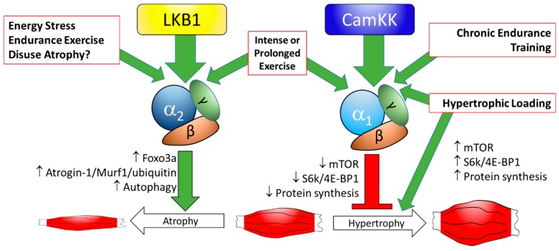
AMPK (5’-adenosine monophosphate-activated protein kinase) is heavily involved in skeletal muscle metabolic control through its regulation of many downstream targets. Because of their effects on anabolic and catabolic cellular processes, AMPK plays an important role in the control of skeletal muscle development and growth. In this review, the effects of AMPK signaling, and those of its upstream activator, liver kinase B1 (LKB1), on skeletal muscle growth and atrophy are reviewed. The effect of AMPK activity on satellite cell-mediated muscle growth and regeneration after injury is also reviewed. Together, the current data indicate that AMPK does play an important role in regulating muscle mass and regeneration, with AMPKα1 playing a prominent role in stimulating anabolism and in regulating satellite cell dynamics during regeneration, and AMPKα2 playing a potentially more important role in regulating muscle degradation during atrophy.
Overview
The claims and statistics of this paper haven’t been checked yet.
Run the check — free, no account needed. It reads the open-access text and checks the sources it cites.
Integrity signals
Registry
Cites
Statistics
Figures
Verification
Claims the paper makes
The claims and statistics of this paper haven’t been checked yet.
Run the check — free, no account needed. It reads the open-access text and checks the sources it cites.
Claims other papers make about this one
Each is checked against this paper’s own text: does it say what the citing paper says it does?
The check wasn’t confident either way, so it gives no verdict — never disputed.
In the citing paper
“AMPK can inhibit RNA polymerase I activity through multiple mechanisms, including suppression of TIF-1A function and modulation of chromatin accessibility at rDNA loci”
From Ribosome Biogenesis as a Putative Bottleneck to Skeletal Muscle Hypertrophy: Mechanisms, Human Evidence, and Practical Modulators (2026) · cites this paper as [62]
Experimental — accuracy not yet measuredThe check wasn’t confident either way, so it gives no verdict — never disputed.
In the citing paper
“A novel biomechanical stimulus might successfully initiate Pol I, but concurrent metabolic stress might abort snoRNA-guided processing downstream, leading to aborted assembly despite massive precursor transcripts [60,62].”
From Ribosome Biogenesis as a Putative Bottleneck to Skeletal Muscle Hypertrophy: Mechanisms, Human Evidence, and Practical Modulators (2026) · cites this paper as [62]
Experimental — accuracy not yet measured
Self-check
Percentages, sums and reported differences re-computed from the paper’s own numbers.
The self-check needs the paper’s full text.
Citations
Cites
Repertoires of Autophagy in the Pathogenesis of Ocular Diseases
Li, Y., Jiang, Q. L., Cao, G.-F., Yao, J., & Yan, B. (2015) · Cellular Physiology and Biochemistry · doi:10.1159/000373980 · Cited by 16,351
AMPK and mTOR regulate autophagy through direct phosphorylation of Ulk1
Kim, J., Kundu, M., Viollet, B., & Guan, K.-L. (2011) · Nature Cell Biology · doi:10.1038/ncb2152 · Cited by 7,227
Role of AMP-Activated Protein Kinase in Mechanism of Metformin Action
Zhou, G., Myers, R., Li, Y., Chen, Y., Shen, X., Fenyk-Melody, J., Wu, M., Ventre, J., Doebber, T., Fujii, N., et al. (2001) · Journal of Clinical Investigation · doi:10.1172/jci13505 · Cited by 5,652
AMPK: guardian of metabolism and mitochondrial homeostasis
Herzig, S., & Shaw, R. J. (2017) · Nature Reviews Molecular Cell Biology · doi:10.1038/nrm.2017.95 · Cited by 4,192
AMPK Phosphorylation of Raptor Mediates a Metabolic Checkpoint
Gwinn, D. M., Shackelford, D. B., Egan, D. F., Mihaylova, M. M., Mery, A., Vasquez, D. S., Turk, B. E., & Shaw, R. J. (2008) · Molecular Cell · doi:10.1016/j.molcel.2008.03.003 · Cited by 3,849
AMPK regulates energy expenditure by modulating NAD+ metabolism and SIRT1 activity
Cantó, C., Gerhart-Hines, Z., Feige, J. N., Lagouge, M., Noriega, L., Milne, J. C., Elliott, P. J., Puigserver, P., & Auwerx, J. (2009) · Nature · doi:10.1038/nature07813 · Cited by 3,300
The mechanisms of action of metformin
Rena, G., Hardie, D. G., & Pearson, E. R. (2017) · Diabetologia · doi:10.1007/s00125-017-4342-z · Cited by 2,287
Corton, J. M., Gillespie, J. G., Hawley, S. A., & Hardie, D. G. (1995) · European Journal of Biochemistry · doi:10.1111/j.1432-1033.1995.tb20498.x · Cited by 1,232
Merrill, G. F., Kurth, E. J., Hardie, D. G., & Winder, W. W. (1997) · American Journal of Physiology-Endocrinology and Metabolism · doi:10.1152/ajpendo.1997.273.6.e1107 · Cited by 1,048
Rheb Binds and Regulates the mTOR Kinase
Long, X., Lin, Y., Ortiz-Vega, S., Yonezawa, K., & Avruch, J. (2005) · Current Biology · doi:10.1016/j.cub.2005.02.053 · Cited by 1,005
Cited by
Sorted by citations
Mechanisms of IGF-1-Mediated Regulation of Skeletal Muscle Hypertrophy and Atrophy
Yoshida, T., & Delafontaine, P. (2020) · Cells · doi:10.3390/cells9091970 · Cited by 772
“Importantly, IGF-1 level is reduced in both of these atrophying conditions and Akt is inhibited in skeletal muscle, although the role of AMPK (which is known to inhibit Akt/mTOR and activate FoxO [78,92]) in relation to IGF-1 signaling in atrophying conditions is not clear.”
BackgroundSarcopenia and Muscle Aging: A Brief Overview
Dao, T., Green, A. E., Kim, Y.-A., Bae, S., Ha, K., Gariani, K., Lee, M.-R., Menzies, K. J., & Ryu, D. (2020) · Endocrinology and Metabolism · doi:10.3803/enm.2020.405 · Cited by 249
Huang, Y., Zhu, X., Chen, K., Lang, H., Zhang, Y., Hou, P., Ran, L., Zhou, M., Zheng, J., Yi, L., et al. (2019) · Aging · doi:10.18632/aging.101910 · Cited by 167
“AMPK, a potential longevity target [66], plays an important role in the regulation of skeletal muscle mass and regeneration [67].”
BackgroundVarghese, S., Rao, S., Khattak, A., Zamir, F., & Chaari, A. (2024) · Nutrients · doi:10.3390/nu16213663 · Cited by 152
The flesh texture of teleost fish: Characteristics and interventional strategies
Wang, Z., Qiao, F., Zhang, W., Parisi, G., Du, Z., & Zhang, M. (2023) · Reviews in Aquaculture · doi:10.1111/raq.12849 · Cited by 139
Garbern, J. C., & Lee, R. T. (2021) · Stem Cell Research & Therapy · doi:10.1186/s13287-021-02252-6 · Cited by 146
“of AMPK, which can in turn inhibit the mTOR signaling pathway, and p70S6K is immediately downstream of mTOR complex 1 (mTORC1) [108].”
BackgroundRoles of mTOR Signaling in Tissue Regeneration
Wei, X., Luo, L., & Chen, J.-Z. (2019) · Cells · doi:10.3390/cells8091075 · Cited by 136
“Their progenies either maintain the satellite cells pool through self-renewal or differentiate into myoblasts expressing myogenic markers Myf5 (myogenic factor 5), MyoD (myogenic differentiation 1), myogenin and MRF4 (myogenic regulatory factor 4) [130,133] (Figure 3C).”
BackgroundLiu, D., Wang, S., Liu, S., Wang, Q., Che, X., & Wu, G. (2024) · Molecular Aspects of Medicine · doi:10.1016/j.mam.2024.101270 · Cited by 157
Organ protection by SGLT2 inhibitors: role of metabolic energy and water conservation
Marton, A., Kaneko, T., Kovalik, J., Yasui, A., Nishiyama, A., Kitada, K., & Titze, J. M. (2020) · Nature Reviews Nephrology · doi:10.1038/s41581-020-00350-x · Cited by 136
Hyatt, H. W., & Powers, S. K. (2021) · Antioxidants · doi:10.3390/antiox10040588 · Cited by 134
“Moreover, low energy levels in muscle fibers are also associated with increased AMP-kinase (AMPK) activity, which is associated with inhibition of mTORC directly or indirectly (reviewed in [49]).”
BackgroundGuo, A., Li, K., Tian, H., Fan, Z., Chen, Q., Yang, Y., Yu, J., Wu, Y., & Xiao, Q. (2021) · Journal of Cellular and Molecular Medicine · doi:10.1111/jcmm.16448 · Cited by 118
Metformin induces muscle atrophy by transcriptional regulation of myostatin via HDAC6 and FoxO3a
Kang, M. J., Moon, J. W., Lee, J. O., Kim, J. H., Jung, E. J., Kim, S. J., Oh, J. Y., Wu, S. W., Lee, P. R., Park, S. H., et al. (2021) · Journal of Cachexia Sarcopenia and Muscle · doi:10.1002/jcsm.12833 · Cited by 102
Wang, S., Jin, J., Xu, Z., & Zuo, B. (2019) · Cells · doi:10.3390/cells8091107 · Cited by 89
Ubiquitin Ligases at the Heart of Skeletal Muscle Atrophy Control
Peris-Moreno, D., Cussonneau, L., Combaret, L., Polge, C., & Taillandier, D. (2021) · Molecules · doi:10.3390/molecules26020407 · Cited by 90
“Additionally, AMPK also promotes skeletal muscle autophagy [88].”
Sekar, P., Hsiao, G., Hsu, S.-H., Huang, D.-Y., Lin, W., & Chan, C.-M. (2023) · Redox Biology · doi:10.1016/j.redox.2023.102786 · Cited by 84
The hallmarks of myotonic dystrophy type 1 muscle dysfunction
Ozimski, L. L., Sabater-Arcis, M., Bargiela, A., & Artero, R. D. (2020) · Biological reviews/Biological reviews of the Cambridge Philosophical Society · doi:10.1111/brv.12674 · Cited by 81
“AMPK has been shown to play a vital role in maintaining and remodelling skeletal muscle (Thomson, 2018).”
BackgroundYan, Y., Li, M., Lin, J., Ji, Y., Wang, K., Yan, D., Shen, Y., Wang, W., Huang, Z., Jiang, H., et al. (2022) · Frontiers in Pharmacology · doi:10.3389/fphar.2022.947387 · Cited by 84
“AMPK inhibits the protein synthesis by inhibiting the activities of mechanistic target of rapamycin, complex 1 (mTORC1) and eukaryotic elongation factor 2 (eEF2) (Thomson, 2018).”
BackgroundVignaud, J., Loiseau, C., Hérault, J., Mayer, C., Côme, M., Martin, I., & Ulmann, L. (2023) · Antioxidants · doi:10.3390/antiox12051050 · Cited by 72
“In contrast, during muscle disease associated with mitochondrial dysfunction, AMPK suppresses the MPS via an inhibition of mTORC1 [49].”
BackgroundRybalka, E., Timpani, C. A., Debruin, D. A., Bagaric, R. M., Campelj, D. G., & Hayes, A. (2020) · Cells · doi:10.3390/cells9122657 · Cited by 69
“In this regard, myostatin may act as an environmental sensor/signaler of nutritional status (particularly in a low amino acid environment) [10] in synergy with the cellular energy sensor, adenosine monophosphate-activated protein kinase (AMPK) [11], promoting a negative feedback loop that inhibits ribosomal biogenesis…”
BackgroundInfluentialMishra, S., Cosentino, C., Tamta, A. K., Khan, D., Srinivasan, S., Ravi, V., Abbotto, E., Arathi, B. P., Kumar, S. A., Jain, A., et al. (2022) · Nature Communications · doi:10.1038/s41467-022-32905-w · Cited by 58
Livshits, G., & Kalinkovich, A. (2023) · Ageing Research Reviews · doi:10.1016/j.arr.2023.101852 · Cited by 56
Post‐translational regulation of muscle growth, muscle aging and sarcopenia
Zhong, Q., Zheng, K., Li, W., An, K., Liu, Y., Xiao, X., Hai, S., Dong, B., Li, S., An, Z., et al. (2023) · Journal of Cachexia Sarcopenia and Muscle · doi:10.1002/jcsm.13241 · Cited by 59
“tion of mTOR in response to energy expenditure and reduced protein synthesis.(30) In damaged muscle, the activation of the AMPK signalling pathway enhances the transcriptional activ-”
BackgroundThe Role of Mitophagy in Skeletal Muscle Damage and Regeneration
Chatzinikita, E., Maridaki, M. D., Palikaras, K., Koutsilieris, M., & Philippou, Α. (2023) · Cells · doi:10.3390/cells12050716 · Cited by 63
SIRT1 signaling pathways in sarcopenia: Novel mechanisms and potential therapeutic targets
Yang, L., Liu, D., Jiang, S., Li, H., Chen, L., Wu, Y., Essien, A. E., Opoku, M., Naranmandakh, S., Liu, S., et al. (2024) · Biomedicine & Pharmacotherapy · doi:10.1016/j.biopha.2024.116917 · Cited by 62
Daidzein alleviates cisplatin‐induced muscle atrophy by regulating Glut4/ AMPK / FoxO pathway
Zhang, H., Chi, M., Chen, L., Sun, X., Wan, L., Yang, Q., & Guo, C. (2021) · Phytotherapy Research · doi:10.1002/ptr.7132 · Cited by 52
“skeletal muscle atrophy, and activation of AMPK can promote the expression of FoxO in the nucleus (Greer et al., 2007; Thomson, 2018).”
BackgroundToda, K., Yamauchi, Y., Tanaka, A., Kuhara, T., Odamaki, T., Yoshimoto, S., & Xiao, J. (2020) · Nutrients · doi:10.3390/nu12010219 · Cited by 54
“B-3HK significantly promoted the activation of AMPK (Figure 4A,B), which regulates energy balance at both the cellular and physiological levels as a master regulator of metabolism [22,23].”
BackgroundAshtary-Larky, D., Bagheri, R., Asbaghi, O., Tinsley, G. M., Kooti, W., Abbasnezhad, A., Afrisham, R., & Wong, A. (2022) · Critical Reviews in Food Science and Nutrition · doi:10.1080/10408398.2021.1890689 · Cited by 50
“Importantly, differences in stored glycogen and the associated water content could influence BM results, along with other body composition outcomes, when comparing KD and non-KD regimens (Tinsley and Willoughby 2016).”
BackgroundResultInfluentialmiR-7 Restores Phenotypes in Myotonic Dystrophy Muscle Cells by Repressing Hyperactivated Autophagy
Sabater-Arcis, M., Bargiela, A., Furling, D., & Artero, R. D. (2019) · Molecular Therapy — Nucleic Acids · doi:10.1016/j.omtn.2019.11.012 · Cited by 48
“AMPK is involved in the regulation of several metabolic pathways that regulate muscle size, such as autophagy and UPS activation or inhibition of protein synthesis via mTOR blockade.(49) However, our data suggest that this pathway remains unaltered in DM1 and that, in consequence, the activation of autophagy and UPS s…”
BackgroundHuang, Y., Wang, C., Cui, H., Sun, G., Qi, X., & Yao, X. (2025) · Frontiers in Cell and Developmental Biology · doi:10.3389/fcell.2025.1590524 · Cited by 54
“AMPK can be strongly activated in skeletal muscle by repeated muscle contractions and exercise (Thomson, 2018).”
Xie, Y., Gu, Y., Li, Z., He, B., & Zhang, L. (2024) · Nutrients · doi:10.3390/nu16173007 · Cited by 57
“However, there is a lack of literature directly comparing the four dietary interventions combined with exercise, which makes the results more susceptible to the influence of indirect comparisons.”
BackgroundTherapeutic Implications of miRNAs for Muscle-Wasting Conditions
Yedigaryan, L., & Sampaolesi, M. (2021) · Cells · doi:10.3390/cells10113035 · Cited by 43
“Additionally, AMPK suppresses anabolic processes such as protein synthesis [76].”
BackgroundEffects of Various Muscle Disuse States and Countermeasures on Muscle Molecular Signaling
Sharlo, K. A., Tyganov, S. A., Tomilovskaya, E., Popov, D. V., Saveko, A. A., & Shenkman, B. S. (2021) · International Journal of Molecular Sciences · doi:10.3390/ijms23010468 · Cited by 47
“When activated, AMPK can suppress mTORC1 signaling through the phosphorylation and activation of TSC2 [116] or the phosphorylation of Raptor (regulatory-associated protein of mammalian target of rapamycin, a part of the mTORC1 complex), which leads to its subsequent sequestration by regulatory proteins of the 14-3-3 f…”
BackgroundHuang, C., Ge, F., Ma, X., Dai, R., Dingkao, R., Zhuoma, Z., Burenchao, G., Bao, P., Wu, X., Guo, X., et al. (2021) · Frontiers in Genetics · doi:10.3389/fgene.2021.772557 · Cited by 38
“A total of 117 DEMs were identified that were related to the differentiation and proliferation of myoblasts, AMPK signaling pathway, MAPK signaling pathway, PI3K–Akt signaling pathway, etc. (Neri et al., 2002; Anderson, 2006; Gehart et al., 2010; Thomson, 2018).”
BackgroundNAD+ centric mechanisms and molecular determinants of skeletal muscle disease and aging
Wagner, S., Manickam, R., Brotto, M. A. P., & Tipparaju, S. M. (2022) · Molecular and Cellular Biochemistry · doi:10.1007/s11010-022-04408-1 · Cited by 39
Regenerating Myofibers after an Acute Muscle Injury: What Do We Really Know about Them?
Pizza, F. X., & Buckley, K. H. (2023) · International Journal of Molecular Sciences · doi:10.3390/ijms241612545 · Cited by 37
Skeletal muscle atrophy after sciatic nerve damage: Mechanistic insights
Yadav, A., & Dabur, R. (2024) · European Journal of Pharmacology · doi:10.1016/j.ejphar.2024.176506 · Cited by 38
A transition to degeneration triggered by oxidative stress in degenerative disorders
Stern, M., & McNew, J. A. (2020) · Molecular Psychiatry · doi:10.1038/s41380-020-00943-9 · Cited by 33
Pathophysiological changes of muscle after ischemic stroke: a secondary consequence of stroke injury
Hu, Q., Tian, D., Luan, F., Yang, R., & Zeng, N. (2023) · Neural Regeneration Research · doi:10.4103/1673-5374.382221 · Cited by 35
“…proliferators-activated receptor γ coactivator 1 alpha (PGC-1α) plays a role in promoting slow muscle fiber expression and pro-mitotic mitochondrial biosynthesis to improve motility (Boström et al., 2012), and changes in AMPK levels are positively correlated with PGC-1α expression (Thomson, 2018).”
Fukada, S., & Ito, N. (2021) · Experimental Cell Research · doi:10.1016/j.yexcr.2021.112907 · Cited by 32
Fovet, T., Guilhot, C., Delobel, P., Chopard, A., Py, G., & Brioche, T. (2022) · Frontiers in Physiology · doi:10.3389/fphys.2022.834597 · Cited by 31
“This is consistent with the lower metabolic stress observed in ET muscles, because such stress normally delays protein synthesis activation after physical activity (MacDougall et al., 1995; Bolster et al., 2002; Dreyer et al., 2006; Thomson, 2018).”
ResultVerpoorten, S., Sfyri, P., Scully, D., Mitchell, R., Tzimou, A., Mougios, V., Patel, K., & Matsakas, A. (2019) · Acta Physiologica · doi:10.1111/apha.13395 · Cited by 31
Slc20a1/Pit1 and Slc20a2/Pit2 are essential for normal skeletal myofiber function and survival
Chande, S., Caballero, D., Ho, B., Fetene, J. A., Serna, J., Pesta, D. H., Nasiri, A. R., Jurczak, M. J., Chavkin, N. W., Hernando, N., et al. (2020) · Scientific Reports · doi:10.1038/s41598-020-59430-4 · Cited by 34
Huang, Y., Xie, H., Pan, P., Qu, Q., Qin, X., Gao, X., Zhang, S., & Jiang, Q. (2021) · Cell Stress and Chaperones · doi:10.1007/s12192-021-01201-9 · Cited by 30
Sun, C., Yang, D. K., Chen, Z., Xiao, J., Xiao, Q., Li, C., Zhou, Z., Peng, X., Tang, C., & Zheng, L. (2022) · FEBS Journal · doi:10.1111/febs.16637 · Cited by 34
Sataranatarajan, K., Pharaoh, G. A., Brown, J. L., Ranjit, R., Piekarz, K. M., Street, K., Wren, J. D., Georgescu, C., Kinter, C. S., Kinter, M. T., et al. (2020) · GeroScience · doi:10.1007/s11357-020-00189-x · Cited by 31
LncEDCH1 improves mitochondrial function to reduce muscle atrophy by interacting with SERCA2
Cai, B., Ma, M., Zhang, J., Wang, Z., Kong, S., Zhou, Z., Lian, L., Zhang, J., Li, J., Wang, Y., et al. (2021) · Molecular Therapy — Nucleic Acids · doi:10.1016/j.omtn.2021.12.004 · Cited by 29
Salucci, S., Bartoletti‐Stella, A., Bavelloni, A., Aramini, B., Blalock, W. L., Fabbri, F., Vannini, I., Sambri, V., Stella, F., & Faenza, I. (2022) · Nutrients · doi:10.3390/nu14173567 · Cited by 30
“Moreover, mTORC1 inhibition blocks muscle hypertrophy in post-natal development and muscle regeneration [51].”
BackgroundGinseng and ginsenosides: Therapeutic potential for sarcopenia
Zha, W., Sun, Y., Gong, W., Li, L., Kim, W., & Li, H. (2022) · Biomedicine & Pharmacotherapy · doi:10.1016/j.biopha.2022.113876 · Cited by 31
Molecular mechanisms of post‐burn muscle wasting and the therapeutic potential of physical exercise
Dombrecht, D., Daele, U. V., Asbroeck, B. V., Schieffelers, D. R., Guns, P., Gebruers, N., Meirte, J., & van Breda, E. (2023) · Journal of Cachexia Sarcopenia and Muscle · doi:10.1002/jcsm.13188 · Cited by 28
“tein kinase (AMPK), a key regulator of energy homoeostasis with a critical role as energy sensor during exercise and metabolic disorders.(32) AMPK is involved in both anabolic and catabolic pathways, maintaining energy stores and ensuring a balance between energy supply and demand, whereas in skeletal muscle AMPK inhi…”
BackgroundLiu, X., Zhao, L., Gao, Y., Chen, Y., Tian, Q., Son, J. S., Chae, S. A., de Avila, J. M., Zhu, M. J., & Du, . (2022) · Journal of Cachexia Sarcopenia and Muscle · doi:10.1002/jcsm.13150 · Cited by 27
“Notably, AMPK also regulates skeletal muscle development and regeneration.(18) During muscle regeneration, inflammatory response is meticulously modulated.”
BackgroundJun, L., Tao, Y.-X., Geetha, T., & Babu, J. R. (2024) · Current Nutrition Reports · doi:10.1007/s13668-024-00555-7 · Cited by 27
“Specifically, phosphorylation of AMPKα1 subunit at Thr183 plays a prominent role in stimulating anabolism during regeneration process, while phosphorylation of AMPKα2 subunit at Thr172 plays a role in regulating muscle degradation during atrophy [37].”
BackgroundHan, J.-Y., Park, M., & Lee, H. (2023) · BMC Complementary Medicine and Therapies · doi:10.1186/s12906-023-04033-5 · Cited by 27
“AMP-activated protein kinase-α (AMPKα), a central regulator of mitochondrial biogenesis, controls intracellular energy balance and is an essential factor in skeletal muscle atrophy [17, 18].”
BackgroundShimizu, J., & Kawano, F. (2022) · The Journal of Physiology · doi:10.1113/jp282917 · Cited by 26
“AMPK is activated via phosphorylation by metabolic stress and functions to switch cellular fuel utilization from glucose to fatty acids, stimulating mitochondrial biogenesis (Thomson, 2018).”
BackgroundAlcohol, Resistance Exercise, and mTOR Pathway Signaling: An Evidence-Based Narrative Review
Levitt, D. E., Luk, H., & Vingren, J. L. (2022) · Biomolecules · doi:10.3390/biom13010002 · Cited by 33
Effects of Fucoxanthin on the Inhibition of Dexamethasone-Induced Skeletal Muscle Loss in Mice
Yoshikawa, M., Hosokawa, M., Miyashita, K., Nishino, H., & Hashimoto, T. (2021) · Nutrients · doi:10.3390/nu13041079 · Cited by 25
“Mitochondrial dysfunction increases AMP and ROS and activates AMPK, which suppresses protein synthesis via inhibition of mTOR signaling [35] and hence results in muscle atrophy [36].”
BackgroundMechanisms of the NAD+ salvage pathway in enhancing skeletal muscle function
Su, M., Qiu, F., Li, Y., Che, T., Li, N., & Zhang, S. (2024) · Frontiers in Cell and Developmental Biology · doi:10.3389/fcell.2024.1464815 · Cited by 27
“Studies have shown that the absence of AMPK α 1 leads to a reduction in satellite cell numbers and decreased expression of myogenic factors such as Myf5 and Myogenin (Fu et al., 2015; Thomson, 2018).”
BackgroundDuan, Z., Tu, C., Liu, Q., Li, S., Li, Y., Xie, P., & Li, Z. (2020) · Journal of Cellular Biochemistry · doi:10.1002/jcb.29605 · Cited by 25
“Accumulating evidence suggests that the AMPK‐mTOR pathway might act as the primary signal axis for autophagy.(37,38) Our results show that AdipoRon promoted the phosphorylation of the AMPK protein and then inhibited the phosphorylation of mTOR protein in chondrocytes.”
BackgroundLi, Y., Feng, Y., Chen, X., He, J., Luo, Y., Yu, B., Chen, D., & Huang, Z. (2024) · Meat Science · doi:10.1016/j.meatsci.2024.109436 · Cited by 23
Tsukita, K., Yano, T., Tamura, A., & Tsukita, S. (2019) · International Journal of Molecular Sciences · doi:10.3390/ijms20236012 · Cited by 25
The Roles of AMPK in Revascularization
Chen, M.-H., & Fu, Q.-M. (2020) · Cardiology Research and Practice · doi:10.1155/2020/4028635 · Cited by 22
“Zhenkun Lou et al. have found that AMPKα1 or AMPKα2 ubiquitination blocks its phosphorylation by LKB1, which could be rescued by the deubiquitinase ubiquitin specific peptidase 10 (USP10) [54].”
BackgroundInfluentialMiRNA Profiling in Pectoral Muscle Throughout Pre- to Post-Natal Stages of Chicken Development
Chen, M., Zhang, S., Xu, Z., Gao, J., Mishra, S. K., Zhu, Q., Zhao, X., Wang, Y., Yin, H., Fan, X., et al. (2020) · Frontiers in Genetics · doi:10.3389/fgene.2020.00570 · Cited by 22
“These pathways had been reported to be involved in muscle growth and development (Schiaffino et al., 2013; Thomson, 2018; Du et al., 2019).”
BackgroundHigh-Frequency Stimulation on Skeletal Muscle Maintenance in Female Cachectic Mice
Sato, S., Gao, S., Puppa, M. J., Kostek, M., Wilson, L., & Carson, J. (2019) · Medicine & Science in Sports & Exercise · doi:10.1249/mss.0000000000001991 · Cited by 15
Elevated CO2 Levels Delay Skeletal Muscle Repair by Increasing Fatty Acid Oxidation
Ceco, E., Celli, D. A., Weinberg, S. E., Shigemura, M., Welch, L. C., Volpe, L., Chandel, N. S., Bharat, A., Lecuona, E., & Sznajder, J. I. (2021) · Frontiers in Physiology · doi:10.3389/fphys.2020.630910 · Cited by 20
“With regards to muscle regeneration, AMPKα1 plays a prominent role in regulating satellite cell dynamics, with AMPKα2 playing a potentially more important role in regulating muscle degradation during atrophy (Thomson, 2018).”
BackgroundWheel running improves fasting‐induced AMPK signaling in skeletal muscle from tumor‐bearing mice
Fix, D. K., Counts, B. R., Smuder, A. J., Sarzynski, M. A., Koh, H., & Carson, J. A. (2021) · Physiological Reports · doi:10.14814/phy2.14924 · Cited by 19
“Nevertheless, many different catabolic conditions have investigated skeletal muscle atrophy regulation by metabolic disruptions that induce AMPK signaling (Bujak et al., 2015; Di Magno et al., 2016; Nakashima & Yakabe, 2007; Sanchez, Candau, et al., 2012; Sanchez, Csibi, et al., 2012; Thomson, 2018).”
BackgroundMinari, A. L. A., & dos Santos, R. V. T. (2021) · American Journal of Physiology-Regulatory, Integrative and Comparative Physiology · doi:10.1152/ajpregu.00038.2021 · Cited by 21
Malila, Y., Thanatsang, K. V., Sanpinit, P., Arayamethakorn, S., Soglia, F., Zappaterra, M., Bordini, M., Sirri, F., Rungrassamee, W., Davoli, R., et al. (2022) · PLoS ONE · doi:10.1371/journal.pone.0275160 · Cited by 21
“In addition, AMPK plays a key role in regulating skeletal muscle growth, development, and repair [21].”
BackgroundRavel‐Chapuis, A., Duchesne, É., & Jasmin, B. J. (2022) · The Journal of Physiology · doi:10.1113/jp282725 · Cited by 19
“This last element further obscures the investigation of AMPK in the context of exercise because the molecular responses to different exercise modalities are less clear in humans than in animals (Thomson, 2018).”
BackgroundGamberi, T., Magherini, F., & Fiaschi, T. (2019) · International Journal of Molecular Sciences · doi:10.3390/ijms20071544 · Cited by 18
“Full AMPK activation occurs through both phosphorylation by liver kinase B1 (LKB1) and AMP binding [8].”
BackgroundTransient changes to metabolic homeostasis initiate mitochondrial adaptation to endurance exercise
Dent, J. R., Stocks, B., Campelj, D. G., & Philp, A. (2022) · Seminars in Cell and Developmental Biology · doi:10.1016/j.semcdb.2022.03.022 · Cited by 19
Aoki, Y., Ozawa, T., Numata, O., & Takemasa, T. (2019) · Nutrients · doi:10.3390/nu11092131 · Cited by 18
“Additionally, dominant-negative Ampk transgenic mice in which a dominant-negative Ampkα2 subunit was overexpressed under the muscle creatine kinase promoter (expressed in heart and skeletal muscle) showed that EDL muscles tended to be larger than in wild-type mice, suggesting that Ampk might negatively regulate basal…”
BackgroundDou, Y., Wei, Y., Zhang, Z., Li, C., Song, C., Liu, Y., Qi, K., Li, X., Li, X., Qiao, R., et al. (2023) · BMC Genomics · doi:10.1186/s12864-023-09346-w · Cited by 17
“Among them, the AMPK signaling pathway is an important signaling pathway of energy metabolism that is heavily involved in skeletal muscle metabolic processes; controls skeletal muscle growth and development by regulating many downstream targets; is related to myocyte energy metabolism, protein synthesis, and catabolis…”
BackgroundAMPK/mTOR balance during exercise: implications for insulin resistance in aging muscle
Mingzheng, X., & You, W. (2025) · Molecular and Cellular Biochemistry · doi:10.1007/s11010-025-05362-4 · Cited by 22
“As a result of the energetic challenge, intracellular AMP/ATP and ADP/ATP ratios increase, serving as crucial triggers for AMPK activation [39].”
Effect of Age and Lipoperoxidation in Rat and Human Adipose Tissue-Derived Stem Cells
Muñoz, M., Argüelles, S., Marotta, F., Barbagallo, M., Cano, M., & Ayala, A. (2020) · Oxidative Medicine and Cellular Longevity · doi:10.1155/2020/6473279 · Cited by 17
“Recent studies have shown that AMPK activation, in response to various stimuli, such as exercise and muscle contraction, gradually declines during aging [77].”
LKB1 signaling is altered in skeletal muscle of a Duchenne muscular dystrophy mouse model
Boccanegra, B., Mantuano, P., Conte, E., Cerchiara, A. G., Tulimiero, L., Quarta, R., Caputo, E., Sanarica, F., Forino, M., Spadotto, V., et al. (2023) · Disease Models & Mechanisms · doi:10.1242/dmm.049930 · Cited by 16
“Nonetheless, chronic activation in basal conditions may have deleterious effects on muscle homeostasis, inhibiting muscle growth and stimulating pro-atrophic pathways (Thomson, 2018).”
Beta-hydroxybutyric acid attenuates neuronal damage in epileptic mice
Guo, Q.-H., Liu, S., Wang, S., Wu, M., Li, Z., & Wang, Y. (2019) · Acta Histochemica · doi:10.1016/j.acthis.2019.03.009 · Cited by 16
Gombos, Z., Koltai, E., Torma, F., Bakonyi, P., Kolonics, A., Aczél, D., Ditrói, T., Nagy, P., Kawamura, T., & Radak, Z. O. (2021) · International Journal of Molecular Sciences · doi:10.3390/ijms22147588 · Cited by 18
“Hence, as a result of intensive investigation a lot is known about the molecular pathways that are involved in increased protein synthesis and attenuated catabolic processes, which happen to occur during the hypertrophy of skeletal muscle [6,7].”
BackgroundLiu, J., Zhao, H., Yang, L., Wang, X., Yang, L., Xing, Y., Lv, X., Ma, H., & Song, G. (2022) · BMC Endocrine Disorders · doi:10.1186/s12902-021-00921-4 · Cited by 17
“The fatty acid synthesis can be increased by reducing the inhibitory effect of malonyl-CoA on CPT-1α oxidative metabolism [31].”
BackgroundTranscriptome Analysis of Differentially Expressed mRNA Related to Pigeon Muscle Development
Ding, H., Lin, Y., Zhang, T., Chen, L., Zhang, G., Wang, J., Xie, K., & Dai, G. (2021) · Animals · doi:10.3390/ani11082311 · Cited by 15
“AMPK plays an important role in controlling skeletal muscle development and growth due to its effects on anabolism and catalytic cellular processes [27].”
BackgroundLi, K., Wang, M., Wang, R., Wang, X., Jiao, H., Zhao, J., Zhou, Y., Li, H., & Lin, H. (2023) · Journal of Nutrition · doi:10.1016/j.tjnut.2023.08.024 · Cited by 15
Epigenetics of Skeletal Muscle Atrophy
Du, J., Wu, Q., & Bae, E. J. (2024) · International Journal of Molecular Sciences · doi:10.3390/ijms25158362 · Cited by 18
“The AMPK pathway is essential in skeletal muscle atrophy, as it can inhibit protein synthesis by modulating the phosphorylation of mTOR and eukaryotic initiation factor 2 [43].”
BackgroundGrönholdtKlein, M., Altun, M., Becklén, M., Kahm, E. D., Fahlström, A., Rullman, E., & Ulfhake, B. (2019) · Acta Physiologica · doi:10.1111/apha.13335 · Cited by 15
Transcriptional Changes Involved in Atrophying Muscles during Prolonged Fasting in Rats
Ibrahim, M., Wasselin, T., Challet, É., DORSSELAER, A. V., Maho, Y. L., Raclot, T., & Bertile, F. (2020) · International Journal of Molecular Sciences · doi:10.3390/ijms21175984 · Cited by 15
“AMPK generally activates catabolic factors while inactivating anabolic ones, thereby controlling energy metabolism and muscle growth or atrophy [83].”
BackgroundChernyavskij, D. A., Pletjushkina, O. Y., Kashtanova, A. V., Galkin, I. I., Karpukhina, A. A., Chernyak, B. V., Vassetzky, Y. S., & Popova, E. N. (2023) · Antioxidants · doi:10.3390/antiox12030602 · Cited by 15
Mechanisms of endurance and resistance exercise in type 2 diabetes mellitus: A Narrative review
Zhao, X., Huang, F., Sun, Y., & Li, L. (2025) · Biochemical and Biophysical Research Communications · doi:10.1016/j.bbrc.2025.151731 · Cited by 14
Regulatory Role of N6-Methyladenosine in Longissimus Dorsi Development in Yak
Ma, X., La, Y., Bao, P., Chu, M., Guo, X., Wu, X., Pei, J., Ding, X., Liang, C., & Yan, P. (2022) · Frontiers in Veterinary Science · doi:10.3389/fvets.2022.757115 · Cited by 15
“In addition, AMPK not only plays a key regulatory role in muscle energy metabolism but also participates in the transformation of muscle fiber types (44).”
BackgroundJaitovich, A. (2022) · American Journal of Physiology-Cell Physiology · doi:10.1152/ajpcell.00292.2022 · Cited by 16
Pudjihartono, N., Ho, D., Golovina, E., Fadason, T., Kempa-Liehr, A. W., & O'Sullivan, J. M. (2023) · Journal of Autoimmunity · doi:10.1016/j.jaut.2023.103046 · Cited by 13
Chan, C.-M., Sekar, P., Huang, D.-Y., Hsu, S.-H., & Lin, W. (2020) · Antioxidants · doi:10.3390/antiox9111057 · Cited by 15
Ho, J., Baskaran, R., Wang, M.-F., Mohammedsaleh, Z. M., Yang, H., Balasubramanian, B., & Lin, W. (2022) · International Journal of Molecular Sciences · doi:10.3390/ijms23158167 · Cited by 15
“In addition, swimming exercise protects mice against myocardial and liver apoptosis [18,19].”
BackgroundSu, L.-Y., Huang, W.-C., Kan, N., Tung, T.-H., Huynh, L. B. P., & Huang, S. (2023) · Nutrients · doi:10.3390/nu15010249 · Cited by 14
“Studies have also supported that resveratrol is involved in the activation of AMP-activated protein kinase (AMPK) during muscle contraction over a longer exercise period [11].”
BackgroundThe Role of Natural Products in the Improvement of Cancer-Associated Cachexia
Han, Y., Kim, H. I., & Park, J. (2023) · International Journal of Molecular Sciences · doi:10.3390/ijms24108772 · Cited by 13
“In cancer-induced cachexia, it has been reported that AMPK could play a key role in regulating energy production and consumption, and it has been suggested to contribute to the development of muscle wasting and weight loss seen in cachexia [21,83,84].”
BackgroundWei, X., Wang, J., Sun, Y., Zhao, T., Luo, X., Lu, J., Hou, W., Yu, X., Xue, L., Yan, Y. L., et al. (2023) · Journal of Cellular Biochemistry · doi:10.1002/jcb.30453 · Cited by 13
Yu, H., He, Y., Qin, M., Wang, L., Rong, K., & Zhang, X. (2024) · Aquaculture · doi:10.1016/j.aquaculture.2024.740591 · Cited by 14
Fine-tuning AMPK in physiology and disease using point-mutant mouse models
Ashraf, N., & Nostrand, J. L. V. (2024) · Disease Models & Mechanisms · doi:10.1242/dmm.050798 · Cited by 14
“…muscle, AMPK facilitates glucose uptake, stimulates fatty acid β -oxidation (FAO, see Glossary, Box 2) and promotes mitochondrial biogenesis, thereby enhancing insulin sensitivity and maintaining a healthy body weight (Jørgensen et al., 2021; Lee et al., 2006; Rhein et al., 2021; Thomson, 2018).”
BackgroundZhang, Y., Li, T., Liu, Y., Wang, C., Wang, D., Xu, L., Zhao, H., Bai, X., Li, Z., & Wang, Y.-C. (2024) · Shock · doi:10.1097/shk.0000000000002430 · Cited by 13
“Muscle ring finger protein 1 (MuRF1) and F-box protein Atrogin-1 are two indicators that are commonly employed to assess skeletal muscle atrophy due to their high expression and sensitivity (3,7).”
InfluentialEffects of Maca on Muscle Hypertrophy in C2C12 Skeletal Muscle Cells
Yi, D., Yoshikawa, M., Sugimoto, T., Tomoo, K., Okada, Y., & Hashimoto, T. (2022) · International Journal of Molecular Sciences · doi:10.3390/ijms23126825 · Cited by 13
“AMPK greatly affects skeletal muscle development and growth [32].”
BackgroundYakabe, M., Hosoi, T., Sasakawa, H., Akishita, M., & Ogawa, S. (2022) · BMC Complementary Medicine and Therapies · doi:10.1186/s12906-022-03812-w · Cited by 12
“In addition, 5’ adenosine monophosphate-activated protein kinase (AMPK) has two kinds of effects on metabolism: the inhibition of anabolism to minimize ATP consumption and the stimulation of catabolism to stimulate ATP production [6], and it is involved in skeletal muscle metabolic control [7].”
BackgroundAnalysis of the mechanism of skeletal muscle atrophy from the pathway of decreased protein synthesis
Chen, P., Jia, F., Wang, M., & Yang, S. (2025) · Frontiers in Physiology · doi:10.3389/fphys.2025.1533394 · Cited by 17
“This inhibits the binding of elongation factors to ribosomes, resulting in decreased translation efficiency and reduced protein synthesis (Thomson, 2018) (Figure 1).”
Valini, G. A. D. C., Duarte, M. D. S., Rodrigues, G. D. A., Veroneze, R., Saraiva, A., Hausman, G. J., & Rocha, G. C. (2020) · Ciência Rural · doi:10.1590/0103-8478cr20190948 · Cited by 12
Direct or indirect regulation of muscle protein synthesis by energy status?
Moinard, C., & Fontaine, E. M. (2020) · Clinical Nutrition · doi:10.1016/j.clnu.2020.07.015 · Cited by 12
Metadata
- Authors
- Thomson, D. M.
- Affiliations
- Brigham Young University (US)
- Corresponding author
- Thomson, David M.
- ORCID
- Thomson, David M.
- Venue
- International Journal of Molecular Sciences
- Publisher
- MDPI AG
- Published
- 2018-10-11
- Volume
- 19
- Issue
- 10
- Pages
- 3125
- ISSN
- 1422-0067 (ISSN-L)
- PMC
- PMC6212977
- OpenAlex
- W2896374260
- Cited by
- 267
- References
- 142
- Citations by year
- 2026: 29 · 2025: 36 · 2024: 41 · 2023: 52 · 2022: 40 · 2021: 31 · 2020: 24 · 2019: 13 · 2018: 1
- FWCI
- 1.68
- Type
- journal article
- Language
- en
- Open access
- gold
- dx.doi.org
- Licence
- Locations
- International Journal of Molecular Sciences (publishedVersion, cc-by)
- Europe PMC (PubMed Central) (publishedVersion, other-oa)
- PubMed Central (publishedVersion, cc-by)
- PubMed (publishedVersion)
- DOAJ (DOAJ: Directory of Open Access Journals) (submittedVersion, cc-by-sa)
- Topics
- Life Sciences › Biochemistry, Genetics and Molecular Biology › Molecular Biology › Muscle Physiology and Disorders
- Life Sciences › Biochemistry, Genetics and Molecular Biology › Molecular Biology › Metabolism, Diabetes, and Cancer
- Health Sciences › Nursing › Nutrition and Dietetics › Muscle metabolism and nutrition
- Keywords
- AMPK, skeletal muscle growth, muscle hypertrophy, muscle atrophy, muscle regeneration, satellite cells, LKB1, AMPKα1, AMPKα2, muscle metabolism, myogenesis
- MeSH terms
- Animals, Energy Metabolism, Enzyme Activation, Humans, Hypertrophy, Organ Size, Regeneration, Wound Healing, Signal Transduction, Biomarkers, Muscle, Skeletal, Muscle Fibers, Skeletal, Muscle Development, AMP-Activated Protein Kinases
About these checks
Registry, retraction, Cites and statistics results are computed, not judged, with one exception: a printed reference that no index lists is read once by a model before it counts as a citation error. Claims are extracted by a model and kept only when their quote appears verbatim in the text; each cited source is then checked, with a model’s help, for whether it says what the claim relies on. Those verdicts can be wrong — read the claim’s quote and the cited source before relying on one.
Statistics and arithmetic are recomputed, not judged.
Confidence bands are shown only for fields calibrated on our annotated test set. None is calibrated yet, so no band is shown.
What counts as a claim ›How we measure confidence ›How we check this ›
Are you an author, or do you see a problem with this page? Request a review
