

The DOI matches its Crossref record; 9 of the 9 references it cites were found in a registry or an index.
The Machinery of Exosomes: Biogenesis, Release, and Uptake
Abstract
Exosomes are a subtype of membrane-contained vesicles 40–200 nm in diameter that are secreted by cells into their surroundings. By transporting proteins, lipids, mRNA, miRNA, lncRNA, and DNA, exosomes are able to perform such vital functions as maintaining cellular homeostasis, removing cellular debris, and facilitating intercellular and interorgan communication. Exosomes travel in all body fluids and deliver their molecular messages in autocrine, paracrine as well as endocrine manners. In recent years, there has been an increased interest in studying exosomes as diagnostic markers and therapeutic targets, since in many disease conditions this machinery becomes dysregulated or hijacked by pathological processes. Additionally, delivery of exosomes and exosomal miRNA has already been shown to improve systemic metabolism and inhibit progression of cancer development in mice. However, the subcellular machinery of exosomes, including their biogenesis, release and uptake, remains largely unknown. This review will bring molecular details of these processes up to date with the goal of expanding the knowledge basis for designing impactful exosome experiments in the future.
Overview
The claims and statistics of this paper haven’t been checked yet.
Run the check — free, no account needed. It reads the open-access text and checks the sources it cites.
Integrity signals
Registry
Cites
Statistics
Figures
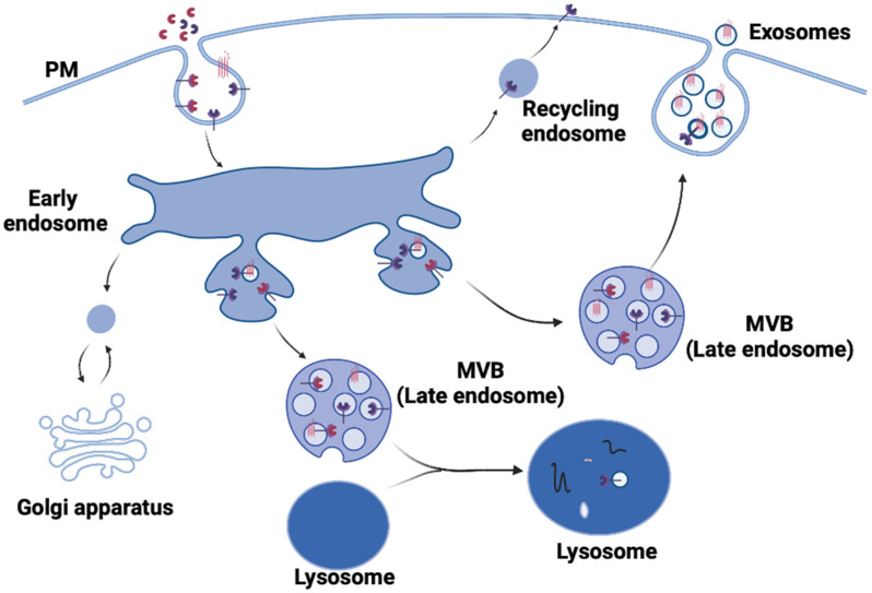
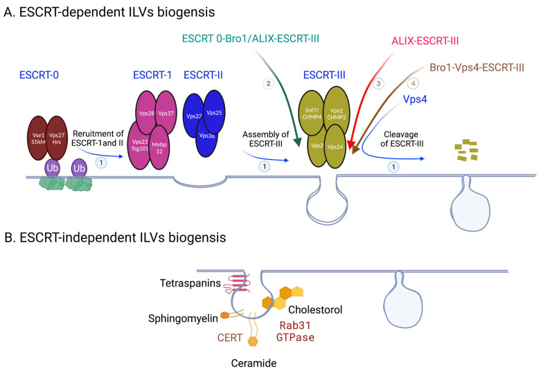
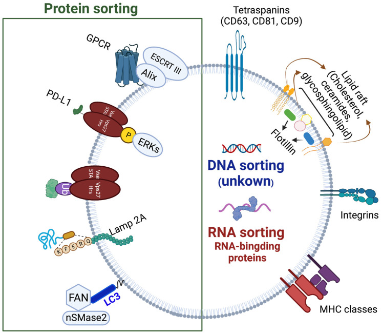
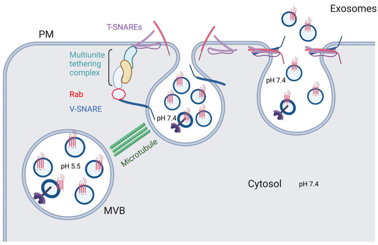
Verification
Claims the paper makes
The claims and statistics of this paper haven’t been checked yet.
Run the check — free, no account needed. It reads the open-access text and checks the sources it cites.
Claims other papers make about this one
Each is checked against this paper’s own text: does it say what the citing paper says it does?
In the citing paper
“Exosome biogenesis and secretion are thus seen as potential therapeutic targets, as modulating these processes could disrupt pathological interactions between tumor cells and their microenvironment.”
From Emerging role of exosomes in cancer therapy: progress and challenges (2025) · cites this paper as [19]
In the citing paper
“For instance, in cancer, exosome biogenesis can be upregulated, leading to increased secretion of tumor-derived exosomes that carry oncogenic factors, which can promote tumor growth, metastasis, and immune evasion.”
From Emerging role of exosomes in cancer therapy: progress and challenges (2025) · cites this paper as [19]
Self-check
Percentages, sums and reported differences re-computed from the paper’s own numbers.
The self-check needs the paper’s full text.
Citations
Cites
Isolation and Characterization of Exosomes from Cell Culture Supernatants and Biological Fluids
Théry, C., Amigorena, S., Raposo, G., & Clayton, A. (2006) · Current Protocols in Cell Biology · doi:10.1002/0471143030.cb0322s30 · Cited by 5,766
Rab GTPases as coordinators of vesicle traffic
Stenmark, H. (2009) · Nature Reviews Molecular Cell Biology · doi:10.1038/nrm2728 · Cited by 3,413
Pegtel, D. M., & Gould, S. J. (2019) · Annual Review of Biochemistry · doi:10.1146/annurev-biochem-013118-111902 · Cited by 2,682
Syndecan–syntenin–ALIX regulates the biogenesis of exosomes
Baietti, M. F., Zhang, Z., Mortier, E., Melchior, A., Degeest, G., Geeraerts, A., Ivarsson, Y., Depoortere, F., Coomans, C., Vermeiren, E., et al. (2012) · Nature Cell Biology · doi:10.1038/ncb2502 · Cited by 1,899
Exosomes Mediate Stromal Mobilization of Autocrine Wnt-PCP Signaling in Breast Cancer Cell Migration
Luga, V., Zhang, L., Viloria-Petit, A. M., Ogunjimi, A. A., Inanlou, M. R., Chiu, E., Buchanan, M., Hosein, A. N., Basik, M., & Wrana, J. L. (2012) · Cell · doi:10.1016/j.cell.2012.11.024 · Cited by 1,311
Tian, T., Zhu, Y.-L., Zhou, Y.-Y., Liang, G.-F., Wang, Y.-Y., Hu, F.-H., & Xiao, Z.-D. (2014) · Journal of Biological Chemistry · doi:10.1074/jbc.m114.588046 · Cited by 726
Banks, W. A., Sharma, P., Bullock, K. M., Hansen, K. M., Ludwig, N., & Whiteside, T. L. (2020) · International Journal of Molecular Sciences · doi:10.3390/ijms21124407 · Cited by 561 · cited 1×
“Adsorptive transcytosis, a mechanism that facilitates transport through the cell by interactions of positive and negative molecular charges, has also been suggested [106].”
BackgroundTargeting and Crossing the Blood-Brain Barrier with Extracellular Vesicles
Saint-Pol, J., Gosselet, F., Duban-Deweer, S., Pottiez, G., & Karamanos, Y. (2020) · Cells · doi:10.3390/cells9040851 · Cited by 503
A platform for actively loading cargo RNA to elucidate limiting steps in EV‐mediated delivery
Hung, M. E., & Leonard, J. N. (2016) · Journal of Extracellular Vesicles · doi:10.3402/jev.v5.31027 · Cited by 251
Research works printed but not linked: 109
- Woodman, 2008, Multivesicular bodies: Co-ordinated progression to maturity, Curr. Opin. Cell Biol., 20, 408, 10.1016/j.ceb.2008.04.001 · DOI
- Grant, 2009, Pathways and mechanisms of endocytic recycling, Nat. Rev. Mol. Cell Biol., 10, 597, 10.1038/nrm2755 · DOI · Paper page
- Megha, 2004, Ceramide selectively displaces cholesterol from ordered lipid domains (rafts): Implications for lipid raft structure and function, J. Biol. Chem., 279, 9997, 10.1074/jbc.M309992200 · DOI
- Sonnichsen, 2000, Distinct membrane domains on endosomes in the recycling pathway visualized by multicolor imaging of Rab4, Rab5, and Rab11, J. Cell Biol., 149, 901, 10.1083/jcb.149.4.901 · DOI · Paper page
- Charrin, 2011, The tetraspanin CD63 regulates ESCRT-independent and -dependent endosomal sorting during melanogenesis, Dev. Cell, 21, 708, 10.1016/j.devcel.2011.08.019 · DOI · Paper page
- Hurley, 2015, ESCRTs are everywhere, EMBO J., 34, 2398, 10.15252/embj.201592484 · DOI
- Schoneberg, 2017, Reverse-topology membrane scission by the ESCRT proteins, Nat. Rev. Mol. Cell Biol., 18, 5, 10.1038/nrm.2016.121 · DOI
- McCullough, 2018, Structures, Functions, and Dynamics of ESCRT-III/Vps4 Membrane Remodeling and Fission Complexes, Annu. Rev. Cell Dev. Biol., 34, 85, 10.1146/annurev-cellbio-100616-060600 · DOI
- Winter, 2006, Exploring the ESCRTing machinery in eukaryotes, Trends Plant Sci., 11, 115, 10.1016/j.tplants.2006.01.008 · DOI
- Raiborg, 2001, FYVE and coiled-coil domains determine the specific localisation of Hrs to early endosomes, J. Cell Sci., 114, 2255, 10.1242/jcs.114.12.2255 · DOI
- Kostelansky, 2006, Structural and functional organization of the ESCRT-I trafficking complex, Cell, 125, 113, 10.1016/j.cell.2006.01.049 · DOI · Paper page
- Morvan, 2012, Pkh1/2-dependent phosphorylation of Vps27 regulates ESCRT-I recruitment to endosomes, Mol. Biol. Cell, 23, 4054, 10.1091/mbc.e12-01-0001 · DOI
- Babst, 2002, Escrt-III: An endosome-associated heterooligomeric protein complex required for mvb sorting, Dev. Cell, 3, 271, 10.1016/S1534-5807(02)00220-4 · DOI
- Chiaruttini, 2015, Relaxation of Loaded ESCRT-III Spiral Springs Drives Membrane Deformation, Cell, 163, 866, 10.1016/j.cell.2015.10.017 · DOI
- Lata, 2008, Helical structures of ESCRT-III are disassembled by VPS4, Science, 321, 1354, 10.1126/science.1161070 · DOI
- Su, 2017, Mechanism of Vps4 hexamer function revealed by cryo-EM, Sci. Adv., 3, e1700325, 10.1126/sciadv.1700325 · DOI
- Adell, 2014, Coordinated binding of Vps4 to ESCRT-III drives membrane neck constriction during MVB vesicle formation, J. Cell Biol., 205, 33, 10.1083/jcb.201310114 · DOI
- Tang, 2016, ESCRT-III activation by parallel action of ESCRT-I/II and ESCRT-0/Bro1 during MVB biogenesis, eLife, 5, e15507, 10.7554/eLife.15507 · DOI
- Larios, 2020, ALIX- and ESCRT-III-dependent sorting of tetraspanins to exosomes, J. Cell Biol., 219, e201904113, 10.1083/jcb.201904113 · DOI · Paper page
- Tseng, 2021, Bro1 stimulates Vps4 to promote intralumenal vesicle formation during multivesicular body biogenesis, J. Cell Biol., 220, e202102070, 10.1083/jcb.202102070 · DOI
- White, 2006, EGF stimulates annexin 1-dependent inward vesiculation in a multivesicular endosome subpopulation, EMBO J., 25, 1, 10.1038/sj.emboj.7600759 · DOI
- Futter, 2001, Human VPS34 is required for internal vesicle formation within multivesicular endosomes, J. Cell Biol., 155, 1251, 10.1083/jcb.200108152 · DOI
- Zhu, 2013, Mutation of SIMPLE in Charcot-Marie-Tooth 1C alters production of exosomes, Mol. Biol. Cell, 24, 1619, 10.1091/mbc.e12-07-0544 · DOI
- Misra, 1999, Crystal structure of a phosphatidylinositol 3-phosphate-specific membrane-targeting motif, the FYVE domain of Vps27p, Cell, 97, 657, 10.1016/S0092-8674(00)80776-X · DOI
- Fyfe, 2011, Association of the endosomal sorting complex ESCRT-II with the Vps20 subunit of ESCRT-III generates a curvature-sensitive complex capable of nucleating ESCRT-III filaments, J. Biol. Chem., 286, 34262, 10.1074/jbc.M111.266411 · DOI
- Jorge, 2013, The intracellular interactome of tetraspanin-enriched microdomains reveals their function as sorting machineries toward exosomes, J. Biol. Chem., 288, 11649, 10.1074/jbc.M112.445304 · DOI
- Chairoungdua, 2010, Exosome release of beta-catenin: A novel mechanism that antagonizes Wnt signaling, J. Cell Biol., 190, 1079, 10.1083/jcb.201002049 · DOI
- Hurwitz, 2016, Nanoparticle analysis sheds budding insights into genetic drivers of extracellular vesicle biogenesis, J. Extracell Vesicles, 5, 31295, 10.3402/jev.v5.31295 · DOI
- Kajimoto, 2013, Ongoing activation of sphingosine 1-phosphate receptors mediates maturation of exosomal multivesicular endosomes, Nat. Commun., 4, 2712, 10.1038/ncomms3712 · DOI
- Trajkovic, 2008, Ceramide triggers budding of exosome vesicles into multivesicular endosomes, Science, 319, 1244, 10.1126/science.1153124 · DOI
- Wei, 2021, RAB31 marks and controls an ESCRT-independent exosome pathway, Cell Res., 31, 157, 10.1038/s41422-020-00409-1 · DOI
- Crivelli, 2022, Function of ceramide transfer protein for biogenesis and sphingolipid composition of extracellular vesicles, J. Extracell Vesicles, 11, e12233, 10.1002/jev2.12233 · DOI
- Polo, 2002, A single motif responsible for ubiquitin recognition and monoubiquitination in endocytic proteins, Nature, 416, 451, 10.1038/416451a · DOI · Paper page
- Hicke, 2003, Regulation of membrane protein transport by ubiquitin and ubiquitin-binding proteins, Annu. Rev. Cell Dev. Biol., 19, 141, 10.1146/annurev.cellbio.19.110701.154617 · DOI
- Raiborg, 2001, Hrs recruits clathrin to early endosomes, EMBO J., 20, 5008, 10.1093/emboj/20.17.5008 · DOI
- Raiborg, 2002, Hrs sorts ubiquitinated proteins into clathrin-coated microdomains of early endosomes, Nat. Cell Biol., 4, 394, 10.1038/ncb791 · DOI · Paper page
- Hurley, 2006, The ESCRT complexes: Structure and mechanism of a membrane-trafficking network, Annu. Rev. Biophys. Biomol. Struct., 35, 277, 10.1146/annurev.biophys.35.040405.102126 · DOI · Paper page
- Luhtala, 2004, Bro1 coordinates deubiquitination in the multivesicular body pathway by recruiting Doa4 to endosomes, J. Cell Biol., 166, 717, 10.1083/jcb.200403139 · DOI
- Guan, 2022, HRS phosphorylation drives immunosuppressive exosome secretion and restricts CD8(+) T-cell infiltration into tumors, Nat. Commun., 13, 4078, 10.1038/s41467-022-31713-6 · DOI
- Reggiori, 2001, Sorting of proteins into multivesicular bodies: Ubiquitin-dependent and -independent targeting, EMBO J., 20, 5176, 10.1093/emboj/20.18.5176 · DOI · Paper page
- Epple, 2003, Intravacuolar membrane lysis in Saccharomyces cerevisiae. Does vacuolar targeting of Cvt17/Aut5p affect its function?, J. Biol. Chem., 278, 7810, 10.1074/jbc.M209309200 · DOI
- Yamashita, 2008, Ubiquitin-independent binding of Hrs mediates endosomal sorting of the interleukin-2 receptor beta-chain, J. Cell Sci., 121, 1727, 10.1242/jcs.024455 · DOI
- Dores, M.R., Grimsey, N.J., Mendez, F., and Trejo, J. (2016). ALIX Regulates the Ubiquitin-Independent Lysosomal Sorting of the P2Y1 Purinergic Receptor via a YPX3L Motif. PLoS ONE, 11. · DOI
- Ferreira, 2022, LAMP2A regulates the loading of proteins into exosomes, Sci. Adv., 8, eabm1140, 10.1126/sciadv.abm1140 · DOI
- Leidal, 2020, The LC3-conjugation machinery specifies the loading of RNA-binding proteins into extracellular vesicles, Nat. Cell Biol., 22, 187, 10.1038/s41556-019-0450-y · DOI
- Vazquez, 2013, Sumoylated hnRNPA2B1 controls the sorting of miRNAs into exosomes through binding to specific motifs, Nat. Commun., 4, 2980, 10.1038/ncomms3980 · DOI · Paper page
- Cha, 2015, KRAS-dependent sorting of miRNA to exosomes, eLife, 4, e07197, 10.7554/eLife.07197 · DOI
- McKenzie, 2016, KRAS-MEK Signaling Controls Ago2 Sorting into Exosomes, Cell Rep., 15, 978, 10.1016/j.celrep.2016.03.085 · DOI
- Teng, 2017, MVP-mediated exosomal sorting of miR-193a promotes colon cancer progression, Nat. Commun., 8, 14448, 10.1038/ncomms14448 · DOI · Paper page
- Mukherjee, 2016, Reversible HuR-microRNA binding controls extracellular export of miR-122 and augments stress response, EMBO Rep., 17, 1184, 10.15252/embr.201541930 · DOI · Paper page
- Pastuzyn, 2018, The Neuronal Gene Arc Encodes a Repurposed Retrotransposon Gag Protein that Mediates Intercellular RNA Transfer, Cell, 172, 275, 10.1016/j.cell.2017.12.024 · DOI · Paper page
- Fabbiano, 2020, RNA packaging into extracellular vesicles: An orchestra of RNA-binding proteins?, J. Extracell Vesicles, 10, e12043, 10.1002/jev2.12043 · DOI
- Perrin, 2021, Retrofusion of intralumenal MVB membranes parallels viral infection and coexists with exosome release, Curr. Biol., 31, 3884, 10.1016/j.cub.2021.06.022 · DOI
- Kim, 2019, Exosomal release through TRPML1-mediated lysosomal exocytosis is required for adipogenesis, Biochem. Biophys. Res. Commun., 510, 409, 10.1016/j.bbrc.2019.01.115 · DOI
- Mobius, 2002, Immunoelectron microscopic localization of cholesterol using biotinylated and non-cytolytic perfringolysin O, J. Histochem. Cytochem., 50, 43, 10.1177/002215540205000105 · DOI
- Baixauli, 2016, ISGylation controls exosome secretion by promoting lysosomal degradation of MVB proteins, Nat. Commun., 7, 13588, 10.1038/ncomms13588 · DOI
- Majumder, 2015, Mahogunin regulates fusion between amphisomes/MVBs and lysosomes via ubiquitination of TSG101, Cell Death Dis., 6, e1970, 10.1038/cddis.2015.257 · DOI
- Bucci, 2000, Rab7: A key to lysosome biogenesis, Mol. Biol. Cell, 11, 467, 10.1091/mbc.11.2.467 · DOI
- Fei, 2021, Neddylation of Coro1a determines the fate of multivesicular bodies and biogenesis of extracellular vesicles, J. Extracell Vesicles, 10, e12153, 10.1002/jev2.12153 · DOI
- Guo, 2018, Autophagy-independent effects of autophagy-related-5 (Atg5) on exosome production and metastasis, Mol. Cell. Oncol., 5, e1445941, 10.1080/23723556.2018.1445941 · DOI
- Ponpuak, 2015, Secretory autophagy, Curr. Opin. Cell Biol., 35, 106, 10.1016/j.ceb.2015.04.016 · DOI
- Hessvik, 2018, Current knowledge on exosome biogenesis and release, Cell Mol. Life Sci., 75, 193, 10.1007/s00018-017-2595-9 · DOI · Paper page
- Sinha, 2016, Cortactin promotes exosome secretion by controlling branched actin dynamics, J. Cell Biol., 214, 197, 10.1083/jcb.201601025 · DOI
- Savina, 2002, The exosome pathway in K562 cells is regulated by Rab11, J. Cell Sci., 115, 2505, 10.1242/jcs.115.12.2505 · DOI
- Koles, 2012, Mechanism of evenness interrupted (Evi)-exosome release at synaptic boutons, J. Biol. Chem., 287, 16820, 10.1074/jbc.M112.342667 · DOI
- Ostrowski, 2010, Rab27a and Rab27b control different steps of the exosome secretion pathway, Nat. Cell Biol., 12, 19, 10.1038/ncb2000 · DOI
- Cabezas, 2005, Alix regulates cortical actin and the spatial distribution of endosomes, J. Cell Sci., 118, 2625, 10.1242/jcs.02382 · DOI
- Mittelbrunn, 2011, Endosomal clathrin drives actin accumulation at the immunological synapse, J. Cell Sci., 124, 820, 10.1242/jcs.078832 · DOI
- Pfeffer, 2007, Unsolved mysteries in membrane traffic, Annu. Rev. Biochem., 76, 629, 10.1146/annurev.biochem.76.061705.130002 · DOI
- Brocker, 2010, Multisubunit tethering complexes and their role in membrane fusion, Curr. Biol., 20, R943, 10.1016/j.cub.2010.09.015 · DOI
- Vats, 2022, Role of SNAREs in Unconventional Secretion-Focus on the VAMP7-Dependent Secretion, Front. Cell Dev. Biol., 10, 884020, 10.3389/fcell.2022.884020 · DOI
- Chaineau, 2009, Multiple roles of the vesicular-SNARE TI-VAMP in post-Golgi and endosomal trafficking, FEBS Lett., 583, 3817, 10.1016/j.febslet.2009.10.026 · DOI
- Pilliod, 2020, Clearance of intracellular tau protein from neuronal cells via VAMP8-induced secretion, J. Biol. Chem., 295, 17827, 10.1074/jbc.RA120.013553 · DOI
- Ren, 2017, Identification of syntaxin 4 as an essential factor for the hepatitis C virus life cycle, Eur. J. Cell Biol., 96, 542, 10.1016/j.ejcb.2017.06.002 · DOI
- Yu, 2020, Reduced oligodendrocyte exosome secretion in multiple system atrophy involves SNARE dysfunction, Brain, 143, 1780, 10.1093/brain/awaa110 · DOI
- Peak, 2020, Syntaxin 6-mediated exosome secretion regulates enzalutamide resistance in prostate cancer, Mol. Carcinog., 59, 62, 10.1002/mc.23129 · DOI
- Qian, 2021, Reduced O-GlcNAcylation of SNAP-23 promotes cisplatin resistance by inducing exosome secretion in ovarian cancer, Cell Death Discov., 7, 112, 10.1038/s41420-021-00489-x · DOI
- Verweij, 2018, Quantifying exosome secretion from single cells reveals a modulatory role for GPCR signaling, J. Cell Biol., 217, 1129, 10.1083/jcb.201703206 · DOI
- Wei, 2017, Pyruvate kinase type M2 promotes tumour cell exosome release via phosphorylating synaptosome-associated protein 23, Nat. Commun., 8, 14041, 10.1038/ncomms14041 · DOI
- Navarro, 2016, YKT6 expression, exosome release, and survival in non-small cell lung cancer, Oncotarget, 7, 51515, 10.18632/oncotarget.9862 · DOI
- Sun, 2020, LncRNA PVT1 promotes exosome secretion through YKT6, RAB7, and VAMP3 in pancreatic cancer, Aging, 12, 10427, 10.18632/aging.103268 · DOI
- Yang, 2019, Long non-coding RNA HOTAIR promotes exosome secretion by regulating RAB35 and SNAP23 in hepatocellular carcinoma, Mol. Cancer, 18, 78, 10.1186/s12943-019-0990-6 · DOI
- Mulcahy, 2014, Routes and mechanisms of extracellular vesicle uptake, J. Extracell Vesicles, 3, 24641, 10.3402/jev.v3.24641 · DOI · Paper page
- Montecalvo, 2012, Mechanism of transfer of functional microRNAs between mouse dendritic cells via exosomes, Blood, 119, 756, 10.1182/blood-2011-02-338004 · DOI · Paper page
- Feng, 2010, Cellular internalization of exosomes occurs through phagocytosis, Traffic, 11, 675, 10.1111/j.1600-0854.2010.01041.x · DOI
- Schiffelers, 2017, Cellular uptake of extracellular vesicles is mediated by clathrin-independent endocytosis and macropinocytosis, J. Control. Release, 266, 100, 10.1016/j.jconrel.2017.09.019 · DOI
- Nanbo, 2013, Exosomes derived from Epstein-Barr virus-infected cells are internalized via caveola-dependent endocytosis and promote phenotypic modulation in target cells, J. Virol., 87, 10334, 10.1128/JVI.01310-13 · DOI
- Tkach, 2017, Qualitative differences in T-cell activation by dendritic cell-derived extracellular vesicle subtypes, EMBO J., 36, 3012, 10.15252/embj.201696003 · DOI
- Sung, 2019, Extracellular vesicles from CLEC2-activated platelets enhance dengue virus-induced lethality via CLEC5A/TLR2, Nat. Commun., 10, 2402, 10.1038/s41467-019-10360-4 · DOI
- Gurung, 2021, The exosome journey: From biogenesis to uptake and intracellular signalling, Cell Commun. Signal., 19, 47, 10.1186/s12964-021-00730-1 · DOI
- Somiya, 2020, Where does the cargo go?: Solutions to provide experimental support for the “extracellular vesicle cargo transfer hypothesis”, J. Cell Commun. Signal., 14, 135, 10.1007/s12079-020-00552-9 · DOI
- Ughetto, 2022, Uptake, functionality, and re-release of extracellular vesicle-encapsulated cargo, Cell Rep., 39, 110651, 10.1016/j.celrep.2022.110651 · DOI
- Joshi, 2020, Endocytosis of Extracellular Vesicles and Release of Their Cargo from Endosomes, ACS Nano, 14, 4444, 10.1021/acsnano.9b10033 · DOI
- Polanco, 2021, Exosomes induce endolysosomal permeabilization as a gateway by which exosomal tau seeds escape into the cytosol, Acta Neuropathol., 141, 235, 10.1007/s00401-020-02254-3 · DOI
- Kanada, 2015, Differential fates of biomolecules delivered to target cells via extracellular vesicles, Proc. Natl. Acad. Sci. USA, 112, E1433, 10.1073/pnas.1418401112 · DOI
- Morad, 2019, Tumor-Derived Extracellular Vesicles Breach the Intact Blood-Brain Barrier via Transcytosis, ACS Nano, 13, 13853, 10.1021/acsnano.9b04397 · DOI
- Joshi, 2021, Heparan sulfate proteoglycan-mediated dynamin-dependent transport of neural stem cell exosomes in an in vitro blood-brain barrier model, Eur. J. Neurosci., 53, 706, 10.1111/ejn.14974 · DOI
- Royo, F., Thery, C., Falcon-Perez, J.M., Nieuwland, R., and Witwer, K.W. (2020). Methods for Separation and Characterization of Extracellular Vesicles: Results of a Worldwide Survey Performed by the ISEV Rigor and Standardization Subcommittee. Cells, 9. · DOI
- Carter, 2022, Challenges and directions in studying cell-cell communication by extracellular vesicles, Nat. Rev. Mol. Cell Biol., 23, 369, 10.1038/s41580-022-00460-3 · DOI · Paper page
- Sansone, 2017, Packaging and transfer of mitochondrial DNA via exosomes regulate escape from dormancy in hormonal therapy-resistant breast cancer, Proc. Natl. Acad. Sci. USA, 114, E9066, 10.1073/pnas.1704862114 · DOI
- Jang, 2019, Mitochondrial protein enriched extracellular vesicles discovered in human melanoma tissues can be detected in patient plasma, J. Extracell Vesicles, 8, 1635420, 10.1080/20013078.2019.1635420 · DOI
- Wang, 2020, Detection of mitochondria-pertinent components in exosomes, Mitochondrion, 55, 100, 10.1016/j.mito.2020.09.006 · DOI
- Crewe, 2021, Extracellular vesicle-based interorgan transport of mitochondria from energetically stressed adipocytes, Cell Metab., 33, 1853, 10.1016/j.cmet.2021.08.002 · DOI
- Rosina, 2022, Ejection of damaged mitochondria and their removal by macrophages ensure efficient thermogenesis in brown adipose tissue, Cell Metab., 34, 533, 10.1016/j.cmet.2022.02.016 · DOI
- Kim, 2021, Mitovesicles are a novel population of extracellular vesicles of mitochondrial origin altered in Down syndrome, Sci. Adv., 7, eabe5085, 10.1126/sciadv.abe5085 · DOI
- Karn, V., Ahmed, S., Tsai, L.W., Dubey, R., Ojha, S., Singh, H.N., Kumar, M., Gupta, P.K., Sadhu, S., and Jha, N.K. (2021). Extracellular Vesicle-Based Therapy for COVID-19: Promises, Challenges and Future Prospects. Biomedicines, 9. · DOI
- McCann, 2020, Reporter mice for isolating and auditing cell type-specific extracellular vesicles in vivo, Genesis, 58, e23369, 10.1002/dvg.23369 · DOI
- Neckles, 2019, A transgenic inducible GFP extracellular-vesicle reporter (TIGER) mouse illuminates neonatal cortical astrocytes as a source of immunomodulatory extracellular vesicles, Sci. Rep., 9, 3094, 10.1038/s41598-019-39679-0 · DOI · Paper page
- Sung, 2020, A live cell reporter of exosome secretion and uptake reveals pathfinding behavior of migrating cells, Nat. Commun., 11, 2092, 10.1038/s41467-020-15747-2 · DOI
Cited by
Sorted by citations
Emerging role of exosomes in cancer therapy: progress and challenges
Li, J., Wang, J., & Chen, Z. (2025) · Molecular Cancer · doi:10.1186/s12943-024-02215-4 · Cited by 297
Metadata
- Authors
- Krylova, S. V., & Feng, D.
- Venue
- International Journal of Molecular Sciences
- Publisher
- MDPI AG
- Published
- 2023-01-10
- Volume
- 24
- Issue
- 2
- Pages
- 1337
- ISSN
- 1422-0067
- PMC
- PMC9865891
- Cited by
- 678
- References
- 9
- Language
- en
About these checks
Registry, retraction, Cites and statistics results are computed, not judged, with one exception: a printed reference that no index lists is read once by a model before it counts as a citation error. Claims are extracted by a model and kept only when their quote appears verbatim in the text; each cited source is then checked, with a model’s help, for whether it says what the claim relies on. Those verdicts can be wrong — read the claim’s quote and the cited source before relying on one.
Statistics and arithmetic are recomputed, not judged.
Confidence bands are shown only for fields calibrated on our annotated test set. None is calibrated yet, so no band is shown.
What counts as a claim ›How we measure confidence ›How we check this ›
Are you an author, or do you see a problem with this page? Request a review
