

The DOI matches its Crossref record; 5 of the 5 references it cites were found in a registry or an index.
Altered Extracellular Vesicle miRNA Profile in Prodromal Alzheimer’s Disease
Abstract
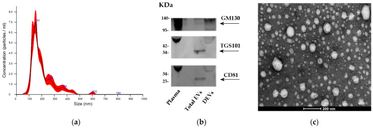
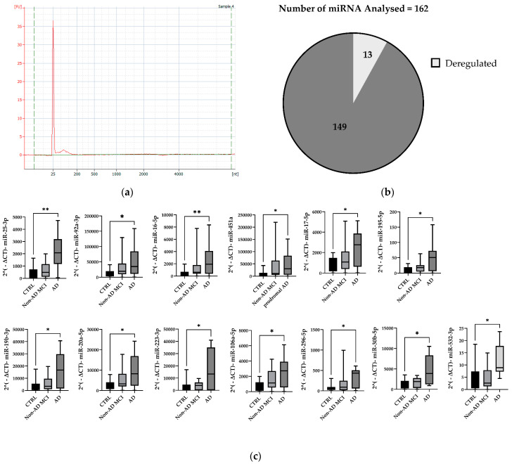
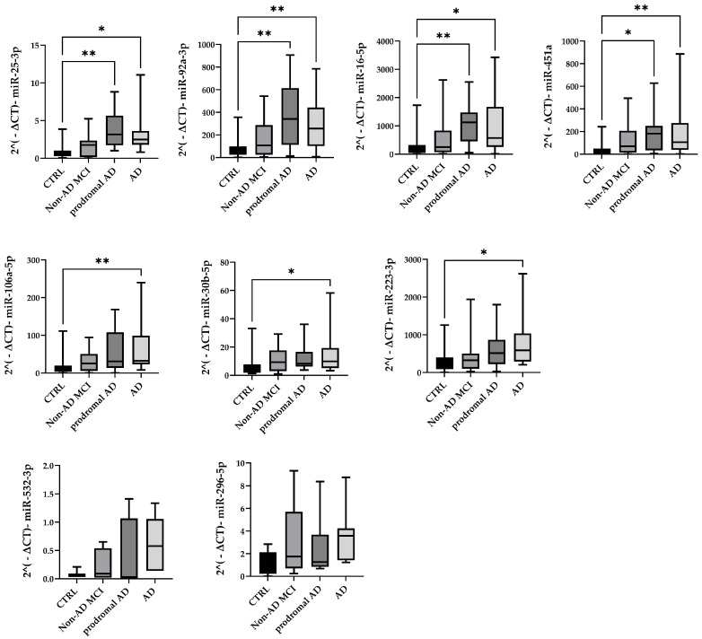
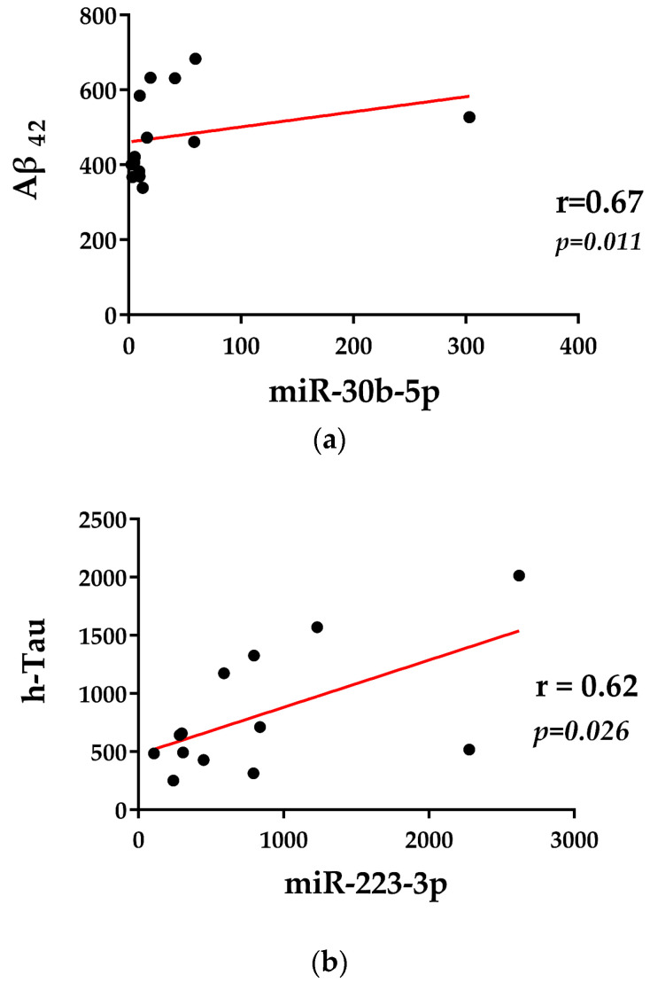
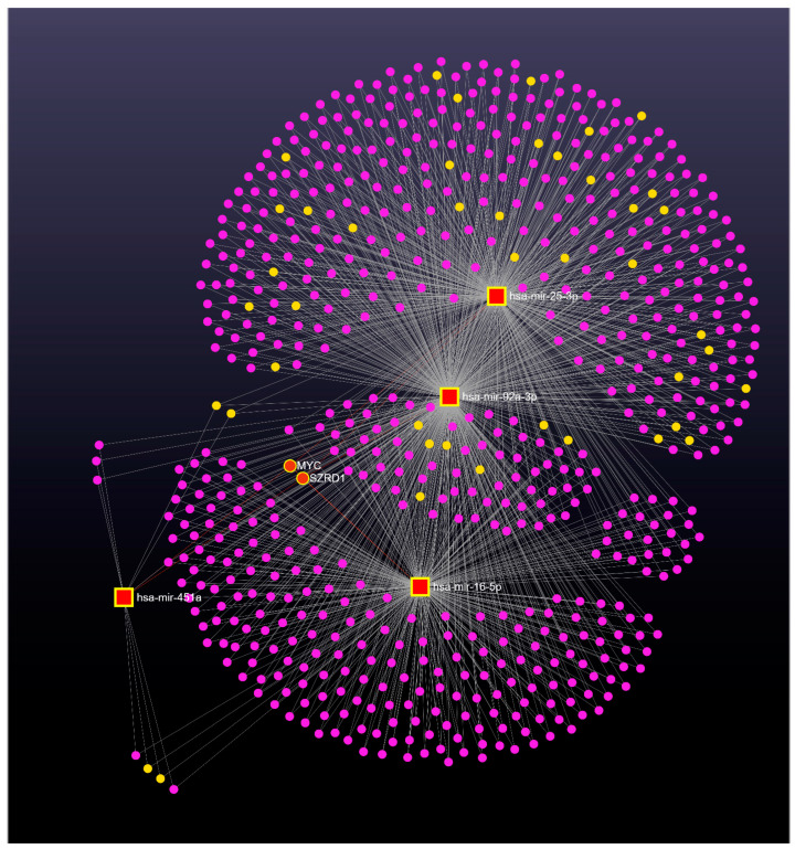
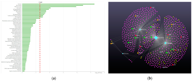
Extracellular vesicles (EVs) are nanosized vesicles released by almost all body tissues, representing important mediators of cellular communication, and are thus promising candidate biomarkers for neurodegenerative diseases like Alzheimer’s disease (AD). The aim of the present study was to isolate total EVs from plasma and characterize their microRNA (miRNA) contents in AD patients. We isolated total EVs from the plasma of all recruited subjects using ExoQuickULTRA exosome precipitation solution (SBI). Subsequently, circulating total EVs were characterized using Nanosight nanoparticle tracking analysis (NTA), transmission electron microscopy (TEM), and Western blotting. A panel of 754 miRNAs was determined with RT-qPCR using TaqMan OpenArray technology in a QuantStudio 12K System (Thermo Fisher Scientific). The results demonstrated that plasma EVs showed widespread deregulation of specific miRNAs (miR-106a-5p, miR-16-5p, miR-17-5p, miR-195-5p, miR-19b-3p, miR-20a-5p, miR-223-3p, miR-25-3p, miR-296-5p, miR-30b-5p, miR-532-3p, miR-92a-3p, and miR-451a), some of which were already known to be associated with neurological pathologies. A further validation analysis also confirmed a significant upregulation of miR-16-5p, miR-25-3p, miR-92a-3p, and miR-451a in prodromal AD patients, suggesting these dysregulated miRNAs are involved in the early progression of AD.
Overview
The claims and statistics of this paper haven’t been checked yet.
Run the check — free, no account needed. It reads the open-access text and checks the sources it cites.
Integrity signals
Registry
Cites
Statistics
Figures
Verification
Claims the paper makes
The claims and statistics of this paper haven’t been checked yet.
Run the check — free, no account needed. It reads the open-access text and checks the sources it cites.
Claims other papers make about this one
Each is checked against this paper’s own text: does it say what the citing paper says it does?
In the citing paper
“In the last year, Visconte et al. identified a panel of EVs-derived miRNAs deregulated in the plasma of AD patients, including miR-92a-3p, which was up-regulated also in prodromal AD [61].”
From miR-92a-3p and miR-320a are Upregulated in Plasma Neuron-Derived Extracellular Vesicles of Patients with Frontotemporal Dementia (2025) · cites this paper as [61]
Self-check
Percentages, sums and reported differences re-computed from the paper’s own numbers.
The self-check needs the paper’s full text.
Citations
Cites
Age-dependent instability of mature neuronal fate in induced neurons from Alzheimer’s patients
Mertens, J., Herdy, J. R., Traxler, L., Schafer, S. T., Schlachetzki, J. C. M., Böhnke, L., Reid, D. A., Lee, H., Zangwill, D., Fernandes, D. P., et al. (2021) · Cell Stem Cell · doi:10.1016/j.stem.2021.04.004 · Cited by 240
Polycomb repressive complex 2 (PRC2) silences genes responsible for neurodegeneration
von Schimmelmann, M., Feinberg, P. A., Sullivan, J. M., Ku, S. M., Badimon, A., Duff, M. K., Wang, Z., Lachmann, A., Dewell, S., Ma'ayan, A., et al. (2016) · Nature Neuroscience · doi:10.1038/nn.4360 · Cited by 225
Circulating miRNAs as Potential Biomarkers in Alzheimer's Disease
Galimberti, D., Villa, C., Fenoglio, C., Serpente, M., Ghezzi, L., Cioffi, S. M. G., Arighi, A., Fumagalli, G., & Scarpini, E. (2014) · Journal of Alzheimer’s Disease · doi:10.3233/jad-140756 · Cited by 222
Self-citationHarrison, E. B., Hochfelder, C. G., Lamberty, B. G., Meays, B. M., Morsey, B. M., Kelso, M. L., Fox, H. S., & Yelamanchili, S. V. (2016) · FEBS Open Bio · doi:10.1002/2211-5463.12092 · Cited by 151
Fitz, N. F., Wang, J., Kamboh, M. I., Koldamova, R., & Lefterov, I. (2021) · Neurobiology of Disease · doi:10.1016/j.nbd.2021.105481 · Cited by 49
Research works printed but not linked: 66
- Liss, 2021, Practical recommendations for timely, accurate diagnosis of symptomatic Alzheimer’s disease (MCI and dementia) in primary care: A review and synthesis, J. Intern. Med., 290, 310, 10.1111/joim.13244 · DOI · Paper page
- Blennow, 2017, A Review of Fluid Biomarkers for Alzheimer’s Disease: Moving from CSF to Blood, Neurol. Ther., 6, 15, 10.1007/s40120-017-0073-9 · DOI · Paper page
- Aharon, 2020, Extracellular Vesicles of Alzheimer’s Disease Patients as a Biomarker for Disease Progression, Mol. Neurobiol., 57, 4156, 10.1007/s12035-020-02013-1 · DOI · Paper page
- Aulston, 2019, Extracellular Vesicles Isolated from Familial Alzheimer’s Disease Neuronal Cultures Induce Aberrant Tau Phosphorylation in the Wild-Type Mouse Brain, J. Alzheimer’s Dis. JAD, 72, 575, 10.3233/JAD-190656 · DOI
- Duggan, 2022, Exosomes in Age-Related Cognitive Decline: Mechanistic Insights and Improving Outcomes, Front. Aging Neurosci., 14, 834775, 10.3389/fnagi.2022.834775 · DOI
- Xiao, 2017, The Role of Exosomes in the Pathogenesis of Alzheimer’ Disease, Transl. Neurodegener., 6, 3, 10.1186/s40035-017-0072-x · DOI
- Pathan, 2019, Vesiclepedia 2019: A Compendium of RNA, Proteins, Lipids and Metabolites in Extracellular Vesicles, Nucleic Acids Res., 47, D516, 10.1093/nar/gky1029 · DOI
- Kalluri, 2020, The Biology, Function, and Biomedical Applications of Exosomes, Science, 367, eaau6977, 10.1126/science.aau6977 · DOI · Paper page
- Witwer, 2018, Minimal Information for Studies of Extracellular Vesicles 2018 (MISEV2018): A Position Statement of the International Society for Extracellular Vesicles and Update of the MISEV2014 Guidelines, J. Extracell. Vesicles, 7, 1535750, 10.1080/20013078.2018.1535750 · DOI · Paper page
- Herschlik, 2015, Emerging Roles of Exosomes in Normal and Pathological Conditions: New Insights for Diagnosis and Therapeutic Applications, Front. Immunol., 6, 203
- Kim, 2017, RNA in Extracellular Vesicles, Wiley Interdiscip. Rev. RNA, 8, e1413, 10.1002/wrna.1413 · DOI
- Abels, 2016, Introduction to Extracellular Vesicles: Biogenesis, RNA Cargo Selection, Content, Release, and Uptake, Cell. Mol. Neurobiol., 36, 301, 10.1007/s10571-016-0366-z · DOI
- Riva, 2016, The Long Non-Coding RNAs in Neurodegenerative Diseases: Novel Mechanisms of Pathogenesis, Curr. Alzheimer Res., 13, 1219, 10.2174/1567205013666160622112234 · DOI
- Kumar, 2016, Are Circulating microRNAs Peripheral Biomarkers for Alzheimer’s Disease?, Biochim. Biophys. Acta (BBA) Mol. Basis Dis., 1862, 1617, 10.1016/j.bbadis.2016.06.001 · DOI
- Campdelacreu, 2019, Exploratory Study on microRNA Profiles from Plasma-Derived Extracellular Vesicles in Alzheimer’s Disease and Dementia with Lewy Bodies, Transl. Neurodegener., 8, 1
- Sproviero, D., Gagliardi, S., Zucca, S., Arigoni, M., Giannini, M., Garofalo, M., Olivero, M., Dell’Orco, M., Pansarasa, O., and Bernuzzi, S. (2021). Different miRNA Profiles in Plasma Derived Small and Large Extracellular Vesicles from Patients with Neurodegenerative Diseases. Int. J. Mol. Sci., 22. · DOI
- Martinez, 2019, MicroRNAs as Diagnostic and Therapeutic Tools for Alzheimer’s Disease: Advances and Limitations, Neural Regen. Res., 14, 242, 10.4103/1673-5374.244784 · DOI
- Serpente, M., Fenoglio, C., D’Anca, M., Arcaro, M., Sorrentino, F., Visconte, C., Arighi, A., Fumagalli, G.G., Porretti, L., and Cattaneo, A. (2020). MiRNA Profiling in Plasma Neural-Derived Small Extracellular Vesicles from Patients with Alzheimer’s Disease. Cells, 9. · DOI · Paper page
- Hulse, 2013, Genetic Variants Contribute to Gene Expression Variability in Humans, Genetics, 193, 95, 10.1534/genetics.112.146779 · DOI
- Lukiw, 2013, Variability in Micro RNA (miRNA) Abundance, Speciation and Complexity amongst Different Human Populations and Potential Relevance to Alzheimer’s Disease (AD), Front. Cell Neurosci., 7, 133, 10.3389/fncel.2013.00133 · DOI
- Visconte, 2023, Plasma Microglial-Derived Extracellular Vesicles Are Increased in Frail Patients with Mild Cognitive Impairment and Exert a Neurotoxic Effect, Geroscience, 45, 1557, 10.1007/s11357-023-00746-0 · DOI
- Mogilyansky, 2013, The miR-17/92 Cluster: A Comprehensive Update on Its Genomics, Genetics, Functions and Increasingly Important and Numerous Roles in Health and Disease, Cell Death Differ., 20, 1603, 10.1038/cdd.2013.125 · DOI · Paper page
- Khuu, 2016, The Three Paralogous MicroRNA Clusters in Development and Disease, miR-17-92, miR-106a-363, and miR-106b-25, Scientifica, 2016, 1379643, 10.1155/2016/1379643 · DOI
- Mendell, 2008, miRiad Roles for the miR-17-92 Cluster in Development and Disease, Cell, 133, 217, 10.1016/j.cell.2008.04.001 · DOI
- McKeever, 2018, MicroRNA Expression Levels Are Altered in the Cerebrospinal Fluid of Patients with Young-Onset Alzheimer’s Disease, Mol. Neurobiol., 55, 8826, 10.1007/s12035-018-1032-x · DOI · Paper page
- Riancho, 2017, MicroRNA Profile in Patients with Alzheimer’s Disease: Analysis of miR-9-5p and miR-598 in Raw and Exosome Enriched Cerebrospinal Fluid Samples, J. Alzheimer’s Dis. JAD, 57, 483, 10.3233/JAD-161179 · DOI · Paper page
- Liu, 2014, MicroRNA-193b Is a Regulator of Amyloid Precursor Protein in the Blood and Cerebrospinal Fluid Derived Exosomal microRNA-193b Is a Biomarker of Alzheimer’s Disease, Mol. Med. Rep., 10, 2395, 10.3892/mmr.2014.2484 · DOI · Paper page
- Trindade, 2020, Diagnostic and Therapeutic Potential of Exosomes in Alzheimer’s Disease, J. Neurochem., 156, 162
- Cheng, 2015, Prognostic Serum miRNA Biomarkers Associated with Alzheimer’s Disease Shows Concordance with Neuropsychological and Neuroimaging Assessment, Mol. Psychiatry, 20, 1188, 10.1038/mp.2014.127 · DOI · Paper page
- Cheng, L., Sharples, R.A., Scicluna, B.J., and Hill, A.F. (2014). Exosomes Provide a Protective and Enriched Source of miRNA for Biomarker Profiling Compared to Intracellular and Cell-Free Blood. J. Extracell. Vesicles, 3. · DOI · Paper page
- Cheng, 2020, Small RNA Fingerprinting of Alzheimer’s Disease Frontal Cortex Extracellular Vesiclesand Their Comparison with Peripheral Extracellular Vesicles, J. Extracell. Vesicles, 9, 1766822, 10.1080/20013078.2020.1766822 · DOI
- Lugli, G., Cohen, A.M., Bennett, D.A., Shah, R.C., Fields, C.J., Hernandez, A.G., and Smalheiser, N.R. (2015). Plasma Exosomal miRNAs in Persons with and without Alzheimer Disease: Altered Expression and Prospects for Biomarkers. PLoS ONE, 10. · DOI · Paper page
- Nie, 2020, Differential Expression of Plasma Exo-miRNA in Neurodegenerative Diseases by Next-Generation Sequencing, Front. Neurosci., 14, 438, 10.3389/fnins.2020.00438 · DOI · Paper page
- Li, 2020, Profile of Pathogenic Proteins and MicroRNAs in Plasma-Derived Extracellular Vesicles in Alzheimer’s Disease: A Pilot Study, Neuroscience, 432, 240, 10.1016/j.neuroscience.2020.02.044 · DOI
- Bleazard, 2015, Bias in microRNA Functional Enrichment Analysis, Bioinformatics, 31, 1592, 10.1093/bioinformatics/btv023 · DOI
- Friedman, 2009, Most Mammalian mRNAs Are Conserved Targets of microRNAs, Genome Res., 19, 92, 10.1101/gr.082701.108 · DOI · Paper page
- Tanzer, 2004, Molecular Evolution of a microRNA Cluster, J. Mol. Biol., 339, 327, 10.1016/j.jmb.2004.03.065 · DOI
- Ventura, 2008, Targeted Deletion Reveals Essential and Overlapping Functions of the miR-17 through 92 Family of miRNA Clusters, Cell, 132, 875, 10.1016/j.cell.2008.02.019 · DOI
- Mestdagh, 2010, The miR-17-92 microRNA Cluster Regulates Multiple Components of the TGF-β Pathway in Neuroblastoma, Mol. Cell, 40, 762, 10.1016/j.molcel.2010.11.038 · DOI · Paper page
- Jin, 2022, Circular RNA Rbms1 Inhibited the Development of Myocardial Ischemia Reperfusion Injury by Regulating miR-92a/BCL2L11 Signaling Pathway, Bioengineered, 13, 3082, 10.1080/21655979.2022.2025696 · DOI
- Xiu, 2022, MicroRNA-17-5p Protects against Propofol Anesthesia-Induced Neurotoxicity and Autophagy Impairment via Targeting BCL2L11, Comput. Math. Methods Med., 2022, 6018037, 10.1155/2022/6018037 · DOI
- Petrocca, 2008, Emerging Role of miR-106b-25/miR-17-92 Clusters in the Control of Transforming Growth Factor Beta Signaling, Cancer Res., 68, 8191, 10.1158/0008-5472.CAN-08-1768 · DOI
- Bergmans, 2009, MicroRNA Regulation of Alzheimer’s Amyloid Precursor Protein Expression, Neurobiol. Dis., 33, 422, 10.1016/j.nbd.2008.11.009 · DOI · Paper page
- Rani, 2017, miRNA in Circulating Microvesicles as Biomarkers for Age-Related Cognitive Decline, Front. Aging Neurosci., 9, 323, 10.3389/fnagi.2017.00323 · DOI
- Grillari, 2010, miR-17-92 Cluster: Ups and Downs in Cancer and Aging, Biogerontology, 11, 501, 10.1007/s10522-010-9272-9 · DOI
- Hackl, 2010, miR-17, miR-19b, miR-20a, and miR-106a Are down-Regulated in Human Aging, Aging Cell, 9, 291, 10.1111/j.1474-9726.2010.00549.x · DOI
- Feng, 2023, Decreased miR-451a in Cerebrospinal Fluid, a Marker for Both Cognitive Impairment and Depressive Symptoms in Alzheimer’s Disease, Theranostics, 13, 3021, 10.7150/thno.81826 · DOI
- Xie, 2015, Serum miR-206 and miR-132 as Potential Circulating Biomarkers for Mild Cognitive Impairment, J. Alzheimers Dis., 45, 721, 10.3233/JAD-142847 · DOI · Paper page
- Liang, 2023, Exosomal miR-532-5p Induced by Long-Term Exercise Rescues Blood-Brain Barrier Function in 5XFAD Mice via Downregulation of EPHA4, Aging Cell, 22, e13748, 10.1111/acel.13748 · DOI
- Cao, 2021, MicroRNA-195 Rescues ApoE4-Induced Cognitive Deficits and Lysosomal Defects in Alzheimer’s Disease Pathogenesis, Mol. Psychiatry, 26, 4687, 10.1038/s41380-020-0824-3 · DOI
- Wei, 2018, Serum Exosomal miR-223 Serves as a Potential Diagnostic and Prognostic Biomarker for Dementia, Neuroscience, 379, 167, 10.1016/j.neuroscience.2018.03.016 · DOI · Paper page
- Song, 2019, A Novel Mechanism of Synaptic and Cognitive Impairments Mediated via microRNA-30b in Alzheimer’s Disease, EBioMedicine, 39, 409, 10.1016/j.ebiom.2018.11.059 · DOI
- Kim, 2020, miR-16-5p Is Upregulated by Amyloid β Deposition in Alzheimer’s Disease Models and Induces Neuronal Cell Apoptosis through Direct Targeting and Suppression of BCL-2, Exp. Gerontol., 136, 110954, 10.1016/j.exger.2020.110954 · DOI
- Zhang, 2015, MiR-16 Regulates Cell Death in Alzheimer’s Disease by Targeting Amyloid Precursor Protein, Eur. Rev. Med. Pharmacol. Sci., 19, 4020
- Baudry, 2010, miR-16 Targets the Serotonin Transporter: A New Facet for Adaptive Responses to Antidepressants, Science, 329, 1537, 10.1126/science.1193692 · DOI · Paper page
- Zhu, 2012, MicroRNA-195 Downregulates Alzheimer’s Disease Amyloid-β Production by Targeting BACE1, Brain Res. Bull., 88, 596, 10.1016/j.brainresbull.2012.05.018 · DOI · Paper page
- Kanduri, 2011, Kcnq1ot1: A Chromatin Regulatory RNA, Semin. Cell Dev. Biol., 22, 343, 10.1016/j.semcdb.2011.02.020 · DOI
- Zhao, 2017, SZRD1 Is a Novel Protein That Functions as a Potential Tumor Suppressor in Cervical Cancer, J. Cancer, 8, 2132, 10.7150/jca.18806 · DOI
- Laugesen, 2016, Role of the Polycomb Repressive Complex 2 (PRC2) in Transcriptional Regulation and Cancer, Cold Spring Harb. Perspect. Med., 6, a026575, 10.1101/cshperspect.a026575 · DOI
- Blackledge, 2015, Targeting Polycomb Systems to Regulate Gene Expression: Modifications to a Complex Story, Nat. Rev. Mol. Cell Biol., 16, 643, 10.1038/nrm4067 · DOI
- Kouznetsova, 2019, Polycomb Repressive 2 Complex—Molecular Mechanisms of Function, Protein Sci., 28, 1387, 10.1002/pro.3647 · DOI
- Pollard, 1995, Ion Channel Hypothesis for Alzheimer Amyloid Peptide Neurotoxicity, Cell. Mol. Neurobiol., 15, 513, 10.1007/BF02071314 · DOI
- Koh, 2008, Amyloid-Beta-Induced Neurotoxicity Is Reduced by Inhibition of Glycogen Synthase Kinase-3, Brain Res., 1188, 254, 10.1016/j.brainres.2007.10.064 · DOI
- Lee, N., Youn, K., Yoon, J.-H., Lee, B., Kim, D.H., and Jun, M. (2023). The Role of Fucoxanthin as a Potent Nrf2 Activator via Akt/GSK-3β/Fyn Axis against Amyloid-β Peptide-Induced Oxidative Damage. Antioxidants, 12. · DOI
- Arcaro, M., Fenoglio, C., Serpente, M., Arighi, A., Fumagalli, G.G., Sacchi, L., Floro, S., D’Anca, M., Sorrentino, F., and Visconte, C. (2022). A Novel Automated Chemiluminescence Method for Detecting Cerebrospinal Fluid Amyloid-Beta 1-42 and 1-40, Total Tau and Phosphorylated-Tau: Implications for Improving Diagnostic Performance in Alzheimer’s Disease. Biomedicines, 10. · DOI
- Dubois, 2014, Advancing Research Diagnostic Criteria for Alzheimer’s Disease: The IWG-2 Criteria, Lancet Neurol., 13, 614, 10.1016/S1474-4422(14)70090-0 · DOI · Paper page
Cited by
Sorted by citations
Manzini, V., Cappelletti, P., Orefice, N. S., Brentari, I., Rigby, M. J., Lo Giudice, M., Feligioni, M., Rivabene, R., Crestini, A., Manfredi, F., et al. (2025) · Molecular Neurobiology · doi:10.1007/s12035-024-04386-z · Cited by 16
Rethinking Alzheimer’s: Novel miRNAs Illuminate a Disease Beyond the Brain
Novotný, J., Čarná, M., Dammer, E., Mao, Z., & Stokin, G. (2025) · doi:10.1101/2025.11.27.690929 · Cited by 1
Metadata
- Authors
- Visconte, C., Fenoglio, C., Serpente, M., Muti, P., Sacconi, A., Rigoni, M., Arighi, A., Borracci, V., Arcaro, M., Arosio, B., Ferri, E., Golia, M. T., Scarpini, E., & Galimberti, D.
- Affiliations
- Department of Biomedical, Surgical and Dental Sciences, University of Milan, 20122 Milan, Italy
- Neurodegenerative Diseases Unit, Fondazione IRCCS Ca’ Granda, Ospedale Maggiore Policlinico, 20122 Milan, Italy
- UOSD Clinical Trial Center, Biostatistics and Bioinformatics, Regina Elena National Cancer Institute—IRCCS, 00144 Rome, Italy
- Department of Clinical Sciences and Community Health, University of Milan, 20122 Milan, Italy
- Geriatric Unit, Fondazione IRCCS Ca’ Granda, Ospedale Maggiore Policlinico, 20122 Milan, Italy
- Venue
- International Journal of Molecular Sciences
- Publisher
- MDPI AG
- Published
- 2023-09-29
- Volume
- 24
- Issue
- 19
- Pages
- 14749
- ISSN
- 1422-0067
- PMC
- PMC10572781
- Cited by
- 57
- References
- 5
- Language
- en
- Funders
- Italian Ministry of Health (RF-2018-12365333, GR-2019-12369100, 825664)
- Dino Ferrari Center (RF-2018-12365333, GR-2019-12369100, 825664)
- Fondazione Gigi (RF-2018-12365333, GR-2019-12369100, 825664)
- Pupa Ferrari Onlus (RF-2018-12365333, GR-2019-12369100, 825664)
- Italian Ministry of University and Research (RF-2018-12365333, GR-2019-12369100, 825664)
About these checks
Registry, retraction, Cites and statistics results are computed, not judged, with one exception: a printed reference that no index lists is read once by a model before it counts as a citation error. Claims are extracted by a model and kept only when their quote appears verbatim in the text; each cited source is then checked, with a model’s help, for whether it says what the claim relies on. Those verdicts can be wrong — read the claim’s quote and the cited source before relying on one.
Statistics and arithmetic are recomputed, not judged.
Confidence bands are shown only for fields calibrated on our annotated test set. None is calibrated yet, so no band is shown.
What counts as a claim ›How we measure confidence ›How we check this ›
Are you an author, or do you see a problem with this page? Request a review
