

The DOI matches its Crossref record; 171 of the 183 references it cites were found in a registry or an index.
Current strategies for the design of PROTAC linkers: a critical review
Open accessAbstract
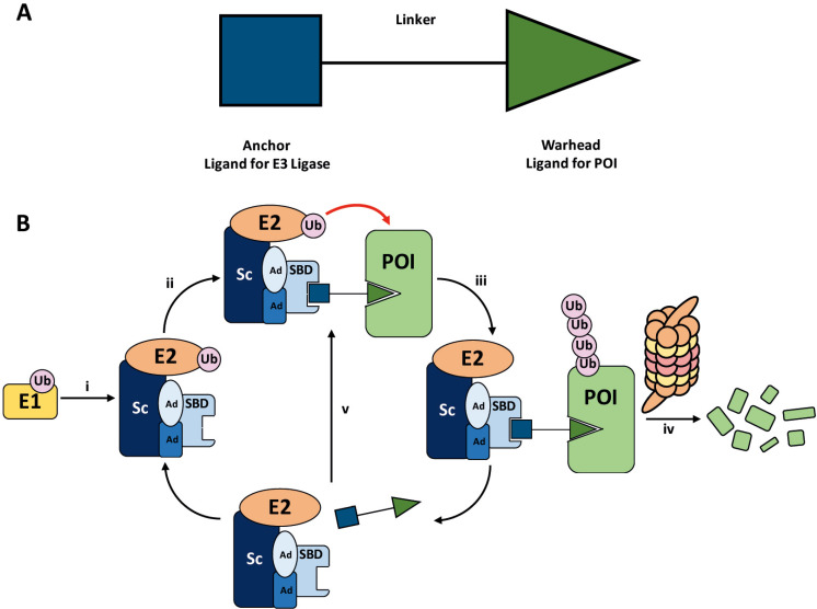
PROteolysis TArgeting Chimeras (PROTACs) are heterobifunctional molecules consisting of two ligands; an “anchor” to bind to an E3 ubiquitin ligase and a “warhead” to bind to a protein of interest, connected by a chemical linker. Targeted protein degradation by PROTACs has emerged as a new modality for the knock down of a range of proteins, with the first agents now reaching clinical evaluation. It has become increasingly clear that the length and composition of the linker play critical roles on the physicochemical properties and bioactivity of PROTACs. While linker design has historically received limited attention, the PROTAC field is evolving rapidly and currently undergoing an important shift from synthetically tractable alkyl and polyethylene glycol to more sophisticated functional linkers. This promises to unlock a wealth of novel PROTAC agents with enhanced bioactivity for therapeutic intervention. Here, the authors provide a timely overview of the diverse linker classes in the published literature, along with their underlying design principles and overall influence on the properties and bioactivity of the associated PROTACs. Finally, the authors provide a critical analysis of current strategies for PROTAC assembly. The authors highlight important limitations associated with the traditional “trial and error” approach around linker design and selection, and suggest potential future avenues to further inform rational linker design and accelerate the identification of optimised PROTACs. In particular, the authors believe that advances in computational and structural methods will play an essential role to gain a better understanding of the structure and dynamics of PROTAC ternary complexes, and will be essential to address the current gaps in knowledge associated with PROTAC design.
Overview
The claims and statistics of this paper haven’t been checked yet.
Run the check — free, no account needed. It reads the open-access text and checks the sources it cites.
Integrity signals
Registry
Cites
Statistics
Figures
Verification
Claims the paper makes
The claims and statistics of this paper haven’t been checked yet.
Run the check — free, no account needed. It reads the open-access text and checks the sources it cites.
Claims other papers make about this one
Each is checked against this paper’s own text: does it say what the citing paper says it does?
The cited source says what the claim attributes to it.
In the citing paper
“The composition and length of linkers can affect the efficiency of degraders40,41.”
From An HDAC4-specific PROTAC degrader achieves radiation sensitization by enhancing ferroptosis in lung cancer (2026) · cites this paper as [41]
In the citing paper
“Short linkers can be used to modulate PROTAC molecular weights67 and properties65”
From PROTAC targeted protein degraders: the past is prologue (2022) · cites this paper as [67]
Self-check
Percentages, sums and reported differences re-computed from the paper’s own numbers.
The self-check needs the paper’s full text.
Citations
Cites
Lipinski, C. A., Lombardo, F., Dominy, B. W., & Feeney, P. J. (2001) · Advanced Drug Delivery Reviews · doi:10.1016/s0169-409x(00)00129-0 · Cited by 18,139
Molecular properties that influence the oral bioavailability of drug candidates
Veber, D. F., Johnson, S. R., Cheng, H.-Y., Smith, B. R., Ward, K. W., & Kopple, K. D. (2002) · Journal of Medicinal Chemistry · doi:10.1021/jm020017n · Cited by 8,420
Lipinski, C. A., Lombardo, F., Dominy, B. W., & Feeney, P. J. (2012) · Advanced Drug Delivery Reviews · doi:10.1016/j.addr.2012.09.019 · Cited by 3,720
Peptide therapeutics: current status and future directions
Fosgerau, K., & Hoffmann, T. (2014) · Drug Discovery Today · doi:10.1016/j.drudis.2014.10.003 · Cited by 3,160
RING Domain E3 Ubiquitin Ligases
Deshaies, R. J., & Joazeiro, C. A. P. (2009) · Annual Review of Biochemistry · doi:10.1146/annurev.biochem.78.101807.093809 · Cited by 2,679
Sakamoto, K. M., Kim, K. B., Kumagai, A., Mercurio, F., Crews, C. M., & Deshaies, R. J. (2001) · Proceedings of the National Academy of Sciences · doi:10.1073/pnas.141230798 · Cited by 2,609
The growing applications of click chemistry
Moses, J. E., & Moorhouse, A. D. (2007) · Chemical Society Reviews · doi:10.1039/b613014n · Cited by 2,376
Phthalimide conjugation as a strategy for in vivo target protein degradation
Winter, G. E., Buckley, D. L., Paulk, J., Roberts, J. M., Souza, A., Dhe-Paganon, S., & Bradner, J. E. (2015) · Science · doi:10.1126/science.aab1433 · Cited by 1,760
Tetrazine Ligation: Fast Bioconjugation Based on Inverse-Electron-Demand Diels−Alder Reactivity
Blackman, M. L., Royzen, M., & Fox, J. M. (2008) · Journal of the American Chemical Society · doi:10.1021/ja8053805 · Cited by 1,691
Honigberg, L. A., Smith, A. M., Sirisawad, M., Verner, E., Loury, D., Chang, B., Li, S., Pan, Z., Thamm, D. H., Miller, R. A., et al. (2010) · Proceedings of the National Academy of Sciences · doi:10.1073/pnas.1004594107 · Cited by 1,561
Cited by
Sorted by citations
Items 101–200 of 429
Breaking Bad Proteins—Discovery Approaches and the Road to Clinic for Degraders
Bouvier, C., Lawrence, R., Cavallo, F., Xolalpa, W., Jordan, A. M., Hjerpe, R., & Rodríguez, M. S. (2024) · Cells · doi:10.3390/cells13070578 · Cited by 28
Targeted protein degradation in CNS disorders: a promising route to novel therapeutics?
Kuemper, S., Cairns, A. G., Birchall, K., Yao, Z., & Large, J. M. (2024) · Frontiers in Molecular Neuroscience · doi:10.3389/fnmol.2024.1370509 · Cited by 32
Discovery of a Natural Ent-Kaurene Diterpenoid Oridonin as an E3 Ligase Recruiter for PROTACs
Huang, J., Fu, X., Qiu, F., Liang, Z., Cao, C., Wang, Z., Chen, H., Yue, S., Xie, D., Liang, Y., et al. (2024) · Journal of the American Chemical Society · doi:10.1021/jacs.4c14650 · Cited by 27
Development of Rapid and Facile Solid‐Phase Synthesis of PROTACs via a Variety of Binding Styles
Xu, H., Kurohara, T., Takano, R., Yokoo, H., Shibata, N., Ohoka, N., Inoué, T., Naito, M., & Demizu, Y. (2022) · ChemistryOpen · doi:10.1002/open.202200131 · Cited by 26
Recent advances in dual PROTACs degrader strategies for disease treatment
Liu, J., Liu, Y., Tang, J., Gong, Q., Yan, G., Fan, H., Zhang, X., & Pu, C. (2024) · European Journal of Medicinal Chemistry · doi:10.1016/j.ejmech.2024.116901 · Cited by 25
CRBN‐PROTACs in Cancer Therapy: From Mechanistic Insights to Clinical Applications
Thapa, R., Bhat, A. A., Gupta, G., Jyothi, S. R., Kaur, I., Kumar, S., Sharma, N., Prasad, G. V. S., Pramanik, A., & Ali, H. (2024) · Chemical Biology & Drug Design · doi:10.1111/cbdd.70009 · Cited by 26
Design, Synthesis and In Vitro Investigation of Cabozantinib-Based PROTACs to Target c-Met Kinase
Sachkova, A. A., Andreeva, D. V., Tikhomirov, A. S., Scherbakov, A. M., Salnikova, D. I., Sorokin, D. V., Bogdanov, F. B., Rysina, Y. D., Shchekotikhin, A. E., Shchegravina, E. S., et al. (2022) · Pharmaceutics · doi:10.3390/pharmaceutics14122829 · Cited by 24
Ligandability and druggability assessment via machine learning
Palma, F. D., Abate, C., Decherchi, S., & Cavalli, A. (2023) · Wiley Interdisciplinary Reviews Computational Molecular Science · doi:10.1002/wcms.1676 · Cited by 26
Synthesis of non-equivalent diamides and amido-esters via Pd-catalysed carbonylation
Ge, Y., Huang, W.-H., Ahrens, S., Spannenberg, A., Jackstell, R., & Beller, M. (2023) · Nature Synthesis · doi:10.1038/s44160-023-00411-6 · Cited by 24
Antermite, D., Friis, S. D., Johansson, J. R., Putra, O. D., Ackermann, L., & Johansson, M. J. (2023) · Nature Communications · doi:10.1038/s41467-023-43789-9 · Cited by 24
Shaik, S., Gayam, P. K. R., Chaudhary, M., Singh, G., & Pai, A. (2024) · Bioorganic Chemistry · doi:10.1016/j.bioorg.2024.107868 · Cited by 26
Navigating PROTACs in Cancer Therapy: Advancements, Challenges, and Future Horizons
Ibrahim, S., Khan, M. U., Khurram, I., Rehman, R., Rauf, A., Ahmad, Z., Aljohani, A. S. M., Abdulmonem, W. A., & Quradha, M. M. (2025) · Food Science & Nutrition · doi:10.1002/fsn3.70011 · Cited by 27
Zhu, C., Luo, X., Tian, T., Rao, Z., Wang, H., Zhou, Z., Gao, Z., Mi, T., Chen, D., Wu, Y., et al. (2022) · European Journal of Medicinal Chemistry · doi:10.1016/j.ejmech.2022.114459 · Cited by 24
Hamada, K., Hashimoto, T., Iwashita, R., Yamada, Y., Kikkawa, Y., & Nomizu, M. (2023) · Cell Reports Physical Science · doi:10.1016/j.xcrp.2023.101296 · Cited by 24
Yue, J., Peng, B., Chen, Y., Jin, J., Zhao, X., Shen, C., Ji, X., Hsieh, C., Song, J., Hou, T., et al. (2024) · Chemical Science · doi:10.1039/d4sc03744h · Cited by 25
He, Y., Zhu, C., Peng, L., Chen, Y., Zhang, Y., Zheng, Y., & Diao, X. (2024) · Journal of Pharmaceutical Analysis · doi:10.1016/j.jpha.2024.101175 · Cited by 25
Chen, Y., Xia, Z., Suwal, U., Rappu, P., Heino, J., Wever, O. D., & Geest, B. G. D. (2024) · Chemical Science · doi:10.1039/d4sc03555k · Cited by 21
Balzarini, M., Tong, J., Gui, W., Jayalath, I. M., Schell, B.-B., & Kodadek, T. J. (2024) · ACS Chemical Biology · doi:10.1021/acschembio.4c00422 · Cited by 33
Arndt, C. M., Bitai, J., Brunner, J., Opatz, T., Martinelli, P., Gollner, A., Sokol, K. R., & Krumb, M. (2023) · Journal of Medicinal Chemistry · doi:10.1021/acs.jmedchem.3c01613 · Cited by 22
High‐Throughput Miniaturized Synthesis of PROTAC‐Like Molecules
Tian, Y., Seifermann, M., Bauer, L., Luchena, C., Wiedmann, J. J., Schmidt, S., Geisel, A., Afonin, S., Höpfner, J., Brehm, M., et al. (2024) · Small · doi:10.1002/smll.202307215 · Cited by 20
Targeted Protein Degradation in Cancer: PROTACs, New Targets, and Clinical Mechanisms
Faryal, B., Abideen, Z. U., Irfan, M., Ahmed, H., Jalilov, F. S., Abduraximova, L., & Ashraf, G. A. (2026) · Biomolecules · doi:10.3390/biom16020325 · Cited by 24
Exploring PROTAC Cooperativity with Coarse-Grained Alchemical Methods
Mai, H., Zimmer, M. H., & Miller, T. F. (2023) · The Journal of Physical Chemistry B · doi:10.1021/acs.jpcb.2c05795 · Cited by 22
Adem, Ş., Yırtıcı, Ü., Aydın, M., Rawat, R., & Eyüpoğlu, V. (2023) · Archiv der Pharmazie · doi:10.1002/ardp.202300326 · Cited by 20
Design, Synthesis, and Biological Evaluation of HDAC Degraders with CRBN E3 Ligase Ligands
Lu, Y., Sun, D., Xiao, D., Shao, Y.-Y., Su, M., Zhou, Y., Li, J. X., Zhu, S., & Lu, W. P. (2021) · Molecules · doi:10.3390/molecules26237241 · Cited by 21
Proteolysis Targeting Chimeras (PROTACs) in Breast Cancer Therapy
Jin, Y., & Lee, Y. (2024) · ChemMedChem · doi:10.1002/cmdc.202400267 · Cited by 21
Zhou, Y., Xu, S., López‐Carrobles, N., Ding, D., Liu, X., Menéndez‐Arias, L., & Zhan, P. (2023) · Acta Materia Medica · doi:10.15212/amm-2023-0019 · Cited by 20
Novel technologies are turning a dream into reality: conditionally replicating viruses as vaccines
Tang, Y., Yu, C., & Cai, X. (2023) · Trends in Microbiology · doi:10.1016/j.tim.2023.09.002 · Cited by 20
Liu, T., Wang, Q., Zhou, Y., Ye, B., Liu, T., Yan, L., Fan, J., Xu, J., Zhou, Y., Xia, Z., et al. (2024) · Journal of Medicinal Chemistry · doi:10.1021/acs.jmedchem.4c00454 · Cited by 21
Pan, B., Mountford, S. J., Kiso, M., Anderson, D. E., Papadakis, G., Jarman, K. E., Tilmanis, D., Maher, B. M., Tran, T., Shortt, J., et al. (2025) · Communications Medicine · doi:10.1038/s43856-025-00863-1 · Cited by 19
Selective and potent PROTAC degraders of c-Src kinase
Mao, W., Vandecan, N., Bingham, C., Tsang, P.-K., Ulintz, P., Sexton, R., Bochar, D., Merajver, S., & Soellner, M. (2023) · ACS Chemical Biology · doi:10.1021/acschembio.3c00548 · Cited by 16
Huang, J., Ma, Z., Peng, X., Yang, Z., Wu, Y., Zhong, G., Ouyang, T., Chen, Z., Liu, Y., Wang, Q., et al. (2024) · Journal of Medicinal Chemistry · doi:10.1021/acs.jmedchem.3c01414 · Cited by 18
ZX703: A Small-Molecule Degrader of GPX4 Inducing Ferroptosis in Human Cancer Cells
Hu, M., Li, X., Wang, L., Zhang, Y., Sun, Y., Hua, H., Liu, H., Cai, T., Zhu, D., & Xiang, Q. (2024) · ACS Medicinal Chemistry Letters · doi:10.1021/acsmedchemlett.3c00571 · Cited by 18
Bayesian optimization for ternary complex prediction (BOTCP)
Rao, A., Tunjic, T. M., Brunsteiner, M., Müller, M., Fooladi, H., Gasbarri, C., & Weber, N. (2023) · Artificial Intelligence in the Life Sciences · doi:10.1016/j.ailsci.2023.100072 · Cited by 25
He, X., Zeng, S., Wen, Y., Yang, T., Huang, C., Li, Y.-F., Zhang, Z., Ding, K., Liu, T., Tan, Y., et al. (2025) · Journal of the American Chemical Society · doi:10.1021/jacs.4c18083 · Cited by 20
Xie, S., Zhu, J., Zhan, F., Feng, D., He, C., Liu, L., Xie, J., Liu, J. Y., Zhong, M., Zhang, X., et al. (2025) · Journal of Medicinal Chemistry · doi:10.1021/acs.jmedchem.5c00488 · Cited by 17
Zhai, J., Li, C., Sun, B., Wang, S., Cui, Y., Gao, Q., & Sang, F. (2022) · Bioorganic & Medicinal Chemistry Letters · doi:10.1016/j.bmcl.2022.129041 · Cited by 16
Zaman, S. U., Pagare, P. P., Huang, B., Rilee, G. J., Ma, Z., Zhang, Y., & Li, J. (2024) · Bioorganic Chemistry · doi:10.1016/j.bioorg.2024.107613 · Cited by 18
Qiu, Y., Wiewiora, R., Izaguirre, J. A., Xu, H., Sherman, W., Tang, W., & Huang, X. (2024) · JACS Au · doi:10.1021/jacsau.4c00503 · Cited by 20
Racioppo, B., Pechalrieu, D., Abegg, D., Dwyer, B. G., Ramseier, N. T., Hu, Y., & Adibekian, A. (2025) · Journal of the American Chemical Society · doi:10.1021/jacs.4c17827 · Cited by 18
Li, Y., Hwang, N., Snedeker, A., Lemon, S. M., Noe, D., Sun, L., Clement, J. A., Zhou, T., Tang, L., Block, T. M., et al. (2024) · Bioorganic & Medicinal Chemistry Letters · doi:10.1016/j.bmcl.2024.129680 · Cited by 16
Tanimoto, H., & Tomohiro, T. (2024) · Chemical Communications · doi:10.1039/d4cc03359k · Cited by 16
Targeted degradation of α-synuclein by arginine-based PROTACs
Shen, L., Zhang, J., Wang, Z., Liu, Y., Cui, S., & Rao, H. (2025) · Journal of Biological Chemistry · doi:10.1016/j.jbc.2025.110449 · Cited by 18
Modern Strategies of Drug Therapy: Multi-Target Drug Delivery, Bioimaging, Diagnostics
Fedorova, O. V., Ovchinnikova, I. G., Rusinov, G. L., Avdeeva, V. V., Zhdanov, А. P., Zhizhin, K. Y., Kuznetsov, N. T., Zakharova, L. Y., Kuznetsova, D. A., Razuvayeva, Y. S., et al. (2025) · Russian Journal of General Chemistry · doi:10.1134/s1070363225606726 · Cited by 16
Regulation of eDHFR-tagged proteins with trimethoprim PROTACs
Etersque, J. M., Lee, I. K., Sharma, N., Xu, K., Ruff, A., Northrup, J. D., Sarkar, S., Nguyen, T., Lauman, R., Burslem, G. M., et al. (2023) · Nature Communications · doi:10.1038/s41467-023-42820-3 · Cited by 17
High, P. C., Guernsey-Biddle, C., Subramanian, S., Jacob, J., & Carmon, K. S. (2024) · Pharmaceutics · doi:10.3390/pharmaceutics16070890 · Cited by 15
Feng, Z., Xie, D., Qiu, F., Huang, J., Wang, Z., & Liang, C. (2024) · European Journal of Medicinal Chemistry · doi:10.1016/j.ejmech.2024.117027 · Cited by 15
Colarusso, E., Ceccacci, S., Monti, M. C., Gazzillo, E., Giordano, A., Chini, M. G., Ferraro, M. G., Piccolo, M., Ruggiero, D., Irace, C., et al. (2022) · European Journal of Medicinal Chemistry · doi:10.1016/j.ejmech.2022.115018 · Cited by 17
Development of Versatile Solid-Phase Methods for Syntheses of PROTACs with Diverse E3 Ligands
Xu, H., Kurohara, T., Ohoka, N., Tsuji, G., Inoué, T., Naito, M., & Demizu, Y. (2023) · Bioorganic & Medicinal Chemistry · doi:10.1016/j.bmc.2023.117293 · Cited by 16
Recent Advances in the Development of Pro-PROTAC for Selective Protein Degradation
Hakem, F. T., Abdelwaly, A., Alshaman, R., Alattar, A., Alanazi, F. E., Zaitone, S. A., & Helal, M. A. (2025) · Pharmaceutics · doi:10.3390/pharmaceutics17091160 · Cited by 14
Manipulating autophagic degradation in human diseases: from mechanisms to interventions
Zhang, Y., Liu, X., Klionsky, D. J., Lu, B., & Zhong, Q. (2022) · Life Medicine · doi:10.1093/lifemedi/lnac043 · Cited by 14
Discovery of Novel Bruton’s Tyrosine Kinase PROTACs with Enhanced Selectivity and Cellular Efficacy
Li, Y.-Q., Lannigan, W. G., Davoodi, S., Daryaee, F., Corrionero, A., Alfonso, P., Rodríguez‐Santamaría, J. A., Wang, N., Haley, J. D., & Tonge, P. J. (2023) · Journal of Medicinal Chemistry · doi:10.1021/acs.jmedchem.3c00176 · Cited by 15
Joshi, M., Dey, P., & De, A. (2023) · Exploration of Targeted Anti-tumor Therapy · doi:10.37349/etat.2023.00194 · Cited by 15
PROTAC Beyond Cancer- Exploring the New Therapeutic Potential of Proteolysis Targeting Chimeras
Bhole, R. P., Patil, S., Kapare, H. S., Chikhale, R. V., & Gurav, S. S. (2024) · Current Topics in Medicinal Chemistry · doi:10.2174/0115680266309968240621072550 · Cited by 15
Zaman, S. U., Pagare, P. P., Ma, H., Hoyle, R. G., Zhang, Y., & Li, J. (2024) · RSC Medicinal Chemistry · doi:10.1039/d4md00122b · Cited by 15
Omar, E. A., Rajesh, R., Das, P. K., Pal, R., Matada, G. S. P., & Maji, L. (2024) · European Journal of Medicinal Chemistry · doi:10.1016/j.ejmech.2024.117034 · Cited by 14
Pedrucci, F., Pappalardo, C., Marzaro, G., Ferri, N., Ferlin, A., & Toni, L. D. (2022) · International Journal of Molecular Sciences · doi:10.3390/ijms23126630 · Cited by 14
An abiotic, tetrameric, eight-helix bundle
Menke, F. S., Wicher, B., Allmendinger, L., Maurizot, V., & Huc, I. (2023) · Chemical Science · doi:10.1039/d3sc00267e · Cited by 13
Hypoxia-Activated Theragnostic Prodrugs (HATPs): Current State and Future Perspectives
Bhuniya, S., & Vrettos, E. I. (2024) · Pharmaceutics · doi:10.3390/pharmaceutics16040557 · Cited by 13
Shen, H., Xu, H., Jin, W., Wu, T., Hu, J., Zhang, C., Zhong, Z., Li, J., Mao, R., Zhang, S., et al. (2025) · Journal of Medicinal Chemistry · doi:10.1021/acs.jmedchem.4c02205 · Cited by 15
Targeting Tau Protein with Proximity Inducing Modulators: A New Frontier to Combat Tauopathies
Uliassi, E., Bolognesi, M. L., & Milelli, A. (2025) · ACS Pharmacology & Translational Science · doi:10.1021/acsptsci.4c00733 · Cited by 13
Recent advances in medicinal chemistry strategies for the development of METTL3 inhibitors
Li, G., Chen, W., Liu, D., & Tang, S. (2025) · European Journal of Medicinal Chemistry · doi:10.1016/j.ejmech.2025.117560 · Cited by 13
PROTACs: The Future of Leukemia Therapeutics
Anwar, Z., Ali, M. S., Galvano, A., Perez, A., Mantia, M. L., Bukhari, I., & Świątczak, B. (2022) · Frontiers in Cell and Developmental Biology · doi:10.3389/fcell.2022.851087 · Cited by 13
Danazumi, A. U., Ishmam, I. T., Idris, S., Izert, M. A., Balogun, E. O., & Górna, M. W. (2023) · European Journal of Pharmaceutical Sciences · doi:10.1016/j.ejps.2023.106451 · Cited by 14
Kudo, G., Hirao, T., Harada, R., Shigeta, Y., Hirokawa, T., & Yoshino, R. (2025) · Journal of Chemical Information and Modeling · doi:10.1021/acs.jcim.5c00102 · Cited by 18
TrypPROTACs Unlocking New Therapeutic Strategies for Chagas Disease
Gini, A. L. R., da Cunha, P. S. T., João, E. E., Chin, C. M., dos Santos, J. L., Serra, E. C., & Scarim, C. B. (2025) · Pharmaceuticals · doi:10.3390/ph18060919 · Cited by 12
BacPROTACs targeting Clp protease: a promising strategy for anti-mycobacterial drug discovery
Bonjorno, A. F., Pavan, A. R., Fernandes, G., Scarim, C. B., Castagnolo, D., & dos Santos, J. L. (2024) · Frontiers in Chemistry · doi:10.3389/fchem.2024.1358539 · Cited by 12
Pierri, M., Liu, X., Kroupova, A., Rutter, Z. J., Hallatt, A. J., & Ciulli, A. (2024) · Bioorganic & Medicinal Chemistry Letters · doi:10.1016/j.bmcl.2024.129861 · Cited by 13
Androgen receptor inhibitors in treating prostate cancer
Cole, R. N., Fang, Q., Matsuoka, K., & Wang, Z. (2024) · Asian Journal of Andrology · doi:10.4103/aja202494 · Cited by 14
Ugurlu, S. Y., McDonald, D., Enisoglu, R., Zhu, Z., & He, S. (2025) · Scientific Reports · doi:10.1038/s41598-024-83558-2 · Cited by 12
PROTACting the kinome with covalent warheads
Chatterjee, D. R., Kapoor, S., Jain, M., Das, R., Chowdhury, M. G., & Shard, A. (2022) · Drug Discovery Today · doi:10.1016/j.drudis.2022.103417 · Cited by 12
Chemically Induced Degradation of Native Proteins by Direct Recruitment to the 26S ProteasomePreprint
Balzarini, M., Gui, W., Jayalath, I. M., Schell, B.-B., Tong, J., & Kodadek, T. J. (2023) · bioRxiv (Cold Spring Harbor Laboratory) · doi:10.1101/2023.07.19.549534 · Cited by 12
Fan, Y., Ma, R., Njobvu, K. M., Wang, Y., Ren, Y., Fu, R., Zhang, C., Liu, F., Jiang, Z., Rao, Y., et al. (2025) · Journal of Medicinal Chemistry · doi:10.1021/acs.jmedchem.5c01620 · Cited by 11
Ayinde, O. R., Sharpe, C., Stahl, E. H., Tokarski, R. J., Lerma, J. R., Muthusamy, N., Byrd, J. C., & Fuchs, J. R. (2023) · ACS Medicinal Chemistry Letters · doi:10.1021/acsmedchemlett.3c00082 · Cited by 14
A one-pot photocatalytic triazole-based linkerology for PROTACs
Liu, J., Deng, Y., Yin, J., Ji, J., Guan, C., Chen, X., Wu, X., Zhu, T., & Liu, S. (2024) · Cell Reports Physical Science · doi:10.1016/j.xcrp.2024.102139 · Cited by 11
Li, Y., Zhu, R., He, X., Song, Y., Fan, T., Ma, J., Xiang, G., & Xiang, G. (2024) · Bioorganic Chemistry · doi:10.1016/j.bioorg.2024.107943 · Cited by 11
A comprehensive primer and review of PROTACs and their In Silico design
Zattoni, J., Vottero, P., Carena, G., Uliveto, C., Pozzati, G., Morabito, B., Gitari, E., Tuszyński, J. A., & Aminpour, M. (2025) · Computer Methods and Programs in Biomedicine · doi:10.1016/j.cmpb.2025.108687 · Cited by 14
Design and development of PROTACs: A new paradigm in anticancer drug discovery
Mathur, V., Jha, M., Zai, I., Mahajan, M., Nazar, S., Ali, S., Ilyas, A., Tanweer, S., Ali, J., & Alam, O. (2025) · Medicine in Drug Discovery · doi:10.1016/j.medidd.2025.100221 · Cited by 11
Mapping the energy landscape of PROTAC-mediated protein-protein interactions
Villegas, J. A., Vaid, T. M., Johnson, M. E., & Moore, T. W. (2023) · Computational and Structural Biotechnology Journal · doi:10.1016/j.csbj.2023.02.049 · Cited by 11
Heterobifunctional Ligase Recruiters Enable pan-Degradation of Inhibitor of Apoptosis Proteins
Ng, Y. L. D., Bricelj, A., Jansen, J. A., Murgai, A., Peter, K., Donovan, K. A., Gütschow, M., Krönke, J., Steinebach, C., & Sosič, I. (2023) · Journal of Medicinal Chemistry · doi:10.1021/acs.jmedchem.2c01817 · Cited by 10
Bifunctional robots inducing targeted protein degradation
Sobhia, M. E., Kumar, H., & Kumari, S. (2023) · European Journal of Medicinal Chemistry · doi:10.1016/j.ejmech.2023.115384 · Cited by 11
First-in-class metallo-PROTAC as an effective degrader of select Pt-binding proteins
O'Dowd, P., Sullivan, G. P., Rodrigues, D. A., Chonghaile, T. N., & Griffith, D. M. (2023) · Chemical Communications · doi:10.1039/d3cc03340f · Cited by 9
Wang, Z., Yue, S., Chen, X., Li, J., Zhu, P., Chen, H., Qiu, F., Xie, D., Liang, Y., Li, D., et al. (2024) · Journal of Medicinal Chemistry · doi:10.1021/acs.jmedchem.4c01228 · Cited by 11
Rational design and discovery of potent PROTAC degraders of ASK1: a targeted therapy in MASH
Sarkar, H. S., Sen, A., Hoque, I., Dastidar, U. G., Pal, S., Sarkar, D., Chakrabarti, P., & Talukdar, A. (2025) · RSC Medicinal Chemistry · doi:10.1039/d5md00252d · Cited by 10
Design of Class I/IV Bromodomain-Targeting Degraders for Chromatin Remodeling Complexes
Zahid, H., Costello, J. P., Li, Y., Kimbrough, J. R., Actis, M., Ranković, Z., Yan, Q., & Pomerantz, W. C. K. (2023) · ACS Chemical Biology · doi:10.1021/acschembio.2c00902 · Cited by 9
Design, Synthesis, and Evaluation of BCL‐2 Targeting PROTACs
Bricelj, A., Ng, Y. L. D., Gobec, M., Kuchta, R. D., Hu, W., Javornik, Š., Rožič, M., Gütschow, M., Zheng, G., Krönke, J., et al. (2024) · Chemistry - A European Journal · doi:10.1002/chem.202400430 · Cited by 10
Xu, M., Fu, J., Pei, Y., Li, M., Kan, W., Yan, R., Xia, C., Ma, J., Wang, P., Zhang, Y., et al. (2024) · Journal of Medicinal Chemistry · doi:10.1021/acs.jmedchem.4c01134 · Cited by 9
Brønsted acid-mediated thiazole synthesis from sulfoxonium ylides
Smy, J. L., Ifill, R., & Hassell‐Hart, S. (2024) · Chemical Communications · doi:10.1039/d4cc03905j · Cited by 9
Investigating protein degradability through site-specific ubiquitin ligase recruitment
Shade, O., Ryan, A., Belsito, G., & Deiters, A. (2024) · RSC Chemical Biology · doi:10.1039/d4cb00273c · Cited by 9
Proteolysis targeting chimera of BI-2536 induces potent dual degradation of PLK1 and BET proteins
Song, S., Yang, W., & Tai, W. (2025) · Bioorganic & Medicinal Chemistry · doi:10.1016/j.bmc.2025.118087 · Cited by 9
Lu, X., Sabbasani, V. R., Hassan, B. A., Swenson, R. E., & Walters, K. J. (2025) · Journal of Biological Chemistry · doi:10.1016/j.jbc.2025.108520 · Cited by 9
Kotagiri, S., Wang, Y., Han, Y., Liang, X., Blazanin, N., Mazhar, H., Sebastian, M. M., Nguyen, P. K., Jiang, Y., & Lissanu, Y. (2025) · Journal of Medicinal Chemistry · doi:10.1021/acs.jmedchem.4c02577 · Cited by 9
Advancing active compound discovery for novel drug targets: insights from AI-driven approaches
Wang, X.-Y., Chen, Y., Li, Y.-F., Wei, C., Liu, M.-Y., Yuan, C.-X., Zheng, Y.-Y., Qin, M.-H., Sheng, Y.-F., Tong, X., et al. (2025) · Acta Pharmacologica Sinica · doi:10.1038/s41401-025-01591-x · Cited by 12
Nafie, M. S., Diab, M. K., Yassen, A. S. A., Elshamy, A. M., Tohamy, M. R. E., Tawfik, H. O., & Fahmy, S. A. (2025) · Drug Development Research · doi:10.1002/ddr.70192 · Cited by 9
Inganäs, E., Lavén, H., Caraballo, R., Klingegård, F., Eklund, M., Andersson, L., Hilgendorf, C., & Matsson, P. (2025) · Journal of Medicinal Chemistry · doi:10.1021/acs.jmedchem.5c01499 · Cited by 9
Ruthenium‐Catalyzed Aminocarbonylation with Isocyanates Through Weak Coordinating Groups
Lai, Y., Yuan, B., Ackermann, L., & Johansson, M. J. (2023) · Chemistry - A European Journal · doi:10.1002/chem.202302023 · Cited by 9
Apprato, G., D’Agostini, G., Rossetti, P., Ermondi, G., & Caron, G. (2023) · Molecules · doi:10.3390/molecules28031206 · Cited by 9
Almodóvar‐Rivera, C. M., Zhang, Z., Li, J., Xie, H., Zhao, Y. Q., Guo, L., Mannhardt, M. G., & Tang, W. (2023) · ChemBioChem · doi:10.1002/cbic.202300482 · Cited by 9
Development of Dual Aurora‐A and Aurora‐B Degrading PROTACs for MYCN‐Amplified Neuroblastoma
Nelson, S. E., Tucker, J. R., Prado, M. G., Tierney, L. C., Quigley, S. L., Lumpkin, A. T., Dodd, C. J., Hasko, V., Howie, S. L., Aastha, A., et al. (2024) · ChemMedChem · doi:10.1002/cmdc.202400703 · Cited by 9
Bazzacco, A., Mercorelli, B., & Loregian, A. (2025) · FEMS Microbiology Reviews · doi:10.1093/femsre/fuaf046 · Cited by 11
Synthetic approaches to constructing proteolysis targeting chimeras (PROTACs)
Bakulina, O., Sapegin, A., Bunev, A. S., & Krasavin, M. (2022) · Mendeleev Communications · doi:10.1016/j.mencom.2022.07.001 · Cited by 8
Metadata
- Authors
- Troup, R. I., Fallan, C., & Baud, M. G. J.
- Affiliations
- University of Southampton (GB)
- AstraZeneca (United Kingdom) (GB)
- Corresponding author
- Fallan, Charlene
- Baud, Matthias G. J.
- Venue
- Exploration of Targeted Anti-tumor Therapy
- Publisher
- Open Exploration Publishing
- Published
- 2020-10-11
- Volume
- 1
- Issue
- 5
- Pages
- 273-312
- ISSN
- 2692-3114 (ISSN-L)
- PMC
- PMC9400730
- OpenAlex
- W3093395956
- Cited by
- 424
- References
- 183
- Citations by year
- 2026: 91 · 2025: 102 · 2024: 96 · 2023: 66 · 2022: 48 · 2021: 19 · 2020: 1
- FWCI
- 3.28
- Type
- journal article
- Language
- en
- Open access
- diamond
- doi.org
- Locations
- Exploration of Targeted Anti-tumor Therapy (publishedVersion, cc-by)
- PubMed Central (publishedVersion, cc-by)
- PubMed (publishedVersion)
- ePrints Soton (University of Southampton) (acceptedVersion)
- DOAJ (DOAJ: Directory of Open Access Journals) (submittedVersion, cc-by-sa)
- Funders
- AstraZeneca
- Engineering and Physical Sciences Research Council (EP/R513325/1)
- Topics
- Life Sciences › Biochemistry, Genetics and Molecular Biology › Molecular Biology › Protein Degradation and Inhibitors
- Physical Sciences › Chemistry › Organic Chemistry › Chemical Synthesis and Analysis
- Life Sciences › Biochemistry, Genetics and Molecular Biology › Molecular Biology › Ubiquitin and proteasome pathways
- Keywords
- PROTACs, linker design, targeted protein degradation, E3 ubiquitin ligase, heterobifunctional molecules, ternary complex structure, rational drug design, polyethylene glycol linker, alkyl linkers
About these checks
Registry, retraction, Cites and statistics results are computed, not judged, with one exception: a printed reference that no index lists is read once by a model before it counts as a citation error. Claims are extracted by a model and kept only when their quote appears verbatim in the text; each cited source is then checked, with a model’s help, for whether it says what the claim relies on. Those verdicts can be wrong — read the claim’s quote and the cited source before relying on one.
Statistics and arithmetic are recomputed, not judged.
Confidence bands are shown only for fields calibrated on our annotated test set. None is calibrated yet, so no band is shown.
What counts as a claim ›How we measure confidence ›How we check this ›
Are you an author, or do you see a problem with this page? Request a review
