

The DOI matches its Crossref record; 57 of the 58 references it cites were found in a registry or an index.
Aging of Skeletal Muscle Fibers
Open accessAbstract
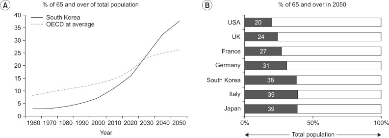
Aging has become an important topic for scientific research because life expectancy and the number of men and women in older age groups have increased dramatically in the last century. This is true in most countries of the world including the Republic of Korea and the United States. From a rehabilitation perspective, the most important associated issue is a progressive decline in functional capacity and independence. Sarcopenia is partly responsible for this decline. Many changes underlying the loss of muscle mass and force-generating capacity of skeletal muscle can be understood at the cellular and molecular levels. Muscle size and architecture are both altered with advanced adult age. Further, changes in myofibers include impairments in several physiological domains including muscle fiber activation, excitation-contraction coupling, actin-myosin cross-bridge interaction, energy production, and repair and regeneration. A thorough understanding of these alterations can lead to the design of improved preventative and rehabilitative interventions, such as personalized exercise training programs.
Overview
The claims and statistics of this paper haven’t been checked yet.
Run the check — free, no account needed. It reads the open-access text and checks the sources it cites.
Integrity signals
Registry
Cites
Statistics
Figures
Verification
Claims the paper makes
The claims and statistics of this paper haven’t been checked yet.
Run the check — free, no account needed. It reads the open-access text and checks the sources it cites.
Claims other papers make about this one
Each is checked against this paper’s own text: does it say what the citing paper says it does?
In the citing paper
“These changes are also accompanied by a diminished regenerative capacity of the muscle due to a loss in the number and activity of satellite cells in type II “fast” fibers [14,16,17].”
From Potential Roles of n-3 PUFAs during Skeletal Muscle Growth and Regeneration (2018) · cites this paper as [17]
Self-check
Percentages, sums and reported differences re-computed from the paper’s own numbers.
The self-check needs the paper’s full text.
Citations
Cites
Sarcopenia: European consensus on definition and diagnosis
Cruz-Jentoft, A. J., Baeyens, J. P., Bauer, J. M., Boirie, Y., Cederholm, T., Landi, F., Martin, F. C., Michel, J.-P., Rolland, Y., Schneider, S. M., et al. (2010) · Age and Ageing · doi:10.1093/ageing/afq034 · Cited by 11,601 · cited 1×
“More recently, the European Working Group on Sarcopenia in Older People has expanded the definition and suggested criteria for diagnosis [12] including the presence of muscle weakness (for example handgrip strength), a lower muscle mass (determined by bioelec1960 40”
BackgroundSkeletal muscle mass and distribution in 468 men and women aged 18–88 yr
Janssen, I., Heymsfield, S. B., Wang, Z., & Ross, R. (2000) · Journal of Applied Physiology · doi:10.1152/jappl.2000.89.1.81 · Cited by 3,087 · cited 1×
“In contrast, type I muscle fiber size is largely unaffected [22,28,29].”
BackgroundStudenski, S. A., Peters, K. W., Alley, D. E., Cawthon, P. M., McLean, R. R., Harris, T. B., Ferrucci, L., Guralnik, J. M., Fragala, M. S., Kenny, A. M., et al. (2014) · The Journals of Gerontology: Series A · doi:10.1093/gerona/glu010 · Cited by 2,545 · cited 2×
“The FNIH Sarcopenia Project Group [13] recently recommended normative values for muscle strength and lean body mass as follows: 1) grip strength in men <26 kg and in women <16 kg, or alternate grip strength adjusted for BMI <1.0 for men and <0.56 for women; and 2) appendicular lean body mass in men <19.75 kg and <15.0…”
MethodInfluentialSarcopenia: origins and clinical relevance
Rosenberg, I. H. (1997) · The Journal of Nutrition · doi:10.1093/jn/127.5.990s · Cited by 2,399 · cited 1×
“The term sarcopenia was used for the first time by Rosenberg [10,11] to refer to the loss of lean body mass with aging.”
MethodSatellite Cells and the Muscle Stem Cell Niche
Yin, H., Price, F., & Rudnicki, M. A. (2013) · Physiological Reviews · doi:10.1152/physrev.00043.2011 · Cited by 2,166 · cited 1×
“Pax7-expressing adult stem cells are normally quiescent, but when stimulated by damage or stress, they become activated and enter the cell cycle to either form new muscle fibers or self-renew and replenish the satellite cell pool, that will be used in the future [19-21].”
BackgroundLongitudinal study of muscle strength, quality, and adipose tissue infiltration
Delmonico, M. J., Harris, T. B., Visser, M., Park, S. W., Conroy, M. B., Velasquez-Mieyer, P., Boudreau, R., Manini, T. M., Nevitt, M., Newman, A. B., et al. (2009) · The American Journal of Clinical Nutrition · doi:10.3945/ajcn.2009.28047 · Cited by 1,446 · cited 1×
“However, a direct relationship between increased intermuscular fat and age-related muscle weakness has not been established [51].”
BackgroundAging of skeletal muscle: a 12-yr longitudinal study
Frontera, W. R., Hughes, V. A., Fielding, R. A., Fiatarone, M. A., Evans, W. J., & Roubenoff, R. (2000) · Journal of Applied Physiology · doi:10.1152/jappl.2000.88.4.1321 · Cited by 1,443 · cited 3×
“The loss of muscle mass and strength in older people is of greater magnitude in longitudinal compared to cross-sectional studies [15].”
BackgroundResultHuman aging, muscle mass, and fiber type composition.
Evans, W. J., & Lexell, J. (1995) · The Journals of Gerontology Series A: Biological Sciences and Medical Sciences · doi:10.1093/gerona/50a.special_issue.11 · Cited by 1,308 · cited 1×
“The decline in muscle mass, most prominent in the lower limbs, is accompanied by a 30% to 40% decrease in muscle fiber number between the second and eight decade [25].”
BackgroundRosenberg, I. H. (1989) · American Journal of Clinical Nutrition · doi:10.1093/ajcn/50.5.1231 · Cited by 1,234
Attenuation of skeletal muscle and strength in the elderly: the Health ABC Study
Goodpaster, B. H., Carlson, C. L., Visser, M., Kelley, D. E., Scherzinger, A., Harris, T. B., Stamm, E., & Newman, A. B. (2001) · Journal of Applied Physiology · doi:10.1152/jappl.2001.90.6.2157 · Cited by 1,226
Molecular regulation of stem cell quiescence
Cheung, T. H., & Rando, T. A. (2013) · Nature Reviews Molecular Cell Biology · doi:10.1038/nrm3591 · Cited by 1,148 · cited 1×
“Pax7-expressing adult stem cells are normally quiescent, but when stimulated by damage or stress, they become activated and enter the cell cycle to either form new muscle fibers or self-renew and replenish the satellite cell pool, that will be used in the future [19-21].”
BackgroundGoodpaster, B. H., Kelley, D. E., Thaete, F. L., He, J., & Ross, R. (2000) · Journal of Applied Physiology · doi:10.1152/jappl.2000.89.1.104 · Cited by 956 · cited 1×
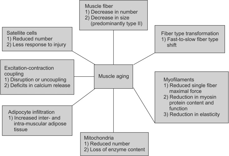
“Adipocyte infiltration Aging is associated with increases in both intra- and inter-muscular adipose tissue and it has been showed that increased muscle fat content is associated with reduced muscle strength [49,50].”
BackgroundHUGHES, V. A., Frontera, W. R., Wood, M., Evans, W. J., Dallal, G. E., Roubenoff, R., & Singh, M. A. F. (2001) · The Journals of Gerontology Series A · doi:10.1093/gerona/56.5.b209 · Cited by 935 · cited 1×
“It must be noted that the loss of strength with advancing age is not a universal phenomenon and some individuals maintain muscle strength after 10 years of follow-up [17].”
BackgroundDynapenia and aging: an update
Manini, T. M., & Clark, B. C. (2012) · The Journals of Gerontology: Series A · doi:10.1093/gerona/glr010 · Cited by 875 · cited 1×
“Disruption or uncoupling at any step of this process may result in reduced muscle fiber activation, force generation, and lower whole muscle strength [41].”
BackgroundMuscle type and fiber type specificity in muscle wasting
Ciciliot, S., Rossi, C. A., Dyar, K. A., Blaauw, B., & Schiaffino, S. (2013) · The International Journal of Biochemistry & Cell Biology · doi:10.1016/j.biocel.2013.05.016 · Cited by 644
Rosenberg, I. H. (1989) · American Journal of Clinical Nutrition · Cited by 589
Satellite-cell pool size does matter: Defining the myogenic potency of aging skeletal muscle
Shefer, G., de Mark, D. P. V., Richardson, J. B., & Yablonka‐Reuveni, Z. (2006) · Developmental Biology · doi:10.1016/j.ydbio.2006.02.022 · Cited by 526 · cited 1×
“Pax7-expressing adult stem cells are normally quiescent, but when stimulated by damage or stress, they become activated and enter the cell cycle to either form new muscle fibers or self-renew and replenish the satellite cell pool, that will be used in the future [19-21].”
BackgroundThe effect of ageing and immobilization on structure and function of human skeletal muscle fibres
D’Antona, G., Pellegrino, M. A., Adami, R., Rossi, R., Carlizzi, C. N., Canepari, M., Saltin, B., & Bottinelli, R. (2003) · The Journal of Physiology · doi:10.1113/jphysiol.2003.046276 · Cited by 500 · cited 3×
“Many molecular mechanisms have been proposed to explain such dysfunction including a reduction in myosin protein content; this may be particularly true in immobilized muscle [32].”
BackgroundSatellite cell content is specifically reduced in type II skeletal muscle fibers in the elderly
Verdijk, L. B., Koopman, R., Schaart, G., Meijer, K., Savelberg, H. H. C. M., & van Loon, L. J. C. (2007) · American Journal of Physiology-Endocrinology and Metabolism · doi:10.1152/ajpendo.00278.2006 · Cited by 489 · cited 2×
“At the level of the individual muscle fiber, sarcopenia may be associated with a reduced number of satellite cells; especially those associated with fibers expressing type II (fast) myosin heavy chain [22].”
BackgroundMechanisms for fiber-type specificity of skeletal muscle atrophy
Wang, Y., & Pessin, J. E. (2013) · Current Opinion in Clinical Nutrition & Metabolic Care · doi:10.1097/mco.0b013e328360272d · Cited by 432 · cited 2×
“pha (PGC1a) protects type I fibers from atrophy while the transforming growth factor beta (TGFb) family and the nuclear factor kappaB (NF-kB) predominantly affect type IIx fibers [39].”
BackgroundSkeletal muscle fiber quality in older men and women
Frontera, W. R., Suh, D., Krivickas, L. S., HUGHES, V. A., Goldstein, R., & Roubenoff, R. (2000) · American Journal of Physiology-Cell Physiology · doi:10.1152/ajpcell.2000.279.3.c611 · Cited by 424 · cited 2×
“The reduction in muscle fiber size is fiber type specific, with 10%–40% smaller type II fibers observed in muscle tissue collected from elderly compared with young controls [27].”
BackgroundPress, J. M., & Young, J. L. (2003) · Elsevier eBooks · doi:10.1016/b978-1-56053-493-8.50021-0 · Cited by 389
Apoptosis in skeletal muscle with aging
Dirks, A., & Leeuwenburgh, C. (2002) · American Journal of Physiology-Regulatory, Integrative and Comparative Physiology · doi:10.1152/ajpregu.00458.2001 · Cited by 377 · cited 1×
“Further evidence has suggested that apoptosis may play a considerable role in mediating the progression of muscle fiber loss in aging [26].”
BackgroundRecovery From Disability Among Community-Dwelling Older Persons
Hardy, S. E. (2004) · JAMA · doi:10.1001/jama.291.13.1596 · Cited by 365
Reid, M. B., Lännergren, J., & Westerblad, H. (2002) · American Journal of Respiratory and Critical Care Medicine · doi:10.1164/rccm.2202005 · Cited by 322
Muscle fiber size and function in elderly humans: a longitudinal study
Frontera, W. R., Reid, K. F., Phillips, E. M., Krivickas, L. S., HUGHES, V. A., Roubenoff, R., & Fielding, R. A. (2008) · Journal of Applied Physiology · doi:10.1152/japplphysiol.90332.2008 · Cited by 285
Knee Extension Strength Cutpoints for Maintaining Mobility
Manini, T. M., Visser, M., Won‐Park, S., Patel, K. V., Strotmeyer, E. S., Chen, H., Goodpaster, B., De Rekeneire, N., Newman, A. B., Simonsick, E. M., et al. (2007) · Journal of the American Geriatrics Society · doi:10.1111/j.1532-5415.2007.01087.x · Cited by 249 · cited 1×
“1-fold greater risk of mortality compared to older adults with high muscle strength [8].”
BackgroundTransitions between States of Disability and Independence among Older Persons
Hardy, S. E., Dubin, J. A., Holford, T. R., & Gill, T. M. (2005) · American Journal of Epidemiology · doi:10.1093/aje/kwi083 · Cited by 249 · cited 1×
“These findings suggest the road to disability in elderly is a dynamic process and offers several opportunities to restore function and recover independence [4-7].”
BackgroundHIONA, A. (2008) · Experimental Gerontology · doi:10.1016/j.exger.2007.10.001 · Cited by 247 · cited 1×
“There are two possible mechanisms underlying these intrinsic mitochondrial changes: 1) mitochondrial genome damage that accumulates over the years and leads to impaired synthesis of mitochondria or synthesis of mitochondria with impaired function [55] and 2) disruption of mitochondrial turnover—mitochondrial removal a…”
BackgroundReid, K. F., Pasha, E., Doros, G., Clark, D. J., Patten, C., Phillips, E. M., Frontera, W. R., & Fielding, R. A. (2014) · European Journal of Applied Physiology · doi:10.1007/s00421-013-2728-2 · Cited by 246 · cited 2×
“It is interesting that longitudinal studies suggest that clusters of fibers that are not lost during aging try to compensate for these agerelated differences in fiber size and quality [18,46,47] and although whole muscle performance is reduced, single muscle fiber properties may be relatively well preserved.”
BackgroundThe Dynamic Nature of Mobility Disability in Older Persons
Gill, T. M., Allore, H. G., Hardy, S. E., & Guo, Z. (2006) · Journal of the American Geriatrics Society · doi:10.1111/j.1532-5415.2005.00586.x · Cited by 215 · cited 2×
“Several studies [4,5] have shown that older men and women transition more frequently from intermittent to continuous mobility limitation, than from no mobility limitation to intermittent mobility limitation.”
BackgroundAge-Related Changes in Motor Cortical Properties and Voluntary Activation of Skeletal Muscle
Clark, B. C., & Taylor, J. L. (2011) · Current Aging Science · doi:10.2174/1874609811104030192 · Cited by 201 · cited 1×
“In contrast, type I muscle fiber size is largely unaffected [22,28,29].”
BackgroundRyu, M., Jo, J., Lee, Y., Chung, Y.-S., Kim, K.-M., & Baek, W.-C. (2013) · Age and Ageing · doi:10.1093/ageing/aft063 · Cited by 168
Joseph, A.-M., Adhihetty, P. J., Wawrzyniak, N. R., Wohlgemuth, S. E., Picca, A., Kujoth, G. C., Prolla, T. A., & Leeuwenburgh, C. (2013) · PLoS ONE · doi:10.1371/journal.pone.0069327 · Cited by 161 · cited 1×
“There are two possible mechanisms underlying these intrinsic mitochondrial changes: 1) mitochondrial genome damage that accumulates over the years and leads to impaired synthesis of mitochondria or synthesis of mitochondria with impaired function [55] and 2) disruption of mitochondrial turnover—mitochondrial removal a…”
BackgroundMitochondrial Involvement and Impact in Aging Skeletal Muscle
Hepple, R. T. (2014) · Frontiers in Aging Neuroscience · doi:10.3389/fnagi.2014.00211 · Cited by 161 · cited 2×
“Apart from this intrinsic alteration in mitochondrial function, extrinsic factors known as ‘aging milieu’ including oxidative stress and muscle fiber denervation may play a very important role in mitochondrial dysfunction [54].”
BackgroundSkeletal Muscle Mitochondria in the Elderly: Effects of Physical Fitness and Exercise Training
Broskey, N. T., Greggio, C., Boss, A., Boutant, M., Dwyer, A., Schlueter, L., Hans, D., Gremion, G., Kreis, R., Boesch, C., et al. (2014) · The Journal of Clinical Endocrinology & Metabolism · doi:10.1210/jc.2013-3983 · Cited by 157
Dihydropyridine Receptor-Ryanodine Receptor Uncoupling in Aged Skeletal Muscle
Renganathan, M., Messi, M. L., & Delbono, O. (1997) · The Journal of Membrane Biology · doi:10.1007/s002329900233 · Cited by 156 · cited 3×
“The mechanism underlying this reduction includes agerelated alterations in cellular and molecular processes such as changes in the satellite cell population, excitation-contraction coupling, myofilament interaction, mitochondrial function, and adipocyte infiltration [37,38] (see below).”
BackgroundBrady, A. O., Straight, C. R., & Evans, E. M. (2014) · Journal of Aging and Physical Activity · doi:10.1123/japa.2013-0009 · Cited by 149 · cited 1×
“Functional changes in elderly Aging is associated with changes in body composition (increase in body fat and decreases in muscle and bone mass) which together with a decline in cognitive, visual, and hearing function, sleeping disorders, depression, and increased fatigue lead to a decline in physical function and sign…”
BackgroundGill, T. M., & Kurland, B. F. (2003) · The Journals of Gerontology Series A · doi:10.1093/gerona/58.1.m70 · Cited by 148 · cited 2×
“Several studies [4,5] have shown that older men and women transition more frequently from intermittent to continuous mobility limitation, than from no mobility limitation to intermittent mobility limitation.”
BackgroundSingle Skeletal Muscle Fiber Elastic and Contractile Characteristics in Young and Older Men
Ochala, J., Frontera, W. R., Dorer, D. J., Hoecke, J. V., & Krivickas, L. S. (2007) · The Journals of Gerontology Series A · doi:10.1093/gerona/62.4.375 · Cited by 145 · cited 2×
“An increased in instantaneous stiffness (reduction in elasticity) has been reported in whole muscle as well as in single fibers [33].”
BackgroundEffects of ageing and gender on contractile properties in human skeletal muscle and single fibres
Yu, F., Hedström, M., Cristea, A., Dalén, N., & Larsson, L. (2007) · Acta Physiologica · doi:10.1111/j.1748-1716.2007.01699.x · Cited by 127
Yamada, M., Moriguch, Y., Mitani, T., Aoyama, T., & Arai, H. (2014) · Geriatrics and gerontology international/Geriatrics & gerontology international · doi:10.1111/ggi.12209 · Cited by 118
Development of Physical Disability in Older Adults
Manini, T. M. (2011) · Current Aging Science · doi:10.2174/1874609811104030184 · Cited by 112 · cited 1×
“Several recent research studies show that other factors such as changes in central nervous system drive, peripheral nerve dysfunction, alterations in the neuromuscular junction structure and function, fat infiltration, and a number of complex cellular and molecular changes at the level of single muscle fibers impair m…”
BackgroundNew records in aerobic power among octogenarian lifelong endurance athletes
Trappe, S. W., Hayes, E., Galpin, A. J., Kaminsky, L. A., Jemioło, B., Fink, W., Trappe, T. A., Jansson, A., Gustafsson, T. P., & Tesch, P. A. (2012) · Journal of Applied Physiology · doi:10.1152/japplphysiol.01107.2012 · Cited by 111 · cited 1×
“It is interesting to note that the loss in mitochondrial function can be partially reversed by exercise training [57].”
BackgroundElectrodiagnostic medicine, 2nd edition
Engstrom, J. W. (2003) · Muscle & Nerve · doi:10.1002/mus.10399 · Cited by 110
Reid, K. F., Doros, G., Clark, D. J., Patten, C., Carabello, R. J., Cloutier, G. J., Phillips, E. M., Krivickas, L. S., Frontera, W. R., & Fielding, R. A. (2012) · European Journal of Applied Physiology · doi:10.1007/s00421-011-2200-0 · Cited by 100 · cited 1×
“It is interesting that longitudinal studies suggest that clusters of fibers that are not lost during aging try to compensate for these agerelated differences in fiber size and quality [18,46,47] and although whole muscle performance is reduced, single muscle fiber properties may be relatively well preserved.”
BackgroundD’Antona, G., Pellegrino, M. A., Carlizzi, C. N., & Bottinelli, R. (2007) · European Journal of Applied Physiology · doi:10.1007/s00421-007-0402-2 · Cited by 100 · cited 1×
“In other words, loss of muscle strength and power with aging can in part be explained by reductions in intrinsic force-generating capacity of skeletal muscle fibers [32-36].”
BackgroundExpression and Regulation of Excitation-Contraction Coupling Proteins in Aging Skeletal Muscle
Delbono, O. (2011) · Current Aging Science · doi:10.2174/1874609811104030248 · Cited by 92 · cited 2×
“Changes in muscle fiber quality (force corrected for size) have been reported by several investigators [15,27,30,31].”
BackgroundMcKay, B. R., Ogborn, D. I., Baker, J. M., Toth, K., Tarnopolsky, M. A., & Parise, G. (2013) · American Journal of Physiology-Cell Physiology · doi:10.1152/ajpcell.00305.2012 · Cited by 87 · cited 1×
“org after acute exercise-induced trauma [23].”
Force generation, but not myosin ATPase activity, declines with age in rat muscle fibers
Lowe, D. A., Thomas, D. D., & Thompson, L. V. (2002) · American Journal of Physiology-Cell Physiology · doi:10.1152/ajpcell.00008.2002 · Cited by 83 · cited 1×
“Other than receptor changes, structural alterations of myosin causing a change in the kinetics of the crossbridge cycle, has been suggested to contribute to ageassociated muscle weakness [45].”
BackgroundRuss, D. W., Grandy, J. S., Toma, K., & Ward, C. W. (2011) · Acta Physiologica · doi:10.1111/j.1748-1716.2010.02191.x · Cited by 61 · cited 3×
“[34] showed that older skeletal muscle exhibits a 34% reduction in its intrinsic force-generating capacity.”
BackgroundWhat Is the Role of Titin in Active Muscle?
Monroy, J. A., Powers, K. L., Gilmore, L. A., Uyeno, T. A., Lindstedt, S. L., & Nishikawa, K. C. (2011) · Exercise and Sport Sciences Reviews · doi:10.1097/jes.0b013e31824580c6 · Cited by 55 · cited 1×
“As previously noted, the role of this important protein in muscle actions has been recently reviewed [48].”
BackgroundMyofilament Protein Alterations Promote Physical Disability in Aging and Disease
Miller, M. S., & Toth, M. J. (2013) · Exercise and Sport Sciences Reviews · doi:10.1097/jes.0b013e31828bbcd8 · Cited by 42 · cited 1×
“Myofilament aging The importance of age-related changes at the level of the myofilaments has been recently discussed [43].”
BackgroundAnderson, A. A., Altafaj, X., Zheng, Z., Wang, Z., Delbono, O., Ronjat, M., Treves, S., & Zorzato, F. (2006) · Journal of Cell Science · doi:10.1242/jcs.02935 · Cited by 36 · cited 3×
“One mechanism to explain this sequence of events is the reduction in the expression of the sarcoplasmic reticulum junctional-face membrane (JP-45) [42] that alters the levels of specific dihydropyridine receptor 159www.e-arm.org subunits [38,42] interfering with the protein-protein interactions involved in excitation-…”
BackgroundDelbono, O. (2000) · PubMed · Cited by 32
Electron Paramagnetic Resonance Resolves Effects of Oxidative Stress on Muscle Proteins
Moen, R. J., Klein, J. C., & Thomas, D. D. (2013) · Exercise and Sport Sciences Reviews · doi:10.1249/jes.0000000000000004 · Cited by 19 · cited 1×
“Oxidative modification of myosin may disrupt the binding of the myosin head to the actin filament and, thus, reduce the number of actin-myosin cross-bridges in the strongbinding state [44].”
BackgroundAge-Related Changes in Adult Muscle Morphology
Kostek, M. C., & Delmonico, M. J. (2011) · Current Aging Science · doi:10.2174/1874609811104030221 · Cited by 19 · cited 2×
“Changes in muscle fiber quality (force corrected for size) have been reported by several investigators [15,27,30,31].”
Background
Research works printed but not linked: 2
- Lexell, 1995, J Gerontol A Biol Sci Med Sci, 50 Spec No, 11
- Delbono, 2000, J Nutr Health Aging, 4, 162
Cited by
Sorted by citations
Nishikawa, H., Shiraki, M., Hiramatsu, A., Moriya, K., Hino, K., & Nishiguchi, S. (2016) · Hepatology Research · doi:10.1111/hepr.12774 · Cited by 656
The bystander effect contributes to the accumulation of senescent cells in vivo
da Silva, P. F. L., Ogrodnik, M., Kucheryavenko, O., Glibert, J., Miwa, S., Cameron, K., Ishaq, A., Saretzki, G., Nagaraja‐Grellscheid, S., Nelson, G., et al. (2019) · Aging Cell · doi:10.1111/acel.12848 · Cited by 247
Roman, M. A., Rossiter, H. B., & Casaburi, R. H. (2016) · European Respiratory Journal · doi:10.1183/13993003.00347-2016 · Cited by 208
“Muscle size, architecture and metabolism are altered with advancing adult age.[23] Limb muscles, particularly large locomotor muscles, are 25335% smaller in older age and have more fat and connective tissue than those of younger individuals.”
BackgroundDiagnosing sarcopenia: Functional perspectives and a new algorithm from the ISarcoPRM
Kara, M., Kaymak, B., Frontera, W. R., Ata, A. M., Ricci, V., Ekiz, T., Chang, K. C., Han, D., Michail, X., Quittan, M., et al. (2021) · Journal of Rehabilitation Medicine · doi:10.2340/16501977-2851 · Cited by 190
“Although it is assumed that type I fibres are protected from atrophy by the peroxisome proliferator-activated receptor-gamma coactivator 1-α (52), it has been reported that the number of type I fibres in pectoralis minor muscle decreased after 60 years of age.”
BackgroundAngulo, J., Assar, M. E., & Rodríguez‐Mañas, L. (2016) · Molecular Aspects of Medicine · doi:10.1016/j.mam.2016.06.001 · Cited by 195
Metadata
- Authors
- Miljkovic, N., Lim, J.-Y., Miljkovic, I., & Frontera, W. R.
- Affiliations
- Vanderbilt University (US)
- Seoul National University (KR)
- University of Pittsburgh (US)
- University of Puerto Rico, Medical Sciences Campus (PR)
- Corresponding author
- Frontera, Walter R.
- Venue
- Annals of Rehabilitation Medicine
- Published
- 2015
- Volume
- 39
- Issue
- 2
- Pages
- 155
- ISSN
- 2234-0645 (ISSN-L)
- 2234-0653
- PMC
- PMC4414960
- OpenAlex
- W2049942632
- Cited by
- 309
- References
- 58
- Citations by year
- 2026: 21 · 2025: 20 · 2024: 32 · 2023: 30 · 2022: 24 · 2021: 42 · 2020: 44 · 2019: 28 · 2018: 30 · 2017: 14 · 2016: 23 · 2015: 1
- FWCI
- 2.20
- Type
- journal article
- Language
- en
- Open access
- gold
- doi.org
- Licence
- Locations
- Annals of Rehabilitation Medicine (publishedVersion, cc-by-nc)
- Europe PMC (PubMed Central) (publishedVersion, cc-by-nc)
- PubMed Central (publishedVersion, cc-by-nc)
- PubMed (publishedVersion)
- Topics
- Life Sciences › Biochemistry, Genetics and Molecular Biology › Molecular Biology › Muscle Physiology and Disorders
- Health Sciences › Medicine › Physiology (medical) › Nutrition and Health in Aging
- Health Sciences › Nursing › Nutrition and Dietetics › Muscle metabolism and nutrition
- Keywords
- skeletal muscle aging, sarcopenia, muscle fiber atrophy, muscle fiber activation, excitation-contraction coupling, actin-myosin cross-bridges, muscle energy metabolism, muscle regeneration, muscle architecture, functional capacity decline, exercise training intervention
About these checks
Registry, retraction, Cites and statistics results are computed, not judged, with one exception: a printed reference that no index lists is read once by a model before it counts as a citation error. Claims are extracted by a model and kept only when their quote appears verbatim in the text; each cited source is then checked, with a model’s help, for whether it says what the claim relies on. Those verdicts can be wrong — read the claim’s quote and the cited source before relying on one.
Statistics and arithmetic are recomputed, not judged.
Confidence bands are shown only for fields calibrated on our annotated test set. None is calibrated yet, so no band is shown.
What counts as a claim ›How we measure confidence ›How we check this ›
Are you an author, or do you see a problem with this page? Request a review
