

The DOI matches its Crossref record; 47 of the 47 references it cites were found in a registry or an index.
Progenitor cell-derived exosomes endowed with VEGF plasmids enhance osteogenic induction and vascular remodeling in large segmental bone defects
Open accessAbstract
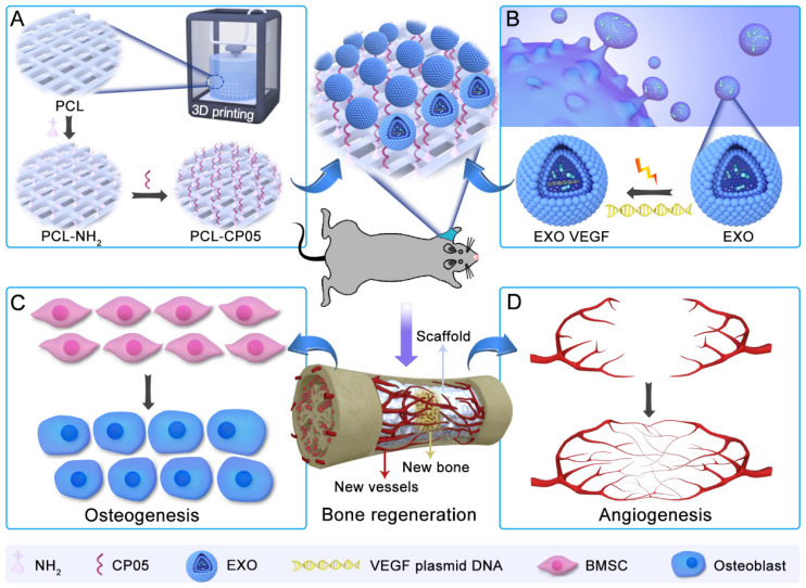
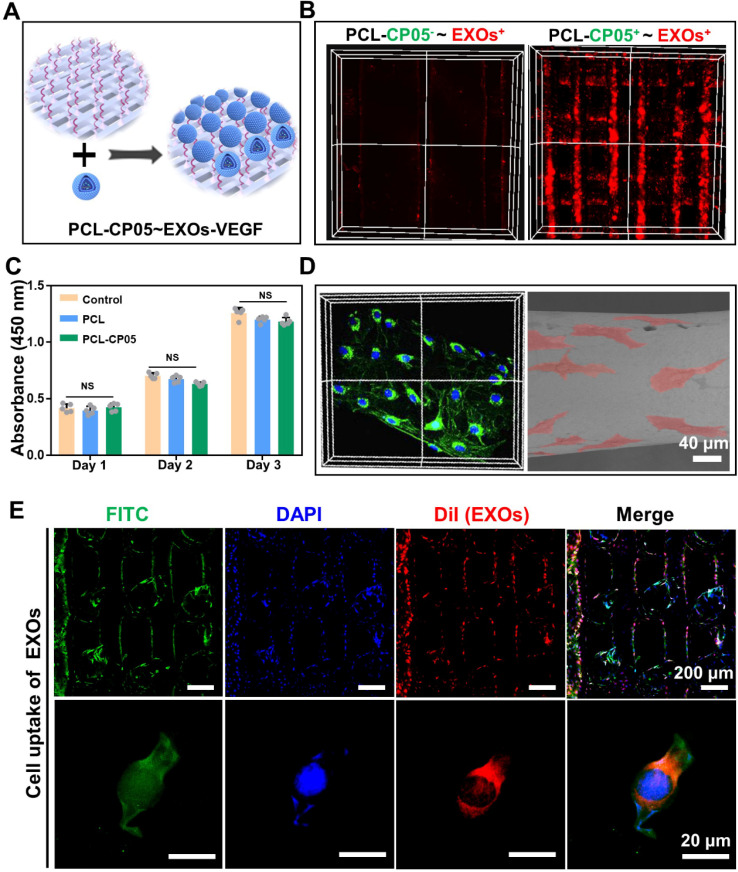
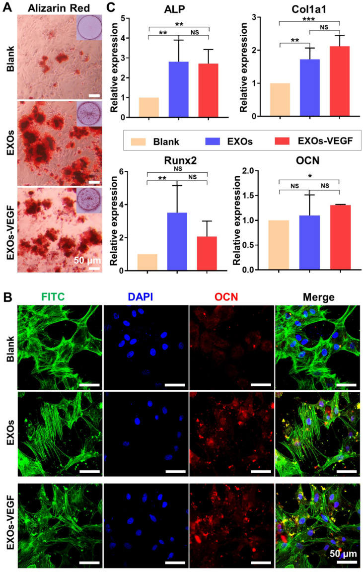
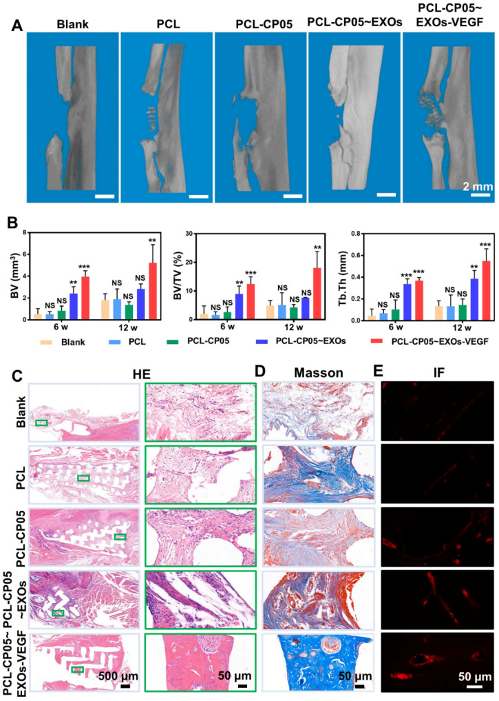
In our current work, we designed specifically engineered exosomes based on the requirements of vascularized bone repair in segmental bone defects. This work simultaneously illuminates the potential of functional exosomes in acellular tissue engineering.
Overview
The claims and statistics of this paper haven’t been checked yet.
Run the check — free, no account needed. It reads the open-access text and checks the sources it cites.
Integrity signals
Registry
Cites
Statistics
Figures
Verification
Claims the paper makes
The claims and statistics of this paper haven’t been checked yet.
Run the check — free, no account needed. It reads the open-access text and checks the sources it cites.
Claims other papers make about this one
Each is checked against this paper’s own text: does it say what the citing paper says it does?
In the citing paper
“Zha et al. extracted ATDC5 (chondrogenic progenitor cell line)-derived exosomes to load the vascular endothelial growth factor (VEGF) gene, which can effectively restore segmental bone defects by increasing osteogenesis and angiogenesis [86].”
From The Therapeutic Potential and Clinical Significance of Exosomes as Carriers of Drug Delivery System (2022) · cites this paper as [86]
Self-check
Percentages, sums and reported differences re-computed from the paper’s own numbers.
The self-check needs the paper’s full text.
Citations
Cites
The biology , function , and biomedical applications of exosomes
Kalluri, R., & LeBleu, V. S. (2020) · Science · doi:10.1126/science.aau6977 · Cited by 10,770
Shedding light on the cell biology of extracellular vesicles
van Niel, G., D'Angelo, G., & Raposo, G. (2018) · Nature Reviews Molecular Cell Biology · doi:10.1038/nrm.2017.125 · Cited by 8,369
Melanoma exosomes educate bone marrow progenitor cells toward a pro-metastatic phenotype through MET
Peinado, H., Alečković, M., Lavotshkin, S., Matei, I., Costa-Silva, B., Moreno-Bueno, G., Hergueta-Redondo, M., Williams, C., García-Santos, G., Ghajar, C. M., et al. (2012) · Nature Medicine · doi:10.1038/nm.2753 · Cited by 3,715
Extracellular vesicles: biology and emerging therapeutic opportunities
EL Andaloussi, S., Mäger, I., Breakefield, X. O., & Wood, M. J. A. (2013) · Nature Reviews Drug Discovery · doi:10.1038/nrd3978 · Cited by 3,571
Regulation of immune responses by extracellular vesicles
Robbins, P. D., & Morelli, A. E. (2014) · Nature Reviews Immunology · doi:10.1038/nri3622 · Cited by 2,260
Bone tissue engineering using 3D printing
Bose, S., Vahabzadeh, S., & Bandyopadhyay, A. K. (2013) · Materials Today · doi:10.1016/j.mattod.2013.11.017 · Cited by 1,873
3D bioprinting for engineering complex tissues
Mandrycky, C., Wang, Z., Kim, K., & Kim, D. (2015) · Biotechnology Advances · doi:10.1016/j.biotechadv.2015.12.011 · Cited by 1,818
Extracellular Vesicles: Unique Intercellular Delivery Vehicles
Maas, S. L. N., Breakefield, X. O., & Weaver, A. M. (2016) · Trends in Cell Biology · doi:10.1016/j.tcb.2016.11.003 · Cited by 1,466
The Smart Drug Delivery System and Its Clinical Potential
Liu, D., Yang, F., Xiong, F., & Gu, N. (2016) · Theranostics · doi:10.7150/thno.14858 · Cited by 1,066
Exosomes: Therapy delivery tools and biomarkers of diseases
Barile, L., & Vassalli, G. (2017) · Pharmacology & Therapeutics · doi:10.1016/j.pharmthera.2017.02.020 · Cited by 1,053
Cited by
Sorted by citations
Al-Shalawi, F. D., Ariff, A. H. M., Jung, D. W., Ariffin, M. K. A. M., Looi, C. S. K., Brabazon, D., & Al-Osaimi, M. O. (2023) · Polymers · doi:10.3390/polym15122601 · Cited by 250
Treatment of inflammatory bone loss in periodontitis by stem cell-derived exosomes
Lei, F., Li, M., Lin, T., Zhou, H., Wang, F., & Su, X. (2022) · Acta Biomaterialia · doi:10.1016/j.actbio.2021.12.035 · Cited by 213
Ding, J., Zhang, Y., Cai, X., Zhang, Y., Yan, S., Wang, J., Zhang, S., Yin, T., Yang, C., & Yang, J. (2021) · Theranostics · doi:10.7150/thno.58731 · Cited by 172
Cell-free exosome-laden scaffolds for tissue repair
Huang, J., Xiong, J., Yang, L., Zhang, J., Sun, S., & Liang, Y. (2021) · Nanoscale · doi:10.1039/d1nr01314a · Cited by 152
Ling, Z., Chen, Z., Deng, J. G., Wang, Y., Yuan, B., Yang, X., Lin, H., Cao, J., Zhu, X., & Zhang, X. (2021) · Chemical Engineering Journal · doi:10.1016/j.cej.2021.130302 · Cited by 141
Advances in materials-based therapeutic strategies against osteoporosis
Chen, L., Song, J., Li, S., Zhu, Y., Liu, M., Wan, M., 高木, 昭., Tay, F. R., & Niu, L. (2023) · Biomaterials · doi:10.1016/j.biomaterials.2023.122066 · Cited by 127
Todd, E. A., Mirsky, N. A., Silva, B. L. G., Shinde, A. R., Arakelians, A. R. L., Nayak, V. V., Marcantonio, R. A. C., Gupta, N., Witek, L., & Coelho, P. G. (2024) · Journal of Functional Biomaterials · doi:10.3390/jfb15100280 · Cited by 130
Neuro–bone tissue engineering: emerging mechanisms, potential strategies, and current challenges
Sun, W., Ye, B., Chen, S., Zeng, L., Lu, H., Wan, Y., Gao, Q. H., Chen, K., Qu, Y., Wu, B., et al. (2023) · Bone Research · doi:10.1038/s41413-023-00302-8 · Cited by 137
4D printing: The spotlight for 3D printed smart materials
Chen, J., Virrueta, C., Zhang, S., Mao, C., & Wang, J. (2024) · Materials Today · doi:10.1016/j.mattod.2024.06.004 · Cited by 124
Engineering exosomes and biomaterial-assisted exosomes as therapeutic carriers for bone regeneration
Lu, Y., Mai, Z., Cui, L., & Zhao, X. (2023) · Stem Cell Research & Therapy · doi:10.1186/s13287-023-03275-x · Cited by 119
Intelligent Vascularized 3D/4D/5D/6D-Printed Tissue Scaffolds
Han, X., Saiding, Q., Cai, X., Xiao, Y., Wang, P., Cai, Z., Gong, X., Gong, W., Zhang, X., & Cui, W. (2023) · Nano-Micro Letters · doi:10.1007/s40820-023-01187-2 · Cited by 130
Hao, Z., Ren, L., Zhang, Z., Yang, Z., Wu, S., Liu, G., Cheng, B., Wu, J., & Xia, J. (2022) · Bioactive Materials · doi:10.1016/j.bioactmat.2022.10.018 · Cited by 113
The Therapeutic Potential and Clinical Significance of Exosomes as Carriers of Drug Delivery System
Li, T., Li, X., Han, G., Liang, M., Yang, Z., Zhang, C., Huang, S., Tai, S., & Yu, S. (2022) · Pharmaceutics · doi:10.3390/pharmaceutics15010021 · Cited by 110
Li, F., Wu, J., Li, D., Hao, L., Li, Y., Yi, D., Yeung, K. W. K., Chen, D., Lu, W. W., Pan, H., et al. (2022) · Journal of Nanobiotechnology · doi:10.1186/s12951-022-01347-3 · Cited by 99
Extracellular vesicles in nanomedicine and regenerative medicine: A review over the last decade
Moghassemi, S., Dadashzadeh, A., Sousa, M. J., Vlieghe, H., Yang, J., León-Félix, C. M., & Amorim, C. A. (2024) · Bioactive Materials · doi:10.1016/j.bioactmat.2024.02.021 · Cited by 113
Shafiei, M., Ansari, M. N. M., Razak, S. I. A., & Khan, M. U. A. (2021) · Polymers · doi:10.3390/polym13152529 · Cited by 100
Liu, H., Geng, Z., & Su, J. (2022) · Extracellular Vesicles and Circulating Nucleic Acids · doi:10.20517/evcna.2022.04 · Cited by 95
Bone‐Targeted Exosomes: Strategies and Applications
Wang, J., Li, X., Wang, S., Cui, J., Ren, X., & Su, J. (2023) · Advanced Healthcare Materials · doi:10.1002/adhm.202203361 · Cited by 92
OTUB1 promotes osteoblastic bone formation through stabilizing FGFR2
Zhu, Q. N., Fu, Y., Cui, C., Ding, Y., Deng, Z., Ning, C., Hu, F., Qiu, C., Yu, B., Zhou, X., et al. (2023) · Signal Transduction and Targeted Therapy · doi:10.1038/s41392-023-01354-2 · Cited by 81
Engineered bacterial extracellular vesicles for osteoporosis therapy
Liu, H., Li, M., Zhang, T., Liu, X., Zhang, H., Geng, Z., & Su, J. (2022) · Chemical Engineering Journal · doi:10.1016/j.cej.2022.138309 · Cited by 78
Tumor cell membrane‐based vaccines: A potential boost for cancer immunotherapy
Yang, M., Zhou, J., Lu, L., Deng, D., Huang, J., Tang, Z., Shi, X., Lo, P., Lovell, J. F., Zheng, Y., et al. (2024) · Exploration · doi:10.1002/exp.20230171 · Cited by 74
Engineered extracellular vesicles for bone therapy
Jiang, Y., Li, J., Xu, X. J., Yin, Z., Xu, K., & Su, J. (2022) · Nano Today · doi:10.1016/j.nantod.2022.101487 · Cited by 68
Hu, H., Zhang, H., Bu, Z., Liu, Z., Lv, F., Pan, M., Huang, X., & Cheng, L. (2022) · International Journal of Nanomedicine · doi:10.2147/ijn.s389471 · Cited by 69
Clinical Applications of Adipose-Derived Stem Cell (ADSC) Exosomes in Tissue Regeneration
Papadopoulos, K., Piperi, C., & Korkolopoulou, P. A. (2024) · International Journal of Molecular Sciences · doi:10.3390/ijms25115916 · Cited by 72
Production and Utility of Extracellular Vesicles with 3D Culture Methods
Ester, M. C., & Day, R. M. (2023) · Pharmaceutics · doi:10.3390/pharmaceutics15020663 · Cited by 69
Mishra, A., Singh, P., Qayoom, I., Prasad, A., & Kumar, A. (2021) · Journal of Materials Chemistry B · doi:10.1039/d1tb01088c · Cited by 70
Extracellular Vesicles in Tissue Engineering: Biology and Engineered Strategy
Pan, Z., Sun, W., Chen, Y., Tang, H., Lin, W., Chen, J., & Chen, C. (2022) · Advanced Healthcare Materials · doi:10.1002/adhm.202201384 · Cited by 69
Sahebalzamani, M., Ziminska, M., McCarthy, H. O., Levingstone, T. J., Dunne, N., & Hamilton, A. (2022) · Biomaterials Science · doi:10.1039/d1bm01756j · Cited by 70
Extracellular vesicle-embedded materials
Ma, Y., Brocchini, S., & Williams, G. R. (2023) · Journal of Controlled Release · doi:10.1016/j.jconrel.2023.07.059 · Cited by 73
Recent advancement in vascularized tissue-engineered bone based on materials design and modification
Liu, H. R., Chen, H., Han, Q., Sun, B., Liu, Y., Zhang, A., Fan, D., Xia, P., & Wang, J. (2023) · Materials Today Bio · doi:10.1016/j.mtbio.2023.100858 · Cited by 72
Li, Z., Liu, J., Song, J., Yin, Z., Zhou, F., Shen, H., Wang, G., & Su, J. (2024) · Theranostics · doi:10.7150/thno.97317 · Cited by 68
Gu, X., Zha, Y., Li, Y., Chen, J., Liu, S., Du, Y., Zhang, S., & Wang, J. (2022) · Acta Biomaterialia · doi:10.1016/j.actbio.2022.01.021 · Cited by 64
Bioactive Scaffolds Integrated with Liposomal or Extracellular Vesicles for Bone Regeneration
Kang, M., Lee, C., & Lee, M. (2021) · Bioengineering · doi:10.3390/bioengineering8100137 · Cited by 65
Jiang, W., Zhan, Y., Zhang, Y., Sun, D., Guo, Z., Wang, Z., Chen, L., & Sun, J. (2024) · Biomaterials · doi:10.1016/j.biomaterials.2024.122566 · Cited by 64
Hyaluronic acid-based hydrogels: As an exosome delivery system in bone regeneration
Deng, H., Wang, J., & An, R. (2023) · Frontiers in Pharmacology · doi:10.3389/fphar.2023.1131001 · Cited by 62
Wang, W., Liang, X., Zheng, K., Ge, G., Chen, X., Xu, Y., Bai, J., Pan, G., & Geng, D. (2022) · Materials Today Bio · doi:10.1016/j.mtbio.2022.100355 · Cited by 59
Approaches and materials for endocytosis-independent intracellular delivery of proteins
Chen, N., He, Y., Zang, M., Zhang, Y., Lü, H., Zhao, Q., Wang, S., & Gao, Y. (2022) · Biomaterials · doi:10.1016/j.biomaterials.2022.121567 · Cited by 59
Exosomes as Potential Functional Nanomaterials for Tissue Engineering
Yao, Y., Jiang, Y., Song, J., Wang, R., Li, Z., Yang, L., Wu, W., Zhang, L., & Peng, Q. (2022) · Advanced Healthcare Materials · doi:10.1002/adhm.202201989 · Cited by 59
Organoid Vascularization: Strategies and Applications
Gao, Q., Wang, J., Zhang, H., Wang, J., Jing, Y., & Su, J. (2025) · Advanced Healthcare Materials · doi:10.1002/adhm.202500301 · Cited by 63
Technological Advances of 3D Scaffold-Based Stem Cell/Exosome Therapy in Tissues and Organs
Gu, C., Feng, J., Waqas, A., Deng, Y., Zhang, Y., Chen, W., Long, J., Huang, S., & Chen, L. (2021) · Frontiers in Cell and Developmental Biology · doi:10.3389/fcell.2021.709204 · Cited by 52
Wang, Q., Liu, K., Cao, X., Rong, W., Shi, W., Yu, Q., Deng, W., Yu, J., & Xu, X. (2024) · Bioengineering & Translational Medicine · doi:10.1002/btm2.10646 · Cited by 51
Organoids and organoid extracellular vesicles-based disease treatment strategies
Zhou, G., Li, R., Sheng, S., Huang, J., Zhou, F., Wei, Y., Liu, H., & Su, J. (2024) · Journal of Nanobiotechnology · doi:10.1186/s12951-024-02917-3 · Cited by 58
Liu, X., Zhou, C., Xie, Q., Xia, L., Liu, L., Bao, W., Lin, H., Xiong, X., Zhang, H., Zheng, Z., et al. (2024) · Journal of Translational Medicine · doi:10.1186/s12967-024-05809-0 · Cited by 56
Fan, X., Zhang, Y., Liu, W., Shao, M., Gong, Y., Wang, T., Xue, S., & Nian, R. (2024) · Biomaterials Science · doi:10.1039/d4bm00558a · Cited by 50
Li, N., Wang, J., Feng, G., Liu, Y., Shi, Y., Wang, Y., & Chen, L. (2024) · Regenerative Biomaterials · doi:10.1093/rb/rbae078 · Cited by 48
Wu, X., Huo, Y., Ci, Z., Wang, Y., Xu, W., Bai, B., Hao, J., Hu, G., Yu, M., Ren, W., et al. (2022) · Applied Materials Today · doi:10.1016/j.apmt.2022.101478 · Cited by 49
Lan, Y., Xie, H., Jin, Q., Zhao, X., Shi, Y., Zhou, Y., Hu, Z., Ye, Y., Huang, X., Sun, Y., et al. (2022) · Bioactive Materials · doi:10.1016/j.bioactmat.2022.01.019 · Cited by 47
Sun, X., Mao, Y., Liu, B., Gu, K., Liu, H., Du, W., Li, R., & Zhang, J. (2023) · Journal of Personalized Medicine · doi:10.3390/jpm13020180 · Cited by 50
Yu, L., Wei, Q., Li, J., Wang, H., Meng, Q., Xie, E., Li, Z., Li, K., Shu, W., Wu, J., et al. (2023) · Composites Part B Engineering · doi:10.1016/j.compositesb.2023.110505 · Cited by 46
Frontiers of Hydroxyapatite Composites in Bionic Bone Tissue Engineering
Shi, J., Dai, W., Gupta, A., Zhang, B., Wu, Z., Zhang, Y., Pan, L., & Wang, L. (2022) · Materials · doi:10.3390/ma15238475 · Cited by 48
Qi, L., Fang, X., Yan, J., Pan, C., Ge, W., Wang, J., Shen, S. G., Lin, K., & Zhang, L. (2023) · Bioactive Materials · doi:10.1016/j.bioactmat.2023.10.024 · Cited by 46
Hao, Z., Zhang, Z., Wang, Z.-P., Wang, Y., Chen, J., Chen, T.-H., Shi, G., Li, H., Wang, J.-W., Dong, M.-C., et al. (2024) · Military Medical Research · doi:10.1186/s40779-024-00576-x · Cited by 51
Biodegradable Implants for Internal Fixation of Fractures and Accelerated Bone Regeneration
Wang, P., Gong, Y., Zhou, G., Ren, W., & Wang, X. (2023) · ACS Omega · doi:10.1021/acsomega.3c02727 · Cited by 44
Research Progress of Exosomes in Bone Diseases: Mechanism, Diagnosis and Therapy
Meng, F., Xu, X. J., Yin, Z., Gao, F., Wang, X., & Geng, Z. (2022) · Frontiers in Bioengineering and Biotechnology · doi:10.3389/fbioe.2022.866627 · Cited by 44
Huang, J., Guo, J., Dong, Y., Xiao, H., Yang, P., Liu, Y., Liu, S., Cheng, S., Song, J., Su, Y., et al. (2022) · International Journal of Biological Macromolecules · doi:10.1016/j.ijbiomac.2022.11.233 · Cited by 43
Liu, T., Li, B., Chen, G., Ye, X., & Zhang, Y. (2022) · International Journal of Biological Macromolecules · doi:10.1016/j.ijbiomac.2022.09.003 · Cited by 42
The Delivery and Activation of Growth Factors Using Nanomaterials for Bone Repair
Li, Y., Xu, C., & Lei, C. (2023) · Pharmaceutics · doi:10.3390/pharmaceutics15031017 · Cited by 43
Exosomes and exosome composite scaffolds in periodontal tissue engineering
Wang, T., Zhou, Y., Zhang, W., Xue, Y., Xiao, Z., Zhou, Y., & Peng, X. (2024) · Frontiers in Bioengineering and Biotechnology · doi:10.3389/fbioe.2023.1287714 · Cited by 43
Zhao, H., Shen, S. H., Zhao, L., Xu, Y., Li, Y., & Zhuo, N. (2021) · BMC Musculoskeletal Disorders · doi:10.1186/s12891-021-04617-7 · Cited by 41
The role of microfluidics and 3D-bioprinting in the future of exosome therapy
Amondarain, M., Gallego, I., Puras, G., del Burgo, L. S., Luzzani, C., & Pedraz, J. L. (2023) · Trends in biotechnology · doi:10.1016/j.tibtech.2023.05.006 · Cited by 41
Engineering exosomes for bone defect repair
Ma, S., Zhang, Y., Li, S., Li, A., Li, Y., & Pei, D. (2022) · Frontiers in Bioengineering and Biotechnology · doi:10.3389/fbioe.2022.1091360 · Cited by 38
Bang, O. Y., & Kim, J.-E. (2022) · BMB Reports · doi:10.5483/bmbrep.2022.55.1.162 · Cited by 39
MSC-Exos: important active factor of bone regeneration
Ren, S., Lin, Y., Liu, W., Yang, L., & Zhao, M. (2023) · Frontiers in Bioengineering and Biotechnology · doi:10.3389/fbioe.2023.1136453 · Cited by 37
Lv, Z., Ji, Y., Wen, G., Liang, X., Zhang, K., & Zhang, W. (2024) · Burns & Trauma · doi:10.1093/burnst/tkae036 · Cited by 35
Chen, X., Gao, C., Chu, X., Zheng, C., Luan, Y., He, X., Yang, K. C., & Zhang, D. (2022) · Frontiers in Bioengineering and Biotechnology · doi:10.3389/fbioe.2022.915181 · Cited by 33
Luo, L., Zheng, W., Li, J., Chen, T., Xue, W., Lin, T., Liu, M., Yan, Z., Yang, J., Li, J., et al. (2025) · Advanced Science · doi:10.1002/advs.202500599 · Cited by 32
Yu, L., Dou, G., Kuang, H., Bao, L., Liu, H., Ye, Q., Wang, Z., Yang, X., Ren, L., Li, Z., et al. (2024) · ACS Nano · doi:10.1021/acsnano.3c11050 · Cited by 31
Propelling Minimally Invasive Tissue Regeneration With Next‐Era Injectable Pre‐Formed Scaffolds
Liao, J., Timoshenko, A. B., Cordova, D. J., Potes, M. D. A., Gaihre, B., Liu, X., Elder, B. D., Lu, L., & Tilton, M. (2024) · Advanced Materials · doi:10.1002/adma.202400700 · Cited by 32
Exosomes to exosome-functionalized scaffolds: a novel approach to stimulate bone regeneration
Deng, L., Liu, Y., Wu, Q., Lai, S., Yang, Q., Mu, Y., & Dong, M. (2024) · Stem Cell Research & Therapy · doi:10.1186/s13287-024-04024-4 · Cited by 32
Li, J., Li, X., Li, X., Liang, Z., Wang, Z., Shahzad, K. A., Xu, M., & Tan, F. (2024) · ACS Applied Materials & Interfaces · doi:10.1021/acsami.4c05004 · Cited by 30
Next-generation probiotics and engineered BEVs for precision therapeutics in osteoporosis
Wei, J., Ding, W., Song, K., Zhang, Y., Luo, Q., & Qi, C. (2025) · Frontiers in Nutrition · doi:10.3389/fnut.2025.1581971 · Cited by 30
Lv, Z., Xu, J., Cai, X., Wang, Y., Yang, X.-D., Wang, H., Zhu, Y., Feng, B., & Weng, X. (2025) · ACS Nano · doi:10.1021/acsnano.5c05416 · Cited by 31
Wu, S., Ma, J., Liu, J., Liu, C., Ni, S., Dai, T., Wang, Y., Weng, Y., Zhao, H., Zhou, D., et al. (2022) · ACS Applied Materials & Interfaces · doi:10.1021/acsami.1c24748 · Cited by 30
Electrospinning-based bone tissue scaffold construction: Progress and trends
Ma, Y., Zhou, R., Yang, M., Zhang, J., Song, W. F., Ma, X., Liu, M., Cui, X., Li, B., Zhang, Y., et al. (2025) · Materials & Design · doi:10.1016/j.matdes.2025.113792 · Cited by 29
Bone Engineering Scaffolds With Exosomes: A Promising Strategy for Bone Defects Repair
Zhang, M., Li, Y., Feng, T., Li, R., Wang, Z., Zhang, L., Yin, P., & Tang, P. (2022) · Frontiers in Bioengineering and Biotechnology · doi:10.3389/fbioe.2022.920378 · Cited by 30
3D bioprinted extracellular vesicles for tissue engineering—a perspective
Han, P., & Ivanovski, S. (2022) · Biofabrication · doi:10.1088/1758-5090/ac9809 · Cited by 32
Ma, W., Wang, W., Liu, F., Kong, Y., Xia, B., Yang, H., Zhao, H., Yang, H., Li, K., Li, Y., et al. (2023) · Composites Part B Engineering · doi:10.1016/j.compositesb.2023.110612 · Cited by 29
Nanoparticle-Based Drug Delivery Systems for Enhancing Bone Regeneration
Xu, H., Cui, Y., Tian, Y., Dou, M., Sun, S., Wang, J., & Wu, D. (2024) · ACS Biomaterials Science & Engineering · doi:10.1021/acsbiomaterials.3c01643 · Cited by 31
Li, Y., Peng, H., Tang, W., Gu, D., Ren, S., Yu, Y., Yang, J., & Miao, L. (2022) · Materials & Design · doi:10.1016/j.matdes.2022.111540 · Cited by 28
Yang, J., Li, L., Wang, L., Chen, R., Yang, X., Wu, J., Feng, G., Ding, J., Diao, L., Chen, J., et al. (2024) · Journal of Translational Medicine · doi:10.1186/s12967-023-04831-y · Cited by 28
Organoid extracellular vesicle-based therapeutic strategies for bone therapy.
Liu, H., & Su, J. (2023) · PubMed Central · doi:10.12336/biomatertransl.2023.04.002 · Cited by 28
Chen, D., Lu, C., Na, N., Yin, R., & Huang, F. (2024) · Cell & Bioscience · doi:10.1186/s13578-024-01255-z · Cited by 29
Liu, H., Li, R., Yang, H., Situ, B., Wang, G., Xu, K., & Su, J. (2024) · Small Science · doi:10.1002/smsc.202400474 · Cited by 30
Harnessing Nucleic Acids Nanotechnology for Bone/Cartilage Regeneration
Han, Y., Cao, L., Li, G., Zhou, F., Bai, L., & Su, J. (2023) · Small · doi:10.1002/smll.202301996 · Cited by 28
Zhang, M., Wan, L., Li, R., Li, X., Zhu, T., & Lu, H. (2023) · Biomaterials Science · doi:10.1039/d3bm01169k · Cited by 26
SHED‐derived exosomes improve the repair capacity and osteogenesis potential of hPDLCs
Wang, M., Li, J., Ye, Y., Chen, D., & Song, J. (2022) · Oral Diseases · doi:10.1111/odi.14153 · Cited by 25
Wang, F., Gu, Z., Yin, Z., Zhang, W., Bai, L., & Su, J. (2023) · Journal of Nanobiotechnology · doi:10.1186/s12951-023-02003-0 · Cited by 24
Efforts to promote osteogenesis–angiogenesis coupling for bone tissue engineering
Xu, Z.-W., Wang, B., Huang, R., Guo, M., Han, D., Yin, L., Zhang, X., Huang, Y., & Li, X. (2024) · Biomaterials Science · doi:10.1039/d3bm02017g · Cited by 26
Chen, J., Li, Y., Quan, X., Chen, J., Han, Y., Li, Y., Zhou, M., Mok, G. S. P., Wang, R., & Zhao, Y. (2024) · Neural Regeneration Research · doi:10.4103/nrr.nrr-d-24-00243 · Cited by 24
Zhang, Q., Wang, Y., Zhu, Z., Waqas, A., Zhou, D., & Chen, L. (2025) · International Journal of Nanomedicine · doi:10.2147/ijn.s505792 · Cited by 23
Liu, F., Sun, T., An, Y., Ming, L., Li, Y., Zhou, Z., & Shang, F. (2023) · Frontiers in Bioengineering and Biotechnology · doi:10.3389/fbioe.2023.1050916 · Cited by 34
Bone aging and extracellular vesicles
Wang, J., Zhang, Y., Wang, S., Wang, X., Jing, Y., & Su, J. (2024) · 中国科学通报:英文版 · doi:10.1016/j.scib.2024.10.013 · Cited by 25
Engineering extracellular vesicles for targeted therapeutics in cardiovascular disease
Fu, E., Pan, K., & Li, Z. (2024) · Frontiers in Cardiovascular Medicine · doi:10.3389/fcvm.2024.1503830 · Cited by 22
Bioprinting of exosomes: Prospects and challenges for clinical applications
Selvam, S., Thomas, M. B., Bhowmick, T., & Chandru, A. (2023) · International Journal of Bioprinting · doi:10.18063/ijb.690 · Cited by 22
Advances in the Study of Extracellular Vesicles for Bone Regeneration
Yao, J., Liu, Y., Du, J., Xu, J., Luo, Z., Liu, Y. F., & Guo, L. (2024) · International Journal of Molecular Sciences · doi:10.3390/ijms25063480 · Cited by 24
Adipose-derived stem cell exosomes: multifaceted therapeutic applications in regenerative medicine
Rau, C., Kuo, P., & Hsieh, C. (2025) · International Journal of Surgery · doi:10.1097/js9.0000000000002841 · Cited by 25
Zhang, J., Zhang, B., Zheng, Z., Cai, Q., Wang, J., Shu, Q., & Wang, L. (2021) · Advanced Functional Materials · doi:10.1002/adfm.202109882 · Cited by 21
Huang, Y. L., & Xie, H. (2025) · International Journal of Nanomedicine · doi:10.2147/ijn.s522198 · Cited by 20
Wu, H., Chen, G., Zhang, G., Lv, Q., Gu, D., & Dai, M. (2023) · Biomedical Materials · doi:10.1088/1748-605x/acb412 · Cited by 20
Delivery of Growth Factors to Enhance Bone Repair
Ball, J. R., Shelby, T., Hernandez, F., Mayfield, C. K., & Lieberman, J. R. (2023) · Bioengineering · doi:10.3390/bioengineering10111252 · Cited by 21
Metadata
- Authors
- Zha, Y., Li, Y., Lin, T., Chen, J., Zhang, S., & Wang, J.
- Affiliations
- Huazhong University of Science and Technology (CN)
- Corresponding author
- Zhang, Shengmin
- Venue
- Theranostics
- Publisher
- Ivyspring International Publisher
- Published
- 2021
- Volume
- 11
- Issue
- 1
- Pages
- 397-409
- ISSN
- 1838-7640 (ISSN-L)
- PMC
- PMC7681080
- OpenAlex
- W3095185855
- Cited by
- 232
- References
- 47
- Citations by year
- 2026: 25 · 2025: 55 · 2024: 55 · 2023: 48 · 2022: 39 · 2021: 10
- FWCI
- 7.86
- Type
- journal article
- Language
- en
- Open access
- gold
- doi.org
- Licence
- cc-by
- Locations
- Theranostics (publishedVersion, cc-by)
- Europe PMC (PubMed Central) (publishedVersion, other-oa)
- PubMed Central (publishedVersion, cc-by)
- PubMed (publishedVersion)
- Funders
- National Natural Science Foundation of China (2017YFC1103900, 31670968, 91939111, SZSM201812055, 31800805, 81601610)
- Sanming Project of Medicine in Shenzhen (SZSM201812055)
- National Key Research and Development Program of China (2018YFC1105700, 2017YFC1103900)
- Fundamental Research Funds for the Central Universities (2019kfyRCPY103)
- Topics
- Life Sciences › Biochemistry, Genetics and Molecular Biology › Molecular Biology › Extracellular vesicles in disease
- Physical Sciences › Engineering › Biomedical Engineering › Bone Tissue Engineering Materials
- Life Sciences › Biochemistry, Genetics and Molecular Biology › Cell Biology › Mesenchymal stem cell research
- Keywords
- exosomes, progenitor cells, vascular endothelial growth factor, osteogenic induction, vascular remodeling, segmental bone defect, gene delivery
- MeSH terms
- Animals, Bone and Bones, Bone Regeneration, Cell Adhesion, Cell Line, Male, Materials Testing, Osteogenesis, Plasmids, Polyesters, Radius, Stem Cells, Tissue Engineering, Vascular Endothelial Growth Factor A, Cell Proliferation, Mice, Rats, Tissue Scaffolds, X-Ray Microtomography, Exosomes, Vascular Remodeling, Printing, Three-Dimensional
About these checks
Registry, retraction, Cites and statistics results are computed, not judged, with one exception: a printed reference that no index lists is read once by a model before it counts as a citation error. Claims are extracted by a model and kept only when their quote appears verbatim in the text; each cited source is then checked, with a model’s help, for whether it says what the claim relies on. Those verdicts can be wrong — read the claim’s quote and the cited source before relying on one.
Statistics and arithmetic are recomputed, not judged.
Confidence bands are shown only for fields calibrated on our annotated test set. None is calibrated yet, so no band is shown.
What counts as a claim ›How we measure confidence ›How we check this ›
Are you an author, or do you see a problem with this page? Request a review
