Cited by 97

The DOI matches its Crossref record; 18 of the 18 references it cites were found in a registry or an index.

Transformative Impact of Nanocarrier‐Mediated Drug Delivery: Overcoming Biological Barriers and Expanding Therapeutic Horizons

Kim, Minhye, Shin, Myeongyeon, Zhao, Yaping, Ghosh, Mrinmoy, Son, Young‐Ok·2024·Small Science

Abstract

Advancing therapeutic progress is centered on developing drug delivery systems (DDS) that control therapeutic molecule release, ensuring precise targeting and optimal concentrations. Targeted DDS enhances treatment efficacy and minimizes off‐target effects, but struggles with drug degradation. Over the last three decades, nanopharmaceuticals have evolved from laboratory concepts into clinical products, highlighting the profound impact of nanotechnology in medicine. Despite advancements, the effective delivery of therapeutics remains challenging because of biological barriers. Nanocarriers offer a solution with a small size, high surface‐to‐volume ratios, and customizable properties. These systems address physiological and biological challenges, such as shear stress, protein adsorption, and quick clearance. They allow targeted delivery to specific tissues, improve treatment outcomes, and reduce adverse effects. Nanocarriers exhibit controlled release, decreased degradation, and enhanced efficacy. Their size facilitates cell membrane penetration and intracellular delivery. Surface modifications increase affinity for specific cell types, allowing precise treatment delivery. This study also elucidates the potential integration of artificial intelligence with nanoscience to innovate future nanocarrier systems.

Overview

The claims and statistics of this paper haven’t been checked yet.

Run the check — free, no account needed. It reads the open-access text and checks the sources it cites.

A few free checks a day · papers already verified open instantly · results are public

Integrity signals

Registry

Crossref · 10.1002/smsc.202400280

Cites

18 of 18 references verified0 citation errors · 0 not checkable

Statistics

Figures

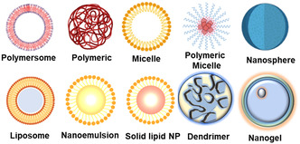
Figure 1 Common biopolymers used in organic nanocarriers. This figure illustrates the diverse array of common biopolymers utilized in the formulation of organic nanocarriers for drug delivery applications. Biopolymers play a critical role in enhancing the stability, biocompatibility, and functionality of nanocarrier systems. Several types of biopolymers, including polymeric NPs, lipid‐based carriers, dendrimers, nanoemulsions, and nanomicelles are highlighted. Each biopolymer offers unique characteristics and functionalities that contribute to their effectiveness in drug delivery. Created with BioRender.com.
Figure 1 Common biopolymers used in organic nanocarriers. This figure illustrates the diverse array of common biopolymers utilized in the formulation of organic nanocarriers for drug delivery applications. Biopolymers play a critical role in enhancing the stability, biocompatibility, and functionality of nanocarrier systems. Several types of biopolymers, including polymeric NPs, lipid‐based carriers, dendrimers, nanoemulsions, and nanomicelles are highlighted. Each biopolymer offers unique characteristics and functionalities that contribute to their effectiveness in drug delivery. Created with BioRender.com.CC BY · PubMed Central
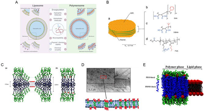
Figure 2 The diagram illustrates the structure of LPNPs, highlighting the interaction between lipids and polymers. A) The structure of a liposome features an aqueous core surrounded by a bilayer membrane. This membrane consists of hydrophobic tails forming the interior of the bilayer, while hydrated hydrophilic heads stabilize the outer and inner surfaces. Polymersomes, which are synthetic counterparts to liposomes, are composed of amphiphilic block copolymer membranes. Although both carriers share many similar properties, polymersomes offer greater versatility and improved stability. Adapted with permission.[ 238 ] Copyright 2014, Elsevier. B)(a) The illustration shows LPNPs created through interactions with (b–d) different polymers, where the lipids (depicted in yellow) are enveloped by a “belt” of polymer (depicted in green). Different polymers can be used to create this polymer–lipid combination, with each polymer influencing the NP's properties, such as its stability, drug release profile, and interaction with biological systems. Hydrophilic groups are highlighted in blue, and hydrophobic groups are highlighted in red. [ 239 ] C) (a) Conformational adaptation at the polymer/lipid domain boundary in hybrid vesicles is expected when domain formation occurs, allowing the components to adjust and integrate within distinct regions. (b) In contrast, the absence of conformational adaptation between the polymer and lipid boundaries results in a homogeneous mixture, where the components are evenly distributed without distinct domain separation.[ 88 ] D) Hybrid lipid/polymer vesicle systems, which incorporate the lipid 1,2‐dioleoyl‐sn‐glycero‐3‐phosphocholine (DOPC) and amphiphilic triblock copolymers composed of poly(ethylene glycol)–poly(dimethylsiloxane)–poly(ethylene glycol) (PEG–PDMS–PEG), are studied to understand how hydrophilic blocks impact vesicle morphology. The hydrophilic mass fraction of the amphiphilic polymer plays a significant role in influencing the membrane thickness, size polydispersity, and lamellarity of these polymer/lipid hybrid vesicles. Changes in the hydrophilic mass fraction can lead to variations in how the vesicles form, including their structural uniformity and stability, making this a crucial factor in the design and application of such hybrid systems. Adapted with permission.[ 92 ] Copyright 2022, ACS. E) The schematic view of a hybrid polymer/lipid membrane with lateral phase separation typically shows a membrane where distinct regions or “domains” of polymers and lipids coexist within the same membrane. Adapted with permission.[ 93 ] Copyright 2022, Elsevier.
Figure 2 The diagram illustrates the structure of LPNPs, highlighting the interaction between lipids and polymers. A) The structure of a liposome features an aqueous core surrounded by a bilayer membrane. This membrane consists of hydrophobic tails forming the interior of the bilayer, while hydrated hydrophilic heads stabilize the outer and inner surfaces. Polymersomes, which are synthetic counterparts to liposomes, are composed of amphiphilic block copolymer membranes. Although both carriers share many similar properties, polymersomes offer greater versatility and improved stability. Adapted with permission.[ 238 ] Copyright 2014, Elsevier. B)(a) The illustration shows LPNPs created through interactions with (b–d) different polymers, where the lipids (depicted in yellow) are enveloped by a “belt” of polymer (depicted in green). Different polymers can be used to create this polymer–lipid combination, with each polymer influencing the NP's properties, such as its stability, drug release profile, and interaction with biological systems. Hydrophilic groups are highlighted in blue, and hydrophobic groups are highlighted in red. [ 239 ] C) (a) Conformational adaptation at the polymer/lipid domain boundary in hybrid vesicles is expected when domain formation occurs, allowing the components to adjust and integrate within distinct regions. (b) In contrast, the absence of conformational adaptation between the polymer and lipid boundaries results in a homogeneous mixture, where the components are evenly distributed without distinct domain separation.[ 88 ] D) Hybrid lipid/polymer vesicle systems, which incorporate the lipid 1,2‐dioleoyl‐sn‐glycero‐3‐phosphocholine (DOPC) and amphiphilic triblock copolymers composed of poly(ethylene glycol)–poly(dimethylsiloxane)–poly(ethylene glycol) (PEG–PDMS–PEG), are studied to understand how hydrophilic blocks impact vesicle morphology. The hydrophilic mass fraction of the amphiphilic polymer plays a significant role in influencing the membrane thickness, size polydispersity, and lamellarity of these polymer/lipid hybrid vesicles. Changes in the hydrophilic mass fraction can lead to variations in how the vesicles form, including their structural uniformity and stability, making this a crucial factor in the design and application of such hybrid systems. Adapted with permission.[ 92 ] Copyright 2022, ACS. E) The schematic view of a hybrid polymer/lipid membrane with lateral phase separation typically shows a membrane where distinct regions or “domains” of polymers and lipids coexist within the same membrane. Adapted with permission.[ 93 ] Copyright 2022, Elsevier.CC BY · PubMed Central
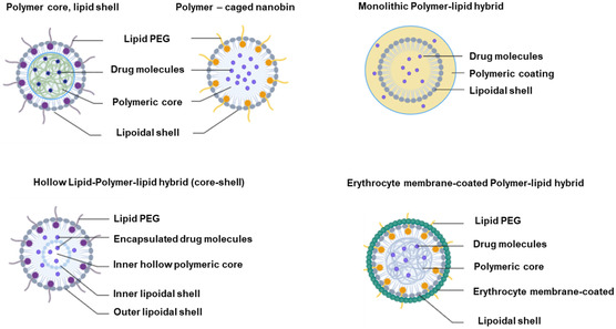
Figure 3 Hybrid structures combining polymers and lipids in nanocarrier design. This figure illustrates the concept of hybrid structures that combine polymers and lipids into a single entity, offering significant potential as versatile and efficient drug delivery carriers. Hybridization of polymers and lipids results in nanocarriers with enhanced properties, including improved stability, biocompatibility, and drug‐loading capacity. The figure depicts the structural composition of these hybrid nanocarriers, highlighting the integration of the polymer chains and lipid bilayers. Created with BioRender.com.
Figure 3 Hybrid structures combining polymers and lipids in nanocarrier design. This figure illustrates the concept of hybrid structures that combine polymers and lipids into a single entity, offering significant potential as versatile and efficient drug delivery carriers. Hybridization of polymers and lipids results in nanocarriers with enhanced properties, including improved stability, biocompatibility, and drug‐loading capacity. The figure depicts the structural composition of these hybrid nanocarriers, highlighting the integration of the polymer chains and lipid bilayers. Created with BioRender.com.CC BY · PubMed Central
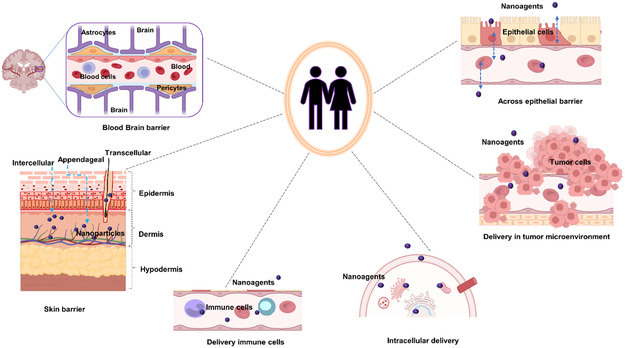
Figure 4 Biological barriers to effective drug delivery. This figure illustrates the major biological barriers that pose challenges to the effective delivery of therapeutics during drug development. These barriers include the BBB, skin layers, intercellular barrier, TME, and epithelial barrier. Each barrier presents unique challenges for drug delivery to target tissues, such as restricting the passage of molecules or altering drug distribution within the body. Created with BioRender.com.
Figure 4 Biological barriers to effective drug delivery. This figure illustrates the major biological barriers that pose challenges to the effective delivery of therapeutics during drug development. These barriers include the BBB, skin layers, intercellular barrier, TME, and epithelial barrier. Each barrier presents unique challenges for drug delivery to target tissues, such as restricting the passage of molecules or altering drug distribution within the body. Created with BioRender.com.CC BY · PubMed Central
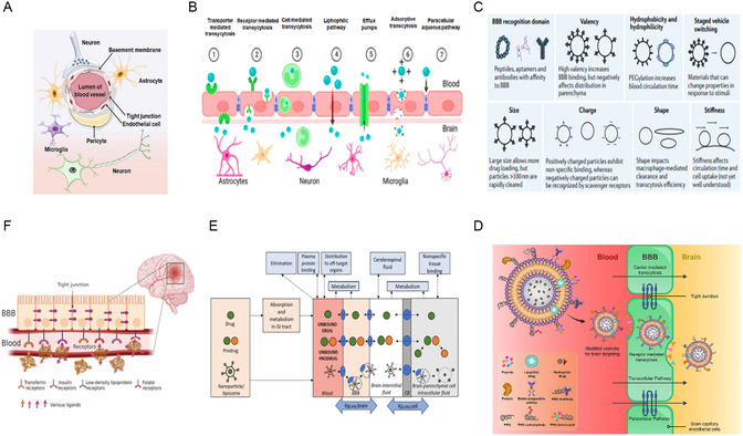
Figure 5 BBB regulation and targeted drug delivery across the barrier. A) Schematic representation of the BBB structure, showing brain capillary endothelial cells in association with astrocytes and pericytes.[ 110 ] B) Diagram illustrating various mechanisms for crossing the BBB, such as passive diffusion, carrier‐mediated transport, receptor‐mediated transcytosis, and AMT.[ 108 ] C) Influence of material properties on the adsorption, distribution, and clearance of DDSs following systemic administration, highlighting factors such as particle size, surface charge, and hydrophobicity.[ 240 ] D) The various biomolecules and their conjugation to the vesicle as BBB‐targeted carriers, including antibodies, peptides, and small molecules The various biomolecules and their conjugation to the vesicle as BBB‐targeted carriers, including antibodies, peptides, and small molecules. Adapted with permission.[ 241 ] Copyright 2022, Elsevier. E) Schematic depiction of the primary pharmacokinetic principles for transporter‐mediated drug delivery to the brain, including drug, prodrug, or nanocarrier transport across the BBB and brain parenchymal cellular barrier.[ 242 ] F) Illustration of receptor‐mediated drug delivery utilizing ligand‐conjugated NPs targeted to the brain, showing the interaction between the ligand and specific receptors on the BBB.[ 108 ] Figures are reproduced with permission.
Figure 5 BBB regulation and targeted drug delivery across the barrier. A) Schematic representation of the BBB structure, showing brain capillary endothelial cells in association with astrocytes and pericytes.[ 110 ] B) Diagram illustrating various mechanisms for crossing the BBB, such as passive diffusion, carrier‐mediated transport, receptor‐mediated transcytosis, and AMT.[ 108 ] C) Influence of material properties on the adsorption, distribution, and clearance of DDSs following systemic administration, highlighting factors such as particle size, surface charge, and hydrophobicity.[ 240 ] D) The various biomolecules and their conjugation to the vesicle as BBB‐targeted carriers, including antibodies, peptides, and small molecules The various biomolecules and their conjugation to the vesicle as BBB‐targeted carriers, including antibodies, peptides, and small molecules. Adapted with permission.[ 241 ] Copyright 2022, Elsevier. E) Schematic depiction of the primary pharmacokinetic principles for transporter‐mediated drug delivery to the brain, including drug, prodrug, or nanocarrier transport across the BBB and brain parenchymal cellular barrier.[ 242 ] F) Illustration of receptor‐mediated drug delivery utilizing ligand‐conjugated NPs targeted to the brain, showing the interaction between the ligand and specific receptors on the BBB.[ 108 ] Figures are reproduced with permission.CC BY · PubMed Central
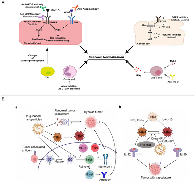
Figure 6 Vascular normalization mechanisms, associated signaling pathways, and the role of drug‐loaded NPs. A) Vascular normalization can be achieved through various strategies, including targeting VEGF signaling, Ang–Tie signaling, phospholipase C (PLC), phosphoinositide 3‐kinase (PI3K), angiopoietin 2 (Ang2), and epidermal growth factor receptor (EGFR) pathways in endothelial cells, as well as disrupting oncogenic signaling in cancer cells. Immune checkpoint blockade also induces vascular normalization by activating CD8+ or CD4+ T cells and promoting eosinophil accumulation. Key abbreviations: AAT (antiangiogenic therapy), IFP (interstitial fluid pressure), CAF (cancer‐associated fibroblast), ECM, VEGF (VEGFR (VEGF receptor).[ 174 ] B) (a) Drug‐loaded NPs can suppress hypoxic tumor growth by targeting and reprogramming abnormal tumor vasculature. They also can convert M2 macrophages into M1 phenotypes, which reactivates the immune response, further inhibiting tumor growth. Additionally, these NPs can activate mature DCs, leading to the activation of B cells via T cells, and the production of antibodies that target and eliminate tumor cells. By stimulating these pathways, NPs can counteract the inhibitory effects of myeloid‐derived suppressor cells on mature DCs and the suppressive effects of regulatory T cells (Tregs) on interferon‐gamma (INF‐γ), ultimately resulting in tumor cell death. (b) TAMs can inhibit tumor vasculature through M1 macrophages and interleukin‐12 (IL‐12). However, hypoxic conditions can induce the conversion of TAM1 into TAM2, which promotes tumor vasculature through interleukin‐10 (IL‐10). Drug‐loaded NPs can prevent this TAM1 to TAM2 conversion, thereby reducing tumor growth. Additionally, siRNA‐loaded NPs can specifically suppress TAM2 activity.[ 243 ] Figures are Reproduced with permission.
Figure 6 Vascular normalization mechanisms, associated signaling pathways, and the role of drug‐loaded NPs. A) Vascular normalization can be achieved through various strategies, including targeting VEGF signaling, Ang–Tie signaling, phospholipase C (PLC), phosphoinositide 3‐kinase (PI3K), angiopoietin 2 (Ang2), and epidermal growth factor receptor (EGFR) pathways in endothelial cells, as well as disrupting oncogenic signaling in cancer cells. Immune checkpoint blockade also induces vascular normalization by activating CD8+ or CD4+ T cells and promoting eosinophil accumulation. Key abbreviations: AAT (antiangiogenic therapy), IFP (interstitial fluid pressure), CAF (cancer‐associated fibroblast), ECM, VEGF (VEGFR (VEGF receptor).[ 174 ] B) (a) Drug‐loaded NPs can suppress hypoxic tumor growth by targeting and reprogramming abnormal tumor vasculature. They also can convert M2 macrophages into M1 phenotypes, which reactivates the immune response, further inhibiting tumor growth. Additionally, these NPs can activate mature DCs, leading to the activation of B cells via T cells, and the production of antibodies that target and eliminate tumor cells. By stimulating these pathways, NPs can counteract the inhibitory effects of myeloid‐derived suppressor cells on mature DCs and the suppressive effects of regulatory T cells (Tregs) on interferon‐gamma (INF‐γ), ultimately resulting in tumor cell death. (b) TAMs can inhibit tumor vasculature through M1 macrophages and interleukin‐12 (IL‐12). However, hypoxic conditions can induce the conversion of TAM1 into TAM2, which promotes tumor vasculature through interleukin‐10 (IL‐10). Drug‐loaded NPs can prevent this TAM1 to TAM2 conversion, thereby reducing tumor growth. Additionally, siRNA‐loaded NPs can specifically suppress TAM2 activity.[ 243 ] Figures are Reproduced with permission.CC BY · PubMed Central
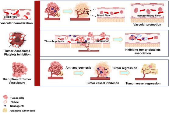
Figure 7 Strategies for overcoming abnormal vasculature barriers in the TME. This figure illustrates the three primary strategies developed to address the abnormal vasculature barriers encountered within the TME and facilitate the administration of nanoagents. Normalization and promotion: This involves interventions to normalize the tumor vasculature to improve blood flow and vessel integrity. Promoting the restoration of a more regular vascular network enhances the delivery of nanoagents to tumor tissues. Permeabilization or disruption: This strategy focuses on temporarily permeabilizing or disrupting the abnormal tumor vasculature to facilitate nanoagent penetration into tumor tissues. By creating transient openings in the vascular barrier, this approach allows for improved delivery of therapeutic agents to the TME. Tumor‐associated platelets inhibition: This strategy targets tumor‐associated platelets, which contribute to tumor growth and metastasis by promoting angiogenesis and thrombosis. By inhibiting the activity of these platelets, this approach aims to prevent abnormal vasculature formation and enhance the efficacy of nanoagent delivery to tumors. Created with BioRender.com.
Figure 7 Strategies for overcoming abnormal vasculature barriers in the TME. This figure illustrates the three primary strategies developed to address the abnormal vasculature barriers encountered within the TME and facilitate the administration of nanoagents. Normalization and promotion: This involves interventions to normalize the tumor vasculature to improve blood flow and vessel integrity. Promoting the restoration of a more regular vascular network enhances the delivery of nanoagents to tumor tissues. Permeabilization or disruption: This strategy focuses on temporarily permeabilizing or disrupting the abnormal tumor vasculature to facilitate nanoagent penetration into tumor tissues. By creating transient openings in the vascular barrier, this approach allows for improved delivery of therapeutic agents to the TME. Tumor‐associated platelets inhibition: This strategy targets tumor‐associated platelets, which contribute to tumor growth and metastasis by promoting angiogenesis and thrombosis. By inhibiting the activity of these platelets, this approach aims to prevent abnormal vasculature formation and enhance the efficacy of nanoagent delivery to tumors. Created with BioRender.com.CC BY · PubMed Central
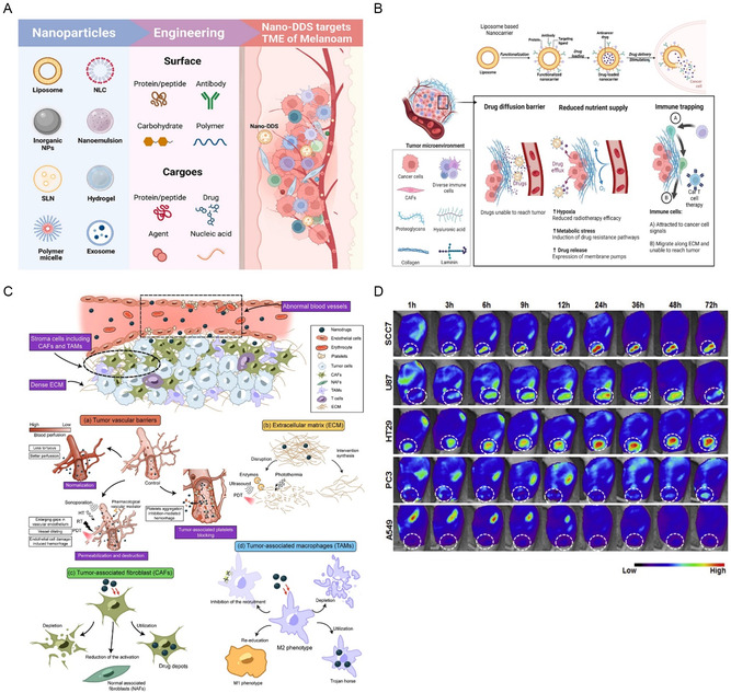
Figure 8 Nanocarriers for targeted DDS to the TME. A) Nano‐DDSs can selectively target TME of melanoma through modifications such as protein/peptide conjugation, antibody‐based targeting, carbohydrate surface decorating, or polymer coating. These carriers can then release their payloads into the TME, exerting antitumor effects. Adapted with permission. [ 244 ] Copyright 2021, Elsevier. B) Nano carrier‐based smart DDS overcomes the barriers in TME and releases drugs in cancer cells. Solid tumors induce high expression of ECM molecules (collagens, proteoglycans, hyaluronic acid, and laminins), which become complex and disordered, altering the characteristics of the TME. The ECM acts as a physical barrier, reducing the delivery of therapeutics, nutrients, and immune cells to solid tumors, leading to poorer prognosis. Created with BioRender.com.[ 245 , 246 ] C) The figure illustrates the biological barriers within the TME that affect nanodrug delivery, along with strategies for TME remodeling to enhance tumor penetration. (a) Techniques such as vasculature normalization, permeabilization, disruption, and blocking tumor‐associated platelets are employed to overcome barriers to transvascular transport. (b) Strategies for disrupting the ECM or inhibiting its synthesis are also depicted. Additionally, the regulation of (c) CAFs and (d) TAMs through depletion, reeducation, or utilization is shown to facilitate deeper infiltration of nanodrugs within the tumor stroma. Adapted with permission.[ 177 ] Copyright 2017, Elsevier. Distribution of chitosan NPs (CNPs) in mice bearing transplanted tumors. Time‐dependent, noninvasive in vivo near‐infrared fluorescence (NIRF) imaging was performed on tumor‐bearing mice following the injection of NIRF dye‐labeled CNPs (200 μg μL−1 head). The distribution of CNPs across these various tumor models reflects the diverse characteristics of the TMEs associated with clinical tumors.[ 247 ] Figures are Reproduced with permission.
Figure 8 Nanocarriers for targeted DDS to the TME. A) Nano‐DDSs can selectively target TME of melanoma through modifications such as protein/peptide conjugation, antibody‐based targeting, carbohydrate surface decorating, or polymer coating. These carriers can then release their payloads into the TME, exerting antitumor effects. Adapted with permission. [ 244 ] Copyright 2021, Elsevier. B) Nano carrier‐based smart DDS overcomes the barriers in TME and releases drugs in cancer cells. Solid tumors induce high expression of ECM molecules (collagens, proteoglycans, hyaluronic acid, and laminins), which become complex and disordered, altering the characteristics of the TME. The ECM acts as a physical barrier, reducing the delivery of therapeutics, nutrients, and immune cells to solid tumors, leading to poorer prognosis. Created with BioRender.com.[ 245 , 246 ] C) The figure illustrates the biological barriers within the TME that affect nanodrug delivery, along with strategies for TME remodeling to enhance tumor penetration. (a) Techniques such as vasculature normalization, permeabilization, disruption, and blocking tumor‐associated platelets are employed to overcome barriers to transvascular transport. (b) Strategies for disrupting the ECM or inhibiting its synthesis are also depicted. Additionally, the regulation of (c) CAFs and (d) TAMs through depletion, reeducation, or utilization is shown to facilitate deeper infiltration of nanodrugs within the tumor stroma. Adapted with permission.[ 177 ] Copyright 2017, Elsevier. Distribution of chitosan NPs (CNPs) in mice bearing transplanted tumors. Time‐dependent, noninvasive in vivo near‐infrared fluorescence (NIRF) imaging was performed on tumor‐bearing mice following the injection of NIRF dye‐labeled CNPs (200 μg μL−1 head). The distribution of CNPs across these various tumor models reflects the diverse characteristics of the TMEs associated with clinical tumors.[ 247 ] Figures are Reproduced with permission.CC BY · PubMed Central
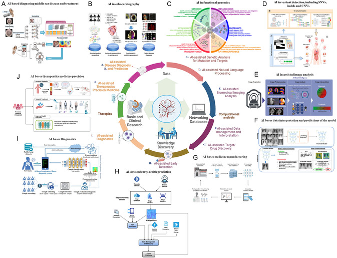
Figure 9 Role of AI in biomedical research. This figure highlights the pivotal role of AI in driving biomedical research and revolutionizing healthcare. AI significantly advances biomedical research by unlocking new insights, accelerating discovery processes, and facilitating personalized healthcare approaches. Key applications include AI‐based disease diagnostics and prediction: A) Developing a smartphone‐based computing program for diagnosing middle ear disease and providing medical suggestions. Adapted with permission.[ 248 ] Copyright 2022, Elsevier. B) AI in echocardiography.[ 249 ] C) AI‐assisted genetic analysis for mutation and target identification, particularly in functional genomics AI‐assisted genetic analysis for mutation and target identification, particularly in functional genomics.[ 250 ] D) AI models for variant detection and prioritization of disease‐causing variants in different genomic regions.[ 251 ] E) AI‐assisted biomedical imaging and processing, enhancing tasks such as preprocessing, image analysis, and interpretation, integrated with ML.[ 252 ] F) AI‐based data interception and model prediction.[ 253 ] G) AI‐assisted targeted drug delivery, Created with BioRender.com.[ 231 ] H) AI‐based early heath prediction.[ 254 ] I) AI‐assisted diagnostics, including AI‐based cough diagnosis systems that encompass every stage with data exchange capabilities for researchers and medical doctors.[ 255 ] J) AI‐assisted therapeutics in precision medicine, particularly in evaluating cancer treatments.[ 256 ] Figures are reproduced with permission. Created with BioRender.com.
Figure 9 Role of AI in biomedical research. This figure highlights the pivotal role of AI in driving biomedical research and revolutionizing healthcare. AI significantly advances biomedical research by unlocking new insights, accelerating discovery processes, and facilitating personalized healthcare approaches. Key applications include AI‐based disease diagnostics and prediction: A) Developing a smartphone‐based computing program for diagnosing middle ear disease and providing medical suggestions. Adapted with permission.[ 248 ] Copyright 2022, Elsevier. B) AI in echocardiography.[ 249 ] C) AI‐assisted genetic analysis for mutation and target identification, particularly in functional genomics AI‐assisted genetic analysis for mutation and target identification, particularly in functional genomics.[ 250 ] D) AI models for variant detection and prioritization of disease‐causing variants in different genomic regions.[ 251 ] E) AI‐assisted biomedical imaging and processing, enhancing tasks such as preprocessing, image analysis, and interpretation, integrated with ML.[ 252 ] F) AI‐based data interception and model prediction.[ 253 ] G) AI‐assisted targeted drug delivery, Created with BioRender.com.[ 231 ] H) AI‐based early heath prediction.[ 254 ] I) AI‐assisted diagnostics, including AI‐based cough diagnosis systems that encompass every stage with data exchange capabilities for researchers and medical doctors.[ 255 ] J) AI‐assisted therapeutics in precision medicine, particularly in evaluating cancer treatments.[ 256 ] Figures are reproduced with permission. Created with BioRender.com.CC BY · PubMed Central
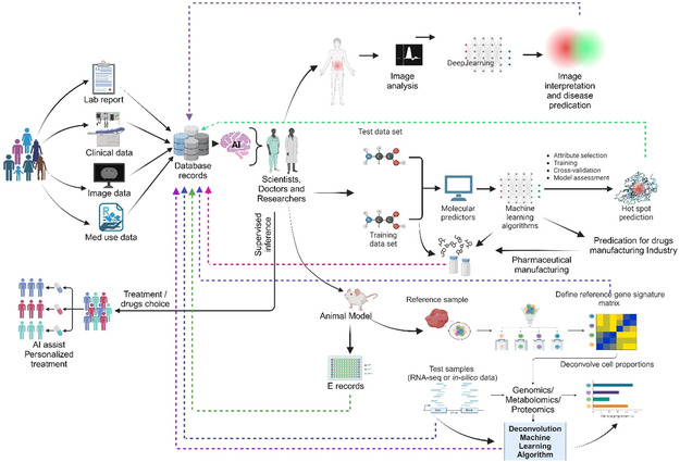
Figure 10 The diagram illustrates the role of AI integrated across various stages of biomedical research, highlighting its impact and challenges. AI‐driven workflows are increasingly explored in digital pathology, drug discovery, and development, showcasing significant potential in personalized care, clinical decision support systems, early disease detection, disease monitoring, and dynamic drug dosing. However, the successful application of AI in these areas also faces challenges, such as the need for high‐quality large datasets, managing the overwhelming influx of data (data deluge), and addressing data drift, which can affect the accuracy and reliability of AI models. These challenges may slow progress and delay the clinical implementation of AI‐driven solutions in healthcare. Created with BioRender.com.
Figure 10 The diagram illustrates the role of AI integrated across various stages of biomedical research, highlighting its impact and challenges. AI‐driven workflows are increasingly explored in digital pathology, drug discovery, and development, showcasing significant potential in personalized care, clinical decision support systems, early disease detection, disease monitoring, and dynamic drug dosing. However, the successful application of AI in these areas also faces challenges, such as the need for high‐quality large datasets, managing the overwhelming influx of data (data deluge), and addressing data drift, which can affect the accuracy and reliability of AI models. These challenges may slow progress and delay the clinical implementation of AI‐driven solutions in healthcare. Created with BioRender.com.CC BY · PubMed Central

Verification

Claims the paper makes

The claims and statistics of this paper haven’t been checked yet.

Run the check — free, no account needed. It reads the open-access text and checks the sources it cites.

A few free checks a day · papers already verified open instantly · results are public

Claims other papers make about this one

Each is checked against this paper’s own text: does it say what the citing paper says it does?

Self-check

Percentages, sums and reported differences re-computed from the paper’s own numbers.

The self-check needs the paper’s full text.

Citations

Cites

Show all 18 references

Research works printed but not linked: 262

  1. 10.1038/s43588-021-00060-9 · DOI
  2. 10.1038/s41540-022-00247-4 · DOI
  3. 10.1016/j.addr.2019.07.010 · DOI
  4. 10.2147/IJN.S236927 · DOI
  5. 10.2147/IJN.S406415 · DOI
  6. 10.1088/1748-605X/abf7d5 · DOI
  7. 10.2147/IJN.S146315 · DOI
  8. 10.1021/acs.chemrev.5b00589 · DOI
  9. 10.1016/j.heliyon.2023.e17488 · DOI · Paper page
  10. 10.3390/nano14070557 · DOI
  11. 10.1038/s41565-019-0567-y · DOI
  12. 10.2174/092986712800784676 · DOI
  13. 10.3390/pharmaceutics16020188 · DOI
  14. 10.1016/j.jddst.2021.102426 · DOI · Paper page
  15. 10.3390/molecules25092193 · DOI
  16. 10.1016/B978-0-323-89925-3.00013-7 · DOI
  17. 10.1186/s12951-018-0392-8 · DOI
  18. 10.2174/1567201818666210609161301 · DOI
  19. Hafner A., 2014, Int. J. Nanomed., 9, 1005
  20. 10.1038/s41577-019-0269-6 · DOI
  21. 10.1016/j.ijbiomac.2022.07.118 · DOI
  22. 10.1021/acsomega.2c05976 · DOI · Paper page
  23. 10.1007/s40089-017-0208-0 · DOI
  24. 10.1039/9781782622345-00001 · DOI
  25. Sabine Gressler C. H., 2024, Nanocarrier. Part I: Overview and Categorization of Nanocarriers
  26. 10.2217/nnm-2018-0120 · DOI
  27. 10.1089/jamp.2023.29089.sb · DOI
  28. 10.1016/j.ijpharm.2020.119098 · DOI
  29. 10.1002/adma.202002047 · DOI
  30. 10.2174/1574891X113089990038 · DOI
  31. 10.3390/pharmaceutics15020413 · DOI
  32. 10.1016/j.ijbiomac.2020.11.052 · DOI
  33. 10.3390/nano14080672 · DOI
  34. 10.1080/17425247.2016.1244187 · DOI
  35. 10.1016/j.ijbiomac.2020.10.104 · DOI
  36. 10.1007/978-981-99-3307-5_7 · DOI
  37. 10.3390/molecules25163731 · DOI · Paper page
  38. 10.1016/j.jare.2021.11.008 · DOI
  39. 10.1016/j.cocis.2017.12.004 · DOI
  40. 10.3389/fphar.2021.683935 · DOI
  41. 10.1016/j.ejpb.2011.07.005 · DOI
  42. 10.1016/j.ijpharm.2019.118796 · DOI
  43. 10.3390/pharmaceutics12090837 · DOI
  44. 10.1016/j.neuron.2018.07.039 · DOI · Paper page
  45. 10.2217/nnm.14.156 · DOI
  46. 10.1080/10717544.2018.1461955 · DOI
  47. 10.1016/j.ijbiomac.2020.11.207 · DOI
  48. 10.1016/j.jconrel.2017.05.019 · DOI · Paper page
  49. 10.1016/j.jconrel.2018.09.001 · DOI
  50. 10.1038/s41578-020-00269-6 · DOI · Paper page
  51. Shazwani S. S., 2024, ACS Omega, 9, 17379
  52. 10.1016/j.ijpharm.2005.04.029 · DOI
  53. 10.3109/02652049509010292 · DOI
  54. Melis Ç., 2014, Application of Nanotechnology in Drug Delivery
  55. 10.3390/pharmaceutics14102195 · DOI
  56. 10.1111/cns.12580 · DOI
  57. 10.1016/j.cis.2015.08.011 · DOI
  58. 10.1016/S0928-0987(99)00071-8 · DOI
  59. Scheffel U., 1972, J. Nucl. Med., 13, 498
  60. 10.1016/j.pneurobio.2014.05.002 · DOI · Paper page
  61. 10.4103/0250-474X.57282 · DOI
  62. 10.1080/17425255.2019.1621289 · DOI
  63. 10.1016/j.drudis.2019.01.004 · DOI
  64. 10.1016/j.ejphar.2014.11.014 · DOI
  65. 10.1155/2021/8844030 · DOI
  66. 10.3390/molecules23040938 · DOI
  67. 10.3389/fphar.2023.1159131 · DOI
  68. 10.1016/S1359-6446(04)03276-3 · DOI
  69. 10.1016/S1367-5931(02)00347-2 · DOI
  70. 10.1021/acsami.2c13129 · DOI
  71. 10.1016/j.drudis.2022.01.001 · DOI
  72. 10.1016/B978-0-323-91924-1.00010-1 · DOI
  73. 10.1186/s13036-022-00298-5 · DOI
  74. 10.1016/j.addr.2023.115005 · DOI
  75. 10.3390/polym10060595 · DOI
  76. 10.1073/pnas.1003103107 · DOI
  77. 10.1039/D1TB02144C · DOI
  78. 10.1021/acschemneuro.7b00301 · DOI
  79. 10.1016/j.colsurfb.2021.111764 · DOI
  80. 10.3390/polym14122510 · DOI
  81. 10.2174/1381612826666200116150426 · DOI
  82. 10.1021/nn800275r · DOI
  83. 10.1016/j.mattod.2013.09.002 · DOI
  84. 10.1021/nn3023602 · DOI
  85. 10.1039/c2sm07188f · DOI
  86. 10.1529/biophysj.104.048223 · DOI
  87. 10.1021/acsapm.2c01272 · DOI
  88. 10.1016/j.bpj.2021.12.005 · DOI
  89. 10.1016/j.biomaterials.2009.01.005 · DOI
  90. 10.3390/pharmaceutics13081291 · DOI
  91. 10.1016/j.biomaterials.2008.12.013 · DOI
  92. 10.1021/ja9017336 · DOI
  93. 10.1016/S0169-409X(99)00035-6 · DOI
  94. 10.1074/jbc.272.4.2382 · DOI
  95. 10.1002/anie.201004867 · DOI
  96. 10.1016/j.mimet.2019.03.017 · DOI
  97. 10.1039/b709566j · DOI
  98. 10.1016/j.bios.2003.10.017 · DOI
  99. 10.1073/pnas.1106634108 · DOI
  100. 10.1039/c2cs35103j · DOI
  101. 10.1007/s11095-011-0568-5 · DOI
  102. 10.1002/anie.201101554 · DOI
  103. 10.1080/17425247.2019.1614911 · DOI
  104. 10.3390/polym15092196 · DOI
  105. 10.1007/s12272-013-0272-6 · DOI
  106. 10.1038/s41573-020-0090-8 · DOI · Paper page
  107. 10.3390/ijms222111654 · DOI
  108. 10.1111/j.1471-4159.2008.05492.x · DOI · Paper page
  109. 10.1016/S0962-8924(02)02282-1 · DOI
  110. 10.1016/j.chemphyslip.2015.03.003 · DOI
  111. 10.1016/j.jconrel.2016.01.046 · DOI
  112. 10.1186/s12951-016-0177-x · DOI
  113. 10.1016/j.jconrel.2009.03.002 · DOI · Paper page
  114. 10.1023/A:1006948027674 · DOI
  115. 10.1016/j.colsurfb.2016.04.041 · DOI
  116. 10.1016/j.jtice.2013.09.024 · DOI
  117. 10.1016/j.lfs.2005.05.085 · DOI
  118. 10.1016/j.ijpharm.2011.05.062 · DOI
  119. 10.1016/j.jconrel.2008.10.016 · DOI
  120. 10.4161/derm.1.4.9501 · DOI
  121. 10.1039/D2NA00530A · DOI
  122. 10.1166/jnn.2015.9185 · DOI
  123. 10.1016/j.jconrel.2004.06.015 · DOI
  124. 10.1016/j.ijpharm.2010.09.037 · DOI
  125. 10.3390/ijms22158319 · DOI
  126. 10.1080/17425247.2020.1727883 · DOI
  127. Uner M., 2006, Pharmazie, 61, 375
  128. 10.1016/B978-0-323-89925-3.00008-3 · DOI
  129. Sakhrani N. M., 2013, Drug Des. Dev. Ther., 2013, 585
  130. 10.1016/j.ijpharm.2015.10.053 · DOI
  131. 10.1016/j.biomaterials.2016.04.026 · DOI
  132. 10.1016/j.drudis.2017.08.011 · DOI
  133. 10.1016/j.nano.2014.02.014 · DOI
  134. 10.1016/j.jconrel.2014.09.031 · DOI
  135. 10.1016/S0168-3659(99)00228-X · DOI
  136. 10.1002/adma.201902791 · DOI
  137. 10.7150/thno.57828 · DOI
  138. 10.3390/pharmaceutics15061610 · DOI
  139. 10.3390/pharmaceutics11120632 · DOI
  140. 10.1016/j.biopha.2018.09.097 · DOI
  141. 10.1016/j.addr.2024.115239 · DOI
  142. 10.3390/biom13121723 · DOI
  143. 10.1016/j.eurpolymj.2020.110058 · DOI
  144. 10.1007/s10895‐023‐03562‐z · DOI
  145. 10.1021/acsami.6b02721 · DOI
  146. 10.1016/j.bioadv.2022.212725 · DOI
  147. 10.1038/s41427-021-00303-1 · DOI
  148. 10.3390/biology13010001 · DOI
  149. 10.1016/j.trac.2022.116714 · DOI
  150. 10.1021/acsnano.8b06924 · DOI
  151. 10.1016/j.polymer.2015.12.040 · DOI
  152. 10.1002/app.42650 · DOI
  153. 10.1073/pnas.1210096109 · DOI
  154. 10.1016/j.drudis.2022.103469 · DOI
  155. 10.1016/j.biomaterials.2022.121656 · DOI
  156. 10.1038/nnano.2016.160 · DOI
  157. 10.1016/j.ijpharm.2022.122570 · DOI
  158. 10.1371/journal.pone.0179545 · DOI
  159. 10.1038/onc.2017.119 · DOI
  160. 10.1186/s11658-018-0088-y · DOI
  161. Tei K., 2002, Cancer Res., 62, 6289
  162. 10.1096/fj.99-1049com · DOI
  163. 10.1096/fj.05-4493com · DOI · Paper page
  164. 10.3389/fonc.2018.00367 · DOI
  165. 10.1080/10717544.2020.1809559 · DOI
  166. 10.3389/fimmu.2018.00455 · DOI
  167. 10.1016/j.addr.2021.02.019 · DOI
  168. 10.1186/s12935-023-02927-5 · DOI
  169. Lupo G., 2016, Front. Pharmacol., 7, 519
  170. 10.1021/acsnano.6b03110 · DOI
  171. 10.1016/j.ijpharm.2013.05.018 · DOI
  172. 10.1016/j.nantod.2017.06.010 · DOI
  173. 10.1002/smll.201903323 · DOI
  174. 10.1002/anie.201506262 · DOI
  175. 10.1007/s11894-010-0131-2 · DOI
  176. 10.1016/S0006-3495(01)75844-4 · DOI
  177. 10.1080/10717544.2018.1501119 · DOI
  178. 10.1080/10717544.2022.2058646 · DOI
  179. 10.1080/10717544.2017.1279238 · DOI · Paper page
  180. 10.1016/j.biomaterials.2007.02.021 · DOI
  181. 10.1016/j.ijmm.2007.05.004 · DOI
  182. 10.1016/j.addr.2008.11.002 · DOI
  183. 10.1016/j.ejpb.2014.12.024 · DOI
  184. 10.1016/j.ijpharm.2014.05.047 · DOI
  185. 10.1016/j.ejpb.2015.01.022 · DOI
  186. 10.1007/s11051-012-1353-z · DOI
  187. 10.1021/mp500439c · DOI
  188. 10.1016/j.biomaterials.2013.04.053 · DOI
  189. 10.1016/j.biomaterials.2008.09.050 · DOI · Paper page
  190. 10.1016/j.biomaterials.2013.04.028 · DOI
  191. 10.1093/toxsci/kfm217 · DOI
  192. 10.1034/j.1600-0854.2001.020301.x · DOI
  193. 10.1002/btm2.10143 · DOI · Paper page
  194. 10.37349/ebmx.2024.00009 · DOI
  195. 10.1007/978-1-61779-052-2_21 · DOI
  196. Pillai G., 2014, Nanomedicines for Cancer Therapy : An Update of FDA Approved and those Under Various Stages of Development
  197. 10.3390/pharmaceutics14010106 · DOI
  198. 10.1093/rb/rbw011 · DOI
  199. 10.1089/apc.1995.9.306 · DOI
  200. FDA approves KS drug, Food and Drug Administration, Florida Department of Health and Rehabilitation Services, 1996, AIDS Alert, 11, 11

200 of 262 shown

Cited by

Sorted by citations

See all 97

Metadata

Authors
Kim, M., Shin, M., Zhao, Y., Ghosh, M., & Son, Y.
Venue
Small Science
Publisher
Wiley
Published
2024-09-17
Volume
4
Issue
11
ISSN
2688-4046
Cited by
97
References
18
Language
en
Funders
National Research Foundation of Korea (2020R1A2C2004128, RS‐2023‐00208776, 2019R1A6A1A10072987, 2022H1D3A2A02053110, 2023RIS‐009, 2020R1A6C101A188)

About these checks

Registry, retraction, Cites and statistics results are computed, not judged, with one exception: a printed reference that no index lists is read once by a model before it counts as a citation error. Claims are extracted by a model and kept only when their quote appears verbatim in the text; each cited source is then checked, with a model’s help, for whether it says what the claim relies on. Those verdicts can be wrong — read the claim’s quote and the cited source before relying on one.

Statistics and arithmetic are recomputed, not judged.

Confidence bands are shown only for fields calibrated on our annotated test set. None is calibrated yet, so no band is shown.

What counts as a claim ›How we measure confidence ›How we check this ›

Are you an author, or do you see a problem with this page? Request a review

How to cite

Cite CoChat for the checks on this page, and the paper for its own findings.

This page
CoChat. (2026). Citation check: Transformative Impact of Nanocarrier‐Mediated Drug Delivery: Overcoming Biological Barriers and Expanding Therapeutic Horizons. https://app.cochat.ai/papers/10.1002/smsc.202400280
The paper
Kim, M., Shin, M., Zhao, Y., Ghosh, M., & Son, Y. (2024). Transformative Impact of Nanocarrier‐Mediated Drug Delivery: Overcoming Biological Barriers and Expanding Therapeutic Horizons. Small Science. https://doi.org/10.1002/smsc.202400280