

Common and specific activations supporting optic flow processing and navigation as revealed by a meta-analysis of neuroimaging studies
Open accessThe DOI matches its Crossref record; 167 of the 167 references it cites were found in a registry or an index.
Abstract
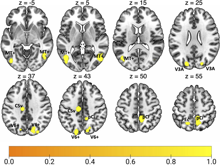
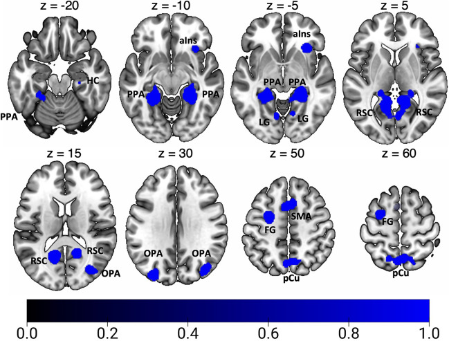
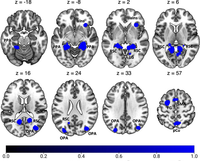
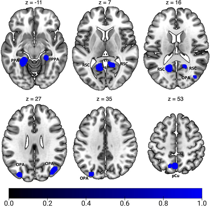
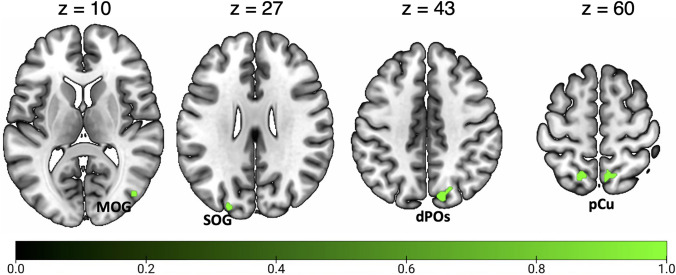
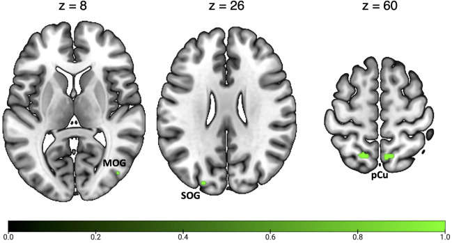
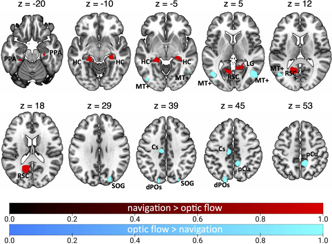
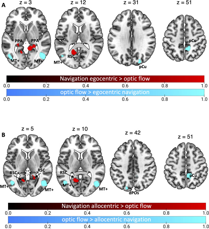
Optic flow provides useful information in service of spatial navigation. However, whether brain networks supporting these two functions overlap is still unclear. Here we used Activation Likelihood Estimation (ALE) to assess the correspondence between brain correlates of optic flow processing and spatial navigation and their specific neural activations. Since computational and connectivity evidence suggests that visual input from optic flow provides information mainly during egocentric navigation, we further tested the correspondence between brain correlates of optic flow processing and that of both egocentric and allocentric navigation. Optic flow processing shared activation with egocentric (but not allocentric) navigation in the anterior precuneus, suggesting its role in providing information about self-motion, as derived from the analysis of optic flow, in service of egocentric navigation. We further documented that optic flow perception and navigation are partially segregated into two functional and anatomical networks, i.e., the dorsal and the ventromedial networks. Present results point to a dynamic interplay between the dorsal and ventral visual pathways aimed at coordinating visually guided navigation in the environment.
Overview
The claims and statistics of this paper haven’t been checked yet.
Free: it reads the open-access text and checks the first 5 claims that cite a source we can check. Create a free account to check every claim.
Integrity signals
Registry
Cites
Statistics
Figures
Verification
Claims the paper makes
The claims and statistics of this paper haven’t been checked yet.
Free: it reads the open-access text and checks the first 5 claims that cite a source we can check. Create a free account to check every claim.
Claims other papers make about this one
Each is checked against this paper’s own text: does it say what the citing paper says it does?
The source backs part of the claim, not all of it — or backs it for a different population, condition or comparison.
In the citing paper
“Previous rs-fMRI studies in healthy individuals have helped clarify the neural circuitry for visually guided movement, identifying the precuneus and middle temporal cortex as key regions that provide visual information about self-motion and integrate sensorimotor signals.[10,11]”
From Brain Rewires to Stabilize Walking During Visual Impairment · cites this paper as [11]
Experimental — accuracy not yet measured
Self-check
Percentages, sums and reported differences re-computed from the paper’s own numbers.
The self-check needs the paper’s full text.
Citations
Cites
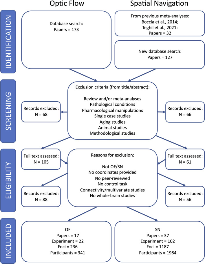
Preferred Reporting Items for Systematic Reviews and Meta-Analyses: the PRISMA statement
Moher, D., Liberati, A., Tetzlaff, J., Altman, D. G., & for the PRISMA Group (2009) · BMJ · doi:10.1136/bmj.b2535 · Cited by 83,823
Preferred reporting items for systematic reviews and meta-analyses: the PRISMA Statement.
(2009) · PubMed Central · Cited by 44,859
Liberati, A., Altman, D. G., Tetzlaff, J., Mulrow, C., Gotzsche, P. C., Ioannidis, J. P. A., Clarke, M., Devereaux, P. J., Kleijnen, J., & Moher, D. (2009) · BMJ · doi:10.1136/bmj.b2700 · Cited by 17,648
Co-planar stereotaxic atlas of the human brain
Laitinen, L. (1989) · Clinical Neurology and Neurosurgery · doi:10.1016/0303-8467(89)90128-5 · Cited by 7,565
Separate visual pathways for perception and action
Goodale, M. A., & Milner, A. D. (1992) · Trends in Neurosciences · doi:10.1016/0166-2236(92)90344-8 · Cited by 6,519
The hippocampus as a spatial map. Preliminary evidence from unit activity in the freely-moving rat
O'Keefe, J., & Dostrovsky, J. (1971) · Brain Research · doi:10.1016/0006-8993(71)90358-1 · Cited by 6,151
Ingle, D., Goodale, M. A., & Mansfield, R. J. W. (1982) · The MIT Press eBooks · Cited by 4,118
Microstructure of a spatial map in the entorhinal cortex
Hafting, T., Fyhn, M., Molden, S., Moser, M.-B., & Moser, E. I. (2005) · Nature · doi:10.1038/nature03721 · Cited by 4,068
A cortical representation of the local visual environment
Epstein, R., & Kanwisher, N. (1998) · Nature · doi:10.1038/33402 · Cited by 2,984
Object vision and spatial vision: Two cortical pathways
Mishkin, M., Ungerleider, L. G., & Macko, K. A. (1983) · Trends in Neurosciences · doi:10.1016/0166-2236(83)90190-x · Cited by 2,947
The Hippocampus as a Cognitive Map
RANCK, J. B. (1979) · American Journal of Psychiatry · doi:10.1176/ajp.136.10.1353 · Cited by 2,346
Functional role of the supplementary and pre-supplementary motor areas
Nachev, P., Kennard, C., & Husain, M. (2008) · Nature Reviews Neuroscience · doi:10.1038/nrn2478 · Cited by 1,928
Eickhoff, S. B., Laird, A. R., Grefkes, C., Wang, L. E., Zilles, K., & Fox, P. T. (2009) · Human Brain Mapping · doi:10.1002/hbm.20718 · Cited by 1,924 · cited 1×
“For a quantitative assessment of inter study convergence, the ALE method (Eickhoff et al. 2009; Laird et al. 2005; Turkeltaub et al. 2002) has been applied.”
MethodMeta-analysis of the functional neuroanatomy of single-word reading: method and validation
Turkeltaub, P. E., Eden, G. F., Jones, K. M., & Zeffiro, T. A. (2002) · NeuroImage · doi:10.1006/nimg.2002.1131 · Cited by 1,604
Cellular networks underlying human spatial navigation
Ekstrom, A. D., Kahana, M. J., Caplan, J. B., Fields, T. A., Isham, E. A., Newman, E. L., & Fried, I. (2003) · Nature · doi:10.1038/nature01964 · Cited by 1,425 · cited 1×
“…i.e., neurons firing at specific positions in space, have been discovered in both freely moving animals (O’Keefe and Dostrovsky 1971), and humans (Ekstrom et al. 2003), supporting the hypothesis that HC contains a metric allocentric representations of the surrounding space (cognitive map)…”
BackgroundA new neural framework for visuospatial processing
Kravitz, D. J., Saleem, K. S., Baker, C. I., & Mishkin, M. (2011) · Nature Reviews Neuroscience · doi:10.1038/nrn3008 · Cited by 1,325 · cited 2×
“…and ventral streams, in both primates and humans, lead to selective deficits in object vision and spatial vision, respectively, leading to their functional characterization as “What” and “Where” pathways (Kravitz et al. 2011; Macko et al. 1982; Mishkin et al. 1983; Ungerleider and Mishkin 1982).”
BackgroundTurkeltaub, P. E., Eickhoff, S. B., Laird, A. R., Fox, M., Wiener, M., & Fox, P. (2012) · Human Brain Mapping · doi:10.1002/hbm.21186 · Cited by 1,119
Remembering the past and imagining the future: A neural model of spatial memory and imagery.
Byrne, P., Becker, S., & Burgess, N. (2007) · Psychological Review · doi:10.1037/0033-295x.114.2.340 · Cited by 1,011
Parahippocampal and retrosplenial contributions to human spatial navigation
Epstein, R. A. (2008) · Trends in Cognitive Sciences · doi:10.1016/j.tics.2008.07.004 · Cited by 928
ALE meta-analysis: controlling the false discovery rate and performing statistical contrasts
Laird, A. R., Fox, P. M., Price, C. J., Glahn, D. C., Uecker, A. M., Lancaster, J. L., Turkeltaub, P. E., Kochunov, P., & Fox, P. T. (2005) · Human Brain Mapping · doi:10.1002/hbm.20136 · Cited by 868
Functional Analysis of V3A and Related Areas in Human Visual Cortex
Tootell, R. B. H., Mendola, J. D., Hadjikhani, N. K., Ledden, P. J., Liu, A. K., Reppas, J. B., Sereno, M. I., & Dale, A. M. (1997) · The Journal of Neuroscience · doi:10.1523/jneurosci.17-18-07060.1997 · Cited by 817
The Parahippocampal Place Area
Epstein, R., Harris, A., Stanley, D., & Kanwisher, N. (1999) · Neuron · doi:10.1016/s0896-6273(00)80758-8 · Cited by 810
Ten simple rules for neuroimaging meta-analysis
Müller, V. I., Cieslik, E. C., Laird, A. R., Fox, P. T., Radua, J., Mataix-Cols, D., Tench, C. R., Yarkoni, T., Nichols, T. E., Turkeltaub, P. E., et al. (2018) · Neuroscience & Biobehavioral Reviews · doi:10.1016/j.neubiorev.2017.11.012 · Cited by 805 · cited 2×
“Recent guidelines for the meta-analysis (Muller et al. 2018) have been used in the current study.”
MethodInfluentialPolymodal Motion Processing in Posterior Parietal and Premotor Cortex
Bremmer, F., Schlack, A., Shah, N. J., Zafiris, O., Kubischik, M., Hoffmann, K.-P., Zilles, K., & Fink, G. R. (2001) · Neuron · doi:10.1016/s0896-6273(01)00198-2 · Cited by 803 · cited 1×
“…and posterior cingulate sulcus area (pCi) (Wall and Smith 2008; Serra et al. 2019), and in two dorsal parietal regions, corresponding to the putative human areas VIP (IPS-mot, Pitzalis et al. 2013c; Bremmer et al. 2001; Sereno and Huang 2006; Cardin and Smith 2010) and PEc (Pitzalis et al. 2019).”
BackgroundTopographical disorientation: a synthesis and taxonomy
Aguirre, G. K. (1999) · Brain · doi:10.1093/brain/122.9.1613 · Cited by 772 · cited 1×
“…in both freely moving animals (O’Keefe and Dostrovsky 1971), and humans (Ekstrom et al. 2003), supporting the hypothesis that HC contains a metric allocentric representations of the surrounding space (cognitive map) (Aguirre and D’Esposito 1999; Byrne et al. 2007; O’Keefe and Nadel 1978).”
BackgroundThe Perception of the Visual World
Prentice, W. C. H., & Gibson, J. J. (1951) · The American Journal of Psychology · doi:10.2307/1418204 · Cited by 772
Optic flow is used to control human walking
Warren, W. H., Kay, B. A., Zosh, W. D., Duchon, A. P., & Sahuc, S. (2001) · Nature Neuroscience · doi:10.1038/84054 · Cited by 744
Eickhoff, S. B., Nichols, T. E., Laird, A. R., Hoffstaedter, F., Amunts, K., Fox, P. T., Bzdok, D., & Eickhoff, C. R. (2016) · NeuroImage · doi:10.1016/j.neuroimage.2016.04.072 · Cited by 683 · cited 1×
“Statistical ALE maps were thresholded using cluster level correction at p < 0.05 (1000 permutation) with a cluster-forming threshold at voxel-level p < 0.001 (uncorrected) (Eickhoff et al. 2016) in line with the recent guidelines for coordinate based meta-analysis (Muller et al. 2018).”
MethodInfluentialMapping of Contralateral Space in Retinotopic Coordinates by a Parietal Cortical Area in Humans
Sereno, M. I., Pitzalis, S., & Martinez, A. (2001) · Science · doi:10.1126/science.1063695 · Cited by 682 · cited 1×
“…from the pIPS to the border of area V6, which is typically activated by both low-level motion stimulation (contrasting motion vs static, see Sereno et al. 2001; Pitzalis et al. 2010; Sulpizio et al. 2020) and high-level motion stimulation (contrasting coherent vs random motion, see…”
Self-citationTootell, R. B. H., Reppas, J. B., Dale, A. M., Look, R. B., Sereno, M. I., Malach, R., Brady, T. J., & Rosen, B. R. (1995) · Nature · doi:10.1038/375139a0 · Cited by 659
The Neural Bases of Multisensory Processes
(2011) · CRC Press eBooks · doi:10.1201/9781439812174 · Cited by 491
Mental navigation along memorized routes activates the hippocampus, precuneus, and insula
Ghaem, O., Mellet, E., Crivello, F., Tzourio, N., Mazoyer, B., Berthoz, A., & Denis, M. (1997) · NeuroReport · doi:10.1097/00001756-199702100-00032 · Cited by 478
A cortical area that responds specifically to optic flow, revealed by fMRI
Morrone, M. C., Tosetti, M., Montanaro, D., Fiorentini, A., Cioni, G., & Burr, D. C. (2000) · Nature Neuroscience · doi:10.1038/81860 · Cited by 392
Selective neural representation of objects relevant for navigation
Janzen, G., & van Turennout, M. (2004) · Nature Neuroscience · doi:10.1038/nn1257 · Cited by 382
Thoughts, behaviour, and brain dynamics during navigation in the real world
Spiers, H. J., & Maguire, E. A. (2006) · NeuroImage · doi:10.1016/j.neuroimage.2006.01.037 · Cited by 371 · cited 1×
“For example, tasks involving computations of the spatial relationship between the navigator’s heading and a specific goal location (spatial updating) have shown the recruitment of the posterior parietal cortex (Howard et al. 2014; Spiers and Maguire 2006) and pCu (Wolbers et al. 2008).”
MethodThe Retinotopic Organization of the Human Middle Temporal Area MT/V5 and Its Cortical Neighbors
Kolster, H., Peeters, R., & Orban, G. A. (2010) · Journal of Neuroscience · doi:10.1523/jneurosci.2069-10.2010 · Cited by 364
Maguire, E. A., Frith, C. D., Burgess, N., Donnett, J. G., & O'Keefe, J. (1998) · Journal of Cognitive Neuroscience · doi:10.1162/089892998563789 · Cited by 357
Vectorial representation of spatial goals in the hippocampus of bats
Sarel, A., Finkelstein, A., Las, L., & Ulanovsky, N. (2017) · Science · doi:10.1126/science.aak9589 · Cited by 339
Committeri, G., Galati, G., Paradis, A.-L., Pizzamiglio, L., Berthoz, A., & LeBihan, D. (2004) · Journal of Cognitive Neuroscience · doi:10.1162/0898929042568550 · Cited by 335
A human parietal face area contains aligned head-centered visual and tactile maps
Sereno, M. I., & Huang, R.-S. (2006) · Nature Neuroscience · doi:10.1038/nn1777 · Cited by 322 · cited 1×
“…and posterior cingulate sulcus area (pCi) (Wall and Smith 2008; Serra et al. 2019), and in two dorsal parietal regions, corresponding to the putative human areas VIP (IPS-mot, Pitzalis et al. 2013c; Bremmer et al. 2001; Sereno and Huang 2006; Cardin and Smith 2010) and PEc (Pitzalis et al. 2019).”
BackgroundWolbers, T., & Büchel, C. (2005) · The Journal of Neuroscience · doi:10.1523/jneurosci.4705-04.2005 · Cited by 307
Howard, L. R., Javadi, A. H., Yu, Y., Mill, R. D., Morrison, L. C., Knight, R., Loftus, M. M., Staskute, L., & Spiers, H. J. (2014) · Current Biology · doi:10.1016/j.cub.2014.05.001 · Cited by 298
Wolbers, T., Wiener, J. M., Mallot, H. A., & Büchel, C. (2007) · The Journal of Neuroscience · doi:10.1523/jneurosci.2146-07.2007 · Cited by 278
MST Neurons Respond to Optic Flow and Translational Movement
Duffy, C. J. (1998) · Journal of Neurophysiology · doi:10.1152/jn.1998.80.4.1816 · Cited by 275
Wide-Field Retinotopy Defines Human Cortical Visual Area V6
Pitzalis, S., Galletti, C., Huang, R.-S., Patria, F., Committeri, G., Galati, G., Fattori, P., & Sereno, M. I. (2006) · The Journal of Neuroscience · doi:10.1523/jneurosci.0178-06.2006 · Cited by 271
Self-citationIaria, G., Chen, J., Guariglia, C., Ptito, A., & Petrides, M. (2007) · European Journal of Neuroscience · doi:10.1111/j.1460-9568.2007.05371.x · Cited by 262
Sensitivity to optic flow in human cortical areas MT and MST
Smith, A. T., Wall, M. B., Williams, A. L., & Singh, K. D. (2006) · European Journal of Neuroscience · doi:10.1111/j.1460-9568.2005.04526.x · Cited by 228
Mellet, E., Bricogne, S., Tzourio-Mazoyer, N., Ghaëm, O., Petit, L., Zago, L., Etard, O., Berthoz, A., Mazoyer, B., & Denis, M. (2000) · NeuroImage · doi:10.1006/nimg.2000.0648 · Cited by 217
Boccia, M., Nemmi, F., & Guariglia, C. (2014) · Neuropsychology Review · doi:10.1007/s11065-014-9247-8 · Cited by 213
Self-citationHuman V6: The Medial Motion Area
Pitzalis, S., Sereno, M. I., Committeri, G., Fattori, P., Galati, G., Patria, F., & Galletti, C. (2010) · Cerebral Cortex · doi:10.1093/cercor/bhp112 · Cited by 213
Self-citationThe Representation of Egomotion in the Human Brain
Wall, M. B., & Smith, A. T. (2008) · Current Biology · doi:10.1016/j.cub.2007.12.053 · Cited by 211
Park, S., & Chun, M. M. (2009) · NeuroImage · doi:10.1016/j.neuroimage.2009.04.058 · Cited by 209
Retrosplenial cortex codes for permanent landmarks
Auger, S. D., Mullally, S. L., & Maguire, E. A. (2012) · PLoS ONE · doi:10.1371/journal.pone.0043620 · Cited by 205 · cited 1×
“…and Büchel 2005), retrieval of environ-ment-centered information (Committeri et al. 2004; Galati et al. 2010; Sulpizio et al. 2013, 2016), visuo-spatial mental imagery of familiar environments (Boccia et al. 2015) and encoding of permanent landmarks (Auger et al. 2012; Auger and Maguire 2013).”
BackgroundCoding of navigational affordances in the human visual system
Bonner, M. F., & Epstein, R. A. (2017) · Proceedings of the National Academy of Sciences · doi:10.1073/pnas.1618228114 · Cited by 204
Distances between real-world locations are represented in the human hippocampus
Morgan, L. K., MacEvoy, S. P., Aguirre, G. K., & Epstein, R. A. (2011) · The Journal of Neuroscience · doi:10.1523/jneurosci.4667-10.2011 · Cited by 203 · cited 2×
“…spatial navigation and/or map-like representations (Ghaem et al. 1997; Maguire et al. 1998; Wolbers and Büchel 2005; Iaria et al. 2007; Wolbers et al. 2007; Baumann et al. 2010, 2012; Brown et al. 2010, 2012; Morgan et al. 2011; Viard et al. 2011; Baumann and Mattingley 2010; Brown and Stern 2014).”
BackgroundResultMultiple reference frames used by the human brain for spatial perception and memory
Galati, G., Pelle, G., Berthoz, A., & Committeri, G. (2010) · Experimental Brain Research · doi:10.1007/s00221-010-2168-8 · Cited by 201
Our sense of direction: progress, controversies and challenges
Cullen, K. E., & Taube, J. S. (2017) · Nature Neuroscience · doi:10.1038/nn.4658 · Cited by 197
Cardin, V., & Smith, A. T. (2010) · Cerebral Cortex · doi:10.1093/cercor/bhp268 · Cited by 194
Ekstrom, A. D., Huffman, D. J., & Starrett, M. (2017) · Journal of Neurophysiology · doi:10.1152/jn.00531.2017 · Cited by 186
Spatial updating: how the brain keeps track of changing object locations during observer motion
Wolbers, T., Hegarty, M., Büchel, C., & Loomis, J. M. (2008) · Nature Neuroscience · doi:10.1038/nn.2189 · Cited by 174
Optic Flow Processing for the Assessment of Object Movement during Ego Movement
Warren, P. A., & Rushton, S. K. (2009) · Current Biology · doi:10.1016/j.cub.2009.07.057 · Cited by 173
Visual Field Map Clusters in Macaque Extrastriate Visual Cortex
Kolster, H., Mandeville, J. B., Arsenault, J. T., Ekstrom, L. B., Wald, L. L., & Vanduffel, W. (2009) · The Journal of Neuroscience · doi:10.1523/jneurosci.0518-09.2009 · Cited by 168
Is Navigation in Virtual Reality with fMRI Really Navigation?
Taube, J. S., Valerio, S., & Yoder, R. M. (2013) · Journal of Cognitive Neuroscience · doi:10.1162/jocn_a_00386 · Cited by 168
Neural Mechanisms of Hierarchical Planning in a Virtual Subway Network
Balaguer, J., Spiers, H., Hassabis, D., & Summerfield, C. (2016) · Neuron · doi:10.1016/j.neuron.2016.03.037 · Cited by 163
The vestibular contribution to the head direction signal and navigation
Yoder, R. M., & Taube, J. S. (2014) · Frontiers in Integrative Neuroscience · doi:10.3389/fnint.2014.00032 · Cited by 161
Schlack, A., Hoffmann, K., & Bremmer, F. (2002) · European Journal of Neuroscience · doi:10.1046/j.1460-9568.2002.02251.x · Cited by 159
A model of episodic memory: Mental time travel along encoded trajectories using grid cells
Hasselmo, M. E. (2009) · Neurobiology of Learning and Memory · doi:10.1016/j.nlm.2009.07.005 · Cited by 157
Spatial updating in virtual reality: the sufficiency of visual information
Riecke, B. E., Cunningham, D. W., & Bülthoff, H. (2006) · Psychological Research · doi:10.1007/s00426-006-0085-z · Cited by 156
The human homologue of macaque area V6A
Pitzalis, S., Sereno, M. I., Committeri, G., Fattori, P., Galati, G., Tosoni, A., & Galletti, C. (2013) · NeuroImage · doi:10.1016/j.neuroimage.2013.06.026 · Cited by 154 · cited 9×
“…flow has been observed in the temporal area MT + (Kolster et al. 2010), in the medial parieto-occipital areas V6 and V6Av (V6 complex or V6 + ; Pitzalis et al. 2006, 2010, 2013b; Cardin and Smith 2010), in the posterior segment of the intraparietal sulcus (pIPS), a location remarkably…”
BackgroundInfluentialSelf-citationVisual Scene Processing in Familiar and Unfamiliar Environments
Epstein, R. A., Higgins, J. S., Jablonski, K., & Feiler, A. M. (2007) · Journal of Neurophysiology · doi:10.1152/jn.00003.2007 · Cited by 153
Epstein, R. A., & Higgins, J. S. (2007) · Cerebral Cortex · doi:10.1093/cercor/bhl079 · Cited by 152
Julian, J. B., Ryan, J., Hamilton, R. H., & Epstein, R. A. (2016) · Current Biology · doi:10.1016/j.cub.2016.02.066 · Cited by 152
Anterior hippocampus and goal-directed spatial decision making
Viard, A., Doeller, C. F., Hartley, T., Bird, C. M., & Burgess, N. (2011) · The Journal of Neuroscience · doi:10.1523/jneurosci.4640-10.2011 · Cited by 144
The parieto-insular vestibular cortex in humans: more than a single area?
Frank, S. M., & Greenlee, M. W. (2018) · Journal of Neurophysiology · doi:10.1152/jn.00907.2017 · Cited by 140
Where is the “where” in the brain? A meta‐analysis of neuroimaging studies on spatial cognition
Cona, G., & Scarpazza, C. (2019) · Human Brain Mapping · doi:10.1002/hbm.24496 · Cited by 138
Medial Parietal Cortex Encodes Perceived Heading Direction in Humans
Baumann, O., & Mattingley, J. B. (2010) · The Journal of Neuroscience · doi:10.1523/jneurosci.3077-10.2010 · Cited by 137 · cited 1×
“…spatial navigation and/or map-like representations (Ghaem et al. 1997; Maguire et al. 1998; Wolbers and Büchel 2005; Iaria et al. 2007; Wolbers et al. 2007; Baumann et al. 2010, 2012; Brown et al. 2010, 2012; Morgan et al. 2011; Viard et al. 2011; Baumann and Mattingley 2010; Brown and Stern 2014).”
BackgroundLinear Vestibular Self‐Motion Signals in Monkey Medial Superior Temporal Area
BREMMER, F., KUBISCHIK, M., PEKEL, M., LAPPE, M., & HOFFMANN, K. ‐. (1999) · Annals of the New York Academy of Sciences · doi:10.1111/j.1749-6632.1999.tb09191.x · Cited by 135
Vestibular Inputs to Human Motion-Sensitive Visual Cortex
Smith, A. T., Wall, M. B., & Thilo, K. V. (2012) · Cerebral Cortex · doi:10.1093/cercor/bhr179 · Cited by 135
Sulpizio, V., Committeri, G., Lambrey, S., Berthoz, A., & Galati, G. (2012) · Behavioural Brain Research · doi:10.1016/j.bbr.2012.12.031 · Cited by 131
Self-citationAbstract Representations of Location and Facing Direction in the Human Brain
Vass, L. K., & Epstein, R. A. (2013) · The Journal of Neuroscience · doi:10.1523/jneurosci.3873-12.2013 · Cited by 131
Brown, T. I., Ross, R. S., Keller, J. B., Hasselmo, M. E., & Stern, C. E. (2010) · The Journal of Neuroscience · doi:10.1523/jneurosci.6021-09.2010 · Cited by 130
Mapping the Primate Visual System with [2- 14 C]Deoxyglucose
Macko, K. A., Jarvis, C. D., Kennedy, C., Miyaoka, M., Shinohara, M., Sololoff, L., & Mishkin, M. (1982) · Science · doi:10.1126/science.7123241 · Cited by 127
The occipital place area represents the local elements of scenes
Kamps, F. S., Julian, J. B., Kubilius, J., Kanwisher, N., & Dilks, D. D. (2016) · NeuroImage · doi:10.1016/j.neuroimage.2016.02.062 · Cited by 123
Mental navigation in humans is processed in the anterior bank of the parieto-occipital sulcus
Ino, T., Inoue, Y., Kage, M., Hirose, S., Kimura, T., & Fukuyama, H. (2002) · Neuroscience Letters · doi:10.1016/s0304-3940(02)00019-8 · Cited by 119
Multisensory integration in the estimation of walked distances
Campos, J. L., Butler, J. S., & Bülthoff, H. (2012) · Experimental Brain Research · doi:10.1007/s00221-012-3048-1 · Cited by 119
The Neurocognitive Basis of Spatial Reorientation
Julian, J. B., Keinath, A. T., Marchette, S. A., & Epstein, R. A. (2018) · Current Biology · doi:10.1016/j.cub.2018.04.057 · Cited by 117
Moving observers, relative retinal motion and the detection of object movement
Rushton, S. K., & Warren, P. A. (2005) · Current Biology · doi:10.1016/j.cub.2005.07.020 · Cited by 116
Neuronal mechanisms for detection of motion in the field of view
Galletti, C., & Fattori, P. (2003) · Neuropsychologia · doi:10.1016/s0028-3932(03)00174-x · Cited by 113
Baumann, O., Chan, E., & Mattingley, J. B. (2009) · NeuroImage · doi:10.1016/j.neuroimage.2009.10.021 · Cited by 112
Scenes, Spaces, and Memory Traces: What Does the Hippocampus Do?
Maguire, E. A., Intraub, H., & Mullally, S. L. (2016) · The Neuroscientist · doi:10.1177/1073858415600389 · Cited by 110
Brain Computations: What and How
Rolls, E. T. (2020) · Oxford University Press eBooks · doi:10.1093/oso/9780198871101.001.0001 · Cited by 106
Patai, E. Z., Javadi, A.-H., Ozubko, J. D., O’Callaghan, A., Ji, S., Robin, J., Grady, C., Winocur, G., Rosenbaum, R. S., Moscovitch, M., et al. (2019) · Cerebral Cortex · doi:10.1093/cercor/bhz044 · Cited by 102 · cited 1×
“Several neuroimaging studies demonstrated its role in encoding distance to the goal during navigation (Balaguer et al. 2016; Howard et al. 2014; Patai et al. 2019; Sarel et al. 2017), and directional information in large-scale environments (Sulpizio et al. 2018) and in constructing coherent spatial…”
BackgroundAssessing the mechanism of response in the retrosplenial cortex of good and poor navigators
Auger, S. D., & Maguire, E. A. (2013) · Cortex · doi:10.1016/j.cortex.2013.08.002 · Cited by 91
Cooperative interactions between hippocampal and striatal systems support flexible navigation
Brown, T. I., Ross, R. S., Tobyne, S. M., & Stern, C. E. (2012) · NeuroImage · doi:10.1016/j.neuroimage.2012.01.046 · Cited by 91
Vestibular and visual responses in human posterior insular cortex
Frank, S. M., Baumann, O., Mattingley, J. B., & Greenlee, M. W. (2014) · Journal of Neurophysiology · doi:10.1152/jn.00078.2014 · Cited by 90
Resting-state connectivity and functional specialization in human medial parieto-occipital cortex
Tosoni, A., Pitzalis, S., Committeri, G., Fattori, P., Galletti, C., & Galati, G. (2014) · Brain Structure and Function · doi:10.1007/s00429-014-0858-x · Cited by 89
Self-citationThe human posterior parietal cortex: effective connectome, and its relation to function
Rolls, E. T., Deco, G., Huang, C., & Feng, J. (2022) · Cerebral Cortex · doi:10.1093/cercor/bhac266 · Cited by 89
Selectivity to Translational Egomotion in Human Brain Motion Areas
Pitzalis, S., Sdoia, S., Bultrini, A., Committeri, G., Di Russo, F., Fattori, P., Galletti, C., & Galati, G. (2013) · PLoS ONE · doi:10.1371/journal.pone.0060241 · Cited by 87
Self-citationBrain Computations: What and How
Rolls, E. T. (2021) · Cited by 86
Structural connectivity and functional properties of the macaque superior parietal lobule
Gamberini, M., Passarelli, L., Fattori, P., & Galletti, C. (2019) · Brain Structure and Function · doi:10.1007/s00429-019-01976-9 · Cited by 84
Rolls, E. T. (2023) · Hippocampus · doi:10.1002/hipo.23467 · Cited by 82
Cardin, V., & Smith, A. T. (2011) · Journal of Neurophysiology · doi:10.1152/jn.01120.2010 · Cited by 78
Brown, T. I., & Stern, C. E. (2014) · Cerebral Cortex · doi:10.1093/cercor/bht041 · Cited by 77
Brain Computations and Connectivity
Rolls, E. T. (2023) · doi:10.1093/oso/9780198887911.001.0001 · Cited by 76
The functional role of the medial motion area V6
Pitzalis, S., Fattori, P., & Galletti, C. (2013) · Frontiers in Behavioral Neuroscience · doi:10.3389/fnbeh.2012.00091 · Cited by 75
Self-citationMultiple cortical visual streams in humans
Rolls, E. T., Deco, G., Huang, C., & Feng, J. (2022) · Cerebral Cortex · doi:10.1093/cercor/bhac276 · Cited by 73
Neural Systems in the Visual Control of Steering
Field, D. T., Wilkie, R. M., & Wann, J. P. (2007) · Journal of Neuroscience · doi:10.1523/jneurosci.2130-07.2007 · Cited by 71
Loomis, J. M., & Beall, A. C. (1998) · Ecological Psychology · doi:10.1080/10407413.1998.9652685 · Cited by 70
Boccia, M., Piccardi, L., Palermo, L., Nemmi, F., Sulpizio, V., Galati, G., & Guariglia, C. (2015) · Human Brain Mapping · doi:10.1002/hbm.22678 · Cited by 66
Self-citationMultisensory and sensorimotor maps
Huang, R., & Sereno, M. I. (2018) · Handbook of clinical neurology · doi:10.1016/b978-0-444-63622-5.00007-3 · Cited by 65
Helfrich, R. F., Becker, H. G. T., & Haarmeier, T. (2012) · Brain Topography · doi:10.1007/s10548-012-0226-1 · Cited by 63
Human Areas V3A and V6 Compensate for Self-Induced Planar Visual Motion
Fischer, E., Bülthoff, H., Logothetis, N. K., & Bartels, A. (2012) · Neuron · doi:10.1016/j.neuron.2012.01.022 · Cited by 63
Multisensory Integration in Self Motion Perception
Greenlee, M. W., Frank, S. M., Kaliuzhna, M., Blanke, O., Bremmer, F., Churan, J., Cuturi, L. F., MacNeilage, P. R., & Smith, A. T. (2016) · Multisensory Research · doi:10.1163/22134808-00002527 · Cited by 61
Sherrill, K. R., Chrastil, E. R., Ross, R. S., Erdem, U. M., Hasselmo, M. E., & Stern, C. E. (2015) · NeuroImage · doi:10.1016/j.neuroimage.2015.06.009 · Cited by 60
Age-Related Changes in Spatial Navigation Are Evident by Midlife and Differ by Sex
Yu, S., Boone, A. P., He, C., Davis, R. C., Hegarty, M., Chrastil, E. R., & Jacobs, E. G. (2021) · Psychological Science · doi:10.1177/0956797620979185 · Cited by 60
A common neural substrate for processing scenes and egomotion-compatible visual motion
Sulpizio, V., Galati, G., Fattori, P., Galletti, C., & Pitzalis, S. (2020) · Brain Structure and Function · doi:10.1007/s00429-020-02112-8 · Cited by 59
Self-citationRolls, E. T. (2019) · Hippocampus · doi:10.1002/hipo.23171 · Cited by 57
Neural bases of self‐ and object‐motion in a naturalistic vision
Pitzalis, S., Serra, C., Sulpizio, V., Committeri, G., de Pasquale, F., Fattori, P., Galletti, C., Sepe, R., & Galati, G. (2020) · Human Brain Mapping · doi:10.1002/hbm.24862 · Cited by 56
Self-citationConnectivity of the Cingulate Sulcus Visual Area (CSv) in the Human Cerebral Cortex
Smith, A. T., Beer, A. L., Furlan, M., & Mars, R. B. (2017) · Cerebral Cortex · doi:10.1093/cercor/bhx002 · Cited by 54
Distributed cognitive maps reflecting real distances between places and views in the human brain
Sulpizio, V., Committeri, G., & Galati, G. (2014) · Frontiers in Human Neuroscience · doi:10.3389/fnhum.2014.00716 · Cited by 51
Self-citationBaumann, O., Chan, E., & Mattingley, J. B. (2012) · NeuroImage · doi:10.1016/j.neuroimage.2012.01.089 · Cited by 47
Modeling Boundary Vector Cell Firing Given Optic Flow as a Cue
Raudies, F., & Hasselmo, M. E. (2012) · PLoS Computational Biology · doi:10.1371/journal.pcbi.1002553 · Cited by 47
Neuronal Responses to Optic Flow in the Monkey Parietal Area PEc
Raffi, M., Squatrito, S., & Maioli, M. G. (2002) · Cerebral Cortex · doi:10.1093/cercor/12.6.639 · Cited by 47
Role of the human retrosplenial cortex/parieto-occipital sulcus in perspective priming
Sulpizio, V., Committeri, G., Lambrey, S., Berthoz, A., & Galati, G. (2015) · NeuroImage · doi:10.1016/j.neuroimage.2015.10.040 · Cited by 47
Self-citationA Representation of Changing Heading Direction in Human Cortical Areas pVIP and CSv
Furlan, M., Wann, J. P., & Smith, A. T. (2014) · Cerebral Cortex · doi:10.1093/cercor/bht132 · Cited by 45
The parahippocampal place area and hippocampus encode the spatial significance of landmark objects
Sun, L., Frank, S., Epstein, R. A., & Tse, P. U. (2021) · NeuroImage · doi:10.1016/j.neuroimage.2021.118081 · Cited by 44
Functional organization of the caudal part of the human superior parietal lobule
Sulpizio, V., Fattori, P., Pitzalis, S., & Galletti, C. (2023) · Neuroscience & Biobehavioral Reviews · doi:10.1016/j.neubiorev.2023.105357 · Cited by 44
Self-citationHuman Path Integration by Optic Flow
Ellmore, T. M., & McNaughton, B. L. (2004) · Spatial Cognition and Computation · doi:10.1207/s15427633scc0403_3 · Cited by 41
Vision for action: thalamic and cortical inputs to the macaque superior parietal lobule
Gamberini, M., Passarelli, L., Filippini, M., Fattori, P., & Galletti, C. (2021) · Brain Structure and Function · doi:10.1007/s00429-021-02377-7 · Cited by 40
Hippocampal spatial view cells, place cells, and concept cells: View representations
Rolls, E. T. (2023) · Hippocampus · doi:10.1002/hipo.23536 · Cited by 40
Teghil, A., Bonavita, A., Guariglia, C., & Boccia, M. (2021) · Neuroscience & Biobehavioral Reviews · doi:10.1016/j.neubiorev.2021.06.012 · Cited by 39
Self-citationWada, A., SAKANO, Y., & Ando, H. (2016) · Frontiers in Psychology · doi:10.3389/fpsyg.2016.00309 · Cited by 38
Vestibular signals in primate cortex for self-motion perception
Gu, Y. (2018) · Current Opinion in Neurobiology · doi:10.1016/j.conb.2018.04.004 · Cited by 38
A putative human homologue of the macaque area PEc
Pitzalis, S., Serra, C., Sulpizio, V., Di Marco, S., Fattori, P., Galati, G., & Galletti, C. (2019) · NeuroImage · doi:10.1016/j.neuroimage.2019.116092 · Cited by 38
Self-citationEgomotion‐related visual areas respond to active leg movements
Serra, C., Galletti, C., Di Marco, S., Fattori, P., Galati, G., Sulpizio, V., & Pitzalis, S. (2019) · Human Brain Mapping · doi:10.1002/hbm.24589 · Cited by 35
Self-citationRamanoël, S., Durteste, M., Bécu, M., Habas, C., & Arleo, A. (2020) · Frontiers in Human Neuroscience · doi:10.3389/fnhum.2020.552111 · Cited by 35
Schindler, A., & Bartels, A. (2018) · NeuroImage · doi:10.1016/j.neuroimage.2018.02.006 · Cited by 33 · cited 3×
“See Table 1 for the abbreviation meaning of the regional labels (Schindler and Bartels 2018a, b; Fischer et al. 2012; Nau et al. 2018) despite concomitant self-motion.”
BackgroundNeural Substrates Underlying the Passive Observation and Active Control of Translational Egomotion
Huang, R.-S., Chen, C.-F., & Sereno, M. I. (2015) · The Journal of Neuroscience · doi:10.1523/jneurosci.2647-14.2015 · Cited by 30
Marco, S. D., Fattori, P., Galati, G., Galletti, C., Lappe, M., Maltempo, T., Serra, C., Sulpizio, V., & Pitzalis, S. (2021) · Cortex · doi:10.1016/j.cortex.2020.12.021 · Cited by 28
Self-citationGalletti, C., Gamberini, M., & Fattori, P. (2022) · Neuroscience & Biobehavioral Reviews · doi:10.1016/j.neubiorev.2022.104823 · Cited by 28
Modeling the influence of optic flow on grid cell firing in the absence of other cues1
Raudies, F., Mingolla, E., & Hasselmo, M. E. (2012) · Journal of Computational Neuroscience · doi:10.1007/s10827-012-0396-6 · Cited by 27
Loomis, J. M., & Beall, A. C. (1998) · Ecological Psychology · doi:10.1207/s15326969eco103&4_6 · Cited by 26
Detecting moving objects in an optic flow field using direction- and speed-tuned operators
Royden, C. S., & Holloway, M. A. (2014) · Vision Research · doi:10.1016/j.visres.2014.02.009 · Cited by 25
Real-motion signals in human early visual cortex
Nau, M., Schindler, A., & Bartels, A. (2018) · NeuroImage · doi:10.1016/j.neuroimage.2018.04.012 · Cited by 25
Smith, A. T. (2021) · Brain Structure and Function · doi:10.1007/s00429-021-02325-5 · Cited by 25
Domain General Sequence Operations Contribute to Pre-SMA Involvement in Visuo-spatial Processing
Leek, E. C., Yuen, K. S. L., & Johnston, S. J. (2016) · Frontiers in Human Neuroscience · doi:10.3389/fnhum.2016.00009 · Cited by 24
Motion sensitivity of human V6: A magnetoencephalography study
von Pföstl, V., Stenbacka, L., Vanni, S., Parkkonen, L. T., Galletti, C., & Fattori, P. (2009) · NeuroImage · doi:10.1016/j.neuroimage.2008.12.058 · Cited by 23
Motion responses in scene-selective regions
Stark, D., & Bartels, A. (2015) · NeuroImage · doi:10.1016/j.neuroimage.2015.06.031 · Cited by 22
Optic flow direction coding in area PEc of the behaving monkey.
Raffi, M., Maioli, M., & Squatrito, S. (2011) · Neuroscience · doi:10.1016/j.neuroscience.2011.07.036 · Cited by 21
Schindler, A., & Bartels, A. (2016) · Cerebral Cortex · doi:10.1093/cercor/bhw136 · Cited by 20
Maltempo, T., Pitzalis, S., Bellagamba, M., Marco, S. D., Fattori, P., Galati, G., Galletti, C., & Sulpizio, V. (2021) · Brain Structure and Function · doi:10.1007/s00429-021-02254-3 · Cited by 20
Self-citationMotion parallax links visual motion areas and scene regions
Schindler, A., & Bartels, A. (2015) · NeuroImage · doi:10.1016/j.neuroimage.2015.10.066 · Cited by 19
Marco, S. D., Sulpizio, V., Bellagamba, M., Fattori, P., Galati, G., Galletti, C., Lappe, M., Maltempo, T., & Pitzalis, S. (2021) · NeuroImage · doi:10.1016/j.neuroimage.2021.118581 · Cited by 18
Self-citationActivation of human visual area V6 during egocentric navigation with and without visual experience
Aggius-Vella, E., Chebat, D., Maidenbaum, S., & Amedi, A. (2023) · Current Biology · doi:10.1016/j.cub.2023.02.025 · Cited by 18
Riemer, M., Achtzehn, J., Kuehn, E., & Wolbers, T. (2022) · NeuroImage · doi:10.1016/j.neuroimage.2022.119336 · Cited by 17
Optic flow selectivity in the macaque parieto-occipital sulcus
Pitzalis, S., Hadj‐Bouziane, F., Bò, G. D., Guedj, C., Strappini, F., Meunier, M., Farnè, A., Fattori, P., & Galletti, C. (2021) · Brain Structure and Function · doi:10.1007/s00429-021-02293-w · Cited by 16
Self-citationSelective neural coding of object, feature, and geometry spatial cues in humans
Ramanoël, S., Durteste, M., Bizeul, A., Ozier‐Lafontaine, A., Bécu, M., Sahel, J., Habas, C., & Arleo, A. (2022) · Human Brain Mapping · doi:10.1002/hbm.26002 · Cited by 15
Noachtar, I. A., Hidalgo‐Lopez, E., & Pletzer, B. A. (2022) · Frontiers in Endocrinology · doi:10.3389/fendo.2022.885617 · Cited by 15
Human V6 Integrates Visual and Extra-Retinal Cues during Head-Induced Gaze Shifts
Schindler, A., & Bartels, A. (2018) · iScience · doi:10.1016/j.isci.2018.09.004 · Cited by 12
Sulpizio, V., Strappini, F., Fattori, P., Galati, G., Galletti, C., Pecchinenda, A., & Pitzalis, S. (2022) · Brain Structure and Function · doi:10.1007/s00429-022-02549-z · Cited by 12
Self-citationSex and strategy effects on brain activation during a 3D-navigation task
Noachtar, I. A., Harris, T., Hidalgo‐Lopez, E., & Pletzer, B. A. (2022) · Communications Biology · doi:10.1038/s42003-022-03147-9 · Cited by 11
Neural Codes for One’s Own Position and Direction in a Real-World “Vista” Environment
Sulpizio, V., Boccia, M., Guariglia, C., & Galati, G. (2018) · Frontiers in Human Neuroscience · doi:10.3389/fnhum.2018.00167 · Cited by 8
Self-citationNeural sensitivity to translational self‐ and object‐motion velocities
Sulpizio, V., von Gal, A., Galati, G., Fattori, P., Galletti, C., & Pitzalis, S. (2024) · Human Brain Mapping · doi:10.1002/hbm.26571 · Cited by 8
Self-citationProcessing of translational, radial and rotational optic flow in older adults
Guénot, J., Trotter, Y., Delaval, A., Baurès, R., Soler, V. J., & Cottereau, B. R. (2023) · Scientific Reports · doi:10.1038/s41598-023-42479-2 · Cited by 7
Effect of optic flow on spatial updating: insight from an immersive virtual reality study
Cardelli, L., Tullo, M. G., Galati, G., & Sulpizio, V. (2023) · Experimental Brain Research · doi:10.1007/s00221-023-06567-z · Cited by 6
Self-citationTask-related connectivity of decision points during spatial navigation in a schematic map
Qi, Q., Weng, Y., Zheng, S., Wang, S., Liu, S., Huang, Q., & Huang, R. (2022) · Brain Structure and Function · doi:10.1007/s00429-022-02466-1 · Cited by 5
Cortico-hippocampal communication by way of parallel parahippocampal-subicular pathways.
Lavenex (2000) · Pure Amsterdam UMC · doi:10.1002/1098-1063(2000)10:4 · Cited by 1
Research works printed but not linked: 1
- Hartley T, Burgess N, Lever C, Cacucci F, O’Keefe J (2000) Modeling place fields in terms of the cortical inputs to the hippocampus. Hippocampus 10:369–379 · doi:10.1002/1098-1063(2000)10:4<369::aid-hipo3>3.0.co;2-0
Cited by
Liloia, D., & Filomena, G. (2025) · Journal of Environmental Psychology · doi:10.1016/j.jenvp.2025.102857 · Cited by 2
Akan, O., Chandreswaran, V., Bierbrauer, A., Soldan, H. D., Axmacher, N., Wolf, O. T., & Merz, C. J. (2026) · PLoS Biology · doi:10.1371/journal.pbio.3003661 · Cited by 2
Li, Y., Baqapuri, H. I., Keller, M., Wolter, S. C., Zhang, C., Cong, F., & Mathiak, K. (2025) · Brain Structure and Function · doi:10.1007/s00429-025-02891-y · Cited by 3
Investigating the Time-Course of Verticality Sensitivity Changes During Visuo-Vestibular Conflict
Perry, E. C., Martin, A. K., & Gallagher, M. (2026) · Multisensory Research · doi:10.1163/22134808-bja10201 · Cited by 1
(n.d.) · Chinese Medical Journal / Neuroscience News · doi:10.1097/cm9.0000000000004040
“[10,11] Recent investigations of functional remodeling and compensatory mechanisms in motor control have largely focused on walking impairments in Parkinson’s disease, [12,13] multiple sclerosis, [14] and age-related motor decline.”
Metadata
- Authors
- Sulpizio, V., Teghil, A., Pitzalis, S., & Boccia, M.
- Affiliations
- University of Molise (IT)
- Fondazione Santa Lucia (IT)
- Foro Italico University of Rome (IT)
- Corresponding author
- Boccia, Maddalena
- Venue
- Brain Structure and Function
- Published
- 2024-04-09
- Volume
- 229
- Issue
- 5
- Pages
- 1021-1045
- ISSN
- 1863-2653 (ISSN-L)
- 1863-2661
- PMC
- PMC11147901
- OpenAlex
- W4394619735
- Cited by
- 7
- References
- 165
- Citations by year
- 2026: 3 · 2025: 3 · 2024: 1
- FWCI
- 0.45
- Type
- journal article
- Language
- en
- Open access
- hybrid
- doi.org
- Licence
- cc-by
- creativecommons.org/licenses/by/4.0 (tdm)
- creativecommons.org/licenses/by/4.0 (vor)
- Locations
- Brain Structure and Function (publishedVersion, cc-by)
- PubMed Central (publishedVersion, cc-by)
- PubMed (publishedVersion)
- IRIS Research product catalog (Sapienza University of Rome) (submittedVersion, other-oa)
- Funders
- Università degli Studi di Roma La Sapienza
- Topics
- Life Sciences › Neuroscience › Cognitive Neuroscience › Visual perception and processing mechanisms
- Social Sciences › Psychology › Experimental and Cognitive Psychology › Spatial Cognition and Navigation
- Life Sciences › Neuroscience › Neurology › Vestibular and auditory disorders
- Keywords
- optical flow, spatial navigation, egocentric navigation, allocentric navigation, self-motion perception, dorsal visual pathway, brain network segregation, visual navigation
- MeSH terms
- Brain, Brain Mapping, Humans, Visual Pathways, Visual Perception, Optic Flow, Neuroimaging, Spatial Navigation
About these checks
Registry, retraction, Cites and statistics results are computed, not judged, with one exception: a printed reference that no index lists is read once by a model before it counts as a citation error. Claims are extracted by a model and kept only when their quote appears verbatim in the text; each cited source is then checked, with a model’s help, for whether it says what the claim relies on. Those verdicts can be wrong — read the claim’s quote and the cited source before relying on one.
Statistics and arithmetic are recomputed, not judged.
Confidence bands are shown only for fields calibrated on our annotated test set. None is calibrated yet, so no band is shown.
What counts as a claim ›How we measure confidence ›How we check this ›
Are you an author, or do you see a problem with this page? Request a review
Data sources
Bibliographic data: Semantic Scholar, OpenAlex, Crossref.
Contains information from the Semantic Scholar Academic Graph Datasets, made available under the ODC Attribution License (ODC-BY 1.0).
Semantic Scholar’s data is described in: Kinney, R., et al. (2023). The Semantic Scholar Open Data Platform. arXiv:2301.10140.
