

The Basics of Brain Development
Open accessThe DOI matches its Crossref record; 128 of the 132 references it cites were found in a registry or an index.
Abstract
Over the past several decades, significant advances have been made in our understanding of the basic stages and mechanisms of mammalian brain development. Studies elucidating the neurobiology of brain development span the levels of neural organization from the macroanatomic, to the cellular, to the molecular. Together this large body of work provides a picture of brain development as the product of a complex series of dynamic and adaptive processes operating within a highly constrained, genetically organized but constantly changing context. The view of brain development that has emerged from the developmental neurobiology literature presents both challenges and opportunities to psychologists seeking to understand the fundamental processes that underlie social and cognitive development, and the neural systems that mediate them. This chapter is intended to provide an overview of some very basic principles of brain development, drawn from contemporary developmental neurobiology, that may be of use to investigators from a wide range of disciplines.
Overview
The claims and statistics of this paper haven’t been checked yet.
Free: it reads the open-access text and checks the first 5 claims that cite a source we can check. Create a free account to check every claim.
Integrity signals
Registry
Cites
Statistics
Figures





Verification
Claims the paper makes
The claims and statistics of this paper haven’t been checked yet.
Free: it reads the open-access text and checks the first 5 claims that cite a source we can check. Create a free account to check every claim.
Claims other papers make about this one
Each is checked against this paper’s own text: does it say what the citing paper says it does?
The source backs part of the claim, not all of it — or backs it for a different population, condition or comparison.
In the citing paper
“We now know from recent studies that it is the birth and migration of neurons derived from oRGCs that play a role in the development of the primary sulci (superior frontal, inferior frontal and precentral) in week 25–26 of gestation [23, 76].”
From Development of prefrontal cortex (2022) · cites this paper as [23]
Experimental — accuracy not yet measuredIn the citing paper
“This intricate structural-functional interplay underscores that the ontogeny of functional networks likely reflects programmed neurodevelopmental events (e.g. neurogenesis, cell death, myelination, pruning, synaptic plasticity, and glial development) (Innocenti and Price, 2005; Stiles and Jernigan, 2010)”
From Development of large-scale functional networks from birth to adulthood: A guide to the neuroimaging literature (2017) · cites this paper as Stiles and Jernigan, 2010
In the citing paper
“The brain’s major fiber pathways become consolidated through myelination, though their presence is largely established prenatally (Stiles and Jernigan, 2010).”
From Development of large-scale functional networks from birth to adulthood: A guide to the neuroimaging literature (2017) · cites this paper as Stiles and Jernigan, 2010
Self-check
Percentages, sums and reported differences re-computed from the paper’s own numbers.
The self-check needs the paper’s full text.
Citations
Cites
Philosophical transactions of the Royal Society of London. B
Press, H. (1886) · Cited by 10,080
MR diffusion tensor spectroscopy and imaging
Basser, P. J., Mattiello, J., & LeBihan, D. (1994) · Biophysical Journal · doi:10.1016/s0006-3495(94)80775-1 · Cited by 5,970
Dynamic mapping of human cortical development during childhood through early adulthood
Gogtay, N., Giedd, J. N., Lusk, L., Hayashi, K. M., Greenstein, D., Vaituzis, A. C., Nugent, T. F., Herman, D. H., Clasen, L. S., Toga, A. W., et al. (2004) · Proceedings of the National Academy of Sciences · doi:10.1073/pnas.0402680101 · Cited by 5,604
(2005) · Choice Reviews Online · doi:10.5860/choice.43-0626 · Cited by 4,674
Neurotrophins: Roles in Neuronal Development and Function
Huang, E. J., & Reichardt, L. F. (2001) · Annual Review of Neuroscience · doi:10.1146/annurev.neuro.24.1.677 · Cited by 4,394
Ferrier lecture. Functional architecture of macaque monkey visual cortex
Hubel, D. H., & Wiesel, T. N. (1977) · Proceedings of the Royal Society of London. Series B. Biological Sciences · doi:10.1098/rspb.1977.0085 · Cited by 2,608 · cited 1×
“The seminal studies of Hubel and Weisel (Hubel and Wiesel 1977; Hubel et al. 1977) showed that monocular visual deprivation in the early postnatal period can substantially alter basic patterns of organization within primary visual cortex (PVC).”
Background(1995) · Choice Reviews Online · doi:10.5860/choice.32-5931 · Cited by 2,311
Mode of cell migration to the superficial layers of fetal monkey neocortex
Rakic, P. (1972) · Journal of Comparative Neurology · doi:10.1002/cne.901450105 · Cited by 2,170 · cited 1×
“Because of the greater distances, neurons require what was originally identified as a special population of cells within the VZ called “radial glial guides” to support their migration (Rakic 1972).”
BackgroundRegional Development of the Brain in Early Life
(1968) · Archives of Disease in Childhood · doi:10.1136/adc.43.229.388 · Cited by 2,090
Neurons derived from radial glial cells establish radial units in neocortex
Noctor, S. C., Flint, A. C., Weissman, T. A., Dammerman, R. S., & Kriegstein, A. R. (2001) · Nature · doi:10.1038/35055553 · Cited by 2,016 · cited 2×
“…radial glial guides were originally thought to be a special, transient population of cells, it has recently been discovered that the cells that provide the scaffolding are actually the neural progenitor cells (Noctor et al. 2001; Noctor et al. 2002; Parnavelas et al. 2002; Weissman et al. 2003).”
Background
Research works printed but not linked: 1
- 56. Lebel, C., & Beaulieu, C. (2009). Longitudinal diffusion tensor imaging of healthy brain development in children. Honolulu: ISMRM Annual Meeting.
Cited by
Items 201–300 of 1,004
Annual Research Review: Early intervention viewed through the lens of developmental neuroscience
Nelson, C. A., Sullivan, E., & Engelstad, A. (2023) · Journal of Child Psychology and Psychiatry · doi:10.1111/jcpp.13858 · Cited by 82
Thomason, M. E., Hect, J., Waller, R., Manning, J. H., Stacks, A. M., Beeghly, M., Boeve, J. L., Wong, K., van den Heuvel, M. I., Hernandez-Andrade, E., et al. (2018) · Development and Psychopathology · doi:10.1017/s095457941800072x · Cited by 74
“At their start, neural systems are highly malleable (Stiles & Jernigan, 2010), enabling a learned fit between the early environment and the brain that will operate, and survive, within it.”
BackgroundNeural correlates of socioeconomic status in early childhood: a systematic review of the literature
Olson, L., Chen, B., & Fishman, I. (2021) · Child Neuropsychology · doi:10.1080/09297049.2021.1879766 · Cited by 82
“Our understanding of brain development in early childhood (from birth to age 5 years) has expanded greatly over the past several decades (Gilmore et al., 2018; Stiles & Jernigan, 2010).”
BackgroundNguyen, V. T., Chong, S., Tieng, Q. M., Mardon, K., Galloway, G. J., & Kurniawan, N. D. (2017) · Magnetic Resonance Imaging · doi:10.1016/j.mri.2017.06.012 · Cited by 79
“Human brain development starts from the third week of gestation and continues postnatally, reaching approximately 90% of adult size at the age of 6, with structural maturation changes continuing until late adolescence [142].”
BackgroundMaternal diet quality during pregnancy and child cognition and behavior in a US cohort
Mahmassani, H. A., Switkowski, K. M., Scott, T. M., Johnson, E. J., Rifas‐Shiman, S. L., Oken, E., & Jacques, P. F. (2021) · American Journal of Clinical Nutrition · doi:10.1093/ajcn/nqab325 · Cited by 81
“Cognitive domains mature at different times and rates, and nutrition at various stages of pregnancy is likely to affect the specific function developing at that time (1, 5).”
BackgroundInfluentialLuo, F., Chen, Q., Yu, G., Huo, X., Wang, H., Nian, M., Tian, Y., Xu, J., Zhang, J., & Zhang, J. (2022) · Environment International · doi:10.1016/j.envint.2022.107384 · Cited by 70
Contrasting pediatric and adult cerebral malaria: The role of the endothelial barrier
Hawkes, M., Elphinstone, R. E., Conroy, A. L., & Kain, K. C. (2013) · Virulence · doi:10.4161/viru.25949 · Cited by 79
“During childhood, rapid myelination and growth of the CNS white matter occurs.(43) It is conceivable that during this time of delicate fine-tuning of the neuronal circuitry and rapid myelination, a major inflammatory insult such as CM could have distinct repercussions for neuro-psychiatric function.”
BackgroundJacobus, J., Squeglia, L. M., Sorg, S. F., Nguyen-Louie, T. T., & Tapert, S. F. (2014) · Journal of Studies on Alcohol and Drugs · doi:10.15288/jsad.2014.75.729 · Cited by 72
Ji, H., Liang, H., Wang, Z., Miao, M., Wang, X., Zhang, X., Wen, S., Chen, A., Sun, X., & Yuan, W. (2019) · Environment International · doi:10.1016/j.envint.2019.105010 · Cited by 78
Egorova, O., Myte, R., Schneede, J., Hägglöf, B., Bölte, S., Domellöf, E., A’roch, B. I., Elgh, F., Ueland, P. M., & Silfverdal, S. (2020) · Molecular Autism · doi:10.1186/s13229-020-0315-z · Cited by 79
Saglam‐Metiner, P., Devamoglu, U., Filiz, Y., Akbari, S., Beceren, G., Bağca, B. G., Yaldiz, B., Yanasik, S., Avcı, Ç. B., Erdal, E., et al. (2023) · Communications Biology · doi:10.1038/s42003-023-04547-1 · Cited by 77
White Matter Integrity Pre- and Post Marijuana and Alcohol Initiation in Adolescence
Jacobus, J., Squeglia, L., Infante, M., Bava, S., & Tapert, S. (2013) · Brain Sciences · doi:10.3390/brainsci3010396 · Cited by 69
“Specifically, the adolescent brain undergoes marked decreases in gray matter along with linear increases in white matter, purportedly associated with myelination and to some degree, regressive changes to axons and dendrites [2–4].”
BackgroundLiving scaffolds for neuroregeneration
Struzyna, L. A., Katiyar, K. S., & Cullen, D. K. (2014) · Current Opinion in Solid State and Materials Science · doi:10.1016/j.cossms.2014.07.004 · Cited by 77
“These glial processes create guided pathways along which the newborn neurons migrate to the neocortex [7].”
Scheyltjens, I., & Arckens, L. (2016) · Neural Plasticity · doi:10.1155/2016/8723623 · Cited by 62
“Early in life, brain plasticity is enhanced during well-defined windows of brain development, or critical periods (CP), when experience-driven activity strongly modifies both structurally and functionally the neuronal basis originally laid out by a genetic blueprint [149, 150].”
BackgroundSubtle left-right asymmetry of gene expression profiles in embryonic and foetal human brains
de Kovel, C. G. F., Lisgo, S., Fisher, S. E., & Francks, C. (2018) · Scientific Reports · doi:10.1038/s41598-018-29496-2 · Cited by 78
Evidence for Brainstem Contributions to Autism Spectrum Disorders
Dadalko, O. I., & Travers, B. G. (2018) · Frontiers in Integrative Neuroscience · doi:10.3389/fnint.2018.00047 · Cited by 67
“Indeed, embryonic brain development resembles a hierarchical threedimensional scaffold: the deep ancestral brain structures (like the brainstem) guide the formation of the evolutionarily more recent regions (like the neocortex) (Stiles and Jernigan, 2010).”
BackgroundRebuilding Brain Circuitry with Living Micro-Tissue Engineered Neural Networks
Struzyna, L. A., Wolf, J. A., Mietus, C. J., Adewole, D. O., Chen, H.-C. I., Smith, D. H., & Cullen, D. K. (2015) · Tissue Engineering Part A · doi:10.1089/ten.tea.2014.0557 · Cited by 76
“71 Although recent studies have demonstrated the utility of axon-facilitated axon outgrowth for axon pathfinding in repair of peripheral nerve lesions, the utility of targeted axon-facilitated axon regeneration in the CNS has not been established.”
Sullivan, E. V., Brumback, T., Tapert, S. F., Fama, R., Prouty, D., Brown, S. A., Cummins, K., Thompson, W. K., Colrain, I. M., Baker, F. C., et al. (2016) · Neuropsychology · doi:10.1037/neu0000259 · Cited by 75
“Stage of pubertal development was found to be another factor to consider in neuropsychological studies of adolescents and provided further evidence, albeit cross-sectional, on the relevance of pubertal development on cognitive and motor functioning (cf., Stiles & Jernigan, 2010).”
Alternative splicing and nonsense‐mediated mRNA decay enforce neural specific gene expression
Zheng, S. (2016) · International Journal of Developmental Neuroscience · doi:10.1016/j.ijdevneu.2016.03.003 · Cited by 76
“The rudimentary structures and major compartments of the human brain are established around eight post-conception weeks (pcw) (Stiles and Jernigan, 2010).”
BackgroundA Network Neuroscience Approach to Typical and Atypical Brain Development
Morgan, S. E., White, S. R., Bullmore, E. T., & Vértes, P. E. (2018) · Biological Psychiatry Cognitive Neuroscience and Neuroimaging · doi:10.1016/j.bpsc.2018.03.003 · Cited by 78
“conception, axons begin to grow and synapses start to form between neurons [19, 32].”
Cell-type-specific epigenetic effects of early life stress on the brain
Rahman, M. F., & McGowan, P. O. (2022) · Translational Psychiatry · doi:10.1038/s41398-022-02076-9 · Cited by 75
“Most neuronal cell death occurs early to late gestation, while glial cell death, synaptic pruning and experience-dependent modification occurs mostly at the post-natal stage [29].”
BackgroundSocial brain circuitry and social cognition in infants born preterm
Fenoglio, A., Georgieff, M., & Elison, J. T. (2017) · Journal of Neurodevelopmental Disorders · doi:10.1186/s11689-017-9206-9 · Cited by 75
“This folding occurs in an orderly fashion, with many primary sulci forming during the second trimester, and tertiary sulci appearing during the third trimester and after term [97].”
BackgroundTorsvik, I., Ueland, P. M., Markestad, T., & Bjørke-Monsen, A.-L. (2013) · The American Journal of Clinical Nutrition · doi:10.3945/ajcn.113.061549 · Cited by 74
“An adequate cobalamin status is crucial for postnatal development of the central nervous system (1), as shown by the clinical picture presented in children with inborn errors of cobalamin metabolism and cobalamin deficiency (2–5).”
BackgroundShin, H. S., Lee, S. H., Moon, H. J., So, Y. H., Lee, H. R., Lee, E.-H., & Jung, E.-M. (2023) · Journal of Hazardous Materials · doi:10.1016/j.jhazmat.2023.131465 · Cited by 74
Sialic acid, the secret gift for the brain
Fan, L., Simpson, A. B., D’Costa, E., Bunn, F. S., & van Leeuwen, S. S. (2022) · Critical Reviews in Food Science and Nutrition · doi:10.1080/10408398.2022.2072270 · Cited by 75
“Human brain development begins after three weeks of gestation, and the fundamental structures are formed at the eighth gestational week, when also the blood-brain barrier is formed (Stiles and Jernigan 2010).”
BackgroundNi, Y., Loftus, C. T., Szpiro, A. A., Young, M. T., Hazlehurst, M. F., Murphy, L. E., Tylavsky, F. A., Mason, W. A., LeWinn, K. Z., Sathyanarayana, S., et al. (2022) · Environmental Health Perspectives · doi:10.1289/ehp10248 · Cited by 75
Deep grey matter growth predicts neurodevelopmental outcomes in very preterm children
Young, J. M., Powell, T. L., Morgan, B. R., Card, D., Lee, W., Smith, M. L., Sled, J. G., & Taylor, M. J. (2015) · NeuroImage · doi:10.1016/j.neuroimage.2015.02.030 · Cited by 64
Simanek, A. M., & Meier, H. C. S. (2015) · Current problems in pediatric and adolescent health care · doi:10.1016/j.cppeds.2015.06.008 · Cited by 75
“During the embryonic period ending at 8 gestational weeks, the basic structures of the brain and central nervous system are generated and the major compartments of the central and peripheral nervous systems are defined.(22) From 8 weeks gestation until birth, brain development, including neuron production, migration,…”
BackgroundDisorganized Gyrification Network Properties During the Transition to Psychosis
Das, T. K., Borgwardt, S., Hauke, D. J., Harrisberger, F., Lang, U. E., Riecher‐Rössler, A., Palaniyappan, L. K., & Schmidt, A. (2018) · JAMA Psychiatry · doi:10.1001/jamapsychiatry.2018.0391 · Cited by 75
Associations of age and sex with brain volumes and asymmetry in 2–5-week-old infants
Lehtola, S. J., Tuulari, J. J., Karlsson, L., Parkkola, R., Merisaari, H., Saunavaara, J., Lähdesmäki, T., Scheinin, N. M., & Karlsson, H. (2019) · Brain Structure and Function · doi:10.1007/s00429-018-1787-x · Cited by 66
“2014; Stiles and Jernigan 2010).”
BackgroundElnaggar, M. A., El‐Fawal, H. A. N., & Allam, N. K. (2020) · Materials Science and Engineering C · doi:10.1016/j.msec.2020.111550 · Cited by 73
The role of Extracellular Vesicles during CNS development
Sangani, N. B., Gomes, A. R., Curfs, L., & Reutelingsperger, C. P. M. (2021) · Progress in Neurobiology · doi:10.1016/j.pneurobio.2021.102124 · Cited by 73
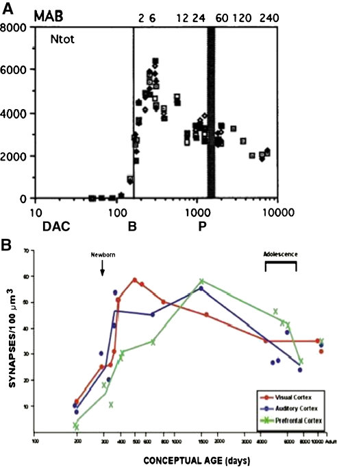
“It is estimated that nearly half of the synapses and neurons will be removed by pruning and apoptosis (Jiang & Nardelli, 2016; Stiles & Jernigan, 2010).”
BackgroundMapping White Matter Microstructure in the One Month Human Brain
Dean, D. C., Planalp, E. M., Wooten, W., Adluru, N., Kecskemeti, S. R., Frye, C., Schmidt, C. K., Schmidt, N. L., Styner, M. A., Goldsmith, H. H., et al. (2017) · Scientific Reports · doi:10.1038/s41598-017-09915-6 · Cited by 68
Diagnostic Approach to Macrocephaly in Children
Accogli, A., Geraldo, A. F., Piccolo, G., Riva, A., Scala, M., Balagura, G., Salpietro, V., Madia, F., Maghnie, M., Zara, F., et al. (2022) · Frontiers in Pediatrics · doi:10.3389/fped.2021.794069 · Cited by 74
“Neural progenitor cells begin to divide and differentiate into neurons and glia, the two cell types that form the basis of the nervous system (15).”
BackgroundMicroRNA-29 is an essential regulator of brain maturation through regulation of CH methylation
Swahari, V., Nakamura, A., Hollville, É., Stroud, H., Simon, J. M., Ptacek, T., Beck, M. V., Flowers, C., Guo, J., Plestant, C., et al. (2021) · Cell Reports · doi:10.1016/j.celrep.2021.108946 · Cited by 72
“During brain maturation, widespread changes in synaptic connectivity are evident (Jiang and Nardelli, 2016; Stiles and Jernigan, 2010), and perturbations of synaptic development or maturation can result in severe neurobehavioral sequelae as seen in autism spectrum disorders (ASD), epilepsy, and schizophrenia (Bourgero…”
The coding and long noncoding single-cell atlas of the developing human fetal striatum
Bocchi, V. D., Conforti, P., Vezzoli, E., Besusso, D., Cappadona, C., Lischetti, T., Galimberti, M., Ranzani, V., Bonnal, R. J. P., De Simone, M., et al. (2021) · Science · doi:10.1126/science.abf5759 · Cited by 70
“During development,MSNs are derived from progenitors in the lateral ganglionic eminence (LGE) of the telencephalon (4).”
BackgroundGuilbert, A., Rolland, M., Pin, I., Thomsen, C., Sakhi, A. K., Sabaredzovic, A., Slama, R., Guichardet, K., & Philippat, C. (2021) · Environment International · doi:10.1016/j.envint.2021.106697 · Cited by 72
“Human brain development starts in the first weeks after conception and is controlled by many factors including genes and hormones (Stiles and Jernigan, 2010; Williams, 2008).”
BackgroundMa, J., Wang, Z., Qin, C., Wang, T., Hu, X., & Ling, W. (2023) · Chemosphere · doi:10.1016/j.chemosphere.2023.138455 · Cited by 72
Lee, J. K., Fandakova, Y., Johnson, E. G., Cohen, N. J., Bunge, S. A., & Ghetti, S. (2020) · Developmental Cognitive Neuroscience · doi:10.1016/j.dcn.2019.100741 · Cited by 70
“Although not yet definitively linked, volumetric increases may reflect ongoing synaptogenesis and dendritic elaboration, while volumetric declines may reflect synaptic pruning (Stiles and Jernigan, 2010).”
BackgroundStagni, F., Giacomini, A., Guidi, S., Emili, M., Uguagliati, B., Salvalai, M. E., Bortolotto, V., Grilli, M., Rimondini, R., & Bartesaghi, R. (2017) · Experimental Neurology · doi:10.1016/j.expneurol.2017.08.018 · Cited by 70
“Most of the brain neurons are produced in the prenatal period, with the notable exception of those involved in the formation of the hippocampus, where neurogenesis continues postnatally and throughout life (Seress et al., 2001; Rice and Barone, 2010; Stiles and Jernigan, 2010; Spalding et al., 2013).”
Frosk, P., Arts, H. H., Philippe, J., Gunn, C. S., Brown, E., Chodirker, B. N., Simard, L. R., Majewski, J., Fahiminiya, S., Russell, C., et al. (2017) · Journal of Medical Genetics · doi:10.1136/jmedgenet-2016-104296 · Cited by 70
“The most likely explanation for the reduction is apoptosis in cells with cep55l disruption, which is supported by TUNEL assay at 2 dpf (see ref.”
BackgroundLiu, S. H., Bobb, J. F., Henn, B. C., Gennings, C., Schnaas, L., Téllez‐Rojo, M. M., Bellinger, D. C., Arora, M., Wright, R. O., & Coull, B. A. (2018) · Statistics in Medicine · doi:10.1002/sim.7947 · Cited by 70
“The hierarchical model is y|h,X,β, σ2,b ∼ N(Xβ + Wh + Ub, σ2I) (6) h|τ2 1 , τ2 2 ∼ N2n {0,Σh = ( τ2 1 K 0 0 τ2 2 K )} (7) b|σ2,D ∼ N2n (0, σ(2)(In ⊗ D)) .”
Verma, V., Paul, A., Vishwanath, A. A., Vaidya, B., & Clement, J. P. (2019) · Open Biology · doi:10.1098/rsob.180265 · Cited by 72
Exposure to endocrine‐disrupting chemicals and implications for neurodevelopment
Özel, F., & Rüegg, J. (2023) · Developmental Medicine & Child Neurology · doi:10.1111/dmcn.15551 · Cited by 71
Human 3D brain organoids: steering the demolecularization of brain and neurological diseases
Adlakha, Y. K. (2023) · Cell Death Discovery · doi:10.1038/s41420-023-01523-w · Cited by 70
“INTRODUCTION Human brain is the most complex and greatly developed organ in multicellular organisms [1].”
BackgroundThe role of nutrition and gut microbiome in childhood brain development and behavior
Jiang, Y., & Li, Y. (2025) · Frontiers in Nutrition · doi:10.3389/fnut.2025.1590172 · Cited by 71
Prem, S., Millonig, J. H., & DiCicco-Bloom, E. M. (2020) · Advances in neurobiology · doi:10.1007/978-3-030-45493-7_5 · Cited by 69
Seal, S. V., & Turner, J. D. (2021) · International Journal of Molecular Sciences · doi:10.3390/ijms22073344 · Cited by 69
Network-specific selectivity of functional connections in the neonatal brain
Sylvester, C. M., Kaplan, S., Myers, M. J., Gordon, E. M., Schwarzlose, R. F., Alexopoulos, D., Nielsen, A. N., Kenley, J. K., Meyer, D., Yu, Q., et al. (2023) · Cerebral Cortex · doi:10.1093/cercor/bhac202 · Cited by 58
Davidsen, N., Lauvås, A. J., Myhre, O., Ropstad, E., Carpi, D., Gyves, E. M., Berntsen, H. F., Dirven, H. A. A. M., Paulsen, R. E., Bal‐Price, A., et al. (2020) · Reproductive Toxicology · doi:10.1016/j.reprotox.2020.12.013 · Cited by 68
Toxoplasmosis and neuropsychiatric diseases: can serological studies establish a clear relationship?
Fabiani, S., Pinto, B., & Bruschi, F. (2012) · Neurological Sciences · doi:10.1007/s10072-012-1197-4 · Cited by 66
“Proliferation and migration of glial progenitors continue after birth, while their differentiation and maturation continue throughout childhood [53].”
BackgroundMetabolic defects in multiple sclerosis
Adiele, R. C., & Adiele, C. A. (2017) · Mitochondrion · doi:10.1016/j.mito.2017.12.005 · Cited by 68
Relationship between diet, the gut microbiota, and brain function
Tengeler, A. C., Kozicz, T., & Kiliaan, A. J. (2018) · Nutrition Reviews · doi:10.1093/nutrit/nuy016 · Cited by 56
Cumulative multiple early life hits- a potent threat leading to neurological disorders
Sarkar, T., Patro, N., & Patro, I. K. (2019) · Brain Research Bulletin · doi:10.1016/j.brainresbull.2019.02.005 · Cited by 69
Early vocal contact and music in the NICU: new insights into preventive interventions
Filippa, M., Lordier, L., de Almeida, J. S., Monaci, M. G., Adam‐Darqué, A., Grandjean, D., Kuhn, P., & Hüppi, P. S. (2019) · Pediatric Research · doi:10.1038/s41390-019-0490-9 · Cited by 68
Rehman, A., & Khalili, Y. A. (2019) · StatPearls · Cited by 67
Wong, C. T., Ahmad, E., Li, H., & Crawford, D. A. (2014) · Cell Communication and Signaling · doi:10.1186/1478-811x-12-19 · Cited by 67
Schaffner, S. L., Lussier, A. A., Baker, J. A., Goldowitz, D., Hamre, K. M., & Kobor, M. S. (2020) · Frontiers in Genetics · doi:10.3389/fgene.2020.00035 · Cited by 67
“During mammalian brain development, periods of neurogenesis, gliogenesis, differentiation, and normal apoptotic cell death occur to establish and maintain a healthy population of neurons and glia (Stiles and Jernigan, 2010).”
BackgroundA shifting role of thalamocortical connectivity in the emergence of cortical functional organization
Park, S., Haak, K. V., Oldham, S., Cho, H., Byeon, K., Park, B., Thomson, P., Chen, H., Gao, W., Xu, T., et al. (2024) · Nature Neuroscience · doi:10.1038/s41593-024-01679-3 · Cited by 66
“In this initial stage, various cellular mechanisms such as synaptogenesis, axonogenesis and dendritic arborization underlie the dynamic changes pertaining to postnatal surface expansion 51–53 .”
Boschen, K. E., & Klintsova, A. Y. (2016) · Vitamins and hormones · doi:10.1016/bs.vh.2016.10.008 · Cited by 69
“Neurotrophic factors represent one group of factors capable of protecting developing neurons against apoptotic cascades (Nijhawan et al., 2000; Stiles & Jernigan, 2010).”
BackgroundDevelopment Period of Prefrontal Cortex
Uytun, M. Ç. (2018) · InTech eBooks · doi:10.5772/intechopen.78697 · Cited by 70
“Primary sulci (superior frontal, inferior frontal, and precentral) are the main regions of the PFC, and develop during gestational weeks 25–26 [42].”
BackgroundWhite matter connectomes at birth accurately predict cognitive abilities at age 2
Girault, J. B., Munsell, B. C., Puechmaille, D., Goldman, B. D., Prieto, J. C., Styner, M. A., & Gilmore, J. H. (2019) · NeuroImage · doi:10.1016/j.neuroimage.2019.02.060 · Cited by 66
“WM connections in the developing brain are built through genetically regulated cascades of cellular events that govern neurogenesis and migration and promote an exuberance of connections via processes of axon guidance, synaptogenesis and dendritic arborization (Stiles and Jernigan, 2010).”
BackgroundMaternal fever during pregnancy and offspring attention deficit hyperactivity disorder
Gustavson, K., Ask, H., Ystrøm, E., Stoltenberg, C., Lipkin, W. I., Surén, P., Håberg, S. E., Magnus, P. M., Knudsen, G. P., Eilertsen, E. M., et al. (2019) · Scientific Reports · doi:10.1038/s41598-019-45920-7 · Cited by 67
Sah, E., Krishnamurthy, S., Ahmidouch, M. Y., Gillispie, G. J., Milligan, C., & Orr, M. E. (2021) · Life · doi:10.3390/life11030229 · Cited by 55
“Along with oligodendrocytes, astrocytes originate from the neural tube [98].”
BackgroundImpact of Maternal Selenium Status on Infant Outcome during the First 6 Months of Life
Varsi, K., Bolann, B. J., Torsvik, I. K., Eik, T. C. R., Høl, P. J., & Bjørke‐Monsen, A. (2017) · Nutrients · doi:10.3390/nu9050486 · Cited by 65
“gestational week and continues after birth [30].”
The Brain before Birth: Using fMRI to Explore the Secrets of Fetal Neurodevelopment
Konkel, L. (2018) · Environmental Health Perspectives · doi:10.1289/ehp2268 · Cited by 65
microRNAs Sculpt Neuronal Communication in a Tight Balance That Is Lost in Neurological Disease
Thomas, K. T., Gross, C., & Bassell, G. J. (2018) · Frontiers in Molecular Neuroscience · doi:10.3389/fnmol.2018.00455 · Cited by 64
“Many of these are maternal exposures that occur during the second trimester of gestation, which coincides with critical events in fetal brain development, including the production of the majority of the brain’s neurons, the migration of neurons within the cerebral cortex, and the formation of the thalamocortical proje…”
BackgroundPorru, S., Esplugues, A., Llop, S., & Delgado-Saborit, J. M. (2024) · Environmental Pollution · doi:10.1016/j.envpol.2024.123732 · Cited by 65
Smith, K. W., Gierski, F., Andre, J., Dowell, N. G., Cercignani, M., Naassila, M., & Duka, T. (2017) · Addiction Biology · doi:10.1111/adb.12332 · Cited by 55
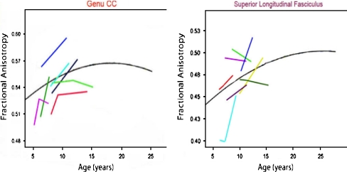
“…matter integrity between men and women (De Bellis et al. 2001) as they were seen not only in regions of the corpus callosum associated with prefrontal brain areas, which reach maturity last, but also in sensorimotor regions, which reach maturity early (Gogtay et al. 2004; Stiles & Jernigan 2010).”
BackgroundPsychosocial Stress and Adversity: Effects from the Perinatal Period to Adulthood
Barrero-Castillero, A., Morton, S. U., Nelson, C. A., & Smith, V. C. (2019) · NeoReviews · doi:10.1542/neo.20-12-e686 · Cited by 54
Maternal Inflammation During Pregnancy and Offspring Brain Development: The Role of Mitochondria
Gyllenhammer, L. E., Rasmussen, J. M., Bertele, N., Halbing, A., Entringer, S., Wadhwa, P. D., & Buß, C. (2021) · Biological Psychiatry Cognitive Neuroscience and Neuroimaging · doi:10.1016/j.bpsc.2021.11.003 · Cited by 64
Vascularized human brain organoid on-chip
Tan, S. Y., Feng, X., Cheng, L. K., & Wu, A. R. (2023) · Lab on a Chip · doi:10.1039/d2lc01109c · Cited by 64
Sullivan, E. V., Pfefferbaum, A., Rohlfing, T., Baker, F. C., Padilla, M. L., & Colrain, I. M. (2011) · NeuroImage · doi:10.1016/j.neuroimage.2011.04.003 · Cited by 60
MiRNA-137-mediated modulation of mitochondrial dynamics regulates human neural stem cell fate
Channakkar, A. S., Singh, T., PATTNAIK, B. R., Gupta, K., Seth, P., & Adlakha, Y. K. (2020) · Stem Cells · doi:10.1002/stem.3155 · Cited by 64
“In addition to gene regulatory networks, post-transcriptional mechanisms play an evident role in neural development.(1,2) Disruptions in the postAbbreviations: ETC, electron transport chain; hiNSCs, human induced neural stem cells; MEF2A, myocyte-specific enhancer factor 2A; NRF2, nuclear factor erythroid 2 (NFE2)rela…”
BackgroundDhamala, E., Ooi, L. Q. R., Chen, J., Kong, R., Anderson, K. M., Chin, R., Yeo, B. T. T., & Holmes, A. J. (2022) · NeuroImage · doi:10.1016/j.neuroimage.2022.119485 · Cited by 63
An introduction to the HEALthy Brain and Child Development Study (HBCD) study
Nelson, C. A., Frankeberger, J., & Chambers, C. (2024) · Developmental Cognitive Neuroscience · doi:10.1016/j.dcn.2024.101441 · Cited by 62
“The human brain begins to develop three weeks after conception and does not reach adult maturity until approximately the third decade of life (Stiles and Jernigan, 2010).”
Visan, A., Hayeß, K., Sittner, D., Pohl, E. E., Riebeling, C., Slawik, B., Gulich, K., Oelgeschläger, M., Luch, A., & Seiler, A. E. M. (2012) · NeuroToxicology · doi:10.1016/j.neuro.2012.06.006 · Cited by 62
“These effects are probably a result of programmed cell death, an important aspect of cell number regulation and neuronal development during brain development (Stiles and Jernigan, 2010).”
BackgroundCrafting the Brain – Role of Histone Acetyltransferases in Neural Development and Disease
Sheikh, B. N. (2014) · Cell and Tissue Research · doi:10.1007/s00441-014-1835-7 · Cited by 62
Wong, C. T., Ussyshkin, N., Ahmad, E., Rai‐Bhogal, R., Li, H., & Crawford, D. A. (2016) · Journal of Neuroscience Research · doi:10.1002/jnr.23759 · Cited by 62
Chen, L., Chen, C., Zhong, X., Sun, X., Zhu, H., Li, X., Yang, H., Shu, Y., Chang, Y., Hu, X., et al. (2018) · Journal of Neuroimmunology · doi:10.1016/j.jneuroim.2018.05.014 · Cited by 62
The impact of the micronutrient iodine in health and diseases
Opazo, M. C., Coronado-Arrázola, I., Vallejos, O. P., Moreno‐Reyes, R., Fardella, C., Mosso, L. M., Kalergis, A. M., Bueno, S. M., & Riedel, C. A. (2020) · Critical Reviews in Food Science and Nutrition · doi:10.1080/10408398.2020.1843398 · Cited by 62
“Maternal thyroid hormones are needed for the development of important structures of the fetus like central nervous system (CNS) (Stiles and Jernigan 2010), cells from the immune system (Foster et al. 1998) and gastrointestinal tract (Sirakov and Plateroti 2011) among other organs (Colony 1983).”
BackgroundCastriotta, L., Rosolen, V., Biggeri, A., Ronfani, L., Catelan, D., Mariuz, M., Bin, M., Brumatti, L. V., Horvat, M., & Barbone, F. (2020) · International Journal of Hygiene and Environmental Health · doi:10.1016/j.ijheh.2020.113604 · Cited by 61
Araneda, R., Ebner‐Karestinos, D., Paradis, J., Klöcker, A., Saussez, G., Démas, J., Bailly, R., Bouvier, S., de Tournai, A. C., Herman, E., et al. (2023) · Archives of Pediatrics and Adolescent Medicine · doi:10.1001/jamapediatrics.2023.4809 · Cited by 61
Ganai, S. A., Banday, S., Farooq, Z., & Altaf, M. (2016) · Pharmacology & Therapeutics · doi:10.1016/j.pharmthera.2016.07.001 · Cited by 61
Gaete, J., & Araya, R. (2017) · Journal of Adolescence · doi:10.1016/j.adolescence.2017.02.011 · Cited by 61
Krefft, O., Jabali, A., Iefremova, V., Koch, P., & Ladewig, J. (2018) · Journal of Visualized Experiments · doi:10.3791/56768 · Cited by 63
Fujisawa, T. X., Shimada, K., Takiguchi, S., Mizushima, S. G., Kosaka, H., Teicher, M. H., & Tomoda, A. (2018) · NeuroImage Clinical · doi:10.1016/j.nicl.2018.07.018 · Cited by 62
“In general, it is well known that brain development in this period is most significant, as the capacity of the brain reaches approximately 90% of adult volume by age 6 years (Stiles and Jernigan, 2010).”
Magnetoencephalography and the infant brain
Chen, Y., Saby, J. N., Kuschner, E. S., Gaetz, W. C., Edgar, J. C., & Roberts, T. P. L. (2019) · NeuroImage · doi:10.1016/j.neuroimage.2019.01.059 · Cited by 61
“As detailed in Stiles and Jernigan (2010), additional knowledge of structural brain development in infants is also based on histological studies from postmortem materials of fetuses, infants and children (Kostovic and Vasung, 2009; Vasung et al., 2016; Huang et al., 2006, 2009) and data acquired in…”
BackgroundUsing a deep recurrent neural network with EEG signal to detect Parkinson’s disease
Xu, S., Wang, Z., Sun, J., Zhang, Z., Wu, Z., Tiezhao, Y., Xue, G., & Cheng, C. (2020) · Annals of Translational Medicine · doi:10.21037/atm-20-5100 · Cited by 61
“( ) ( ) ( ) 1 1 1 1 1 * 1 * a t b W h t U x t = + − + [1]”
RetractedDibble, M., Ang, J. Z., Mariga, L., Molloy, E. J., & Bokde, A. L. W. (2021) · The Journal of Pediatrics · doi:10.1016/j.jpeds.2021.01.008 · Cited by 61
Hepatic Cannabinoid Signaling in the Regulation of Alcohol-Associated Liver Disease
Yang, K. (2021) · Alcohol research · doi:10.35946/arcr.v41.1.11 · Cited by 62
Sabitha, K. R., Shetty, A. K., & Upadhya, D. (2020) · Neuroscience & Biobehavioral Reviews · doi:10.1016/j.neubiorev.2020.12.025 · Cited by 60
“As the brain matures, all of its characteristic features emerge through a highly organized cascade of events, including propagation, migration, differentiation, and functional maturation of the cells through molecular regulation (Sur and Rubenstein, 2005; Stiles and Jernigan, 2010; Tau and Peterson, 2010; Silbereis et…”
BackgroundNeonatal family-centered care: evidence and practice models
Lee, J. Y. (2023) · Korean Journal of Pediatrics · doi:10.3345/cep.2023.00367 · Cited by 61
Adolescents: which risks for their life and health?
Balocchini, E., Chiamenti, G., & Lamborghini, A. (2013) · Journal of preventive medicine and hygiene · doi:10.15167/2421-4248/jpmh2013.54.4.409 · Cited by 62
Altered Regional Brain Cortical Thickness in Pediatric Obstructive Sleep Apnea
Macey, P. M., Gozal, D., Prasad, J. P., Richard, A., Kumar, R., Philby, M. F., & Gozal, D. (2018) · Frontiers in Neurology · doi:10.3389/fneur.2018.00004 · Cited by 60
“30 mm per year as a result of synaptic pruning (52, 69).”
BackgroundPeter, C. J., Saito, A., Hasegawa, Y., Tanaka, Y., Nagpal, M., Perez, G., Alway, E., Espeso-Gil, S., Fayyad, T., Ratner, C., et al. (2019) · Nature Communications · doi:10.1038/s41467-019-12013-y · Cited by 57
The Bidirectional Social-Cognitive Mechanisms of the Social-Attention Symptoms of Autism
Mundy, P. C., & Bullen, J. C. (2022) · Frontiers in Psychiatry · doi:10.3389/fpsyt.2021.752274 · Cited by 61
“Neurocognitive development involves adaptations to different types of environmental demands, which lead to the differentiation of neural structures and functional circuits (284).”
BackgroundMaher, G. M., O’Keeffe, G. W., Kenny, L. C., Kearney, P. M., Dinan, T. G., & Khashan, A. S. (2017) · BMJ Open · doi:10.1136/bmjopen-2017-018313 · Cited by 59
“Furthermore, recent work demonstrated focal patches of abnormal laminar architecture and laminar disorganisation in the prefrontal and temporal cortices of children with ASD suggesting there may be alterations in brain development at prenatal stages, as cortical lamination is ongoing during the second trimester of pre…”
Handryastuti, S., Pusponegoro, H. D., Nurdadi, S., Chandra, A., Pramita, F. A., Soebadi, A., Widjaja, I. R., & Rafli, A. (2022) · Journal of Nutrition and Metabolism · doi:10.1155/2022/9775727 · Cited by 59
Pediatric Brain Development in Down Syndrome: A Field in Its Infancy
Hamner, T., Udhnani, M. D., Osipowicz, K. Z., & Lee, N. R. (2018) · Journal of the International Neuropsychological Society · doi:10.1017/s1355617718000206 · Cited by 58
“That is, TBV is a global measure further segmented into total GM and WM. GM, comprised of cell bodies and synaptic connections (Stiles & Jernigan, 2010), makes up the cerebral cortex and areas deep in the brain (e.g., hippocampus, amygdala, hypothalamus, thalamus, basal ganglia) and in the…”
Background
Metadata
- Authors
- Stiles, J., & Jernigan, T. L.
- Affiliations
- University of California San Diego (US)
- Corresponding author
- Jernigan, Terry L.
- ORCID
- Jernigan, Terry L.
- Venue
- Neuropsychology Review
- Published
- 2010-11-03
- Volume
- 20
- Issue
- 4
- Pages
- 327-348
- ISSN
- 1040-7308 (ISSN-L)
- 1573-6660
- PMC
- PMC2989000
- OpenAlex
- W2115599658
- Cited by
- 2,009
- References
- 132
- Citations by year
- 2026: 142 · 2025: 184 · 2024: 199 · 2023: 209 · 2022: 184 · 2021: 197 · 2020: 195 · 2019: 155 · 2018: 158 · 2017: 122 · 2016: 92 · 2015: 64 · 2014: 49 · 2013: 24 · 2012: 23
- FWCI
- 1.43
- Type
- journal article
- Language
- en
- Open access
- hybrid
- doi.org
- Licence
- cc-by-nc
- Locations
- Neuropsychology Review (publishedVersion, cc-by-nc)
- Europe PMC (PubMed Central) (publishedVersion, cc-by-nc)
- PubMed Central (publishedVersion, cc-by-nc)
- PubMed (publishedVersion)
- eScholarship (California Digital Library) (submittedVersion, public-domain)
- eScholarship (California Digital Library) (submittedVersion)
- Funders
- Lundbeckfonden (R32‐A3161)
- H. Lundbeck A/S
- National Institutes of Health (5r01hd025077-10)
- U.S. Public Health Service (R32-A3161)
- University of California, San Diego (RC2DA029475)
- National Institute on Drug Abuse (RC2DA029475)
- National Institute of Child Health and Human Development
- Eunice Kennedy Shriver National Institute of Child Health and Human Development (R01HD060595, R01 HD 25077)
- Topics
- Life Sciences › Neuroscience › Developmental Neuroscience › Neurogenesis and neuroplasticity mechanisms
- Life Sciences › Biochemistry, Genetics and Molecular Biology › Genetics › Genetics and Neurodevelopmental Disorders
- Social Sciences › Psychology › Developmental and Educational Psychology › Child and Animal Learning Development
- Keywords
- brain development, developmental neuroscience, mammalian brain, neuronal organization, social development (developmental psychology), cognitive development, neural systems, genetic constraints, developmental mechanisms
- MeSH terms
- Animals, Brain, Cell Differentiation, Diagnostic Imaging, Humans, Nerve Net, Neural Pathways, Neurobiology, Neurons, Gene Expression Regulation, Developmental, Body Patterning, Neurogenesis
About these checks
Registry, retraction, Cites and statistics results are computed, not judged, with one exception: a printed reference that no index lists is read once by a model before it counts as a citation error. Claims are extracted by a model and kept only when their quote appears verbatim in the text; each cited source is then checked, with a model’s help, for whether it says what the claim relies on. Those verdicts can be wrong — read the claim’s quote and the cited source before relying on one.
Statistics and arithmetic are recomputed, not judged.
Confidence bands are shown only for fields calibrated on our annotated test set. None is calibrated yet, so no band is shown.
What counts as a claim ›How we measure confidence ›How we check this ›
Are you an author, or do you see a problem with this page? Request a review
Data sources
Bibliographic data: Semantic Scholar, OpenAlex, Crossref.
Contains information from the Semantic Scholar Academic Graph Datasets, made available under the ODC Attribution License (ODC-BY 1.0).
Semantic Scholar’s data is described in: Kinney, R., et al. (2023). The Semantic Scholar Open Data Platform. arXiv:2301.10140.
