

The Basics of Brain Development
Open accessThe DOI matches its Crossref record; 128 of the 132 references it cites were found in a registry or an index.
Abstract
Over the past several decades, significant advances have been made in our understanding of the basic stages and mechanisms of mammalian brain development. Studies elucidating the neurobiology of brain development span the levels of neural organization from the macroanatomic, to the cellular, to the molecular. Together this large body of work provides a picture of brain development as the product of a complex series of dynamic and adaptive processes operating within a highly constrained, genetically organized but constantly changing context. The view of brain development that has emerged from the developmental neurobiology literature presents both challenges and opportunities to psychologists seeking to understand the fundamental processes that underlie social and cognitive development, and the neural systems that mediate them. This chapter is intended to provide an overview of some very basic principles of brain development, drawn from contemporary developmental neurobiology, that may be of use to investigators from a wide range of disciplines.
Overview
The claims and statistics of this paper haven’t been checked yet.
Free: it reads the open-access text and checks the first 5 claims that cite a source we can check. Create a free account to check every claim.
Integrity signals
Registry
Cites
Statistics
Figures





Verification
Claims the paper makes
The claims and statistics of this paper haven’t been checked yet.
Free: it reads the open-access text and checks the first 5 claims that cite a source we can check. Create a free account to check every claim.
Claims other papers make about this one
Each is checked against this paper’s own text: does it say what the citing paper says it does?
The source backs part of the claim, not all of it — or backs it for a different population, condition or comparison.
In the citing paper
“We now know from recent studies that it is the birth and migration of neurons derived from oRGCs that play a role in the development of the primary sulci (superior frontal, inferior frontal and precentral) in week 25–26 of gestation [23, 76].”
From Development of prefrontal cortex (2022) · cites this paper as [23]
Experimental — accuracy not yet measuredIn the citing paper
“This intricate structural-functional interplay underscores that the ontogeny of functional networks likely reflects programmed neurodevelopmental events (e.g. neurogenesis, cell death, myelination, pruning, synaptic plasticity, and glial development) (Innocenti and Price, 2005; Stiles and Jernigan, 2010)”
From Development of large-scale functional networks from birth to adulthood: A guide to the neuroimaging literature (2017) · cites this paper as Stiles and Jernigan, 2010
In the citing paper
“The brain’s major fiber pathways become consolidated through myelination, though their presence is largely established prenatally (Stiles and Jernigan, 2010).”
From Development of large-scale functional networks from birth to adulthood: A guide to the neuroimaging literature (2017) · cites this paper as Stiles and Jernigan, 2010
Self-check
Percentages, sums and reported differences re-computed from the paper’s own numbers.
The self-check needs the paper’s full text.
Citations
Cites
Philosophical transactions of the Royal Society of London. B
Press, H. (1886) · Cited by 10,080
MR diffusion tensor spectroscopy and imaging
Basser, P. J., Mattiello, J., & LeBihan, D. (1994) · Biophysical Journal · doi:10.1016/s0006-3495(94)80775-1 · Cited by 5,970
Dynamic mapping of human cortical development during childhood through early adulthood
Gogtay, N., Giedd, J. N., Lusk, L., Hayashi, K. M., Greenstein, D., Vaituzis, A. C., Nugent, T. F., Herman, D. H., Clasen, L. S., Toga, A. W., et al. (2004) · Proceedings of the National Academy of Sciences · doi:10.1073/pnas.0402680101 · Cited by 5,604
(2005) · Choice Reviews Online · doi:10.5860/choice.43-0626 · Cited by 4,674
Neurotrophins: Roles in Neuronal Development and Function
Huang, E. J., & Reichardt, L. F. (2001) · Annual Review of Neuroscience · doi:10.1146/annurev.neuro.24.1.677 · Cited by 4,394
Ferrier lecture. Functional architecture of macaque monkey visual cortex
Hubel, D. H., & Wiesel, T. N. (1977) · Proceedings of the Royal Society of London. Series B. Biological Sciences · doi:10.1098/rspb.1977.0085 · Cited by 2,608 · cited 1×
“The seminal studies of Hubel and Weisel (Hubel and Wiesel 1977; Hubel et al. 1977) showed that monocular visual deprivation in the early postnatal period can substantially alter basic patterns of organization within primary visual cortex (PVC).”
Background(1995) · Choice Reviews Online · doi:10.5860/choice.32-5931 · Cited by 2,311
Mode of cell migration to the superficial layers of fetal monkey neocortex
Rakic, P. (1972) · Journal of Comparative Neurology · doi:10.1002/cne.901450105 · Cited by 2,170 · cited 1×
“Because of the greater distances, neurons require what was originally identified as a special population of cells within the VZ called “radial glial guides” to support their migration (Rakic 1972).”
BackgroundRegional Development of the Brain in Early Life
(1968) · Archives of Disease in Childhood · doi:10.1136/adc.43.229.388 · Cited by 2,090
Neurons derived from radial glial cells establish radial units in neocortex
Noctor, S. C., Flint, A. C., Weissman, T. A., Dammerman, R. S., & Kriegstein, A. R. (2001) · Nature · doi:10.1038/35055553 · Cited by 2,016 · cited 2×
“…radial glial guides were originally thought to be a special, transient population of cells, it has recently been discovered that the cells that provide the scaffolding are actually the neural progenitor cells (Noctor et al. 2001; Noctor et al. 2002; Parnavelas et al. 2002; Weissman et al. 2003).”
Background
Research works printed but not linked: 1
- 56. Lebel, C., & Beaulieu, C. (2009). Longitudinal diffusion tensor imaging of healthy brain development in children. Honolulu: ISMRM Annual Meeting.
Cited by
Items 101–200 of 1,004
The neuroanatomy of prematurity: Normal brain development and the impact of preterm birth
Ortinau, C. M., & Neil, J. J. (2014) · Clinical Anatomy · doi:10.1002/ca.22430 · Cited by 136
Roles of mitochondria in neuronal development
Son, G., & Han, J. (2018) · BMB Reports · doi:10.5483/bmbrep.2018.51.11.226 · Cited by 117
“end of gastrulation and majority of the brain structure is formed by the end of embryogenesis (39).”
BackgroundSood, D., Chwalek, K., Stuntz, E., Pouli, D., Du, C., Tang-Schomer, M., Georgakoudi, I., Black, L. D., & Kaplan, D. L. (2016) · ACS Biomaterials Science & Engineering · doi:10.1021/acsbiomaterials.5b00446 · Cited by 121
Neuronal migration disorders: Focus on the cytoskeleton and epilepsy
Stouffer, M. A., Golden, J. A., & Francis, F. (2016) · Neurobiology of Disease · doi:10.1016/j.nbd.2015.08.003 · Cited by 115
The Encultured Brain: An Introduction to Neuroanthropology
Lende, D. H., & Downey, G. (2012) · Project Muse (Johns Hopkins University) · Cited by 131
Fumagalli, M., Lecca, D., Abbracchio, M. P., & Ceruti, S. (2017) · Frontiers in Pharmacology · doi:10.3389/fphar.2017.00941 · Cited by 130
“…followed by naturally occurring cell death of damaged or unnecessary cells, formation of synapses, myelination of axons, and generation of neuronal connections are indeed critically controlled by intrinsic and environmental factors at each stage during development (Stiles and Jernigan, 2010).”
BackgroundElectromagnetic field and brain development
Kaplan, S., Deniz, Ö. G., Önger, M. E., Türkmen, A. P., Yurt, K. K., Aydin, I., Altunkaynak, B. Z., & Davis, D. L. (2015) · Journal of Chemical Neuroanatomy · doi:10.1016/j.jchemneu.2015.11.005 · Cited by 127
“In children aged 5–11 and followed for two years via Magnetic Resonance Imaging (MRI), brain development was shown to be 1 mm per year especially in the prefrontal cortex (Sowell et al., 2004; Stiles and Jernigan, 2010).”
BackgroundInfluentialWaking up too early – the consequences of preterm birth on sleep development
Bennet, L., Walker, D. W., & Horne, R. S. C. (2018) · The Journal of Physiology · doi:10.1113/jp274950 · Cited by 128
Advancing models of neural development with biomaterials
Roth, J. G., Huang, M. S., Li, T. L., Feig, V. R., Jiang, Y., Cui, B., Greely, H. T., Bao, Z., Pașca, S. P., & Heilshorn, S. C. (2021) · Nature reviews. Neuroscience · doi:10.1038/s41583-021-00496-y · Cited by 128
Micronutrients and Brain Development
Mattei, D., & Pietrobelli, A. (2019) · Current Nutrition Reports · doi:10.1007/s13668-019-0268-z · Cited by 125
Timing of Early-Life Stress and the Development of Brain-Related Capacities
Hambrick, E. P., Brawner, T. W., & Perry, B. D. (2019) · Frontiers in Behavioral Neuroscience · doi:10.3389/fnbeh.2019.00183 · Cited by 85
Jacobus, J., Squeglia, L. M., Bava, S., & Tapert, S. F. (2013) · Psychiatry Research: Neuroimaging · doi:10.1016/j.pscychresns.2013.07.014 · Cited by 111
“These dynamic changes include substantial cortical remodeling (e.g., decreases in gray matter volume and neural pruning) and increases in white matter volume (e.g., myelination and/or axonal caliber) (Yakovlev and Lecours, 1967; Giedd et al., 1999; Gogtay et al., 2004; Stiles and Jernigan, 2010).”
BackgroundProgesterone Actions During Central Nervous System Development
González-Orozco, J. C., & Camacho-Arroyo, I. (2019) · Frontiers in Neuroscience · doi:10.3389/fnins.2019.00503 · Cited by 106
“In this developmental period, the CNS undergoes indispensable developmental processes, such as synaptogenesis, myelination, organization, and remodeling of the neural circuits (Stiles and Jernigan, 2010).”
BackgroundNurturing the preterm infant brain: leveraging neuroplasticity to improve neurobehavioral outcomes
DeMaster, D., Bick, J. R., Johnson, U., Montroy, J. J., Landry, S. H., & Duncan, A. F. (2018) · Pediatric Research · doi:10.1038/s41390-018-0203-9 · Cited by 122
Neurogenesis impairment: An early developmental defect in Down syndrome
Stagni, F., Giacomini, A., Emili, M., Guidi, S., & Bartesaghi, R. (2018) · Free Radical Biology and Medicine · doi:10.1016/j.freeradbiomed.2017.07.026 · Cited by 120
“In the human forebrain, neocortical neurons are generated during a restricted period that begins at approximately GW 6 and is largely completed by GW 18 [50].”
Shadrin, A. A., Kaufmann, T., van der Meer, D., Palmer, C. E., Makowski, C., Loughnan, R. J., Jernigan, T. L., Seibert, T. M., Hagler, D. J., Smeland, O. B., et al. (2021) · NeuroImage · doi:10.1016/j.neuroimage.2021.118603 · Cited by 120
Self-citationHyperoxia and the Immature Brain
Reich, B., Hoeber, D., Bendix, I., & Felderhoff‐Mueser, U. (2016) · Developmental Neuroscience · doi:10.1159/000454917 · Cited by 119
Bosnjak, Z. J., Logan, S., Liu, Y., & Bai, X. (2016) · Anesthesia & Analgesia · doi:10.1213/ane.0000000000001544 · Cited by 118
Genetic control of postnatal human brain growth
van Dyck, L. I., & Morrow, E. M. (2016) · Current Opinion in Neurology · doi:10.1097/wco.0000000000000405 · Cited by 118
Complement: The Emerging Architect of the Developing Brain
Coulthard, L. G., Hawksworth, O. A., & Woodruff, T. M. (2018) · Trends in Neurosciences · doi:10.1016/j.tins.2018.03.009 · Cited by 119
Hiremath, C., Kommu, J. V. S., Yamini, B. K., Girimaji, A. S., Kumar, K. R., Sravanti, S. L., Padmanabha, H., Raju, K. N. V., Kishore, M. T., Jacob, P., et al. (2021) · Translational Psychiatry · doi:10.1038/s41398-020-01178-6 · Cited by 118
Shared heritability of human face and brain shape
Naqvi, S., Sleyp, Y., Hoskens, H., Indencleef, K., Spence, J. P., Bruffaerts, R., Radwan, A., Eller, R. J., Richmond, S., Shriver, M. D., et al. (2021) · Nature Genetics · doi:10.1038/s41588-021-00827-w · Cited by 103
“Early in embryonic development, the rostral end of the ectodermally derived neural tube gives rise to the forebrain, which in turn gives rise to the cerebrum that encompasses the cerebral cortex 10 .”
Castagna, A., Mascheroni, E., Fustinoni, S., & Montirosso, R. (2022) · Neuroscience & Biobehavioral Reviews · doi:10.1016/j.neubiorev.2022.104623 · Cited by 117
Graph theoretical modeling of baby brain networks
Zhao, T., Xu, Y., & He, Y. (2019) · NeuroImage · doi:10.1016/j.neuroimage.2018.06.038 · Cited by 99
“Before birth, the rapid proliferation and migration of neurons (Bystron et al., 2008; Stiles and Jernigan, 2010), together with exuberant axon growth and synaptogenesis (Webb et al.”
BackgroundBar, E., & Barak, B. (2019) · Glia · doi:10.1002/glia.23637 · Cited by 107
“This was particularly evident in 5- to 23-year-old subjects, an age range that represents the developmental period coinciding with synapse refinement in humans (Stiles & Jernigan, 2010).”
BackgroundFluctuating Asymmetry of Human Populations: A Review
Graham, J. H., & Özener, B. (2016) · Symmetry · doi:10.3390/sym8120154 · Cited by 118
“Dermatoglyphic patterns form during weeks 10–16 of fetal development, just as the population of neurons in the brain is beginning to expand and differentiate [276].”
BackgroundPutative adverse outcome pathways relevant to neurotoxicity
Bal-Price, A., Crofton, K. M., Sachana, M., Shafer, T. J., Behl, M., Forsby, A., Hargreaves, A., Landesmann, B., Lein, P. J., Louisse, J., et al. (2015) · Critical Reviews in Toxicology · doi:10.3109/10408444.2014.981331 · Cited by 114
Brain organoid formation on decellularized porcine brain ECM hydrogels
Simsa, R., Rothenbücher, T., Gürbüz, H., Ghosheh, N., Emneus, J., Jenndahl, L., Kaplan, D. L., Bergh, N., Serrano, A. M., & Fogelstrand, P. (2021) · PLOS ONE · doi:10.1371/journal.pone.0245685 · Cited by 114
“Especially the age of the animal could affect neuronal network formation, as the ECM is dynamically remodeled and shows a different protein composition at different life stages [72], as previously shown in a comparison of fetal and adult pig brain ECM [22].”
BackgroundA novel ecological account of prefrontal cortex functional development.
Werchan, D. M., & Amso, D. (2017) · Psychological Review · doi:10.1037/rev0000078 · Cited by 115
“This period is characterized by large increases in synaptogenesis and neurogenesis, followed by periods of neuronal pruning and synaptic death to accommodate the early increases in neural and synaptic formation (Stiles & Jernigan, 2010).”
Myhre, O., Låg, M., Villanger, G. D., Oftedal, B., Øvrevik, J., Holme, J. A., Aase, H., Paulsen, R. E., Bal‐Price, A., & Dirven, H. A. A. M. (2018) · Toxicology and Applied Pharmacology · doi:10.1016/j.taap.2018.03.015 · Cited by 114
“This will permit evaluation of key neurodevelopmental processes including the commitment and differentiation of neuronal progenitor cells, glial and neuronal cell proliferation, migration, differentiation into various neuronal and glial subtypes, synaptogenesis, pruning, myelination, networking, and terminal and funct…”
MethodThe Power and Promise of Early Learning
Shuey, E. A., & Kankaraš, M. (2018) · OECD education working papers · doi:10.1787/f9b2e53f-en · Cited by 118
Spatial embedding of structural similarity in the cerebral cortex
Song, H. F., Kennedy, H., & Wang, X.-J. (2014) · Proceedings of the National Academy of Sciences · doi:10.1073/pnas.1414153111 · Cited by 107
“Inspired by the growth of real axons that are guided by a variety of attractive and repulsive molecular cues (29, 30), we assume that each area exerts a distance-dependent attractive “ force, ” and the trajectory of each axon is determined by the sum of these forces.”
BackgroundNeurodevelopmental outcomes of children with congenital heart disease: A review
Howell, H. B., Zaccario, M. L., Kazmi, S. H., Desai, P. V., Sklamberg, F. E., & Mally, P. (2019) · Current problems in pediatric and adolescent health care · doi:10.1016/j.cppeds.2019.100685 · Cited by 112
Caspases in the Developing Central Nervous System: Apoptosis and Beyond
Nguyen, T. T. M., Gillet, G., & Popgeorgiev, N. (2021) · Frontiers in Cell and Developmental Biology · doi:10.3389/fcell.2021.702404 · Cited by 114
Vuoksimaa, E., Panizzon, M. S., Chen, C.-H., Fiecas, M., Eyler, L. T., Fennema-Notestine, C., Hagler, D. J., Fischl, B., Franz, C. E., Jak, A., et al. (2015) · Cerebral Cortex · doi:10.1093/cercor/bhu018 · Cited by 108
Price, A. J., Collado-Torres, L., Ivanov, N. A., Xia, W., Burke, E. E., Shin, J. H., Tao, R., Ma, L., Jia, Y., Hyde, T. M., et al. (2019) · Genome Biology · doi:10.1186/s13059-019-1805-1 · Cited by 86
Prenatal PBDE and PCB Exposures and Reading, Cognition, and Externalizing Behavior in Children
Zhang, H., Yolton, K., Webster, G. M., Sjödin, A., Calafat, A. M., Dietrich, K. N., Xu, Y., Xie, C., Braun, J. M., Lanphear, B. P., et al. (2017) · Environmental Health Perspectives · doi:10.1289/ehp478 · Cited by 103
Gut-Brain Axis in the Early Postnatal Years of Life: A Developmental Perspective
Jena, A., Montoya, C. A., Mullaney, J. A., Dilger, R. N., Young, W., McNabb, W. C., & Roy, N. C. (2020) · Frontiers in Integrative Neuroscience · doi:10.3389/fnint.2020.00044 · Cited by 108
“Apoptosis of neuronal cells is largely completed in utero, however, apoptosis of the glial cell population continues to occur in the first few months of after birth (Tau and Peterson, 2009; Stiles and Jernigan, 2010).”
BackgroundHead Growth Trajectory and Neurodevelopmental Outcomes in Preterm Neonates
Raghuram, K., Yang, J., Church, P., Cieslak, Z., Synnes, A. R., Mukerji, A., Shah, P. S., for the Canadian Neonatal Network, & Canadian Neonatal Follow-Up Network Investigators (2017) · PEDIATRICS · doi:10.1542/peds.2017-0216 · Cited by 106
Cao, J., Herman, A. B., West, G. B., Poe, G., & Savage, V. M. (2020) · Science Advances · doi:10.1126/sciadv.aba0398 · Cited by 105
“In addition, after birth, the increase in brain mass derives largely from the increase in glial cell and neuronal spine mass within gray matter and through the myelination of axons within white matter (43).”
BackgroundEssential genetic findings in neurodevelopmental disorders
Cardoso, A. R., Lopes‐Marques, M., Silva, R. M., Serrano, C., Amorim, A., Prata, M. J., & Azevedo, L. (2019) · Human Genomics · doi:10.1186/s40246-019-0216-4 · Cited by 106
“From the ninth week onward, the brain orderly maturates and acquires its typical structure, under a tightly orchestrated chain of events that includes abundant cell proliferation, migration, and differentiation [1, 4, 5].”
BackgroundArzua, T., Yan, Y., Jiang, C., Logan, S., Allison, R. L., Wells, C. W., Kumar, S. N., Schäfer, R., & Bai, X. (2020) · Translational Psychiatry · doi:10.1038/s41398-020-01029-4 · Cited by 104
Alex, A. M., Aguate, F. M., Botteron, K. N., Buß, C., Chong, Y. S., Dager, S. R., Donald, K. A., Entringer, S., Fair, D. A., Fortier, M. V., et al. (2023) · Nature Neuroscience · doi:10.1038/s41593-023-01501-6 · Cited by 104
Unraveling the complexity of human brain: Structure, function in healthy and disease states
Sultana, O. F., Bandaru, M., Islam, M. A., & Reddy, P. H. (2024) · Ageing Research Reviews · doi:10.1016/j.arr.2024.102414 · Cited by 106
Wilde, E. A., Merkley, T. L., Bigler, E. D., Max, J. E., Schmidt, A. T., Ayoub, K. W., McCauley, S. R., Hunter, J. V., Hanten, G., Li, X., et al. (2012) · International Journal of Developmental Neuroscience · doi:10.1016/j.ijdevneu.2012.01.003 · Cited by 95
“…critical periods of development, may alter subsequent maturation and impact neurobehavioral and cognitive development (Catroppa and Anderson, 2005; Suskauer and Huisman, 2009), but little is known regarding the long-term structural and functional consequences of TBI sustained in childhood or…”
BackgroundGhoshal, B., Tucker, A., Sanghera, B., & Wong, W. L. (2020) · Computational Intelligence · doi:10.1111/coin.12411 · Cited by 102
“23,24 We used deep BRUNet architecture that takes advantage of strengths from both deep residual learning 40 and U-Net architecture.”
MethodPutamen gray matter volumes in neuropsychiatric and neurodegenerative disorders.
Luο, X., Mao, Q., Shi, J., Wang, X., & Li, C. R. (2019) · PubMed · Cited by 99
Dumitru, I., Neitz, A., Alfonso, J., & Monyer, H. (2017) · Neuron · doi:10.1016/j.neuron.2017.03.003 · Cited by 99
“The vast majority of neurons are generated during development (Ernst and Frisén, 2015; Rakic, 1985; Stiles and Jernigan, 2010).”
A Blazing Landscape: Neuroinflammation Shapes Brain Metastasis
Doron, H., Pukrop, T., & Erez, N. (2019) · Cancer Research · doi:10.1158/0008-5472.can-18-1805 · Cited by 100
“The development and specialization of neurons during embryogenesis is a very complex and temporally regulated process (20).”
BackgroundBisphenol A Interaction With Brain Development and Functions
Negri‐Cesi, P. (2015) · Dose-Response · doi:10.1177/1559325815590394 · Cited by 98
Copf, T. (2016) · Neuroscience & Biobehavioral Reviews · doi:10.1016/j.neubiorev.2016.04.008 · Cited by 98
“axon and dendrite branching, synapse formation, experience-dependent synapse remodeling and pruning, gliogenesis, as well as myelination [125-128].”
Walsh, M. J. M., Wallace, G. L., Gallegos, S. M., & Braden, B. B. (2021) · NeuroImage: Clinical · doi:10.1016/j.nicl.2021.102719 · Cited by 97
“In NT development, cellular processes of synaptic growth, pruning, and myelination occur across childhood and into adulthood with corresponding changes in brain anatomy, function, and connectivity, in particular during puberty (Kaczkurkin et al., 2019; Stiles and Jernigan, 2010).”
T1 white/gray contrast as a predictor of chronological age, and an index of cognitive performance
Lewis, J. D., Evans, A. C., & Tohka, J. (2018) · NeuroImage · doi:10.1016/j.neuroimage.2018.02.050 · Cited by 98
“A human neonate has approximately the adult cortical layer structure and cortical folding pattern, and the major fiber pathways are complete (Stiles and Jernigan, 2010); but, the brain almost quadruples in size during early childhood (Dekaban and Sadowsky, 1978; Pfefferbaum et al.”
Prenatal exposure to benzophenones, parabens and triclosan and neurocognitive development at 2 years
Jiang, Y., Zhao, H., Xia, W., Li, Y., Liu, H., Hao, K., Chen, J., Sun, X., Liu, W., Li, J., et al. (2019) · Environment International · doi:10.1016/j.envint.2019.01.023 · Cited by 97
Woods, R. M., Lorusso, J. M., Fletcher, J., ElTaher, H., McEwan, F., Harris, I., Kowash, H. M., D'Souza, S. W., Harte, M., Hager, R., et al. (2023) · Neuronal Signaling · doi:10.1042/ns20220064 · Cited by 96
Janbek, J., Sarki, M., Specht, I. O., & Heitmann, B. L. (2019) · European Journal of Clinical Nutrition · doi:10.1038/s41430-019-0400-6 · Cited by 96
Carmona, S., Martínez‐García, M., Paternina‐Die, M., Barba‐Müller, E., Wierenga, L. M., Alemán‐Gómez, Y., Pretus, C., Marcos‐Vidal, L., Beumala, L., Cortizo, R., et al. (2019) · Human Brain Mapping · doi:10.1002/hbm.24513 · Cited by 95
Regulation of miR-34 Family in Neuronal Development
Jauhari, A., Singh, T., Singh, P., Parmar, D., & Yadav, S. (2017) · Molecular Neurobiology · doi:10.1007/s12035-016-0359-4 · Cited by 95
“G1 phase arrest of neurons before they enter in differentiation phase is a common phenomenon, which is regulated at both, transcriptional and posttranscriptional level [3].”
BackgroundPreterm Birth, Poverty, and Cognitive Development
Beauregard, J. L., Drews‐Botsch, C., Sales, J. M., Flanders, W. D., & Kramer, M. R. (2017) · PEDIATRICS · doi:10.1542/peds.2017-0509 · Cited by 95
The neurodevelopment of HIV-infected infants on HAART compared to HIV-exposed but uninfected infants
Whitehead, N. E., Potterton, J. L., & Coovadia, A. H. (2013) · AIDS Care · doi:10.1080/09540121.2013.841828 · Cited by 94
“Stem cells in the epiblast layer of the embryo give rise to neural progenitor cells through a cascade of molecular and genetic signaling (Stiles and Jernigan, 2010; de Graaf-Peters & Hadders-Algra, 2006).”
BackgroundInfluentialInfant gut microbiome composition is associated with non-social fear behavior in a pilot study
Carlson, A. L., Xia, K., Azcárate-Peril, M. A., Rosin, S. P., Fine, J. P., Mu, W., Zopp, J. B., Kimmel, M., Styner, M. A., Thompson, A. L., et al. (2021) · Nature Communications · doi:10.1038/s41467-021-23281-y · Cited by 94
Of microbes and minds: A narrative review on the second brain aging
Calvani, R., Picca, A., Lo Monaco, M. R., Landi, F., Bernabei, R., & Marzetti, E. (2018) · Frontiers in Medicine · doi:10.3389/fmed.2018.00053 · Cited by 92
“Human brain development starts during the third gestational week and lasts through adolescence and into early adulthood in humans under the control of both genetic and environmental factors (36).”
BackgroundPuberty, A Sensitive Window of Hypothalamic Development and Plasticity
Naulé, L., Maione, L., & Kaiser, U. B. (2020) · Endocrinology · doi:10.1210/endocr/bqaa209 · Cited by 93
D'Souza, H., & Karmiloff‐Smith, A. (2017) · WIREs Cognitive Science · doi:10.1002/wcs.1398 · Cited by 73
Neurogenesis, neuronal migration, and axon guidance
Accogli, A., Addour-Boudrahem, N., & Srour, M. (2020) · Handbook of clinical neurology · doi:10.1016/b978-0-444-64150-2.00004-6 · Cited by 90
Microfluidics for Neuronal Cell and Circuit Engineering
Habibey, R., Rojo Arias, J. E., Striebel, J., & Busskamp, V. (2022) · Chemical Reviews · doi:10.1021/acs.chemrev.2c00212 · Cited by 85
The Age of Reason: Functional Brain Network Development during Childhood
Tooley, U. A., Park, A. T., Leonard, J., Boroshok, A. L., McDermott, C. L., Tisdall, M. D., Bassett, D. S., & Mackey, A. P. (2022) · Journal of Neuroscience · doi:10.1523/jneurosci.0511-22.2022 · Cited by 90
Physical activity and brain development
Gomes da Silva, S., & Arida, R. M. (2015) · Expert Review of Neurotherapeutics · doi:10.1586/14737175.2015.1077115 · Cited by 89
“Both gene expression and environmental stimuli are essential for normal brain development [1].”
BackgroundGruskin, D. C., Rosenberg, M. D., & Holmes, A. J. (2019) · NeuroImage · doi:10.1016/j.neuroimage.2019.116217 · Cited by 90
A comprehensive review for chronic disease prediction using machine learning algorithms
Islam, R., Sultana, A., & Islam, M. R. (2024) · Journal of Electrical Systems and Information Technology · doi:10.1186/s43067-024-00150-4 · Cited by 91
Multiscale Neuroscience of Psychiatric Disorders
van den Heuvel, M. P., Scholtens, L. H., & Kahn, R. S. (2019) · Biological Psychiatry · doi:10.1016/j.biopsych.2019.05.015 · Cited by 89
Growth and development in children born very low birthweight
Scharf, R. J., Stroustrup, A., Conaway, M. R., & DeBoer, M. D. (2015) · Archives of Disease in Childhood Fetal & Neonatal · doi:10.1136/archdischild-2015-309427 · Cited by 88
Wong, C. T., Wais, J., & Crawford, D. A. (2015) · European Journal of Neuroscience · doi:10.1111/ejn.13028 · Cited by 73
“This review underlines the importance of avoiding or limiting exposure to these factors during vulnerable periods in development.”
BackgroundInfluentialChung, W. W., & Hudziak, J. J. (2017) · Child and Adolescent Psychiatric Clinics of North America · doi:10.1016/j.chc.2016.12.017 · Cited by 88
E3 Ubiquitin Ligases Neurobiological Mechanisms: Development to Degeneration
Upadhyay, A., Joshi, V., Amanullah, A., Mishra, R., Arora, N., Prasad, A., & Mishra, A. (2017) · Frontiers in Molecular Neuroscience · doi:10.3389/fnmol.2017.00151 · Cited by 87
“It is well accepted that brain development is one of the most complex biological processes, which starts in early gestation period and lasts up to adolescence, and is orchestrated and affected by both genetic and environmental factors (Lenroot and Giedd, 2008; Stiles and Jernigan, 2010).”
BackgroundJang, J.-Y., Hong, Y. J., Lim, J.-S., Choi, J., Choi, E. H., Kang, S., & Rhim, H. (2017) · Biomaterials · doi:10.1016/j.biomaterials.2017.11.045 · Cited by 87
Role of neurotrophins in pregnancy and offspring brain development
Sahay, A. S., Kale, A. A., & Joshi, S. (2020) · Neuropeptides · doi:10.1016/j.npep.2020.102075 · Cited by 86
Błaszczyk, J. W. (2022) · International Journal of Molecular Sciences · doi:10.3390/ijms24010543 · Cited by 86
“To this end, the functional neural networks formed in the cerebral cortex are continuously modified by the streams of sensory stimuli created during interaction with the environment [62,63].”
BackgroundMorgan, Z. E. M., Bailey, M. J., Trifonova, D. I., Naik, N. C., Patterson, W. B., Lurmann, F., Chang, H. H., Peterson, B. S., Goran, M. I., & Alderete, T. L. (2023) · Environmental Health · doi:10.1186/s12940-022-00951-y · Cited by 87
“Additionally, a substantial body of work highlights how disruptive events (environmental, immunological, stress) during pregnancy negatively impact neurodevelopment [27–29], where air pollution research has primarily implicated the mid-late gestational periods, when migration, synaptogenesis, myelination, and circuit…”
BackgroundKing, L., Robins, S., Chen, G., Yerko, V., Zhou, Y., Nagy, C., Feeley, N., Gold, I., Hayton, B. C., Turecki, G., et al. (2017) · Hormones and Behavior · doi:10.1016/j.yhbeh.2017.09.006 · Cited by 86
“…that early life is a critical period for brain development, making the nervous system particularly receptive to incoming signals from its environment thereby “programming” neural pathways in response to these signals (Reynolds, Labad, Buss, Ghaemmaghami, & Raikkonen, 2013; Stiles & Jernigan, 2010).”
Functional properties of resting state networks in healthy full-term newborns
De Asis-Cruz, J., Bouyssi-Kobar, M., Evangelou, I., Vezina, G., & Limperopoulos, C. (2015) · Scientific Reports · doi:10.1038/srep17755 · Cited by 85
Pohl, K. M., Sullivan, E. V., Rohlfing, T., Chu, W., Kwon, D., Nichols, B. N., Zhang, Y., Brown, S. A., Tapert, S. F., Cummins, K., et al. (2016) · NeuroImage · doi:10.1016/j.neuroimage.2016.01.061 · Cited by 85
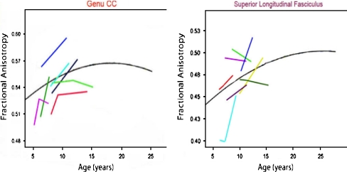
“The anterior/middle cingulate bundle supports communication relevant to high-order, attentional processing, which continues to mature into young adulthood (for review, Stiles and Jernigan, 2010).”
The Neurobiology of Childhood Structural Brain Development: Conception Through Adulthood
Houston, S. M., Herting, M. M., & Sowell, E. R. (2013) · Current Topics in Behavioral Neurosciences · doi:10.1007/7854_2013_265 · Cited by 55
“These cellular changes contribute to the change in the appearance and anatomy of the cortex, which is marked by an orderly and sequential formation of the gyri and sulci of the brain (Stiles 2008; Stiles and Jernigan 2010).”
BackgroundDevelopment and Dysgenesis of the Cerebral Cortex: Malformations of Cortical Development
Raybaud, C., & Widjaja, E. (2011) · Neuroimaging Clinics of North America · doi:10.1016/j.nic.2011.05.014 · Cited by 85
Alural, B., Genc, S., & Haggarty, S. J. (2017) · Progress in Neuro-Psychopharmacology and Biological Psychiatry · doi:10.1016/j.pnpbp.2016.03.010 · Cited by 72
“…development starts during early stages of embryogenesis, and involves proliferation and differentiation of neurons, migration of immature neurons to their ultimate brain region, outgrowth of axons and dendrites, and generation, as well as proper maintenance, of synapses (Stiles and Jernigan, 2010).”
BackgroundSnyder, H. R., Hankin, B. L., Sandman, C. A., Head, K., & Davis, E. P. (2017) · Clinical Psychological Science · doi:10.1177/2167702617714563 · Cited by 77
“Associations of psychopathology with reduced GMV during childhood could reflect any combination of reduced neurogenesis, reduced synaptic proliferation, accelerated synaptic pruning, or loss of cells (e.g., Sandman, Glynn, & Davis, 2016; Stiles & Jernigan, 2010).”
BackgroundGlycosphingolipid metabolic reprogramming drives neural differentiation
Russo, D., Ragione, F. D., Rizzo, R., Sugiyama, E., Scalabrì, F., Hori, K., Capasso, S., Sticco, L., Fioriniello, S., Gregorio, R. D., et al. (2017) · The EMBO Journal · doi:10.15252/embj.201797674 · Cited by 85
“Brain development relies on the execution of morphogenetic programmes that begin at early embryonic stages and are protracted throughout the animal life (Stiles & Jernigan, 2010).”
Investigation of brain structure in the 1-month infant
Dean, D. C., Planalp, E. M., Wooten, W., Schmidt, C. K., Kecskemeti, S. R., Frye, C., Schmidt, N. L., Goldsmith, H. H., Alexander, A. L., & Davidson, R. J. (2018) · Brain Structure and Function · doi:10.1007/s00429-017-1600-2 · Cited by 79
“Initiated in utero, neurodevelopmental processes such as neuronal pruning, synaptogenesis, and myelination operate under the direction of genetic and environmental forces to shape neural architecture and its underlying circuitry (Stiles and Jernigan 2010; Kolb and Gibb 2011).”
BackgroundArdalan, M., Chumak, T., Vexler, Z., & Mallard, C. (2019) · International Journal of Molecular Sciences · doi:10.3390/ijms20092270 · Cited by 80
“Brain development is a sexually dimorphic process that in humans begins in the third gestational week with the differentiation of neural progenitor cells and continues through to late adolescence [18].”
BackgroundImproving Human Health with Milk Fat Globule Membrane, Lactic Acid Bacteria, and Bifidobacteria
Kosmerl, E., Rocha-Mendoza, D., Ortega‐Anaya, J., Jiménez‐Flores, R., & García‐Cano, I. (2021) · Microorganisms · doi:10.3390/microorganisms9020341 · Cited by 83
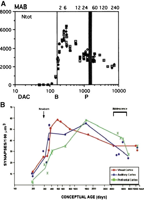
“During postnatal life, the brain rapidly undergoes changes in size, reaching 64% of its adult size within the first three months of life [115], as well as vast changes in neural networks stemming from increased exposure to enriched environments and experiences [116].”
BackgroundMahajan, A., Stavinoha, P. L., Rongthong, W., Brodin, N. P., McGovern, S. L., Naqa, I. E., Palmer, J. D., Vennarini, S., Indelicato, D. J., Aridgides, P. D., et al. (2021) · International Journal of Radiation Oncology*Biology*Physics · doi:10.1016/j.ijrobp.2020.11.073 · Cited by 83
Palaniyappan, L. K. (2017) · Neuroscience & Biobehavioral Reviews · doi:10.1016/j.neubiorev.2017.04.028 · Cited by 83
“A major contributor to the cortical shrinkage that occurs during normal development is synaptic regression or pruning (Stiles and Jernigan, 2010).”
BackgroundNeural Lineage Differentiation From Pluripotent Stem Cells to Mimic Human Brain Tissues
Hong, Y. J., & Do, J. T. (2019) · Frontiers in Bioengineering and Biotechnology · doi:10.3389/fbioe.2019.00400 · Cited by 75
Silva, M. H. (2020) · Birth Defects Research · doi:10.1002/bdr2.1661 · Cited by 83
“Tests conducted at PND 11–14 represent the peak growth period for glial cells (e.g., astrocytes) in rats (Wang & Bordey, 2008), where in humans these events (production and migration) occur prenatally (Stiles & Jernigan, 2010).”
BackgroundLocal Thyroid Hormone Action in Brain Development
Alcaide, M. A., & Mayerl, S. E. (2023) · International Journal of Molecular Sciences · doi:10.3390/ijms241512352 · Cited by 83
“5 and E10 in mice and between E19 and E29 in humans [16,17], a compartmentalization process is initiated that restricts the access to the enclosed space and the neural progenitor cells lining it.”
BackgroundIodine Deficiency and the Brain: Effects and Mechanisms
Redman, K., Ruffman, T., Fitzgerald, P. C. E., & Skeaff, S. A. (2015) · Critical Reviews in Food Science and Nutrition · doi:10.1080/10408398.2014.922042 · Cited by 83
“In the first eight weeks (known as the embryonic period), major compartments and structures are established, such as the neural tube and the ventricles (Stiles and Jernigan, 2010).”
BackgroundInfluentialStagni, F., Giacomini, A., Emili, M., Trazzi, S., Guidi, S., Sassi, M., Ciani, E., Rimondini, R., & Bartesaghi, R. (2016) · Neuroscience · doi:10.1016/j.neuroscience.2016.07.031 · Cited by 82
Garvey, M. (2018) · Open Veterinary Journal · doi:10.4314/ovj.v8i2.10 · Cited by 83
“Nerve cell myelination is one of the most important postnatal developmental events (Stiles and Jernigan, 2010); therefore, it can be assumed that autoimmunity targeting this process will have huge impact on affected individuals.”
BackgroundEarly-Emerging Sulcal Patterns Are Atypical in Fetuses with Congenital Heart Disease
Ortinau, C. M., Rollins, C. K., Gholipour, A., Yun, H. J., Marshall, M., Gagoski, B. A., Afacan, O., Friedman, K. G., Tworetzky, W., Warfield, S. K., et al. (2018) · Cerebral Cortex · doi:10.1093/cercor/bhy235 · Cited by 83
Woods, R. M., Lorusso, J. M., Potter, H. G., Neill, J. C., Glazier, J. D., & Hager, R. (2021) · Neuroscience & Biobehavioral Reviews · doi:10.1016/j.neubiorev.2021.07.015 · Cited by 81
“Importantly, neurodevelopment commences early post-conception and continues into early adulthood (Stiles and Jernigan, 2010; Tau and Peterson, 2010).”
Metadata
- Authors
- Stiles, J., & Jernigan, T. L.
- Affiliations
- University of California San Diego (US)
- Corresponding author
- Jernigan, Terry L.
- ORCID
- Jernigan, Terry L.
- Venue
- Neuropsychology Review
- Published
- 2010-11-03
- Volume
- 20
- Issue
- 4
- Pages
- 327-348
- ISSN
- 1040-7308 (ISSN-L)
- 1573-6660
- PMC
- PMC2989000
- OpenAlex
- W2115599658
- Cited by
- 2,009
- References
- 132
- Citations by year
- 2026: 142 · 2025: 184 · 2024: 199 · 2023: 209 · 2022: 184 · 2021: 197 · 2020: 195 · 2019: 155 · 2018: 158 · 2017: 122 · 2016: 92 · 2015: 64 · 2014: 49 · 2013: 24 · 2012: 23
- FWCI
- 1.43
- Type
- journal article
- Language
- en
- Open access
- hybrid
- doi.org
- Licence
- cc-by-nc
- Locations
- Neuropsychology Review (publishedVersion, cc-by-nc)
- Europe PMC (PubMed Central) (publishedVersion, cc-by-nc)
- PubMed Central (publishedVersion, cc-by-nc)
- PubMed (publishedVersion)
- eScholarship (California Digital Library) (submittedVersion, public-domain)
- eScholarship (California Digital Library) (submittedVersion)
- Funders
- Lundbeckfonden (R32‐A3161)
- H. Lundbeck A/S
- National Institutes of Health (5r01hd025077-10)
- U.S. Public Health Service (R32-A3161)
- University of California, San Diego (RC2DA029475)
- National Institute on Drug Abuse (RC2DA029475)
- National Institute of Child Health and Human Development
- Eunice Kennedy Shriver National Institute of Child Health and Human Development (R01HD060595, R01 HD 25077)
- Topics
- Life Sciences › Neuroscience › Developmental Neuroscience › Neurogenesis and neuroplasticity mechanisms
- Life Sciences › Biochemistry, Genetics and Molecular Biology › Genetics › Genetics and Neurodevelopmental Disorders
- Social Sciences › Psychology › Developmental and Educational Psychology › Child and Animal Learning Development
- Keywords
- brain development, developmental neuroscience, mammalian brain, neuronal organization, social development (developmental psychology), cognitive development, neural systems, genetic constraints, developmental mechanisms
- MeSH terms
- Animals, Brain, Cell Differentiation, Diagnostic Imaging, Humans, Nerve Net, Neural Pathways, Neurobiology, Neurons, Gene Expression Regulation, Developmental, Body Patterning, Neurogenesis
About these checks
Registry, retraction, Cites and statistics results are computed, not judged, with one exception: a printed reference that no index lists is read once by a model before it counts as a citation error. Claims are extracted by a model and kept only when their quote appears verbatim in the text; each cited source is then checked, with a model’s help, for whether it says what the claim relies on. Those verdicts can be wrong — read the claim’s quote and the cited source before relying on one.
Statistics and arithmetic are recomputed, not judged.
Confidence bands are shown only for fields calibrated on our annotated test set. None is calibrated yet, so no band is shown.
What counts as a claim ›How we measure confidence ›How we check this ›
Are you an author, or do you see a problem with this page? Request a review
Data sources
Bibliographic data: Semantic Scholar, OpenAlex, Crossref.
Contains information from the Semantic Scholar Academic Graph Datasets, made available under the ODC Attribution License (ODC-BY 1.0).
Semantic Scholar’s data is described in: Kinney, R., et al. (2023). The Semantic Scholar Open Data Platform. arXiv:2301.10140.
