

The Basics of Brain Development
Open accessThe DOI matches its Crossref record; 128 of the 132 references it cites were found in a registry or an index.
Abstract
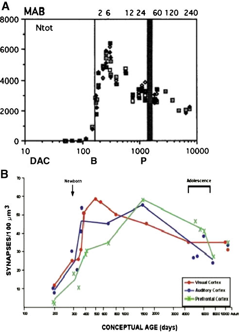
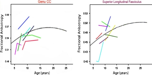
Over the past several decades, significant advances have been made in our understanding of the basic stages and mechanisms of mammalian brain development. Studies elucidating the neurobiology of brain development span the levels of neural organization from the macroanatomic, to the cellular, to the molecular. Together this large body of work provides a picture of brain development as the product of a complex series of dynamic and adaptive processes operating within a highly constrained, genetically organized but constantly changing context. The view of brain development that has emerged from the developmental neurobiology literature presents both challenges and opportunities to psychologists seeking to understand the fundamental processes that underlie social and cognitive development, and the neural systems that mediate them. This chapter is intended to provide an overview of some very basic principles of brain development, drawn from contemporary developmental neurobiology, that may be of use to investigators from a wide range of disciplines.
Overview
The claims and statistics of this paper haven’t been checked yet.
Free: it reads the open-access text and checks the first 5 claims that cite a source we can check. Create a free account to check every claim.
Integrity signals
Registry
Cites
Statistics
Figures





Verification
Claims the paper makes
The claims and statistics of this paper haven’t been checked yet.
Free: it reads the open-access text and checks the first 5 claims that cite a source we can check. Create a free account to check every claim.
Claims other papers make about this one
Each is checked against this paper’s own text: does it say what the citing paper says it does?
The source backs part of the claim, not all of it — or backs it for a different population, condition or comparison.
In the citing paper
“We now know from recent studies that it is the birth and migration of neurons derived from oRGCs that play a role in the development of the primary sulci (superior frontal, inferior frontal and precentral) in week 25–26 of gestation [23, 76].”
From Development of prefrontal cortex (2022) · cites this paper as [23]
Experimental — accuracy not yet measuredIn the citing paper
“This intricate structural-functional interplay underscores that the ontogeny of functional networks likely reflects programmed neurodevelopmental events (e.g. neurogenesis, cell death, myelination, pruning, synaptic plasticity, and glial development) (Innocenti and Price, 2005; Stiles and Jernigan, 2010)”
From Development of large-scale functional networks from birth to adulthood: A guide to the neuroimaging literature (2017) · cites this paper as Stiles and Jernigan, 2010
In the citing paper
“The brain’s major fiber pathways become consolidated through myelination, though their presence is largely established prenatally (Stiles and Jernigan, 2010).”
From Development of large-scale functional networks from birth to adulthood: A guide to the neuroimaging literature (2017) · cites this paper as Stiles and Jernigan, 2010
Self-check
Percentages, sums and reported differences re-computed from the paper’s own numbers.
The self-check needs the paper’s full text.
Citations
Cites
Philosophical transactions of the Royal Society of London. B
Press, H. (1886) · Cited by 10,080
MR diffusion tensor spectroscopy and imaging
Basser, P. J., Mattiello, J., & LeBihan, D. (1994) · Biophysical Journal · doi:10.1016/s0006-3495(94)80775-1 · Cited by 5,970
Dynamic mapping of human cortical development during childhood through early adulthood
Gogtay, N., Giedd, J. N., Lusk, L., Hayashi, K. M., Greenstein, D., Vaituzis, A. C., Nugent, T. F., Herman, D. H., Clasen, L. S., Toga, A. W., et al. (2004) · Proceedings of the National Academy of Sciences · doi:10.1073/pnas.0402680101 · Cited by 5,604
(2005) · Choice Reviews Online · doi:10.5860/choice.43-0626 · Cited by 4,674
Neurotrophins: Roles in Neuronal Development and Function
Huang, E. J., & Reichardt, L. F. (2001) · Annual Review of Neuroscience · doi:10.1146/annurev.neuro.24.1.677 · Cited by 4,394
Ferrier lecture. Functional architecture of macaque monkey visual cortex
Hubel, D. H., & Wiesel, T. N. (1977) · Proceedings of the Royal Society of London. Series B. Biological Sciences · doi:10.1098/rspb.1977.0085 · Cited by 2,608 · cited 1×
“The seminal studies of Hubel and Weisel (Hubel and Wiesel 1977; Hubel et al. 1977) showed that monocular visual deprivation in the early postnatal period can substantially alter basic patterns of organization within primary visual cortex (PVC).”
Background(1995) · Choice Reviews Online · doi:10.5860/choice.32-5931 · Cited by 2,311
Mode of cell migration to the superficial layers of fetal monkey neocortex
Rakic, P. (1972) · Journal of Comparative Neurology · doi:10.1002/cne.901450105 · Cited by 2,170 · cited 1×
“Because of the greater distances, neurons require what was originally identified as a special population of cells within the VZ called “radial glial guides” to support their migration (Rakic 1972).”
BackgroundRegional Development of the Brain in Early Life
(1968) · Archives of Disease in Childhood · doi:10.1136/adc.43.229.388 · Cited by 2,090
Neurons derived from radial glial cells establish radial units in neocortex
Noctor, S. C., Flint, A. C., Weissman, T. A., Dammerman, R. S., & Kriegstein, A. R. (2001) · Nature · doi:10.1038/35055553 · Cited by 2,016 · cited 2×
“…radial glial guides were originally thought to be a special, transient population of cells, it has recently been discovered that the cells that provide the scaffolding are actually the neural progenitor cells (Noctor et al. 2001; Noctor et al. 2002; Parnavelas et al. 2002; Weissman et al. 2003).”
Background
Research works printed but not linked: 1
- 56. Lebel, C., & Beaulieu, C. (2009). Longitudinal diffusion tensor imaging of healthy brain development in children. Honolulu: ISMRM Annual Meeting.
Cited by
Items 301–400 of 1,004
Winkler, C. W., Woods, T. A., Groveman, B. R., Carmody, A., Speranza, E. E., Martens, C., Best, S. M., Haigh, C. L., & Peterson, K. E. (2019) · Journal of Neuroinflammation · doi:10.1186/s12974-019-1614-1 · Cited by 58
The Epigenetic Role of Vitamin C in Neurodevelopment
Coker, S. J., Smith-Díaz, C. C., Dyson, R. M., Vissers, M. C. M., & Berry, M. J. (2022) · International Journal of Molecular Sciences · doi:10.3390/ijms23031208 · Cited by 58
Next generation organoid engineering to replace animals in cancer drug testing
Hockney, S., Parker, J., Turner, J. E., Todd, X., Todryk, S. M., Gieling, R. G., Hilgen, G., Simoes, D. C. M., & Pal, D. (2023) · Biochemical Pharmacology · doi:10.1016/j.bcp.2023.115586 · Cited by 59
“The brain is the most complicated organ in a vertebrate, and its development is controlled in different ways by different parts of the body [113].”
BackgroundBigler, E. D. (2015) · Neuropsychology Review · doi:10.1007/s11065-015-9290-0 · Cited by 59
“9 shows an overall reduction in cortical thickness with age from 7 to 16, inferred from cross-sectional data and thought to reflect neuronal pruning associated with maturation (Stiles and Jernigan 2010).”
BackgroundSustained attention and prediction: distinct brain maturation trajectories during adolescence
Thillay, A., Roux, S., Gissot, V., Carteau‐Martin, I., Knight, R. T., Bonnet‐Brilhault, F., & Bidet‐Caulet, A. (2015) · Frontiers in Human Neuroscience · doi:10.3389/fnhum.2015.00519 · Cited by 58
“…synaptic pruning and dopaminergic innervation of the frontal cortex, enhanced communication between fronto-parietal and fronto-temporal networks, and pubertal hormone changes that can alter brain function or neurotransmitter activity (e.g., Giorgio et al., 2010; Stiles and Jernigan, 2010).”
BackgroundChanges in Visual Object Recognition Precede the Shape Bias in Early Noun Learning
Yee, M., Jones, S. S., & Smith, L. B. (2012) · Frontiers in Psychology · doi:10.3389/fpsyg.2012.00533 · Cited by 58
“As is the case in developmental process more generally (Blumberg, 2006; Stiles, 2008; Stiles and Jernigan, 2010; Sheya and Smith, 2010), the pathways may be complex, multi-causal, possibly bi-directional, and redundant such that there are multiple routes to the same end.”
BackgroundThe Neurobiology of Childhood Structural Brain Development: Conception Through Adulthood
Houston, S. M., Herting, M. M., & Sowell, E. R. (2013) · Current topics in behavioral neurosciences · doi:10.1007/978-3-662-45758-0_265 · Cited by 58
In Vitro Models for Neurogenesis
Azari, H., & Reynolds, B. A. (2015) · Cold Spring Harbor Perspectives in Biology · doi:10.1101/cshperspect.a021279 · Cited by 57
Building brains in a dish: Prospects for growing cerebral organoids from stem cells
Mason, J. O., & Price, D. J. (2016) · Neuroscience · doi:10.1016/j.neuroscience.2016.07.048 · Cited by 57
“Within each layer, 155 neurons tend to share similar patterns of gene expres156 sion, afferent and efferent connectivity and function 157 across species (Stiles and Jernigan, 2010).”
UNPACKING THE BURDEN OF CARE FOR INFANTS IN THE NICU
D’Agata, A. L., Sanders, M. R., Grasso, D. J., Young, E. E., Cong, X. S., & McGrath, J. M. (2017) · Infant Mental Health Journal · doi:10.1002/imhj.21636 · Cited by 57
Kwon, D., Pfefferbaum, A., Sullivan, E. V., & Pohl, K. M. (2018) · Brain Imaging and Behavior · doi:10.1007/s11682-018-9980-3 · Cited by 57
“2008) and functional maturation (Fjell et al. 2012; Muller-Oehring et al. 2017; Stiles and Jernigan 2010) marked the continuation of neurodevelopment throughout adolescence and young adulthood and, as our analysis showed, includes cortical myelin and its functional ramifications in temporal, spatial, and density growt…”
InfluentialWu, W. D., Zhao, A., Liu, B., Ye, W., Su, H.-W., Li, J., & Zhang, Y. (2021) · Nutrients · doi:10.3390/nu13020693 · Cited by 57
“Coinciding with this critical window of gut microbiota shaping, the first years postnatally also represent a crucial time of early brain development [30], when several foundational processes are simultaneously occurring in the brain, including proliferation, apoptosis of the glia, myelination of the axons, and synapto…”
BackgroundNeurological abnormality detection from electroencephalography data: a review
Alvi, A. M., Siuly, S., & Wang, H. (2021) · Artificial Intelligence Review · doi:10.1007/s10462-021-10062-8 · Cited by 57
Curran, E. A., O’Keeffe, G. W., Looney, A.-M., Moloney, G. M., Hegarty, S. V., Murray, D. M., Khashan, A. S., & Kenny, L. C. (2017) · Molecular Neurobiology · doi:10.1007/s12035-017-0794-x · Cited by 57
“This is suggestive of a potential dysregulation of brain development at prenatal developmental stages, as cortical lamination is ongoing during the second trimester of pregnancy [8].”
Pediatric Stroke: Unique Implications of the Immature Brain on Injury and Recovery
Malone, L. A., & Felling, R. J. (2019) · Pediatric Neurology · doi:10.1016/j.pediatrneurol.2019.06.016 · Cited by 57
From Progenitors to Progeny: Shaping Striatal Circuit Development and Function
Knowles, R., Dehorter, N., & Ellender, T. J. (2021) · Journal of Neuroscience · doi:10.1523/jneurosci.0620-21.2021 · Cited by 55
“After closure of the neural tube, distinct rostral, medial, and caudal regions develop to ultimately give rise to the frontal, middle, and hindbrain regions of the brain (Stiles and Jernigan, 2010).”
BackgroundBinter, A.-C., Kusters, M. S. W., van den Dries, M. A., Alonso, L., Lubczynska, M. J., Hoek, G., White, T. J. H., Íñiguez, C., Tiemeier, H. W., & Guxens, M. (2022) · Environmental Pollution · doi:10.1016/j.envpol.2022.120109 · Cited by 55
Probing disorders of the nervous system using reprogramming approaches
Ichida, J. K., & Kiskinis, E. (2015) · The EMBO Journal · doi:10.15252/embj.201591267 · Cited by 50
“During development, early neural progenitors produce neurons whereas late progenitors differentiate into astrocytes (Stiles & Jernigan, 2010). iPSC-directed differentiation recapitulates this developmental process.”
Golgi trafficking defects in postnatal microcephaly: The evidence for “Golgipathies”
Passemard, S., Perez, F., Colin, E., Rasika, S., Gressèns, P., & Ghouzzi, V. E. (2017) · Progress in Neurobiology · doi:10.1016/j.pneurobio.2017.03.007 · Cited by 56
“In line with this prolonged role of glial cells, gliogenesis, though very active 5 around 32-40 weeks of gestation, largely continues after birth, especially during the first two 6 years of life (Stiles and Jernigan, 2010).”
Organs-on-a-chip: A new paradigm for toxicological assessment and preclinical drug development
Bovard, D., Iskandar, A., Luettich, K., Hoeng, J., & Peitsch, M. C. (2017) · Toxicology Research and Application · doi:10.1177/2397847317726351 · Cited by 35
“Prior to this, brain cells reorganize and make new connections essential for learning.(76) Any compound crossing the BBB and affecting brain cell maturation could induce permanent injuries, which poses a risk to the normal brain development of children and adolescents.”
BackgroundThe Association of Maternal Thyroid Autoimmunity During Pregnancy With Child IQ
Derakhshan, A., Korevaar, T. I. M., Taylor, P. N., Levie, D., Guxens, M., Jaddoe, V. W. V., Nelson, S. M., Tiemeier, H. W., & Peeters, R. P. (2018) · The Journal of Clinical Endocrinology & Metabolism · doi:10.1210/jc.2018-00743 · Cited by 56
Synthetic cannabinoids and their impact on neurodevelopmental processes
Alexandre, J., Carmo, H., Carvalho, F. D., & Silva, J. P. (2019) · Addiction Biology · doi:10.1111/adb.12824 · Cited by 55
“As the axon extends, it scans the local environment to find attractive or repulsive molecules.(37) Berghuis et al showed that cannabinoids play an important role in the initial growth of axons through autocrine signaling, while target‐derived endocannabinoid signals were shown to control axonal navigation and position…”
BackgroundInfluentialRodríguez‐Ayllón, M., Derks, I. P. M., van den Dries, M. A., Esteban‐Cornejo, I., Labrecque, J. A., Yang‐Huang, J., Raat, H., Vernooij, M. W., White, T. J. H., Ortega, F. B., et al. (2019) · NeuroImage · doi:10.1016/j.neuroimage.2019.116258 · Cited by 58
“Numerous studies have demonstrated that developmental changes in white matter microstructure occur throughout childhood (Schmithorst and Yuan, 2010; Stiles and Jernigan, 2010), and the technique has been shown to be sensitive to subtle features of psychopathology (Muetzel et al., 2017).”
BackgroundCellular complexity in brain organoids: Current progress and unsolved issues
Mansour, A. A., Schafer, S. T., & Gage, F. H. (2020) · Seminars in Cell and Developmental Biology · doi:10.1016/j.semcdb.2020.05.013 · Cited by 55
Raj, N., McEachin, Z. T., Harousseau, W., Zhou, Y., Zhang, F., Merritt-Garza, M. E., Taliaferro, J. M., Kalinowska, M., Marro, S. G., Hales, C. M., et al. (2021) · Cell Reports · doi:10.1016/j.celrep.2021.108991 · Cited by 54
“The translation of mRNAs is a tightly regulated process that is essential to cellular development and function (Stiles and Jernigan, 2010).”
On Expression Patterns and Developmental Origin of Human Brain Regions
Lior, K., & Chechik, G. (2016) · PLoS Computational Biology · doi:10.1371/journal.pcbi.1005064 · Cited by 56
“These five vesicles are aligned along the rostral-caudal axis of the developing brain and establish the primary organization of the central nervous system (Fig 1A) [7].”
BackgroundInfant gut microbiota contributes to cognitive performance in mice
Cerdó, T., Ruiz-Rodriguez, A., Acuña, I., Torres-Espínola, F. J., Menchén-Márquez, S., Gámiz, F., Gallo, M., Jehmlich, N., Haange, S., von Bergen�, M., et al. (2023) · Cell Host & Microbe · doi:10.1016/j.chom.2023.11.004 · Cited by 54
Meo, S. A., Shaikh, N., Alotaibi, M. S., Alwabel, A. A., & Alqumaidi, H. (2024) · Scientific Reports · doi:10.1038/s41598-024-70646-6 · Cited by 55
“The brain significantly increases in size during pre-school; by age 6, it reaches about 90% of its adult volume 34 .”
BackgroundInfluentialMohamed, R. S., Pettifor, J. M., & Norris, S. A. (2017) · American Journal of Physical Anthropology · doi:10.1002/ajpa.23340 · Cited by 54
Cortical Dysmaturation in Congenital Heart Disease
Leonetti, C., Back, S. A., Gallo, V., & Ishibashi, N. (2019) · Trends in Neurosciences · doi:10.1016/j.tins.2018.12.003 · Cited by 54
“Although cortical neurogenesis at fetal and adult stages has been widely studied and reviewed in detail (for review, see [12,13]), the development of the frontal cortex during the perinatal period, particularly in humans, has only recently received greater attention due to methodological advances and the identificatio…”
BackgroundBrain-on-a-chip Devices for Drug Screening and Disease Modeling Applications
Miccoli, B., Braeken, D., & Li, Y. E. (2019) · Current Pharmaceutical Design · doi:10.2174/1381612825666190220161254 · Cited by 54
Lindeman, B., Johansson, Y., Andreassen, M., Husøy, T., Dirven, H. A. A. M., Hofer, T., Knutsen, H. K., Caspersen, I. H., Vejrup, K., Paulsen, R. E., et al. (2021) · Reproductive Toxicology · doi:10.1016/j.reprotox.2021.02.006 · Cited by 54
Makrygianni, E. A., & Chrousos, G. P. (2021) · Frontiers in Physiology · doi:10.3389/fphys.2021.621970 · Cited by 55
“…different number of layers, dependent on CNS topology and specialization e.g., the medulla, the cerebral cortex and the optic tectum (superior colliculus) have three, six and seven layers, respectively (Stiles and Jernigan, 2010; Lancaster and Knoblich, 2014; Clevers, 2016; Agirman et al., 2017).”
BackgroundBahl, V., Weng, N. J.-H., Schick, S. F., Sleiman, M., Whitehead, J., Ibarra, A. N., & Talbot, P. (2015) · Toxicological Sciences · doi:10.1093/toxsci/kfv327 · Cited by 54
Zou, R., Boer, O. D., Felix, J. F., Muetzel, R. L., Franken, I. H. A., Cecil, C. A. M., & El Marroun, H. (2022) · JAMA Network Open · doi:10.1001/jamanetworkopen.2022.24701 · Cited by 53
Dönertaş, H. M., İzgi, H., Kamacıoğlu, A., He, Z., Khaitovich, P., & Somel, M. (2017) · Scientific Reports · doi:10.1038/s41598-017-05927-4 · Cited by 54
“For the transcription factor binding site (TFBS) determination (1) +/−2000 base pairs of the transcription start site for each gene was extracted using Ensembl annotations, (2) within these sequences, transcription factor binding sites were predicted using the TRANSFAC database and Match algorithm61, (3) for each TFBS…”
MethodGenetic influences on neonatal cortical thickness and surface area
Jha, S. C., Xia, K., Schmitt, J. E., Ahn, M., Girault, J. B., Murphy, V. A., Li, G., Wang, L., Shen, D. G., Zou, F., et al. (2018) · Human Brain Mapping · doi:10.1002/hbm.24340 · Cited by 53
“…are likely involved in neurogenesis, proliferation, and migration of neuronal cell types and genes expressed at high levels during late fetal and early postnatal development likely reflect neuronal and glial differentiation and the robust growth of dendrites and synapses (Stiles & Jernigan, 2010).”
BackgroundZika Virus as a Possible Risk Factor for Autism Spectrum Disorder: Neuroimmunological Aspects
Vianna, P., Gomes, J. D. A., Boquett, J. A., Fraga, L. R., Schuch, J. B., Vianna, F. S. L., & Schüler‐Faccini, L. (2018) · NeuroImmunoModulation · doi:10.1159/000495660 · Cited by 53
Neuroimmunology of the female brain across the lifespan: Plasticity to psychopathology
Barrientos, R. M., Brunton, P. J., Lenz, K. M., Pyter, L. M., & Spencer, S. J. (2019) · Brain Behavior and Immunity · doi:10.1016/j.bbi.2019.03.010 · Cited by 53
(2020) · International Review of Neurobiology · doi:10.1016/bs.irn.2019.12.001 · Cited by 46
Antenatal and Perioperative Mechanisms of Global Neurological Injury in Congenital Heart Disease
Barkhuizen, M., Abella, R. F., Vles, J. S. H., Zimmermann, L. J. I., Gazzolo, D., & Gavilanes, A. W. D. (2020) · Pediatric Cardiology · doi:10.1007/s00246-020-02440-w · Cited by 54
“The brain size only reaches 90% of the adult brain volume by age 6 [72].”
BackgroundO’Shaughnessy, K. L., Fischer, F., & Zenclussen, A. C. (2021) · Best Practice & Research Clinical Endocrinology & Metabolism · doi:10.1016/j.beem.2021.101568 · Cited by 53
Prenatal Stress, Mood, and Gray Matter Volume in Young Adulthood
Marečková, K., Klasnja, A., Bencurova, P., Andrýsková, L., Brázdil, M., & Paus, T. (2019) · Cerebral Cortex · doi:10.1093/cercor/bhy030 · Cited by 52
Targeted drug delivery in neurodegenerative diseases: the role of nanotechnology
Dhariwal, R., Jain, M., Mir, Y. R., Singh, A., Jain, B., Kumar, P., Tariq, M., Verma, D., Deshmukh, K., Yadav, V. K., et al. (2025) · Frontiers in Medicine · doi:10.3389/fmed.2025.1522223 · Cited by 54
“Despite these efforts, many patients remain undiagnosed or face misdiagnoses (50, 51).”
Developmental Exposure to Environmental Toxicants
Falck, A. J., Mooney, S. M., Kapoor, S. S., White, K. M. R., Bearer, C. F., & El‐Metwally, D. (2015) · Pediatric Clinics of North America · doi:10.1016/j.pcl.2015.05.005 · Cited by 53
“Neuronal cell division is complete by 6months, but migration, differentiation, andmyelination continue into adolescence.(14) A newborn’s body fat composition affects distribution of lipid-soluble toxicants.”
BackgroundIntrauterine Viral Infections: Impact of Inflammation on Fetal Neurodevelopment
Ganguli, S., & Chavali, P. L. (2021) · Frontiers in Neuroscience · doi:10.3389/fnins.2021.771557 · Cited by 53
“Neurodevelopment is a complex developmental process that begins during the third week of gestation and continues postnatally until adulthood (Stiles and Jernigan, 2010).”
BackgroundGlia-Driven Brain Circuit Refinement Is Altered by Early-Life Adversity: Behavioral Outcomes
Milbocker, K. A., Campbell, T. S., Collins, N. J., Kim, S., Smith, E. A., Roth, T. L., & Klintsova, A. Y. (2021) · Frontiers in Behavioral Neuroscience · doi:10.3389/fnbeh.2021.786234 · Cited by 52
“Redundant synapses are phagocytosed during the phase of synaptic pruning and putative circuits remain (Stiles and Jernigan, 2010; Faust et al., 2021).”
Cattarinussi, G., Bellani, M., Maggioni, E., Sambataro, F., Brambilla, P., & Delvecchio, G. (2022) · Journal of Affective Disorders · doi:10.1016/j.jad.2022.05.055 · Cited by 52
Auderset, L., Landowski, L. M., Foa, L., & Young, K. M. (2016) · Stem Cells International · doi:10.1155/2016/2108495 · Cited by 49
“Neuroblast generation and their subsequent migration into the developing cortex has been well characterised [112].”
Peterson, E. T., Kwon, D., Luna, B., Larsen, B., Prouty, D., De Bellis, M. D., Voyvodic, J., Liu, C., Li, W., Pohl, K. M., et al. (2019) · Human Brain Mapping · doi:10.1002/hbm.24461 · Cited by 48
Early Actions of Neurotransmitters During Cortex Development and Maturation of Reprogrammed Neurons
Ojeda, J., & Ávila, A. (2019) · Frontiers in Synaptic Neuroscience · doi:10.3389/fnsyn.2019.00033 · Cited by 51
Vitamin B12 - Do You Know Everything?
Butola, L. K., Kute, P. K., Anjankar, A. P., Dhok, A., Gusain, N., & Vagga, A. A. (2020) · Journal of Evolution of Medical and Dental Sciences · doi:10.14260/jemds/2020/688 · Cited by 51
Fan, C. C., Loughnan, R. J., Makowski, C., Pecheva, D., Chen, C., Hagler, D. J., Thompson, W. K., Parker, N., van der Meer, D., Frei, O., et al. (2022) · Nature Communications · doi:10.1038/s41467-022-30110-3 · Cited by 51
Retinal biomarkers provide “insight” into cortical pharmacology and disease
Nguyen, C. T. O., Hui, F., Charng, J., Velaedan, S., van Koeverden, A. K., Lim, J. K. H., He, Z., Wong, V. H. Y., Vingrys, A. J., Bui, B. V., et al. (2017) · Pharmacology & Therapeutics · doi:10.1016/j.pharmthera.2017.02.009 · Cited by 51
The neurobiology of executive functions
Hunter, S. J., Hinkle, C. D., & Edidin, J. P. (2012) · Cambridge University Press eBooks · doi:10.1017/cbo9780511977954.005 · Cited by 49
Dynamic gray matter volume changes in pediatric multiple sclerosis: A 3.5 year MRI study
Meo, E. D., Meani, A., Moiola, L., Ghezzi, A., Veggiotti, P., Filippi, M., & Rocca, M. A. (2019) · Neurology · doi:10.1212/wnl.0000000000007267 · Cited by 49
Beauregard, J. L., Drews‐Botsch, C., Sales, J. M., Flanders, W. D., & Kramer, M. R. (2018) · American Journal of Epidemiology · doi:10.1093/aje/kwy068 · Cited by 49
“In the first months and years of children’s development, sensory enrichment and deprivation have important influences on brain growth and development as children are experiencing, responding to, and learning from stimuli in their environments (8).”
InfluentialBerre, A. L., Pitel, A., Chanraud, S., Beaunieux, H., Eustache, F., Martinot, J. L., Reynaud, M., Martelli, C., Rohlfing, T., Sullivan, E. V., et al. (2014) · Human Brain Mapping · doi:10.1002/hbm.22500 · Cited by 48
“, 2012], when the brain is still undergoing maturation [Stiles and Jernigan, 2010].”
BackgroundJeon, T., Mishra, V. R., Ouyang, M., Chen, M., & Huang, H. (2015) · Frontiers in Neuroanatomy · doi:10.3389/fnana.2015.00158 · Cited by 49
“WM axons undergo dramatic changes during development, including axonal packing and myelination (Yakovlev and Lecours, 1967; Stiles and Jernigan, 2010).”
BackgroundEminian, S., Hajdu, S. D., Meuli, R. A., Maeder, P. P., & Hagmann, P. (2018) · PLoS ONE · doi:10.1371/journal.pone.0198250 · Cited by 49
Brain age prediction: Cortical and subcortical shape covariation in the developing human brain
Zhao, Y., Klein, A., Castellanos, F. X., & Milham, M. P. (2019) · NeuroImage · doi:10.1016/j.neuroimage.2019.116149 · Cited by 48
Mapping brain mechanical property maturation from childhood to adulthood
McIlvain, G., Schneider, J. M., Matyi, M. A., McGarry, M., Qi, Z., Spielberg, J. M., & Johnson, C. L. (2022) · NeuroImage · doi:10.1016/j.neuroimage.2022.119590 · Cited by 47
Modeling Neurodevelopmental Disorders Using Human Pluripotent Stem Cells
Telias, M., & Ben‐Yosef, D. (2014) · Stem Cell Reviews and Reports · doi:10.1007/s12015-014-9507-2 · Cited by 47
“[ 18 1] C or tic al T U J1 ,M A P2 ,T A U T B R 1, C U X 1, B R N 2,”
Bytoft, B., Knorr, S., Vlachova, Z., Jensen, R. B., Mathiesen, E. R., Beck‐Nielsen, H., Gravholt, C. H., Jensen, D. M., Clausen, T. D., Mortensen, E. L., et al. (2016) · Diabetes Care · doi:10.2337/dc16-0168 · Cited by 47
Winkler, C. W., Woods, T. A., Rosenke, R., Scott, D. P., Best, S. M., & Peterson, K. E. (2017) · Scientific Reports · doi:10.1038/s41598-017-07099-7 · Cited by 47
Winje, B. A., Kvestad, I., Krishnamachari, S., Manji, K. P., Taneja, S., Bellinger, D. C., Bhandari, N., Bisht, S., Darling, A. M., Duggan, C., et al. (2018) · BMJ Open · doi:10.1136/bmjopen-2017-018962 · Cited by 47
“The production of myelin is a key component of brain development from gestation, throughout childhood and well into middle age.(10) The myelination of the brain is of importance for multiple brain systems and is highly related to neurodevelopment and subsequent cognitive functioning.”
BackgroundYang, G., & Shcheglovitov, A. (2019) · Developmental Dynamics · doi:10.1002/dvdy.100 · Cited by 47
Tooley, U. A., Bassett, D. S., & Mackey, A. P. (2021) · NeuroImage · doi:10.1016/j.neuroimage.2021.118843 · Cited by 47
Zhen, H., Zhang, F., Cheng, H., Hu, F., Jia, Y., Hou, Y., Shang, M., Yu, H., & Jiang, M. (2023) · Ecotoxicology and Environmental Safety · doi:10.1016/j.ecoenv.2023.114770 · Cited by 47
Covalent growth factor tethering to direct neural stem cell differentiation and self-organization
Ham, T. R., Farrag, M., & Leipzig, N. D. (2017) · Acta Biomaterialia · doi:10.1016/j.actbio.2017.01.068 · Cited by 46
“Self-organization plays a large role in normal CNS development.[44] However, as 570”
BackgroundCraniofacial skeletal response to encephalization: How do we know what we think we know?
Lesciotto, K. M., & Richtsmeier, J. T. (2019) · American Journal of Physical Anthropology · doi:10.1002/ajpa.23766 · Cited by 47
“Although the development, differentiation, and refinement of the brain proceeds along a gradual and continuous pathway throughout the first two decades of postnatal life (Stiles & Jernigan, 2010), the details of prenatal development of the brain provide the map for all future development.”
BackgroundInfluentialControl networks of the frontal lobes
Marek, S. A., & Dosenbach, N. U. F. (2019) · Handbook of clinical neurology · doi:10.1016/b978-0-12-804281-6.00018-5 · Cited by 48
Kaczkurkin, A. N., Moore, T. M., Sotiras, A., Xia, C. H., Shinohara, R. T., & Satterthwaite, T. D. (2019) · Biological Psychiatry · doi:10.1016/j.biopsych.2019.12.015 · Cited by 46
“It is also a time of marked development in brain structure and function (3, 4).”
BackgroundLi, H., Qu, N., Hui, L., Cai, X. M., Zhang, C., Zhong, B., Zhang, S.-F., Chen, J., Xia, B., Wang, L., et al. (2020) · Translational Psychiatry · doi:10.1038/s41398-020-0777-y · Cited by 46
Delagneau, G., Twilhaar, E. S., Testa, R., van Veen, S., & Anderson, P. J. (2023) · Child Development · doi:10.1111/cdev.13885 · Cited by 46
Li, J., Settivari, R. S., LeBaron, M. J., & Marty, M. S. (2019) · NeuroToxicology · doi:10.1016/j.neuro.2019.02.010 · Cited by 45
Parenting with eyes wide open: Young gifted children, early entry and social isolation
Wellisch, M. (2020) · Gifted Education International · doi:10.1177/0261429419899946 · Cited by 45
“Maternal depression and the process of attachment The developing brain of a newborn child takes place within a complex interaction of simultaneous genetic and environmental influences and events that include the cellular and molecular levels (Stiles and Jernigan, 2010).”
BackgroundChoroid Plexus: The Orchestrator of Long-Range Signalling Within the CNS
Kaiser, K., & Bryja, V. (2020) · International Journal of Molecular Sciences · doi:10.3390/ijms21134760 · Cited by 45
“Neuroepithelium consisting of neuronal progenitors, which segregate to the ventricular zone upon neural tube closure and concomitant ventricular system formation, represent the chief source of cells that will give rise to the entire CNS [169].”
BackgroundHuman neural organoids: Models for developmental neurobiology and disease
Guy, B., Zhang, J. S., Duncan, L. H., & Johnston, R. J. (2021) · Developmental Biology · doi:10.1016/j.ydbio.2021.06.012 · Cited by 45
Topal, Z., Tufan, A. E., Karadağ, M., Gökçen, C., Akkaya, C., Sarp, A. S., Bahşi, İ., & Kilinc, M. (2021) · Nordic Journal of Psychiatry · doi:10.1080/08039488.2021.1946712 · Cited by 45
Emerging Roles of RNA-Binding Proteins in Neurodevelopment
Parra, A. S., & Johnston, C. A. (2022) · Journal of Developmental Biology · doi:10.3390/jdb10020023 · Cited by 45
“This process requires accurate and intricate signaling between tissue environments, cells, and proteins [18].”
BackgroundThrush, K., Bennett, D. A., Gaiteri, C., Horvath, S., van Dyck, C. H., Higgins‐Chen, A., & Levine, M. E. (2022) · Aging · doi:10.18632/aging.204196 · Cited by 45
“However, blood or pan-tissue trained clocks may not adequately capture brain aging, due to the brain’s unique methylation profile [48–50], extreme diversity of specialized neuronal [51] and glial [52] cell types, and distinct developmental patterns [53].”
BackgroundCheng, Q., Roth, A., Halgren, E., Klein, D., Chen, J., & Mayberry, R. I. (2023) · Proceedings of the National Academy of Sciences · doi:10.1073/pnas.2215423120 · Cited by 47
“The relation between cortical thickness/volume and biological age is nonlinear, with an initial increase during the proliferation/synaptogenesis stage, followed by a decrease during the pruning stage (42) and a gradual decrease stabilizing at around 30 y (43).”
BackgroundBigler, E. D., Yeates, K. O., Dennis, M., Gerhardt, C. A., Rubin, K. H., Stancin, T., Taylor, H. G., & Vannatta, K. (2013) · NeuroRehabilitation · doi:10.3233/nre-130896 · Cited by 37
“Complex gene-brain-environment interactions shape emotional and social behavior [6–8].”
An Implantable Micro-Caged Device for Direct Local Delivery of Agents
Son, A. I., Opfermann, J. D., McCue, C., Ziobro, J. M., Abrahams, J. H., Jones, K., Morton, P. D., Ishii, S., Oluigbo, C. O., Krieger, A., et al. (2017) · Scientific Reports · doi:10.1038/s41598-017-17912-y · Cited by 45
Prenatal CRH: An integrating signal of fetal distress
Sandman, C. A. (2018) · Development and Psychopathology · doi:10.1017/s0954579418000664 · Cited by 45
“Radial neuronal cell migration begins in the human brain around 42 days gestational age (Stiles & Jernigan, 2010; Takahasi, Folkerth, Galaburda, & Grant, 2012).”
BackgroundBetween Dysbiosis, Maternal Immune Activation and Autism: Is There a Common Pathway?
Suprunowicz, M., Tomaszek, N., Urbaniak, A., Zackiewicz, K., Modzelewski, S., & Waszkiewicz, N. (2024) · Nutrients · doi:10.3390/nu16040549 · Cited by 44
“Inflammation during pregnancy possibly affects the proper proliferation and migration of neurons and glia, formation of synapses, myelination, and establishment of neuronal circuits [170,171].”
BackgroundEffects of small molecules on neurogenesis: Neuronal proliferation and differentiation
Jastrzębski, M. K., Wójcik, P., Stępnicki, P., & Kaczor, A. A. (2023) · Acta Pharmaceutica Sinica B · doi:10.1016/j.apsb.2023.10.007 · Cited by 44
Sasidharan, V., Marepally, S., Elliott, S. A., Baid, S., Lakshmanan, V., Nayyar, N., Bansal, D., Sánchez Alvarado, A., Vemula, P. K., & Palakodeti, D. (2017) · Development · doi:10.1242/dev.144758 · Cited by 42
“This process involves the precise guidance of axons from the neuron to its targets, which, in most animals, occurs during prenatal and early postnatal development (Tessier-Lavigne and Goodman, 1996; Stiles and Jernigan, 2010).”
BackgroundDevelopment of the Cell Population in the Brain White Matter of Young Children
Sigaard, R. K., Kjær, M., & Pakkenberg, B. (2016) · Cerebral Cortex · doi:10.1093/cercor/bhu178 · Cited by 43
Graph Random Forest: A Graph Embedded Algorithm for Identifying Highly Connected Important Features
Tian, L., Wu, W., & Yu, T. (2023) · Biomolecules · doi:10.3390/biom13071153 · Cited by 43
“The fetal brain begins to develop from the third week of gestation [35], neural precursor cells divide and form neurons and glia.”
BackgroundHypoxia and connectivity in the developing vertebrate nervous system
Bonkowsky, J. L., & Son, J. (2018) · Disease Models & Mechanisms · doi:10.1242/dmm.037127 · Cited by 43
“…births from 24-36 weeks PCA, and includes the time when axon and synaptic connections are forming in the human CNS (ten Donkelaar et al., 2004; Ren et al., 2006; Kostovic ́ and Jovanov-Milosevic ́ , 2006; Molyneaux et al., 2007; Stiles and Jernigan, 2010; Vasung et al., 2010; Semple et al., 2013).”
Dynamic N6-Methyladenosine RNA Methylation in Brain and Diseases
Shafik, A. M., Allen, E. G., & Jin, P. (2020) · Epigenomics · doi:10.2217/epi-2019-0260 · Cited by 43
Rechtman, E., Curtin, P. C. P., Papazaharias, D. M., Renzetti, S., Cagna, G., Peli, M., Levin‐Schwartz, Y., Placidi, D., Smith, D. R., Lucchini, R. G., et al. (2020) · Translational Psychiatry · doi:10.1038/s41398-020-01041-8 · Cited by 43
Elp2 mutations perturb the epitranscriptome and lead to a complex neurodevelopmental phenotype
Kojic, M., Gawda, T., Gaik, M., Begg, A., Salerno-Kochan, A., Kurniawan, N. D., Jones, A., Drożdżyk, K., Kościelniak, A., Chramiec‐Głąbik, A., et al. (2021) · Nature Communications · doi:10.1038/s41467-021-22888-5 · Cited by 43
Pérez‐Crespo, L., Kusters, M. S. W., López-Vicente, M., Lubczynska, M. J., Foraster, M., White, T. J. H., Hoek, G., Tiemeier, H. W., Muetzel, R. L., & Guxens, M. (2022) · Environment International · doi:10.1016/j.envint.2022.107275 · Cited by 43
Non-neural Muscle Weakness Has Limited Influence on Complexity of Motor Control during Gait
Goudriaan, M., Shuman, B. R., Steele, K. M., van den Hauwe, M., Goemans, N., Molenaers, G., & Desloovere, K. (2018) · Frontiers in Human Neuroscience · doi:10.3389/fnhum.2018.00005 · Cited by 43
“…on the motor cortex responsible for the distal muscle groups of the lower limb, is closer to the phylogenetic older parts of the brain (Volpe, 2000; Stiles and Jernigan, 2010) and it has been suggested that the older regions of the motor cortex are involved in synergy regulation (Bizzi and Cheung,…”
BackgroundSequential Anomaly Detection Under Sampling Constraints
Tsopelakos, A., & Fellouris, G. (2022) · IEEE Transactions on Information Theory · doi:10.1109/tit.2022.3177142 · Cited by 42
“In brain science, for example, it is desirable to identify groups of cells with large vibration frequency, as this is a symptom for the development of a particular malfunctioning [1].”
BackgroundFrom Womb to Mind: Prenatal Epigenetic Influences on Mental Health Disorders
Álvarez-Mejía, D., Rodas, J. A., & León-Rojas, J. E. (2025) · International Journal of Molecular Sciences · doi:10.3390/ijms26136096 · Cited by 43
“Early gestation is when fundamental neurodevelopmental events (neural tube formation, neuroprogenitor cell proliferation, initial brain patterning) take place [69].”
The impacts of diabetes in pregnancy on hippocampal synaptogenesis in rat neonates
Vafaei‐Nezhad, S., Hami, J., Sadeghi, A., Ghaemi, K., Hosseini, M., Abedini, M. R., & Haghir, H. (2016) · Neuroscience · doi:10.1016/j.neuroscience.2016.01.025 · Cited by 42
“Synaptogenesis is one of the most important events taking place during the development of the CNS (Bury and Sabo 2015; Stiles and Jernigan 2010; Tau and Peterson 2010; Bourgeois 1997; Huttenlocher and Dabholkar 1997).”
Metadata
- Authors
- Stiles, J., & Jernigan, T. L.
- Affiliations
- University of California San Diego (US)
- Corresponding author
- Jernigan, Terry L.
- ORCID
- Jernigan, Terry L.
- Venue
- Neuropsychology Review
- Published
- 2010-11-03
- Volume
- 20
- Issue
- 4
- Pages
- 327-348
- ISSN
- 1040-7308 (ISSN-L)
- 1573-6660
- PMC
- PMC2989000
- OpenAlex
- W2115599658
- Cited by
- 2,009
- References
- 132
- Citations by year
- 2026: 142 · 2025: 184 · 2024: 199 · 2023: 209 · 2022: 184 · 2021: 197 · 2020: 195 · 2019: 155 · 2018: 158 · 2017: 122 · 2016: 92 · 2015: 64 · 2014: 49 · 2013: 24 · 2012: 23
- FWCI
- 1.43
- Type
- journal article
- Language
- en
- Open access
- hybrid
- doi.org
- Licence
- cc-by-nc
- Locations
- Neuropsychology Review (publishedVersion, cc-by-nc)
- Europe PMC (PubMed Central) (publishedVersion, cc-by-nc)
- PubMed Central (publishedVersion, cc-by-nc)
- PubMed (publishedVersion)
- eScholarship (California Digital Library) (submittedVersion, public-domain)
- eScholarship (California Digital Library) (submittedVersion)
- Funders
- Lundbeckfonden (R32‐A3161)
- H. Lundbeck A/S
- National Institutes of Health (5r01hd025077-10)
- U.S. Public Health Service (R32-A3161)
- University of California, San Diego (RC2DA029475)
- National Institute on Drug Abuse (RC2DA029475)
- National Institute of Child Health and Human Development
- Eunice Kennedy Shriver National Institute of Child Health and Human Development (R01HD060595, R01 HD 25077)
- Topics
- Life Sciences › Neuroscience › Developmental Neuroscience › Neurogenesis and neuroplasticity mechanisms
- Life Sciences › Biochemistry, Genetics and Molecular Biology › Genetics › Genetics and Neurodevelopmental Disorders
- Social Sciences › Psychology › Developmental and Educational Psychology › Child and Animal Learning Development
- Keywords
- brain development, developmental neuroscience, mammalian brain, neuronal organization, social development (developmental psychology), cognitive development, neural systems, genetic constraints, developmental mechanisms
- MeSH terms
- Animals, Brain, Cell Differentiation, Diagnostic Imaging, Humans, Nerve Net, Neural Pathways, Neurobiology, Neurons, Gene Expression Regulation, Developmental, Body Patterning, Neurogenesis
About these checks
Registry, retraction, Cites and statistics results are computed, not judged, with one exception: a printed reference that no index lists is read once by a model before it counts as a citation error. Claims are extracted by a model and kept only when their quote appears verbatim in the text; each cited source is then checked, with a model’s help, for whether it says what the claim relies on. Those verdicts can be wrong — read the claim’s quote and the cited source before relying on one.
Statistics and arithmetic are recomputed, not judged.
Confidence bands are shown only for fields calibrated on our annotated test set. None is calibrated yet, so no band is shown.
What counts as a claim ›How we measure confidence ›How we check this ›
Are you an author, or do you see a problem with this page? Request a review
Data sources
Bibliographic data: Semantic Scholar, OpenAlex, Crossref.
Contains information from the Semantic Scholar Academic Graph Datasets, made available under the ODC Attribution License (ODC-BY 1.0).
Semantic Scholar’s data is described in: Kinney, R., et al. (2023). The Semantic Scholar Open Data Platform. arXiv:2301.10140.
