

The Basics of Brain Development
Open accessThe DOI matches its Crossref record; 128 of the 132 references it cites were found in a registry or an index.
Abstract
Over the past several decades, significant advances have been made in our understanding of the basic stages and mechanisms of mammalian brain development. Studies elucidating the neurobiology of brain development span the levels of neural organization from the macroanatomic, to the cellular, to the molecular. Together this large body of work provides a picture of brain development as the product of a complex series of dynamic and adaptive processes operating within a highly constrained, genetically organized but constantly changing context. The view of brain development that has emerged from the developmental neurobiology literature presents both challenges and opportunities to psychologists seeking to understand the fundamental processes that underlie social and cognitive development, and the neural systems that mediate them. This chapter is intended to provide an overview of some very basic principles of brain development, drawn from contemporary developmental neurobiology, that may be of use to investigators from a wide range of disciplines.
Overview
The claims and statistics of this paper haven’t been checked yet.
Free: it reads the open-access text and checks the first 5 claims that cite a source we can check. Create a free account to check every claim.
Integrity signals
Registry
Cites
Statistics
Figures





Verification
Claims the paper makes
The claims and statistics of this paper haven’t been checked yet.
Free: it reads the open-access text and checks the first 5 claims that cite a source we can check. Create a free account to check every claim.
Claims other papers make about this one
Each is checked against this paper’s own text: does it say what the citing paper says it does?
The source backs part of the claim, not all of it — or backs it for a different population, condition or comparison.
In the citing paper
“We now know from recent studies that it is the birth and migration of neurons derived from oRGCs that play a role in the development of the primary sulci (superior frontal, inferior frontal and precentral) in week 25–26 of gestation [23, 76].”
From Development of prefrontal cortex (2022) · cites this paper as [23]
Experimental — accuracy not yet measuredIn the citing paper
“This intricate structural-functional interplay underscores that the ontogeny of functional networks likely reflects programmed neurodevelopmental events (e.g. neurogenesis, cell death, myelination, pruning, synaptic plasticity, and glial development) (Innocenti and Price, 2005; Stiles and Jernigan, 2010)”
From Development of large-scale functional networks from birth to adulthood: A guide to the neuroimaging literature (2017) · cites this paper as Stiles and Jernigan, 2010
In the citing paper
“The brain’s major fiber pathways become consolidated through myelination, though their presence is largely established prenatally (Stiles and Jernigan, 2010).”
From Development of large-scale functional networks from birth to adulthood: A guide to the neuroimaging literature (2017) · cites this paper as Stiles and Jernigan, 2010
Self-check
Percentages, sums and reported differences re-computed from the paper’s own numbers.
The self-check needs the paper’s full text.
Citations
Cites
Philosophical transactions of the Royal Society of London. B
Press, H. (1886) · Cited by 10,080
MR diffusion tensor spectroscopy and imaging
Basser, P. J., Mattiello, J., & LeBihan, D. (1994) · Biophysical Journal · doi:10.1016/s0006-3495(94)80775-1 · Cited by 5,970
Dynamic mapping of human cortical development during childhood through early adulthood
Gogtay, N., Giedd, J. N., Lusk, L., Hayashi, K. M., Greenstein, D., Vaituzis, A. C., Nugent, T. F., Herman, D. H., Clasen, L. S., Toga, A. W., et al. (2004) · Proceedings of the National Academy of Sciences · doi:10.1073/pnas.0402680101 · Cited by 5,604
(2005) · Choice Reviews Online · doi:10.5860/choice.43-0626 · Cited by 4,674
Neurotrophins: Roles in Neuronal Development and Function
Huang, E. J., & Reichardt, L. F. (2001) · Annual Review of Neuroscience · doi:10.1146/annurev.neuro.24.1.677 · Cited by 4,394
Ferrier lecture. Functional architecture of macaque monkey visual cortex
Hubel, D. H., & Wiesel, T. N. (1977) · Proceedings of the Royal Society of London. Series B. Biological Sciences · doi:10.1098/rspb.1977.0085 · Cited by 2,608 · cited 1×
“The seminal studies of Hubel and Weisel (Hubel and Wiesel 1977; Hubel et al. 1977) showed that monocular visual deprivation in the early postnatal period can substantially alter basic patterns of organization within primary visual cortex (PVC).”
Background(1995) · Choice Reviews Online · doi:10.5860/choice.32-5931 · Cited by 2,311
Mode of cell migration to the superficial layers of fetal monkey neocortex
Rakic, P. (1972) · Journal of Comparative Neurology · doi:10.1002/cne.901450105 · Cited by 2,170 · cited 1×
“Because of the greater distances, neurons require what was originally identified as a special population of cells within the VZ called “radial glial guides” to support their migration (Rakic 1972).”
BackgroundRegional Development of the Brain in Early Life
(1968) · Archives of Disease in Childhood · doi:10.1136/adc.43.229.388 · Cited by 2,090
Neurons derived from radial glial cells establish radial units in neocortex
Noctor, S. C., Flint, A. C., Weissman, T. A., Dammerman, R. S., & Kriegstein, A. R. (2001) · Nature · doi:10.1038/35055553 · Cited by 2,016 · cited 2×
“…radial glial guides were originally thought to be a special, transient population of cells, it has recently been discovered that the cells that provide the scaffolding are actually the neural progenitor cells (Noctor et al. 2001; Noctor et al. 2002; Parnavelas et al. 2002; Weissman et al. 2003).”
Background
Research works printed but not linked: 1
- 56. Lebel, C., & Beaulieu, C. (2009). Longitudinal diffusion tensor imaging of healthy brain development in children. Honolulu: ISMRM Annual Meeting.
Cited by
Items 601–700 of 1,004
Zhang, B., Wang, Z., Zhang, J., Dai, Y., Ding, J., Guo, J., Qi, X., Wu, C., & Zhou, Z. (2024) · Environment International · doi:10.1016/j.envint.2024.108720 · Cited by 25
Trajectories of brain and behaviour development in the womb, at birth and through infancy
Weiss, S. M., Aydin, E., Lloyd‐Fox, S., & Johnson, M. H. (2024) · Nature Human Behaviour · doi:10.1038/s41562-024-01896-7 · Cited by 26
“From conception, the brain systems are argued to be malleable 17 , enabling an acquired fit between early in utero activity and the subsequent expectable environment 18 .”
Lutein, a versatile carotenoid: Insight on neuroprotective potential and recent advances
Jayakanthan, M., Manochkumar, J., Efferth, T., & Ramamoorthy, S. (2024) · Phytomedicine · doi:10.1016/j.phymed.2024.156185 · Cited by 26
Strongly reinforced Pólya urns with graph-based competition
van der Hofstad, R., Holmes, M., Kuznetsov, A., & Ruszel, W. M. (2016) · The Annals of Applied Probability · doi:10.1214/16-aap1153 · Cited by 25
“Subsequently, due to environmental factors and learning, synaptic pruning takes place: the brain removes connections which are seldom or never used and reinforces those which are stimulated (see, e.g., [9, 24]).”
Developmental trajectories of macroanatomical structures in common marmoset brain
Seki, F., Hikishima, K., Komaki, Y., Hata, J., Uematsu, A., Okahara, N., Yamamoto, M., Shinohara, H., Sasaki, E., & Okano, H. (2017) · Neuroscience · doi:10.1016/j.neuroscience.2017.09.021 · Cited by 26
“143 Characterization of neuroanatomical substrates using multilevel neurobiological approaches, from the macroanatomic to the cellular or even molecular scale, are essential for gaining a better understanding of changes in the brain during postnatal development (Stiles and Jernigan, 2010).”
BackgroundImpact of anesthesia exposure in early development on learning and sensory functions
Aksenov, D. P., Miller, M. J., Dixon, C. J., & Drobyshevsky, A. (2020) · Developmental Psychobiology · doi:10.1002/dev.21963 · Cited by 26
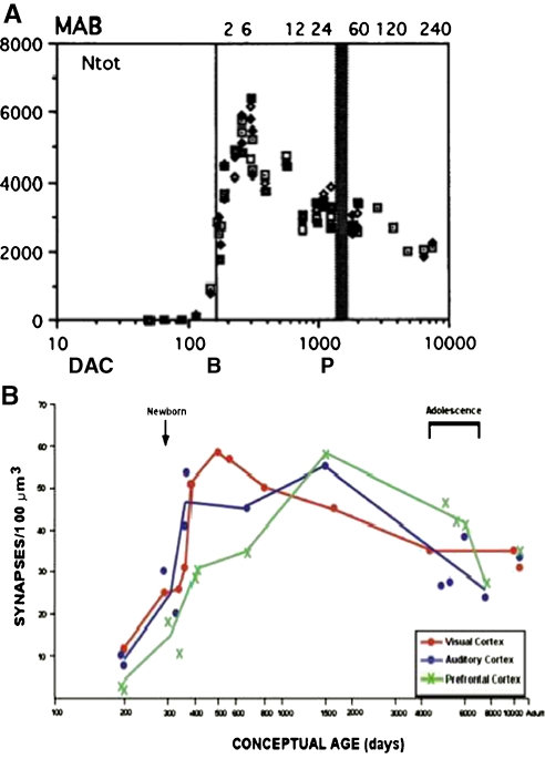
“It was shown that under normal conditions the number of neuronal connections greatly increases in the early postnatal period throughout all brain regions and then then slowly declines to adult levels during childhood and adolescence (Stiles & Jernigan, 2010).”
BackgroundEvolution and transition of expression trajectory during human brain development
Li, M., Tang, H., Shao, Y., Wang, M., Xu, H., Wang, S., Irwin, D. M., Adeola, A. C., Zeng, T., Chen, L., et al. (2020) · BMC Evolutionary Biology · doi:10.1186/s12862-020-01633-4 · Cited by 25
Myhre, O., Zimmer, K. E., Hudecova, A. M., Hansen, K. E. A., Khezri, A., Berntsen, H. F., Berg, V., Lyche, J. L., Mandal, S., Duale, N., et al. (2021) · Chemosphere · doi:10.1016/j.chemosphere.2021.130123 · Cited by 25
Suwannakul, B., Sapbamrer, R., Wiwattanadittakul, N., & Hongsibsong, S. (2021) · Toxics · doi:10.3390/toxics9050099 · Cited by 25
“The rapid growth of the brain during the second trimester is followed by neuronal migration, differentiation, proliferation, and pruning throughout early childhood [33,34].”
BackgroundBuilding Robust Assemblages of Bacteria in the Human Gut in Early Life
Tannock, G. W. (2021) · Applied and Environmental Microbiology · doi:10.1128/aem.01449-21 · Cited by 25
“2 kg and exceeds the rate of growth of any other organ or body tissue (239).”
BackgroundGhosh, S., Chowdhury, S. D., Chandra, A. M., & Ghosh, T. (2014) · American Journal of Physical Anthropology · doi:10.1002/ajpa.22648 · Cited by 26
“The human brain starts its development in the gestational period and continues to grow after birth and reaches the 90% of its adult size by the age of 6 years (Stiles and Jernigan, 2010).”
BackgroundYermolayeva, Y., & Rakison, D. H. (2014) · Psychological Bulletin · doi:10.1037/a0032150 · Cited by 25
“However, it is rare for modelers to implement all of them in a single network, because usually only the neural change that is hypothesized to be responsible for the behavioral change is included in the model.”
BackgroundInfluentialRestorative neurology: Consideration of the new anatomy and physiology of the injured nervous system
Tansey, K. E., McKay, W. B., & Kakulas, B. A. (2012) · Clinical Neurology and Neurosurgery · doi:10.1016/j.clineuro.2012.01.010 · Cited by 25
“For sure, micro-anatomical changes can occur with local prouting and synaptogenesis as well as with structural changes in ynapses [4] but usually the sameneural circuit is capable of particpating in a spectrum of functions.”
BackgroundSullivan, E. V., Lane, B., Kwon, D., Meloy, M. J., Tapert, S. F., Brown, S. A., Colrain, I. M., Baker, F. C., Bellis, M. D. D., Clark, D. B., et al. (2016) · Brain Imaging and Behavior · doi:10.1007/s11682-016-9634-2 · Cited by 25
“Neuroradiological readings of 833 structural MRI from the National Consortium on Alcohol and NeuroDevelopment in Adolescence (NCANDA) cohort found an 11.8 % incidence of brain structural anomalies, represented proportionately across the five collection sites and ethnic groups.”
InfluentialInternational early learning and child well-being study assessment framework
Phair, R. (2021) · OECD education working papers · doi:10.1787/af403e1e-en · Cited by 25
Expression of miR-145 and Its Target Proteins Are Regulated by miR-29b in Differentiated Neurons
Jauhari, A., Singh, T., & Yadav, S. (2018) · Molecular Neurobiology · doi:10.1007/s12035-018-1009-9 · Cited by 24
Neurobehavioral Consequences of Fetal Exposure to Gestational Stress
Sandman, C. A., Glynn, L. M., & Davis, E. P. (2016) · Fetal Development · doi:10.1007/978-3-319-22023-9_13 · Cited by 24
Kruczkowska, W., Gałęziewska, J., Grabowska, K. H., Gromek, P., Czajkowska, K., Rybicki, M., Kciuk, M., & Kłosiński, K. K. (2025) · Molecules · doi:10.3390/molecules30051017 · Cited by 24
“By the eighth week, this structure has differentiated into three primary brain vesicles: the forebrain, midbrain, and hindbrain [43].”
Teramo, K. A., & Piñeiro-Ramos, J. D. (2019) · Free Radical Biology and Medicine · doi:10.1016/j.freeradbiomed.2019.03.012 · Cited by 24
“In addition, intellectua l assessment scales for the outcomes such as composit e intelligence, verbal and nonverbal intelligence, composite memory, reading and writing difficulties, and attendance to classes showed a significant lower scoring for normalized and stan d rdized intelligence indices among offspring of dia…”
BackgroundMaalouf, M., Fearon, M., Lipa, M. C., Chow-Johnson, H. S., Tayeh, L., & Lipa, D. (2021) · Current Neurology and Neuroscience Reports · doi:10.1007/s11910-021-01116-z · Cited by 24
Hosch, A., Oleson, J., Harris, J. L., Goeltz, M. T., Neumann, T., LeBeau, B. C., Hazeltine, E., & Petersen, I. T. (2022) · Developmental Science · doi:10.1111/desc.13280 · Cited by 26
“Brain size increases four-fold during the preschool period, reaching approximately 90% of the adult volume by age 6 (Brown & Jernigan, 2012; Stiles & Jernigan, 2010).”
BackgroundMother’s physical activity during pregnancy and newborn’s brain cortical development
Na, X., Raja, R., Phelan, N. E., Tadros, M. R., Moore, A., Wu, Z., Wang, L., Li, G., Glasier, C. M., Ramakrishnaiah, R. R., et al. (2022) · Frontiers in Human Neuroscience · doi:10.3389/fnhum.2022.943341 · Cited by 24
Surgent, O. J., Riaz, A., Ausderau, K., Adluru, N., Kirk, G. R., Guerrero-Gonzalez, J. M., Skaletski, E. C., Kecskemeti, S., Dean, D., Weismer, S. E., et al. (2022) · Molecular Autism · doi:10.1186/s13229-022-00524-3 · Cited by 24
Howland, M. A. (2023) · Development and Psychopathology · doi:10.1017/s0954579423000998 · Cited by 26
“During prenatal life, the basic architecture of the highly plastic and rapidly developing fetal brain is established (Stiles & Jernigan, 2010), and HPA axis set points appear to be shaped by CRH binds to its receptors on pituitary cortico-tropes, stimulating production of adrenocorti-cotrophic…”
Untreated Early Childhood Caries and Possible Links with Brain Development
Foláyan, M. O., Femi-Akinlosotu, O. M., Adeoti, B., & Olorunmoteni, O. E. (2023) · BioMed · doi:10.3390/biomed3040035 · Cited by 24
“This process plays a vital role in establishing neuronal connections, ensuring proper neural signaling [51].”
BackgroundDeciphering the physiopathology of neurodevelopmental disorders using brain organoids
Dionne, O., Sabatié, S., & Laurent, B. (2024) · Brain · doi:10.1093/brain/awae281 · Cited by 24
Loveson, K. F., & Fillmore, H. L. (2018) · Brain Sciences · doi:10.3390/brainsci8110200 · Cited by 24
“CNS development involves a highly constrained, dynamic, and organised process of stem cell self-renewal and differentiation determined by both genetic and environmental factors in an orderly pattern [89,90].”
BackgroundSegabinazi, E., Spindler, C. D. F., Meireles, A. L. F., Piazza, F. V., Mega, F., Salvalaggio, G. D. S., Achaval, M., & Marcuzzo, S. (2019) · Neuroscience · doi:10.1016/j.neuroscience.2019.08.036 · Cited by 24
Bacha, A. G. B., Al-Orf, N., Al-Osaimi, M., Harrath, A. H., Mansour, L. A., & El-Ansary, A. K. (2019) · Metabolic Brain Disease · doi:10.1007/s11011-019-00496-z · Cited by 24
“The greater vulnerability of the immature brain is attributed to complex and specific developmental processes, such as the commitment and differentiation of neural progenitor cells followed by neuronal and glial cell proliferation, migration, differentiation into glial and neuronal subtypes, pruning, synaptogenesis, m…”
BackgroundScarabello, M., Righini, A., Severino, M., Pinelli, L., Parazzini, C., Scola, E., Palumbo, G., Maurizio, M. D., D’Errico, I., Rossi, A., et al. (2021) · American Journal of Neuroradiology · doi:10.3174/ajnr.a7062 · Cited by 24
Varela, R. B., Cararo, J. H., Tye, S. J., Carvalho, A. F., Valvassori, S. S., Fries, G. R., & Quevedo, J. (2022) · Neuroscience & Biobehavioral Reviews · doi:10.1016/j.neubiorev.2022.104579 · Cited by 24
Ren, Z., Ku, T., Gao, Y., Yang, X., Meng, L., Liu, Q. S., Liang, J., Xu, H., Liao, C., Zhou, Q., et al. (2022) · Environmental Science & Technology · doi:10.1021/acs.est.2c01051 · Cited by 24
Altered Brain Structure in Infants with Turner Syndrome
Davenport, M. L., Cornea, E. A., Xia, K., Crowley, J. J., Halvorsen, M., Goldman, B. D., Reinhartsen, D. B., DeRamus, M., Pretzel, R. E., Styner, M. A., et al. (2019) · Cerebral Cortex · doi:10.1093/cercor/bhz109 · Cited by 23
“…given that the prenatal and early postnatal period is the foundational phase of human brain development, characterized by exuberant neurogenesis, neuronal migration, dendritic arborization, synapto-genesis, gyrification, myelinization, and waves of programmed cell death (Stiles and Jernigan 2010).”
BackgroundKrasnova, A., Tom, S. E., Valeri, L., Crane, P. K., & Bennett, D. A. (2023) · American Journal of Epidemiology · doi:10.1093/aje/kwad033 · Cited by 23
Hernandez‐Castro, I., Eckel, S. P., Chen, X., Yang, T., Vigil, M. J., Foley, H. B., Kannan, K., Robinson, M., Grubbs, B. H., Lerner, D. L., et al. (2023) · Environmental Pollution · doi:10.1016/j.envpol.2023.123131 · Cited by 23
Developmental stage affects cognition in children with recently-diagnosed symptomatic focal epilepsy
Gonzalez, L., Embuldeniya, U. S., Harvey, A. S., Wrennall, J. A., Testa, R., Anderson, V. A., & WOOD, A. G. (2014) · Epilepsy & Behavior · doi:10.1016/j.yebeh.2014.08.006 · Cited by 23
Risky Behaviors: Integrating Adolescent Egocentrism with the Theory of Planned Behavior
Lin, P. S. (2016) · Review of General Psychology · doi:10.1037/gpr0000086 · Cited by 24
“…myelination, which is related to enhanced cognitive functioning, suggesting that adolescents may lack the necessary cognitive skills to fully comprehend risks and potential outcomes when making risky decisions (Fryer et al., 2008; Giedd, 2008; Gogtay et al., 2004; Stiles & Jernigan, 2010).”
BackgroundTorres-Olascoaga, L. A., Watkins, D. J., Schnaas, L., Meeker, J. D., Solano-González, M., Osorio-Valencia, E., Peterson, K. E., Téllez‐Rojo, M. M., & Tamayo‐Ortiz, M. (2020) · International Journal of Environmental Research and Public Health · doi:10.3390/ijerph17218150 · Cited by 23
“Neurodevelopment is a critical life process, beginning in utero and ending in adolescence [1].”
BackgroundRygiel, C. A., Dolinoy, D. C., Bakulski, K. M., Aung, M. T., Perng, W., Jones, T. R., Solano-González, M., Hu, H., Téllez‐Rojo, M. M., Schnaas, L., et al. (2021) · Environmental Epigenetics · doi:10.1093/eep/dvab005 · Cited by 23
New Ultrasound Measurements to Bridge the Gap between Prenatal and Neonatal Brain Growth Assessment
Koning, I. V., Roelants, J. A., Groenenberg, I. A. L., Vermeulen, M. J., Willemsen, S. P., Reiss, I. K. M., Govaert, P., Steegers‐Theunissen, R. P. M., & Dudink, J. (2017) · American Journal of Neuroradiology · doi:10.3174/ajnr.a5278 · Cited by 23
“Brain growth is an important predictor of neurodevelopmental outcome.(1,3,27) We hypothesize that the decreased CC and CCF growth trajectories observed in cases may have consequences for”
BackgroundImpact of DNA repair and stability defects on cortical development
Bianchi, F. T., Berto, G. E., & Cunto, F. D. (2018) · Cellular and Molecular Life Sciences · doi:10.1007/s00018-018-2900-2 · Cited by 23
Määttä, S., Säïsänen, L., Kallioniemi, E., Lakka, T. A., Lintu, N., Haapala, E. A., Koskenkorva, P., Niskanen, E. I., Ferreri, F., & Könönen, M. (2019) · Human Brain Mapping · doi:10.1002/hbm.24525 · Cited by 23
“Exploring how frontal activation and connectivity develop with age can provide insight into why children and adults differ in complex cognitive functions (Stiles & Jernigan, 2010).”
BackgroundMaternal Nutrition during Pregnancy and Offspring Brain Development: Insights from Neuroimaging
Na, X., Mackean, P. P., Cape, G. A., Johnson, J. W., & Ou, X. (2024) · Nutrients · doi:10.3390/nu16193337 · Cited by 22
“Gene expression and environmental input interact to support the ongoing series of events that contribute to brain development [1], which is a complex, dynamic, and adaptive process that supports both brain maturation and functioning.”
BackgroundStructure–function coupling in the first month of life: Associations with age and attention
Tooley, U. A., Kenley, J. K., Camacho, M. C., Latham, A. R., Alexopoulos, D., Nielsen, A. N., Smyser, T. A., Warner, B. B., Shimony, J. S., Neil, J. J., et al. (2025) · Proceedings of the National Academy of Sciences · doi:10.1073/pnas.2412729122 · Cited by 22
“During the postnatal period, brain function is shaped and tuned by the external environment, while the underlying structural connections develop rapidly ( 3 , 7 , 8 ).”
Brooks, J. S., Allison, W., Harriss, A. B., Bian, K., Mao, H., & Dickey, J. P. (2021) · Neurotrauma Reports · doi:10.1089/neur.2021.0014 · Cited by 22
“throughout the brain and makes key contributions to the integration and assimilation of sensory and motor information.(61) Third, the brainstem was selected because of its involvement in motor activity(62) and the observation that standing balance is often impaired in concussed athletes.”
BackgroundSymmetry Perception and Psychedelic Experience
Makin, A. D. J., Roccato, M., Karakashevska, E., Tyson‐Carr, J., & Bertamini, M. (2023) · Symmetry · doi:10.3390/sym15071340 · Cited by 22
TRIM71 mutations cause a neurodevelopmental syndrome featuring ventriculomegaly and hydrocephalus
Duy, P. Q., Jux, B., Zhao, S., Mekbib, K. Y., Dennis, E., Dong, W., Nelson-Williams, C. J., Mehta, N. H., Shohfi, J., Juusola, J., et al. (2024) · Brain · doi:10.1093/brain/awae175 · Cited by 22
Stem Cells in Neurotoxicology/Developmental Neurotoxicology: Current Scenario and Future Prospects
Singh, S., Srivastava, A., Kumar, V., Pandey, A., Kumar, D., Rajpurohit, C. S., Khanna, V. K., Yadav, S., & Pant, A. B. (2015) · Molecular Neurobiology · doi:10.1007/s12035-015-9615-2 · Cited by 22
“and pro-inflammatory factors such as free radicals and cytokines [9].”
BackgroundInfluentialDevelopmental Aspects and Pathological Findings in Spinal Muscular Atrophy
Pérez‐García, M. J., Kong, L., Sumner, C. J., & Tizzano, E. F. (2016) · Elsevier eBooks · doi:10.1016/b978-0-12-803685-3.00002-1 · Cited by 22
Avagliano, L., Grazioli, P., Mariani, M., Bulfamante, G., Selicorni, A., & Massa, V. (2017) · Orphanet Journal of Rare Diseases · doi:10.1186/s13023-017-0723-0 · Cited by 22
“The rhombencephalon is the most posterior vesicle and represents the embryonic hindbrain [9].”
BackgroundInfluentialShyness and Trajectories of Functional Network Connectivity Over Early Adolescence
Sylvester, C. M., Whalen, D. J., Belden, A. C., Sanchez, S. L., Luby, J. L., & Barch, D. M. (2018) · Child Development · doi:10.1111/cdev.13005 · Cited by 22
“The number of physical synaptic connections similarly follows a nonlinear developmental trajectory, with a rapid increase in early childhood, followed by pruning throughout much of adolescence (Stiles & Jernigan, 2010).”
BackgroundKatiyar, K. S., Winter, C. C., Gordián‐Vélez, W. J., O’Donnell, J. C., Song, Y. J., Hernandez, N. S., Struzyna, L. A., & Cullen, D. K. (2018) · Journal of Visualized Experiments · doi:10.3791/55848 · Cited by 22
Wang, N., Lu, Y., Wang, K., Li, W.-S., Lu, P., Lei, S., Li, R., Zhang, H., Zheng, J., Lü, H., et al. (2018) · Cellular Physiology and Biochemistry · doi:10.1159/000488630 · Cited by 22
Gibb, R. L., & Kovalchuk, A. (2017) · Elsevier eBooks · doi:10.1016/b978-0-12-804036-2.00001-7 · Cited by 23
Neurodevelopmental Disorders: Functional Role of Ambra1 in Autism and Schizophrenia
Barbera, L. L., Vedele, F., Nobili, A., D’Amelio, M., & Krashia, P. (2019) · Molecular Neurobiology · doi:10.1007/s12035-019-1557-7 · Cited by 22
Farrell, M. S., Lichtenstein, M. L., Harner, M. K., Crowley, J. J., Filmyer, D. M., Lázaro‐Muñoz, G., Dietterich, T. E., Bruno, L., Shaughnessy, R. A., Biondi, T. F., et al. (2020) · Translational Psychiatry · doi:10.1038/s41398-020-0725-x · Cited by 22
Immune activity at birth and later psychopathology in childhood
Barbosa, S., Khalfallah, O., Forhan, A., Galéra, C., Heude, B., Glaichenhaus, N., & Davidovic, L. (2020) · Brain Behavior & Immunity - Health · doi:10.1016/j.bbih.2020.100141 · Cited by 22
Filippa, M., Casa, E. D., D’Amico, R., Picciolini, O., Lunardi, C., Sansavini, A., & Ferrari, F. (2021) · International Journal of Environmental Research and Public Health · doi:10.3390/ijerph18083915 · Cited by 22
“Therefore, the integrity of the brain processes depends upon the presence of the correct neural elements appearing at the appropriate developmental moments [20], in a delicate equilibrium between environmental stimulation and endogenous brain activity.”
BackgroundLuo, J., & Li, P. (2021) · Cell & Bioscience · doi:10.1186/s13578-021-00617-1 · Cited by 22
“Overview of hPDBOs The complexity of human brain, including the cell type diversity, cellular architecture and functional connectivity, makes understanding its secrets to be one of the most fascinating things in science [1].”
BackgroundRelevance of biometals during neuronal differentiation and myelination: in vitro and in vivo studies
Chin-Chan, M., Montes, S., Blanco-Álvarez, V. M., Aguirre‐Alarcón, H., Rodríguez, I. H., & Bautista, E. (2022) · BioMetals · doi:10.1007/s10534-022-00380-x · Cited by 22
“Gene expression and environmental factors are essential for proper brain development and any alterations might have severe consequences in the brain structure and functionality (Stiles and Jernigan 2010).”
BackgroundInfluentialLobato, M., Vellema, M., Gahr, C. L., Leitão, A., de Lima, S. M. A., Geberzahn, N., & Gahr, M. L. (2015) · Frontiers in Ecology and Evolution · doi:10.3389/fevo.2015.00117 · Cited by 22
“Alternatively, such complicated relationships might indicate that—like in human brain and cognitive development (Stiles and Jernigan, 2010)—the maturation and deterioration of specific brain regions is an insufficient explanatory mechanism for song development of birds.”
BackgroundFOCAL CORTICAL DYSPLASIAS: CLINICAL AND ELECTRO-NEUROIMAGING CHARACTERISTICS
Мukhin, К. Y. (2016) · Russian Journal of Child Neurology · doi:10.17650/2073-8803-2016-11-2-8-24 · Cited by 22
Prenatal and early postnatal measures of brain development and childhood sleep patterns
Kocevska, D., Verhoeff, M. E., Meinderts, S., Jaddoe, V. W. V., Verhulst, F. C., Roza, S. J., Luijk, M. P. C. M., & Tiemeier, H. W. (2017) · Pediatric Research · doi:10.1038/pr.2017.318 · Cited by 22
Subicular hypotrophy in fetuses with Down syndrome and in the Ts65Dn model of Down syndrome
Stagni, F., Giacomini, A., Emili, M., Uguagliati, B., Bonasoni, M. P., Bartesaghi, R., & Guidi, S. (2019) · Brain Pathology · doi:10.1111/bpa.12663 · Cited by 21
“In the human forebrain, neurons are generated during a restricted period that begins at approximately GW 6 and is largely completed by week 18 (32).”
BackgroundCNS Malformations in the Newborn
Baranano, K. W., & Burd, I. D. (2022) · Maternal Health Neonatology and Perinatology · doi:10.1186/s40748-021-00136-4 · Cited by 21
“Pathophysiology/Clinical Features The development of the central nervous system occurs through series of major processes, including neural tube formation, prosencephalic development, neuroblast proliferation and migration, cortical organization and myelination [3].”
BackgroundKim, S., Ochoa, K., Melli, S. E., Yousufzai, F. A. K., Barrera, Z. D., Williams, A. A., McIntyre, G., Delgado, E., Bolish, J. N., Macleod, C. M., et al. (2023) · Scientific Reports · doi:10.1038/s41598-023-32971-0 · Cited by 21
Body as First Teacher: The Role of Rhythmic Visceral Dynamics in Early Cognitive Development
Corcoran, A. W., Perrykkad, K., Feuerriegel, D., & Robinson, J. (2023) · Perspectives on Psychological Science · doi:10.1177/17456916231185343 · Cited by 21
“Blood circulation is established by the 4th week of embryogenesis (Tan & Lewandowski, 2020), while the neural tube is still closing (Stiles & Jernigan, 2010).”
White matter trajectories over the lifespan
Conte, S., Zimmerman, D., & Richards, J. E. (2024) · PLoS ONE · doi:10.1371/journal.pone.0301520 · Cited by 21
“Specifically, the developmental trajectory of brain growth in infancy seems to occur along non-linear patterns that modify the quadratic trend usually used to describe later changes [21, 22].”
Evaluating the association between placenta DNA methylation and cognitive functions in the offspring
Cosín‐Tomás, M., Díez-Ahijado, L., Cilleros‐Portet, A., Fernández, M. F., Fernández, M. F., Guxens, M., Júlvez, J., Llop, S., López-Espinosa, M.-J., Subiza, M., et al. (2024) · Translational Psychiatry · doi:10.1038/s41398-024-03094-5 · Cited by 21
“Early gene expression changes and environmental factors play a crucial role in the normal development of the brain [2].”
BackgroundStructural connectivity of neural reward networks in youth at risk for substance use disorders
Squeglia, L. M., Sorg, S. F., Jacobus, J., Brumback, T., Taylor, C. T., & Tapert, S. F. (2015) · Psychopharmacology · doi:10.1007/s00213-014-3857-y · Cited by 21
“matter coherence and more mature neural processing (Giorgio et al. 2008; Lebel et al. 2012; Schmithorst and Yuan 2010; Stiles and Jernigan 2010; Tamnes et al. 2010).”
Enduring Neuroprotective Effect of Subacute Neural Stem Cell Transplantation After Penetrating TBI
Kassi, A., Mahavadi, A. K., Clavijo, A., Caliz, D., Lee, S. W., Ahmed, A. I., Yokobori, S., Hu, Z., Spurlock, M. S., Wasserman, J. M., et al. (2019) · Frontiers in Neurology · doi:10.3389/fneur.2018.01097 · Cited by 21
“They give rise to striatal and olfactory bulb neurons (274, 276), and hippocampal neurons, respectively.”
BackgroundNeonatal vitamin D levels and cognitive ability in young adulthood
Specht, I. O., Janbek, J., Þorsteinsdóttir, F., Frederiksen, P., & Heitmann, B. L. (2019) · European Journal of Nutrition · doi:10.1007/s00394-019-02042-0 · Cited by 21
The Role of Dynamic miRISC During Neuronal Development
Nawalpuri, B., Ravindran, S., & Muddashetty, R. S. (2020) · Frontiers in Molecular Biosciences · doi:10.3389/fmolb.2020.00008 · Cited by 21
“…al., 2014) Pumilio (Burow et al., 2015; Zahr et al., 2018) ESCRT Factors (Mochida et al., 2012) DDX6 (Nicklas et al., 2015) RBM4 (Tarn et al., 2016; Su et al., 2017) CCR4-NOT (Koch et al., 2014) IPO-8 (Nganou et al., 2018) Hsp-90 (Quintá et al., 2010) FMRP (La Fata et al., 2014) IPO-8 (Nganou et…”
Longitudinal network re-organization across learning and development
McCormick, E. M., Peters, S., Crone, E. A., & Telzer, E. H. (2021) · NeuroImage · doi:10.1016/j.neuroimage.2021.117784 · Cited by 21
Kipp, B. T., Nunes, P. T., Galaj, E., Hitchcock, B., Nasra, T., Poynor, K. R., Heide, S., Reitz, N. L., & Savage, L. M. (2021) · Neuroscience · doi:10.1016/j.neuroscience.2021.08.014 · Cited by 21
Wang, R., Wu, Z., Huang, C., Hashimoto, K., Yang, L., & Yang, C. (2022) · Translational Psychiatry · doi:10.1038/s41398-022-01985-z · Cited by 21
“The fetal nervous system is in a state of development from the third week of gestation and this process continues until adulthood [10].”
BackgroundKitajima, K., Tamura, S., Sasabayashi, D., Nakajima, S., Iwata, Y., Ueno, F., Takai, Y., Takahashi, J., Caravaggio, F., Mar, W., et al. (2022) · Psychiatry and Clinical Neurosciences · doi:10.1111/pcn.13482 · Cited by 21
Multiple benefits of juvenile play: A ground squirrel's perspective
Nunes, S., & Montemayor, M. P. M. (2023) · Neuroscience & Biobehavioral Reviews · doi:10.1016/j.neubiorev.2023.105099 · Cited by 21
Myelination dictates axonal viscoelasticity
Chuang, Y., Alcantara, A., Fabris, G., Abderezaei, J., Lu, T., Melendez‐Vasquez, C. V., & Kurt, M. (2023) · European Journal of Neuroscience · doi:10.1111/ejn.15954 · Cited by 21
“Brain development is a complex biological phenomenon, involving many processes including the growth of neural networks (Baron, 2008; Stiles & Jernigan, 2010), myelination (Chang et al., 2016; Kim et al., 2017) and changes in grey-to-white matter ratios (Giedd et al., 1999; Lenroot & Giedd, 2006).”
BackgroundYawson, N. A., Amankwaa, A. O., Tali, B., Shang, V. O., Batu, E. N., Asiemoah, K., Fuseini, A. D., Tene, L. N., Angaandi, L., Blewusi, I., et al. (2016) · Europe PMC (PubMed Central) · Cited by 21
Jaimes, C. E., Cheng, H. H., Soul, J. S., Ferradal, S. L., Rathi, Y., Gagoski, B. A., Newburger, J. W., Grant, P. E., & Zöllei, L. (2017) · Journal of Magnetic Resonance Imaging · doi:10.1002/jmri.25875 · Cited by 21
“Axonal and synaptic pruning reshape thalamocortical connectivity in the first years of life and also could conceivably affect connectivity-driven parcellation of the thalamus.(40) It is difficult to compare our results with those reported by other authors who used alternative methods to perform structural connectivity…”
ResultToich, J. T. F., Taylor, P. A., Holmes, M., Gohel, S., Cotton, M. F., Dobbels, E., Laughton, B., Little, F., van der Kouwe, A. J. W., Biswal, B. B., et al. (2018) · Frontiers in Human Neuroscience · doi:10.3389/fnhum.2017.00635 · Cited by 22
Kuban, K., Heeren, T., O’Shea, T. M., Joseph, R. M., Fichorova, R. N., Douglass, L. M., Jara, H. J., Frazier, J. A., Hirtz, D., Taylor, H. G., et al. (2018) · The Journal of Pediatrics · doi:10.1016/j.jpeds.2018.05.021 · Cited by 21
“Third, most programmed cell death (apoptosis) in neuronal populations occurs prenatally, whereas cell death in glia populations as well as production and pruning of connections are largely postnatal events.(40) Fourth, to a greater extent than infants born at term, preterm infants encounter a host of potentially prena…”
Comparative analysis reveals distinctive epigenetic features of the human cerebellum
Guevara, E. E., Hopkins, W. D., Hof, P. R., Ely, J. J., Bradley, B. J., & Sherwood, C. C. (2021) · PLoS Genetics · doi:10.1371/journal.pgen.1009506 · Cited by 22
“Indeed, given the known role of methylation in environmental programming [87] and the protracted postnatal development of the brain [88], it is highly likely that growing up human in our highly constructed environments contributes to some of the species differences we observe, as it likely does to the unique cognitive…”
BackgroundMotion correction and volumetric reconstruction for fetal functional magnetic resonance imaging data
Sobotka, D., Ebner, M., Schwartz, E., Nenning, K.-H., Taymourtash, A., Vercauteren, T., Ourselin, S., Kasprian, G., Prayer, D., Langs, G., et al. (2022) · NeuroImage · doi:10.1016/j.neuroimage.2022.119213 · Cited by 20
“These processes start in the third Gestational Week (GW) by the differentiation of neural progenitor cells and proceed in building structural as well as functional trajectories through late adolescence [2].”
BackgroundNBS1 interacts with Notch signaling in neuronal homeostasis
Zhou, Z.-W., Kirtay, M., Schneble, N., Yakoub, G., Ding, M., Rüdiger, T., Siniuk, K., Lu, R., Jiang, Y.-N., Li, T.-L., et al. (2020) · Nucleic Acids Research · doi:10.1093/nar/gkaa716 · Cited by 20
“Brain development is strictly regulated by a concerted serial process of proliferation and differentiation of neuroprogenitors, migration of newborn neurons from origin to final destination, outgrowth of neurites from the soma, and synaptogenesis (20).”
BackgroundBuentello, D. C., García-Corral, M., Santiago, G. T., & Álvarez, M. M. (2022) · IEEE Reviews in Biomedical Engineering · doi:10.1109/rbme.2022.3217486 · Cited by 20
“Interactions of neurons with other cells and their microenvironment exposes them to a plethora of chemical gradients that stimulate synaptic activity in order to guide neurite outgrowth, modulate their transcription activity, and determine neuronal activity [140], [141].”
BackgroundMicroenvironments Matter: Advances in Brain-on-Chip
Akçay, G., & Lüttge, R. (2023) · Biosensors · doi:10.3390/bios13050551 · Cited by 21
“As the new pluripotent cells are produced, they migrate to various brain regions, where they make new connections to establish neural networks upon differenti‐ ation triggered by the local microenvironment [33].”
BackgroundWu, G., Mei, B., Hou, X., Wang, F., Zang, C., Zhang, X., & Zhang, Z. (2023) · BJPsych Open · doi:10.1192/bjo.2023.30 · Cited by 20
The neurological and non-neurological roles of the primary microcephaly-associated protein ASPM
Wu, X., Li, Z., Wang, Z., & Xu, X. (2023) · Frontiers in Neuroscience · doi:10.3389/fnins.2023.1242448 · Cited by 21
“The development of brain relies on neurogenesis, the process by which neural stem cells proliferate, migrate, and differentiate to form neurons, is fundamental to normal brain development (Stiles and Jernigan, 2010; Isaev et al., 2019; Zhou et al., 2020).”
BackgroundA systematic review of interventions to ameliorate the impact of adversity on brain development
McDermott, C. L., Norton, E. S., & Mackey, A. P. (2023) · Neuroscience & Biobehavioral Reviews · doi:10.1016/j.neubiorev.2023.105391 · Cited by 21
An overview of slum upgrading programs in developing countries: Case study in Indonesia
Zubaidah, S., Rusli, B., Saefullah, A. D., & Widianingsih, I. (2023) · Cogent Arts and Humanities · doi:10.1080/23311983.2023.2264021 · Cited by 21
“This program was initiated in 2001, by the Director General of Housing and Settlements, this program was a pilot project between the Netherlands, UNCHS (Habitat)/UNDP, and the Government of Indonesia to build housing for low-income people (Stiles et al., 2010).”
BackgroundGe, M., Jia, K., Gao, R., Wang, X., & Qian, Z. (2024) · Nonlinear Dynamics · doi:10.1007/s11071-024-10349-9 · Cited by 20
Pediatric Traumatic Brain Injury and Microvascular Blood-Brain Barrier Pathology
Fullerton, J. L., Hay, J. R., Bryant-Craig, C., Atkinson, J., Smith, D. H., & Stewart, W. B., Jr. (2024) · JAMA Network Open · doi:10.1001/jamanetworkopen.2024.46767 · Cited by 20
Andonotopo, W., Bachnas, M. A., Dewantiningrum, J., Pramono, M. B. A., Stanojević, M., & Kurjak, A. (2025) · Journal of Perinatal Medicine · doi:10.1515/jpm-2024-0602 · Cited by 20
Tringali, M., Vacher, S. R. W., & Bucci, M. P. (2016) · Acta Paediatrica · doi:10.1111/apa.13609 · Cited by 20
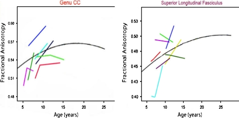
“This was consistent with a study where diffusion tensor imaging was used to explore brain development and showed that there was a continuous maturation of glial populations until adolescence (15).”
ResultEarly human brain development: insights into macroscale connectome wiring
Keunen, K., van der Burgh, H. K., de Reus, M. A., Moeskops, P., Schmidt, R., Stolwijk, L. J., de Lange, S. C., Išgum, I., de Vries, L. S., Benders, M. J. N. L., et al. (2018) · Pediatric Research · doi:10.1038/s41390-018-0138-1 · Cited by 20
“By the time of term birth, all major white matter pathways are in place(4,5) and the neonatal cortex approximates the shape of the adult human brain.(3,6) Diffusion-weighted imaging (DWI) studies in postmortem fetal brains have identified axonal projections from as early as 13 weeks postconceptional age.”
Nelson, E. A., White, D. M., Kraguljac, N. V., & Lahti, A. C. (2018) · Frontiers in Psychiatry · doi:10.3389/fpsyt.2018.00699 · Cited by 20
“Primary cortical folding of the human brain begins as early as the 8th week of gestation with secondary folding having formed by the third trimester and tertiary folding patterns continuing into the postnatal period (7).”
BackgroundAmylin and Leptin interaction: Role During Pregnancy, Lactation and Neonatal Development
Boyle, C. N., & Foll, C. L. (2019) · Neuroscience · doi:10.1016/j.neuroscience.2019.11.034 · Cited by 21
Glial Factors Regulating White Matter Development and Pathologies of the Cerebellum
Revuelta, M., Scheuer, T., Chew, L., & Schmitz, T. (2020) · Neurochemical Research · doi:10.1007/s11064-020-02961-z · Cited by 20
Metadata
- Authors
- Stiles, J., & Jernigan, T. L.
- Affiliations
- University of California San Diego (US)
- Corresponding author
- Jernigan, Terry L.
- ORCID
- Jernigan, Terry L.
- Venue
- Neuropsychology Review
- Published
- 2010-11-03
- Volume
- 20
- Issue
- 4
- Pages
- 327-348
- ISSN
- 1040-7308 (ISSN-L)
- 1573-6660
- PMC
- PMC2989000
- OpenAlex
- W2115599658
- Cited by
- 2,009
- References
- 132
- Citations by year
- 2026: 142 · 2025: 184 · 2024: 199 · 2023: 209 · 2022: 184 · 2021: 197 · 2020: 195 · 2019: 155 · 2018: 158 · 2017: 122 · 2016: 92 · 2015: 64 · 2014: 49 · 2013: 24 · 2012: 23
- FWCI
- 1.43
- Type
- journal article
- Language
- en
- Open access
- hybrid
- doi.org
- Licence
- cc-by-nc
- Locations
- Neuropsychology Review (publishedVersion, cc-by-nc)
- Europe PMC (PubMed Central) (publishedVersion, cc-by-nc)
- PubMed Central (publishedVersion, cc-by-nc)
- PubMed (publishedVersion)
- eScholarship (California Digital Library) (submittedVersion, public-domain)
- eScholarship (California Digital Library) (submittedVersion)
- Funders
- Lundbeckfonden (R32‐A3161)
- H. Lundbeck A/S
- National Institutes of Health (5r01hd025077-10)
- U.S. Public Health Service (R32-A3161)
- University of California, San Diego (RC2DA029475)
- National Institute on Drug Abuse (RC2DA029475)
- National Institute of Child Health and Human Development
- Eunice Kennedy Shriver National Institute of Child Health and Human Development (R01HD060595, R01 HD 25077)
- Topics
- Life Sciences › Neuroscience › Developmental Neuroscience › Neurogenesis and neuroplasticity mechanisms
- Life Sciences › Biochemistry, Genetics and Molecular Biology › Genetics › Genetics and Neurodevelopmental Disorders
- Social Sciences › Psychology › Developmental and Educational Psychology › Child and Animal Learning Development
- Keywords
- brain development, developmental neuroscience, mammalian brain, neuronal organization, social development (developmental psychology), cognitive development, neural systems, genetic constraints, developmental mechanisms
- MeSH terms
- Animals, Brain, Cell Differentiation, Diagnostic Imaging, Humans, Nerve Net, Neural Pathways, Neurobiology, Neurons, Gene Expression Regulation, Developmental, Body Patterning, Neurogenesis
About these checks
Registry, retraction, Cites and statistics results are computed, not judged, with one exception: a printed reference that no index lists is read once by a model before it counts as a citation error. Claims are extracted by a model and kept only when their quote appears verbatim in the text; each cited source is then checked, with a model’s help, for whether it says what the claim relies on. Those verdicts can be wrong — read the claim’s quote and the cited source before relying on one.
Statistics and arithmetic are recomputed, not judged.
Confidence bands are shown only for fields calibrated on our annotated test set. None is calibrated yet, so no band is shown.
What counts as a claim ›How we measure confidence ›How we check this ›
Are you an author, or do you see a problem with this page? Request a review
Data sources
Bibliographic data: Semantic Scholar, OpenAlex, Crossref.
Contains information from the Semantic Scholar Academic Graph Datasets, made available under the ODC Attribution License (ODC-BY 1.0).
Semantic Scholar’s data is described in: Kinney, R., et al. (2023). The Semantic Scholar Open Data Platform. arXiv:2301.10140.
