

The Basics of Brain Development
Open accessThe DOI matches its Crossref record; 128 of the 132 references it cites were found in a registry or an index.
Abstract
Over the past several decades, significant advances have been made in our understanding of the basic stages and mechanisms of mammalian brain development. Studies elucidating the neurobiology of brain development span the levels of neural organization from the macroanatomic, to the cellular, to the molecular. Together this large body of work provides a picture of brain development as the product of a complex series of dynamic and adaptive processes operating within a highly constrained, genetically organized but constantly changing context. The view of brain development that has emerged from the developmental neurobiology literature presents both challenges and opportunities to psychologists seeking to understand the fundamental processes that underlie social and cognitive development, and the neural systems that mediate them. This chapter is intended to provide an overview of some very basic principles of brain development, drawn from contemporary developmental neurobiology, that may be of use to investigators from a wide range of disciplines.
Overview
The claims and statistics of this paper haven’t been checked yet.
Free: it reads the open-access text and checks the first 5 claims that cite a source we can check. Create a free account to check every claim.
Integrity signals
Registry
Cites
Statistics
Figures





Verification
Claims the paper makes
The claims and statistics of this paper haven’t been checked yet.
Free: it reads the open-access text and checks the first 5 claims that cite a source we can check. Create a free account to check every claim.
Claims other papers make about this one
Each is checked against this paper’s own text: does it say what the citing paper says it does?
The source backs part of the claim, not all of it — or backs it for a different population, condition or comparison.
In the citing paper
“We now know from recent studies that it is the birth and migration of neurons derived from oRGCs that play a role in the development of the primary sulci (superior frontal, inferior frontal and precentral) in week 25–26 of gestation [23, 76].”
From Development of prefrontal cortex (2022) · cites this paper as [23]
Experimental — accuracy not yet measuredIn the citing paper
“This intricate structural-functional interplay underscores that the ontogeny of functional networks likely reflects programmed neurodevelopmental events (e.g. neurogenesis, cell death, myelination, pruning, synaptic plasticity, and glial development) (Innocenti and Price, 2005; Stiles and Jernigan, 2010)”
From Development of large-scale functional networks from birth to adulthood: A guide to the neuroimaging literature (2017) · cites this paper as Stiles and Jernigan, 2010
In the citing paper
“The brain’s major fiber pathways become consolidated through myelination, though their presence is largely established prenatally (Stiles and Jernigan, 2010).”
From Development of large-scale functional networks from birth to adulthood: A guide to the neuroimaging literature (2017) · cites this paper as Stiles and Jernigan, 2010
Self-check
Percentages, sums and reported differences re-computed from the paper’s own numbers.
The self-check needs the paper’s full text.
Citations
Cites
Philosophical transactions of the Royal Society of London. B
Press, H. (1886) · Cited by 10,080
MR diffusion tensor spectroscopy and imaging
Basser, P. J., Mattiello, J., & LeBihan, D. (1994) · Biophysical Journal · doi:10.1016/s0006-3495(94)80775-1 · Cited by 5,970
Dynamic mapping of human cortical development during childhood through early adulthood
Gogtay, N., Giedd, J. N., Lusk, L., Hayashi, K. M., Greenstein, D., Vaituzis, A. C., Nugent, T. F., Herman, D. H., Clasen, L. S., Toga, A. W., et al. (2004) · Proceedings of the National Academy of Sciences · doi:10.1073/pnas.0402680101 · Cited by 5,604
(2005) · Choice Reviews Online · doi:10.5860/choice.43-0626 · Cited by 4,674
Neurotrophins: Roles in Neuronal Development and Function
Huang, E. J., & Reichardt, L. F. (2001) · Annual Review of Neuroscience · doi:10.1146/annurev.neuro.24.1.677 · Cited by 4,394
Ferrier lecture. Functional architecture of macaque monkey visual cortex
Hubel, D. H., & Wiesel, T. N. (1977) · Proceedings of the Royal Society of London. Series B. Biological Sciences · doi:10.1098/rspb.1977.0085 · Cited by 2,608 · cited 1×
“The seminal studies of Hubel and Weisel (Hubel and Wiesel 1977; Hubel et al. 1977) showed that monocular visual deprivation in the early postnatal period can substantially alter basic patterns of organization within primary visual cortex (PVC).”
Background(1995) · Choice Reviews Online · doi:10.5860/choice.32-5931 · Cited by 2,311
Mode of cell migration to the superficial layers of fetal monkey neocortex
Rakic, P. (1972) · Journal of Comparative Neurology · doi:10.1002/cne.901450105 · Cited by 2,170 · cited 1×
“Because of the greater distances, neurons require what was originally identified as a special population of cells within the VZ called “radial glial guides” to support their migration (Rakic 1972).”
BackgroundRegional Development of the Brain in Early Life
(1968) · Archives of Disease in Childhood · doi:10.1136/adc.43.229.388 · Cited by 2,090
Neurons derived from radial glial cells establish radial units in neocortex
Noctor, S. C., Flint, A. C., Weissman, T. A., Dammerman, R. S., & Kriegstein, A. R. (2001) · Nature · doi:10.1038/35055553 · Cited by 2,016 · cited 2×
“…radial glial guides were originally thought to be a special, transient population of cells, it has recently been discovered that the cells that provide the scaffolding are actually the neural progenitor cells (Noctor et al. 2001; Noctor et al. 2002; Parnavelas et al. 2002; Weissman et al. 2003).”
Background
Research works printed but not linked: 1
- 56. Lebel, C., & Beaulieu, C. (2009). Longitudinal diffusion tensor imaging of healthy brain development in children. Honolulu: ISMRM Annual Meeting.
Cited by
Items 401–500 of 1,004
Hessel, E. V. S., Staal, Y. C. M., & Piersma, A. H. (2018) · Toxicology and Applied Pharmacology · doi:10.1016/j.taap.2018.03.013 · Cited by 42
Habibey, R., Striebel, J., Schmieder, F., Czarske, J. W., & Busskamp, V. (2022) · Frontiers in Neuroscience · doi:10.3389/fnins.2022.951964 · Cited by 42
“These changes in the shape and activity emerge naturally during a predictable developmental timeline (Stiles and Jernigan, 2010; Gozdas et al., 2019; Martinez and Sprecher, 2020; Kim and Paredes, 2021).”
BackgroundGut microbes in central nervous system development and related disorders
Gan, Y., Chen, Y., Zhong, H., Liu, Z., Geng, J., Wang, H., & Wang, W. (2024) · Frontiers in Immunology · doi:10.3389/fimmu.2023.1288256 · Cited by 41
“These developmental processes entail relevant gene expression and environmental inputs, with disruptions in any of these events fundamentally altering neural outcomes (128).”
BackgroundPeixoto-Rodrigues, M. C., Monteiro-Neto, J. R., Téglás, T., Toborek, M. J., Quinete, N., Hauser‐Davis, R. A., & Adesse, D. (2024) · Journal of Hazardous Materials · doi:10.1016/j.jhazmat.2024.136832 · Cited by 41
“The embryonic period defines the main compartments of both the central and peripheral nervous systems [145].”
Review: Evidence of Neurological Sequelae in Children With Acquired Zika Virus Infection
Lebov, J. F., Brown, L. M., MacDonald, P. D. M., Robertson, K., Bowman, N. M., Hooper, S. R., & Becker-Dreps, S. (2018) · Pediatric Neurology · doi:10.1016/j.pediatrneurol.2018.03.001 · Cited by 35
A Prospective Longitudinal Investigation of Cortical Thickness and Gyrification in Schizophrenia
Nelson, E. A., Kraguljac, N. V., White, D. M., Jindal, R. D., Shin, A. L., & Lahti, A. C. (2020) · The Canadian Journal of Psychiatry · doi:10.1177/0706743720904598 · Cited by 41
“As a measure, gyrification index is of particular interest due to its potential as a marker of abnormal neurodevelopment.(17,18) Altered gyrification patterns have been reported across different illness stages(19-22) and among first-degree relatives.”
BackgroundMorris, A. S., Hays-Grudo, J., Kerr, K. L., & Beasley, L. O. (2021) · Development and Psychopathology · doi:10.1017/s0954579420001595 · Cited by 41
Exposure to prenatal maternal distress and infant white matter neurodevelopment
Demers, C. H., Bagonis, M., Al‐Ali, K., Garcia, S. E., Styner, M. A., Gilmore, J. H., Hoffman, M. C., Hankin, B. L., & Davis, E. P. (2021) · Development and Psychopathology · doi:10.1017/s0954579421000742 · Cited by 41
“During the transformation from a zygote to a human newborn in the 9 months of full-term gestation, cell division and differentiation is both rapid and highly coordinated (Huttenlocher & Dabholkar, 1997; Stiles & Jernigan, 2010).”
BackgroundMuñoz‐Moreno, E., Fischi-Gómez, E., Batallé, D., Borradori-Tolsa, C., Eixarch, E., Thiran, J., Gratacós, E., & Hüppi, P. S. (2016) · Frontiers in Neuroscience · doi:10.3389/fnins.2016.00560 · Cited by 42
“This pruning process extends from the end of gestation to childhood (Innocenti and Price, 2005; Stiles and Jernigan, 2010; Dubois et al., 2014).”
BackgroundCannabis and the developing brain: What does the evidence say?
Jacobus, J., Courtney, K. E., Hodgdon, E. A., & Baca, R. (2019) · Birth Defects Research · doi:10.1002/bdr2.1572 · Cited by 37
“Adolescent brain development is dynamic, with changes in gray and white matter tissue and structural integration occurring well into young adulthood (Gonzalez, PachecoColon, Duperrouzel, & Hawes, 2017; Stiles & Jernigan, 2010).”
BackgroundGlobal Network Organization of the Fetal Functional Connectome
De Asis-Cruz, J., Andersen, N., Kapse, K., Khrisnamurthy, D., Quistorff, J., Lopez, C., Vezina, G., & Limperopoulos, C. (2021) · Cerebral Cortex · doi:10.1093/cercor/bhaa410 · Cited by 40
Oderberg, D. S. (2019) · Metamedicine · doi:10.1007/s11017-019-09479-8 · Cited by 40
Advances and Applications of Brain Organoids
Li, Y., Zeng, P.-M., Wu, J., & Luo, Z. (2023) · Neuroscience Bulletin · doi:10.1007/s12264-023-01065-2 · Cited by 40
“By the sixth gestational week, the difference in the rates of proliferation of cells in rostral regions of the neural tube results in the formation of three brain vesicles, the prospective forebrain, midbrain, and hindbrain [9].”
BackgroundBoerma, T., ter Haar, S. M., Ganga, R., Wijnen, F. N. K., Blom, E., & Wierenga, C. J. (2023) · Neuroscience & Biobehavioral Reviews · doi:10.1016/j.neubiorev.2023.105398 · Cited by 41
Tiwari, S., Phoolmala, Goyal, S., Yadav, R. K., & Chaturvedi, R. K. (2024) · Molecular Neurobiology · doi:10.1007/s12035-024-04160-1 · Cited by 40
“The early stage of development is very important for brain formation [75].”
Assawachananont, J., Kim, S., Kaya, K. D., Fariss, R. N., Roger, J. E., & Swaroop, A. (2018) · Human Molecular Genetics · doi:10.1093/hmg/ddy272 · Cited by 40
Neuropharmacology, pharmacogenetics and pharmacogenomics of aggression: The zebrafish model
de Abreu, M. S., Giacomini, A. C. V. V., Genário, R., dos Santos, B. E., da Rosa, L. G., Demin, K. A., Wappler-Guzzetta, E. A., & Kalueff, A. V. (2019) · Pharmacological Research · doi:10.1016/j.phrs.2019.01.044 · Cited by 41
Fetal neurology: Principles and practice with a life-course perspective
Scher, M. S. (2019) · Handbook of clinical neurology · doi:10.1016/b978-0-444-64029-1.00001-1 · Cited by 41
Mechanical Regulation of Nuclear Translocation in Migratory Neurons
Nakazawa, N., & Kengaku, M. (2020) · Frontiers in Cell and Developmental Biology · doi:10.3389/fcell.2020.00150 · Cited by 41
“Neuronal migration in earlier stages is critical for neuronal network formation in the later stages of brain development (Stiles and Jernigan, 2010; Silva et al., 2019).”
BackgroundMapping Mind-Brain Development: Towards a Comprehensive Theory
Spanoudis, G. C., & Δημητρίου, Α. (2020) · Journal of Intelligence · doi:10.3390/jintelligence8020019 · Cited by 41
“Despite the diversity of brain regions, the brain operates as a single integrated entity governed by multiple processes running within a highly constrained and genetically organized context interacting with the environment (Stiles and Jernigan 2010).”
BackgroundMahmassani, H. A., Switkowski, K. M., Scott, T. M., Johnson, E. J., Rifas‐Shiman, S. L., Oken, E., & Jacques, P. F. (2020) · Journal of Nutrition · doi:10.1093/jn/nxaa348 · Cited by 40
“Different parts and functions of the brain develop at different times during pregnancy (30, 31), and the mechanisms and timing of action of lutein and zeaxanthin in the brain are still not understood.”
BackgroundPrenatal opioid exposure and vulnerability to future substance use disorders in offspring
Abu, Y. F., & Roy, S. (2021) · Experimental Neurology · doi:10.1016/j.expneurol.2021.113621 · Cited by 40
Lizé, M., Monfort, C., Rouget, F., Limon, G., Durand, G., Tillaut, H., & Chevrier, C. (2022) · Environmental Research · doi:10.1016/j.envres.2022.113348 · Cited by 42
Multifaceted atlases of the human brain in its infancy
Ahmad, S., Wu, Y., Wu, Z., Thung, K., Liu, S., Lin, W., Li, G., Wang, L., & Yap, P. (2022) · Nature Methods · doi:10.1038/s41592-022-01703-z · Cited by 40
“5)] months, we estimate displacement field φ(i,τ)→ref(~x) by solving the wave equation: ∂(2)φ(i,τ)→ref(~x) ∂t2 = α ( ∇(2)φ(i,τ)→ref(~x) +∇ ( ∇ · φ(i,τ)→ref(~x) )) + β f vol (i,τ)→ref(~x) + γ f surf (i,τ)→ref(~x), (1) where ∇2 is the vector Laplacian operator and f vol (i,τ)→ref(~x) and f surf (i,τ)→ref(~x) are respect…”
Atazanavir exposure in utero and neurodevelopment in infants: a comparative safety study
Caniglia, E. C., Patel, K., Huo, Y., Williams, P. L., Kapetanovic, S., RICH, K. C., Sirois, P. A., Jacobson, D. L., Hernández–Díaz, S., Hernán, M. Á., et al. (2016) · AIDS · doi:10.1097/qad.0000000000001052 · Cited by 40
Age Effects on Cerebral Oxygenation and Behavior in Children with Sleep-disordered Breathing
Tamanyan, K., Walter, L. M., Weichard, A. J., Davey, M. J., Nixon, G. M., Biggs, S. N., & Horne, R. S. C. (2018) · American Journal of Respiratory and Critical Care Medicine · doi:10.1164/rccm.201709-1825oc · Cited by 40
Lomanowska, A. M., & Melo, Á. I. (2015) · Hormones and Behavior · doi:10.1016/j.yhbeh.2015.05.017 · Cited by 40
“Conversely, experience-dependent plasticity is thought to primarily involve the production of new neuronal synapses stimulated by relevant events (Greenough et al., 1987; Stiles and Jernigan, 2010).”
Telomerase Activity is Downregulated Early During Human Brain Development
Ishaq, A., Hanson, P. S., Morris, C. M., & Saretzki, G. C. (2016) · Genes · doi:10.3390/genes7060027 · Cited by 39
“After reaching their target region in the cortex, the young neurons mature further by forming axons and dendrites in order to form networks and transmit information [40].”
BackgroundInfluentialThe effect of subclinical infantile thiamine deficiency on motor function in preschool children
Harel, Y., Zuk, L., Guindy, M., Nakar, O., Lotan, D., & Fattal‐Valevski, A. (2017) · Maternal and Child Nutrition · doi:10.1111/mcn.12397 · Cited by 39
Congenital olfactory impairment is linked to cortical changes in prefrontal and limbic brain regions
Karstensen, H. G., Vestergaard, M., Baaré, W. F. C., Skimminge, A., Djurhuus, B. D., Ellefsen, B., Brüggemann, N., Klausen, C., Leffers, A.-M., Tommerup, N., et al. (2018) · Brain Imaging and Behavior · doi:10.1007/s11682-017-9817-5 · Cited by 39
“The postnatal brain undergoes protracted maturation lasting well into adulthood characterized by neuronal elimination, synaptic pruning, axonal myelination as well as dendritic and axonal arborization (Lenroot and Giedd 2006; Stiles and Jernigan 2010).”
BackgroundIsoquercetin Improves Inflammatory Response in Rats Following Ischemic Stroke
Shi, Y., Chen, X., Liu, J., Fan, X., Jin, Y., Gu, J., Liang, J., Liang, X., & Wang, C. (2021) · Frontiers in Neuroscience · doi:10.3389/fnins.2021.555543 · Cited by 39
“For convenience, to count the cells in stained cortex sections, the cerebral cortex was divided into seven layers (Figure 2A) according to the reported method (Cooper, 2008; Stiles and Jernigan, 2010).”
MethodZhang, W., Sun, Z., Zhang, Q., Sun, Z., Su, Y., Song, J., Wang, B., & Gao, R. (2021) · BMC Pediatrics · doi:10.1186/s12887-021-02539-w · Cited by 39
Gadaleta, D., Sp̂înu, N., Roncaglioni, A., Cronin, M., & Benfenati, E. (2022) · International Journal of Molecular Sciences · doi:10.3390/ijms23063053 · Cited by 39
Individual differences in human brain development
Brown, T. T. (2016) · Wiley Interdisciplinary Reviews Cognitive Science · doi:10.1002/wcs.1389 · Cited by 39
Anxiogenic effects of a Lactobacillus, inulin and the synbiotic on healthy juvenile rats
Barrera-Bugueño, C., Realini, O., Escobar-Luna, J., Sotomayor-Zárate, R., Gotteland, M., Julio-Pieper, M., & Bravo, J. A. (2017) · Neuroscience · doi:10.1016/j.neuroscience.2017.06.064 · Cited by 38
“…have been obtained in adult individuals, however little has been done in younger animals, at critical stages of development when the brain is still stablishing connections (Stiles and Jernigan, 2010) and the gut is adjusting to changes in diet and luminal composition, including gut symbionts.”
BackgroundInsights into the Brain: Neuroimaging of Brain Development and Maturation
Lyall, A. E., Savadjiev, P., Shenton, M. E., & Kubicki, M. R. (2016) · Journal of Neuroimaging in Psychiatry and Neurology · doi:10.17756/jnpn.2016-003 · Cited by 38
“While the most dramatic brain growth and differentiation occurs prenatally (including the production of neurons, their migration, formation of cortical and subcortical structures and major white matter fiber pathways), it is the early postnatal years that are known to be the most critical for brain development [1, 2].”
BackgroundInfluentialHuang, Y., Wu, Z., Wang, F., Hu, D., Li, T., Guo, L., Wang, L., Lin, W., & Li, G. (2022) · Proceedings of the National Academy of Sciences · doi:10.1073/pnas.2121748119 · Cited by 38
Immune regulatory and neuroprotective properties of preimplantation factor: From newborn to adult
Barnea, E. R., Almogi‐Hazan, O., Or, R., Mueller, M., Ria, F., Weiss, L., & Paidas, M. J. (2015) · Pharmacology & Therapeutics · doi:10.1016/j.pharmthera.2015.10.008 · Cited by 38
Kim, S. Y., Koetzner, C. A., Payne, A. F., Nierode, G. J., Yu, Y., Wang, R., Barr, E., Dordick, J. S., Kramer, L. D., Zhang, F., et al. (2019) · Biochemistry · doi:10.1021/acs.biochem.8b01267 · Cited by 38
Head circumference at birth and school performance: a nationwide cohort study of 536,921 children
Bach, C. C., Henriksen, T. B., Larsen, R. T., Aagaard, K., & Matthiesen, N. B. (2019) · Pediatric Research · doi:10.1038/s41390-019-0683-2 · Cited by 38
Golovina, E., Fadason, T., Lints, T., Walker, C. G., Vickers, M. H., & O'Sullivan, J. M. (2021) · Scientific Reports · doi:10.1038/s41598-021-95447-z · Cited by 39
Edvardson, S., Tian, G., Cullen, H. D., Vanyai, H. K., Ngo, L., Bhat, S., Aran, A. A., Daana, M., Da'amseh, N., Abu‐Libdeh, B., et al. (2016) · Human Molecular Genetics · doi:10.1093/hmg/ddw292 · Cited by 38
“In contrast, postnatal growth largely reflects glial proliferation and the deposition of myelin (2).”
BackgroundImpact of Demographic and Obstetric Factors on Infant Brain Volumes: A Population Neuroscience Study
Knickmeyer, R. C., Xia, K., Lu, Z., Ahn, M., Jha, S. C., Zou, F., Zhu, H., Styner, M., & Gilmore, J. H. (2016) · Cerebral Cortex · doi:10.1093/cercor/bhw331 · Cited by 37
Neurocognitive Outcomes in Pediatric Diabetes: a Developmental Perspective
Schwartz, D. D., Wasserman, R. M., Powell, P. W., & Axelrad, M. E. (2014) · Current Diabetes Reports · doi:10.1007/s11892-014-0533-x · Cited by 38
Hope, K. A., LeDoux, M. S., & Reiter, L. T. (2017) · Neurobiology of Disease · doi:10.1016/j.nbd.2017.09.003 · Cited by 37
Wang, W., Lin, Y., Wang, S., Jones, S., Prayson, R. A., Moosa, A. N. V., McBride, A. F., Gonzalez-Martinez, J. A., Bingaman, W. E., Najm, I., et al. (2019) · European Journal of Neurology · doi:10.1111/ene.13916 · Cited by 37
“As younger brains display significant structural differences compared to mature brains [19,20], performing MRI postprocessing studies dedicated to the pediatric epilepsy population using age-specific normal databases is in principle more accurate and a worthwhile effort.”
BackgroundZhao, X., Shi, J., Dai, F., Lei, W., Zhang, B., Yu, X., Wang, C., Zhu, W., & Wang, H. (2021) · Frontiers in Human Neuroscience · doi:10.3389/fnhum.2021.616132 · Cited by 38
“The human brain undergoes complex anatomical changes from infancy to adolescence, including axonal growth, myelination, dendritic arborization, synapse formation and neuronal pruning (Stiles and Jernigan, 2010).”
BackgroundWiltshire, C. A., Troller‐Renfree, S. V., Giebler, M. A., & Noble, K. G. (2021) · Infant Behavior and Development · doi:10.1016/j.infbeh.2021.101644 · Cited by 37
Developmental Neuropathology and Neurodegeneration of Down Syndrome: Current Knowledge in Humans
Hasina, Z., Wang, N., & Wang, C. C. (2022) · Frontiers in Cell and Developmental Biology · doi:10.3389/fcell.2022.877711 · Cited by 37
“Molecular signals originate from the neural groove (primitive node) and transmit from one to another consecutively, controlled by the expression of genes in the cells (Stiles, 2008; Stiles and Jernigan, 2010).”
BackgroundInfluentialOh, J., Kim, K., Kannan, K., Parsons, P. J., Mlodnicka, A., Schmidt, R. J., Schweitzer, J. B., Hertz‐Picciotto, I., & Bennett, D. H. (2024) · Environmental Health · doi:10.1186/s12940-024-01065-3 · Cited by 38
“While the prenatal period has been recognized as the most sensitive window of neu-rodevelopment, chemical exposure during the early postnatal period has also been a focus due to the continued postnatal development of the brain [10, 11].”
BackgroundHow the brain attunes to sentence processing: Relating behavior, structure, and function
Fengler, A., Meyer, L., & Friederici, A. D. (2016) · NeuroImage · doi:10.1016/j.neuroimage.2016.01.012 · Cited by 38
Tsai, I.-C., McKnight, K., McKinstry, S. U., Maynard, A. T., Tan, P. L., Golzio, C., White, C. T., Price, D. J., Davis, E. E., Amrine‐Madsen, H., et al. (2018) · Scientific Reports · doi:10.1038/s41598-018-28709-y · Cited by 37
Dineva, M., Rayman, M. P., Levie, D., Guxens, M., Peeters, R. P., Vioqué, J., González, L., Espada, M., Ibarluzea, J. M., Sunyer, J., et al. (2019) · European Journal of Nutrition · doi:10.1007/s00394-019-01913-w · Cited by 38
Mornagui, B., Rezg, R., Repond, C., & Pellerin, L. (2019) · Food and Chemical Toxicology · doi:10.1016/j.fct.2019.110670 · Cited by 37
Putative Roles of SETBP1 Dosage on the SET Oncogene to Affect Brain Development
Antonyan, L., & Ernst, C. (2022) · Frontiers in Neuroscience · doi:10.3389/fnins.2022.813430 · Cited by 37
“tube balloons out to become forebrain (telencephalon) and midbrain/hindbrain (mesencephalon/diencephalon) structures derived from proliferating cells from the neuroectoderm (Stiles and Jernigan, 2010).”
BackgroundThe Etiology of Neuronal Development in Craniosynostosis: A Working Hypothesis
Brooks, E. D., Beckett, J. S., Yang, J. F., Timberlake, A. T., Sun, A. H., Chuang, C., & Persing, J. A. (2017) · Journal of Craniofacial Surgery · doi:10.1097/scs.0000000000004040 · Cited by 36
Sequential anomaly detection with observation control
Tsopelakos, A., Fellouris, G., & Veeravalli, V. V. (2019) · IEEE International Symposium on Information Theory (ISIT) · doi:10.1109/isit.2019.8849555 · Cited by 36
Maternal adverse childhood experiences and infant subcortical brain volume
Demers, C. H., Hankin, B. L., Hennessey, E. P., Haase, M. H., Bagonis, M., Kim, S. H., Gilmore, J. H., Hoffman, M. C., Styner, M. A., & Davis, E. P. (2022) · Neurobiology of Stress · doi:10.1016/j.ynstr.2022.100487 · Cited by 36
“newborn, the pace of development exceeds any other stage of the lifespan (Stiles and Jernigan, 2010).”
BackgroundLayrolle, P., Payoux, P., & Chavanas, S. (2022) · Biomolecules · doi:10.3390/biom13010025 · Cited by 36
“1 kPa) promoted differentiation into hindbrain-like neurons [54] Forebrain, midbrain and hindbrain are three specific regions arising from the progressive segmentation of the brain along the rostral–caudal axis during development [55].”
BackgroundThe impact of maternal immune activation on embryonic brain development
McEwan, F., Glazier, J. D., & Hager, R. (2023) · Frontiers in Neuroscience · doi:10.3389/fnins.2023.1146710 · Cited by 36
“Neurodevelopment begins with the formation of the neural tube, where an initial pool of neuroepithelial cells (NECs) divide symmetrically until a sufficient number have formed (Figure 1; Stiles and Jernigan, 2010; Egger et al., 2011; Semple et al., 2013).”
BackgroundMethodKatiyar, K. S., Winter, C. C., Struzyna, L. A., Harris, J. P., & Cullen, D. K. (2016) · Journal of Tissue Engineering and Regenerative Medicine · doi:10.1002/term.2168 · Cited by 36
“Due to the limited capacity for CNS regeneration, neurotrauma and neurodegenerative diseases lack effective treatment options.”
InfluentialZanghi, C. N., & Jevtović‐Todorović, V. (2016) · Neurotoxicology and Teratology · doi:10.1016/j.ntt.2016.12.004 · Cited by 37
“2.1 Neural apoptosis Normal development produces an excess of neurons that are systematically culled via apoptosis, resulting in the eventual elimination of 50% or more of the initial neuronal pool (Bandeira et al., 2009; Stiles and Jernigan, 2010).”
BackgroundInfluentialZika Virus Disease in Children in Colombia, August 2015 to May 2016
Tolosa, N., Tinker, S. C., Pacheco, Ó., Valencia, D., Botero, D. S., Tong, V. T., Mercado, M., Gilboa, S. M., González, M., Nelson, C. A., et al. (2017) · Paediatric and Perinatal Epidemiology · doi:10.1111/ppe.12391 · Cited by 35
Vancamp, P., & Darras, V. M. (2018) · General and Comparative Endocrinology · doi:10.1016/j.ygcen.2017.11.023 · Cited by 33
Naderi, M., Ferrari, M. C. O., Chivers, D. P., & Niyogi, S. (2018) · Environmental Science & Technology · doi:10.1021/acs.est.8b04768 · Cited by 35
Gómez-Verján, J. C., Barrera-Vázquez, O. S., García‐Velázquez, L., Samper‐Ternent, R., & Arroyo, P. (2020) · Clinical Genetics · doi:10.1111/cge.13748 · Cited by 36
“stage and infancy, impacts neural development and cognition significantly.(13,14) There is a strong association between early-life nutrition, maternal diet, microbial infections, and the risk of developing metabolic diseases including type 2 diabetes mellitus (T2DM), neurological and cardiovascular diseases.”
BackgroundLatchney, S. E., & Majewska, A. K. (2021) · Developmental Neurobiology · doi:10.1002/dneu.22825 · Cited by 36
“Given the multifaceted process of neurodevelopment (Stiles & Jernigan, 2010; Tau & Peterson, 2010), it is no surprise that there is a wealth of published literature that reviews the effects of various environmental contaminants on biological processes such as cell proliferation, differentiation,…”
BackgroundNeurogenesis in the adult Drosophila brain
Crocker, K. L., Marischuk, K., Rimkus, S. A., Zhou, H., Yin, J. C. P., & Boekhoff‐Falk, G. (2021) · Genetics · doi:10.1093/genetics/iyab092 · Cited by 35
Sachana, M., Willett, C., Pistollato, F., & Bal-Price, A. (2021) · Reproductive Toxicology · doi:10.1016/j.reprotox.2021.06.006 · Cited by 30
Developmental Timing of Trauma in Women Predicts Unique Extracellular Vesicle Proteome Signatures
Morrison, K. E., Stenson, A. F., Marx-Rattner, R., Carter, S. E., Michopoulos, V., Gillespie, C. F., Powers, A. D., Huang, W., Kane, M. A., Jovanović, T., et al. (2021) · Biological Psychiatry · doi:10.1016/j.biopsych.2021.08.003 · Cited by 35
The Cambridge Handbook of Infant Development: Brain, Behavior, and Cultural Context
Lockman, J. J., Hertz, D., Tamis-LeMonda, C. S., Brain, B. C., Context..........................................., Rey, J., Falck-Ytter, T., Jones, E., Maurer, D., Cirelli, L., et al. (2020) · Cambridge University Press eBooks · doi:10.1017/9781108351959 · Cited by 35
Stagni, F., & Bartesaghi, R. (2022) · Frontiers in Cellular Neuroscience · doi:10.3389/fncel.2022.903729 · Cited by 36
“Key steps of brain development are outlined below (mainly based on Rakic, 2009; Rice and Barone, 2010; Stiles and Jernigan, 2010; Yamaguchi et al., 2016; Kostović et al., 2019), as being instrumental in understanding pathological changes in DS.”
BackgroundZhang, Y., Deng, Q., Wang, J., Wang, H., Li, Q., Zhu, B., Ji, C., Xu, X., & Johnston, L. (2022) · PLoS ONE · doi:10.1371/journal.pone.0272125 · Cited by 35
“As the period from 25 weeks gestation to two years of age is the most rapid growth period of human brain development [10], nutrition is especially important for preterm infants in early life.”
BackgroundDutra, M. L., Dias, P., Freiberger, V., Ventura, L., Comim, C. M., Martins, D. F., & Bobinski, F. (2023) · Neuroscience Letters · doi:10.1016/j.neulet.2022.136974 · Cited by 32
Wu, X., Palaniyappan, L. K., Yu, G., Zhang, K., Seidlitz, J., Liu, Z., Kong, X., Schumann, G., Feng, J., Sahakian, B. J., et al. (2022) · Molecular Psychiatry · doi:10.1038/s41380-022-01896-x · Cited by 36
Developmental Learning Disorders: From Generic Interventions to Individualized Remediation
Moreau, D., & Waldie, K. E. (2016) · Frontiers in Psychology · doi:10.3389/fpsyg.2015.02053 · Cited by 37
“…abnormalities in left parietal and temporal areas involved in phonological processing (Temple, 2002; Démonet et al., 2004), with compensatory engagement of anterior systems around the inferior frontal gyrus and a posterior (right occipitaltemporal) system (Shaywitz et al., 2006; Waldie et al.,…”
BackgroundNeuroanatomy and Neuropathology of Autism Spectrum Disorder in Humans
Ecker, C., Schmeißer, M. J., Loth, E., & Murphy, D. (2017) · Advances in anatomy, embryology and cell biology · doi:10.1007/978-3-319-52498-6_2 · Cited by 35
van’t Westeinde, A., Karlsson, L., Nordenström, A., Padilla, N., & Lajic, S. (2020) · The Journal of Clinical Endocrinology & Metabolism · doi:10.1210/clinem/dgaa340 · Cited by 34
Rodríguez‐Carrillo, A., Verheyen, V. J., van Nuijs, A. L. N., Fernández, M. F., & Rémy, S. (2024) · Frontiers in Toxicology · doi:10.3389/ftox.2023.1319788 · Cited by 34
“By the age of 6, the brain reaches approximately 90% of its adult volume (Stiles and Jernigan, 2010; Konkel, 2018; Ardesch et al., 2019).”
Yadav, A., Tandon, A., Seth, B., Goyal, S., Singh, S. J., Tiwari, S. K., Agarwal, S., Nair, S., & Chaturvedi, R. K. (2020) · Molecular Neurobiology · doi:10.1007/s12035-020-02108-9 · Cited by 34
Melough, M. M., Murphy, L. E., Graff, J. C., Derefinko, K. J., LeWinn, K. Z., Bush, N. R., Enquobahrie, D. A., Loftus, C. T., Koçak, M., Sathyanarayana, S., et al. (2020) · Journal of Nutrition · doi:10.1093/jn/nxaa309 · Cited by 36
Alzheimer's disease amyloid-β pathology in the lens of the eye
Moncaster, J. A., Moir, R. D., Burton, M. A., Chadwick, O., Minaeva, O. V., Alvarez, V. E., Ericsson, M., Clark, J. I., McKee, A. C., Tanzi, R. E., et al. (2022) · Experimental Eye Research · doi:10.1016/j.exer.2022.108974 · Cited by 34
Pescosolido, M. F., Yang, U., Sabbagh, M. F., & Morrow, E. M. (2012) · Dialogues in Clinical Neuroscience · doi:10.1080/19585969.2022.12130883 · Cited by 35
Ben‐Azu, B., del Re, E. C., VanderZwaag, J., Carrier, M., Keshavan, M. S., Khakpour, M., & Tremblay, M. (2023) · Frontiers in Cellular Neuroscience · doi:10.3389/fncel.2023.1139357 · Cited by 34
“Cognition and other complex functions are associated with an intact cortex and ultimately genetics influences the expansion of SA and CT along specific directions (Grove and Fukuchi-Shimogori, 2003; Narr et al., 2005; Stiles and Jernigan, 2010; Alexander-Bloch et al., 2013).”
BackgroundNormalization of brain morphology after surgery in sagittal craniosynostosis
Brooks, E. D., Yang, J. F., Beckett, J. S., Lacadie, C., Scheinost, D., Persing, S. M., Zellner, E., Oosting, D., Keifer, C. M., Friedman, H. E., et al. (2015) · Journal of Neurosurgery Pediatrics · doi:10.3171/2015.7.peds15221 · Cited by 34
Chin-Chan, M., Bautista, E., Alvarado-Cruz, I., Quintanilla‐Vega, B., & Segovia, J. (2017) · Journal of Trace Elements in Medicine and Biology · doi:10.1016/j.jtemb.2017.02.002 · Cited by 34
Bridging the gap: Mechanisms of plasticity and repair after pediatric TBI
Maria, N. S. S., Sargolzaei, S., Prins, M. L., Dennis, E. L., Asarnow, R. F., Hovda, D. A., Harris, N. G., & Giza, C. C. (2019) · Experimental Neurology · doi:10.1016/j.expneurol.2019.04.016 · Cited by 34
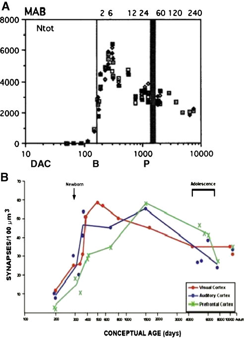
“Maximum dendritic growth occurs around P14 in rats, which corresponds to around 8 months in humans; whereas synaptogenesis peaks around P35 in rats and beginning at 1 year of age to 5-6 years in humans, depending on the brain region (Kolb et al., 2000; Pressler and Auvin, 2013; Stiles and Jernigan, 2010).”
BackgroundDoherty, B. T., Romano, M. E., Gui, J., Punshon, T., Jackson, B. P., Karagas, M. R., & Korrick, S. (2020) · Environmental Epidemiology · doi:10.1097/ee9.0000000000000106 · Cited by 34
“Maternal age (years), median (IQR) 31 (29, 34) 31 (29, 34) Maternal BMI (kg/m(2)), median (IQR) 25 (22, 28) 25 (22, 28) Highest parental education High school or less 55 (15) 51 (16) Any college 157 (42) 133 (42) Any graduate 159 (43) 134 (42) Maternal race White non-Hispanic 354 (95) 306 (96) Any other 17 (5) 12 (4)…”
Fair, D. A., Dosenbach, N. U. F., Moore, A. H., Satterthwaite, T. D., & Milham, M. P. (2021) · Annual Review of Developmental Psychology · doi:10.1146/annurev-devpsych-121318-085124 · Cited by 35
“The brain’s major fiber pathways become consolidated through myelination, though their presence is largely established prenatally (Stiles & Jernigan 2010).”
BackgroundReaching into the toolbox: Stem cell models to study neuropsychiatric disorders
Whiteley, J. T., Fernandes, S., Sharma, A., Mendes, A. P. D., Racha, V., Benassi, S. K., & Marchetto, M. C. (2022) · Stem Cell Reports · doi:10.1016/j.stemcr.2021.12.015 · Cited by 33
“Unlike the large transcription factor cascades responsible for influencing the transition and specification of cells during development, advances in cell reprogramming have identified a subset of transcriptional regulators capable of reprogramming a fully differentiated cell to a distantly related cell type (Stiles an…”
BackgroundCaramaschi, D., Neumann, A., Cárdenas, A., Tindula, G. N., Alemany, S., Zillich, L., Pesce, G., Lahti, J. M. T., Havdahl, A., Mulder, R. H., et al. (2022) · Molecular Psychiatry · doi:10.1038/s41380-022-01441-w · Cited by 33
“Since there is considerable environmental influence on cognitive ability in early childhood [1], it is plausible that environmental exposures that occur in early life, either prenatally or in early childhood, play a role in shaping children’s cognitive”
Volqvartz, T., Andersen, H. H., Pedersen, L. H., & Larsen, A. (2023) · European Journal of Neuroscience · doi:10.1111/ejn.16184 · Cited by 33
“The fetal brain is particularly sensitive and may be affected throughout the gestation (Fitzgerald et al., 2020; Kelley & Pas¸ca, 2022; Stiles & Jernigan, 2010) as the brain relies on neurotrophic signals from the placenta until the parturition (Miranda & Sousa, 2018; Sahay et al., 2017, 2020).”
BackgroundNaspolini, N. F., Schüroff, P. A., Figueiredo, M. J., Sbardellotto, G. E., Ferreira, F. R., Fatori, D., Polanczyk, G. V., Campos, A. C., & Taddei, C. R. (2024) · Microorganisms · doi:10.3390/microorganisms12030424 · Cited by 34
Nassar, R., Kaczkurkin, A. N., Xia, C. H., Sotiras, A., Pehlivanova, M., Moore, T. M., de la Garza, Á. G., Roalf, D. R., Rosen, A. F. G., Lorch, S. A., et al. (2018) · Cerebral Cortex · doi:10.1093/cercor/bhy091 · Cited by 33
Almenar-Queralt, A., Merkurjev, D., Kim, H. S., Navarro, M., Ma, Q., Chaves, R. S., Allegue, C., Driscoll, S. P., Chen, A. G., Kohlnhofer, B., et al. (2019) · Nature Genetics · doi:10.1038/s41588-019-0526-4 · Cited by 32
Radiographic Severity of Metopic Craniosynostosis Correlates with Long-Term Neurocognitive Outcomes
Gabrick, K. S., Wu, R. T., Singh, A., Persing, J. A., & Alperovich, M. A. (2020) · Plastic & Reconstructive Surgery · doi:10.1097/prs.0000000000006746 · Cited by 33
Bridging the Divide: Brain and Behavior in Developmental Language Disorder
Abbott, N. T., & Love, T. E. (2023) · Brain Sciences · doi:10.3390/brainsci13111606 · Cited by 32
“Diffusivity in Child Language-Impaired Populations If the transmission of signals between different neural regions is impaired or slowed in any way, it could lead to disruptions of the In contrast to these results, Girbau-Mas-sana et al. (2014) did not fi nd signi fi cant di ff erences in white ma tt…”
BackgroundMethodResultInfluentialFunctional genomics of human brain development and implications for autism spectrum disorders
Ziats, M. N., Grosvenor, L. P., & Rennert, O. M. (2015) · Translational Psychiatry · doi:10.1038/tp.2015.153 · Cited by 32
Auditory brainstem function in microcephaly related to Zika virus infection
Abramov, D. M., Saad, T., Gomes, S. C., Silva, D. D. S. E., Araújo, I., Moreira, M. E. L., & Lazarev, V. V. (2018) · Neurology · doi:10.1212/wnl.0000000000004974 · Cited by 32
“of cerebral cortex formation extending from the first to the third trimester.(32,33)”
BackgroundHolmes, K. (2019) · Interchange · doi:10.1007/s10780-019-09360-6 · Cited by 32
“We know that the processes that underlie and guide brain development involve the ongoing interplay of genetic and environmental factors” (Stiles and Jerrigan 2010).”
BackgroundInfluential
Metadata
- Authors
- Stiles, J., & Jernigan, T. L.
- Affiliations
- University of California San Diego (US)
- Corresponding author
- Jernigan, Terry L.
- ORCID
- Jernigan, Terry L.
- Venue
- Neuropsychology Review
- Published
- 2010-11-03
- Volume
- 20
- Issue
- 4
- Pages
- 327-348
- ISSN
- 1040-7308 (ISSN-L)
- 1573-6660
- PMC
- PMC2989000
- OpenAlex
- W2115599658
- Cited by
- 2,009
- References
- 132
- Citations by year
- 2026: 142 · 2025: 184 · 2024: 199 · 2023: 209 · 2022: 184 · 2021: 197 · 2020: 195 · 2019: 155 · 2018: 158 · 2017: 122 · 2016: 92 · 2015: 64 · 2014: 49 · 2013: 24 · 2012: 23
- FWCI
- 1.43
- Type
- journal article
- Language
- en
- Open access
- hybrid
- doi.org
- Licence
- cc-by-nc
- Locations
- Neuropsychology Review (publishedVersion, cc-by-nc)
- Europe PMC (PubMed Central) (publishedVersion, cc-by-nc)
- PubMed Central (publishedVersion, cc-by-nc)
- PubMed (publishedVersion)
- eScholarship (California Digital Library) (submittedVersion, public-domain)
- eScholarship (California Digital Library) (submittedVersion)
- Funders
- Lundbeckfonden (R32‐A3161)
- H. Lundbeck A/S
- National Institutes of Health (5r01hd025077-10)
- U.S. Public Health Service (R32-A3161)
- University of California, San Diego (RC2DA029475)
- National Institute on Drug Abuse (RC2DA029475)
- National Institute of Child Health and Human Development
- Eunice Kennedy Shriver National Institute of Child Health and Human Development (R01HD060595, R01 HD 25077)
- Topics
- Life Sciences › Neuroscience › Developmental Neuroscience › Neurogenesis and neuroplasticity mechanisms
- Life Sciences › Biochemistry, Genetics and Molecular Biology › Genetics › Genetics and Neurodevelopmental Disorders
- Social Sciences › Psychology › Developmental and Educational Psychology › Child and Animal Learning Development
- Keywords
- brain development, developmental neuroscience, mammalian brain, neuronal organization, social development (developmental psychology), cognitive development, neural systems, genetic constraints, developmental mechanisms
- MeSH terms
- Animals, Brain, Cell Differentiation, Diagnostic Imaging, Humans, Nerve Net, Neural Pathways, Neurobiology, Neurons, Gene Expression Regulation, Developmental, Body Patterning, Neurogenesis
About these checks
Registry, retraction, Cites and statistics results are computed, not judged, with one exception: a printed reference that no index lists is read once by a model before it counts as a citation error. Claims are extracted by a model and kept only when their quote appears verbatim in the text; each cited source is then checked, with a model’s help, for whether it says what the claim relies on. Those verdicts can be wrong — read the claim’s quote and the cited source before relying on one.
Statistics and arithmetic are recomputed, not judged.
Confidence bands are shown only for fields calibrated on our annotated test set. None is calibrated yet, so no band is shown.
What counts as a claim ›How we measure confidence ›How we check this ›
Are you an author, or do you see a problem with this page? Request a review
Data sources
Bibliographic data: Semantic Scholar, OpenAlex, Crossref.
Contains information from the Semantic Scholar Academic Graph Datasets, made available under the ODC Attribution License (ODC-BY 1.0).
Semantic Scholar’s data is described in: Kinney, R., et al. (2023). The Semantic Scholar Open Data Platform. arXiv:2301.10140.
