

The DOI matches its Crossref record; 16 of the 16 references it cites were found in a registry or an index.
Lipids and membrane-associated proteins in autophagy
Abstract
Autophagy is essential for the maintenance of cellular homeostasis and its dysfunction has been linked to various diseases. Autophagy is a membrane driven process and tightly regulated by membrane-associated proteins. Here, we summarized membrane lipid composition, and membrane-associated proteins relevant to autophagy from a spatiotemporal perspective. In particular, we focused on three important membrane remodeling processes in autophagy, lipid transfer for phagophore elongation, membrane scission for phagophore closure, and autophagosome-lysosome membrane fusion. We discussed the significance of the discoveries in this field and possible avenues to follow for future studies. Finally, we summarized the membrane-associated biochemical techniques and assays used to study membrane properties, with a discussion of their applications in autophagy.
Overview
The claims and statistics of this paper haven’t been checked yet.
Run the check — free, no account needed. It reads the open-access text and checks the sources it cites.
Integrity signals
Registry
Cites
Statistics
Figures
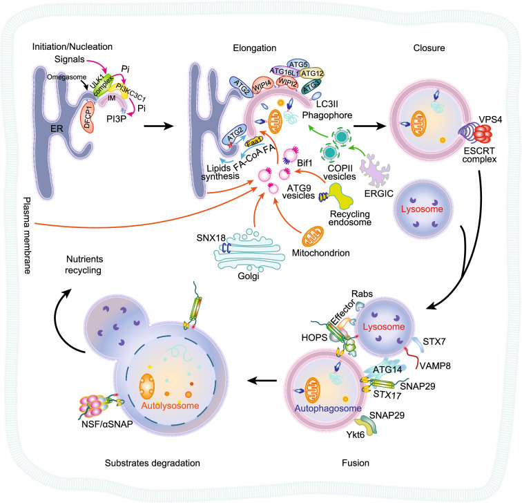
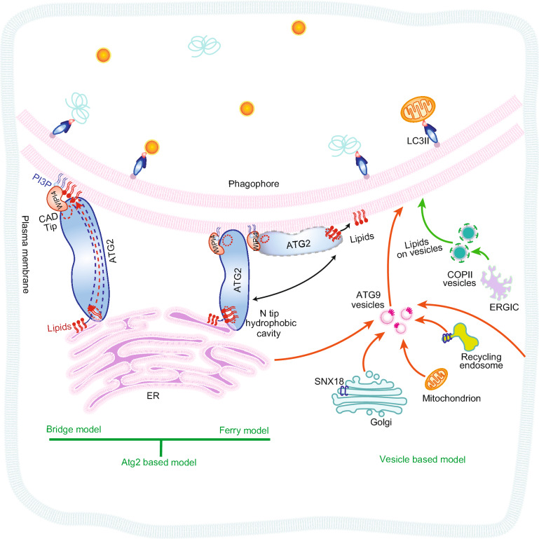
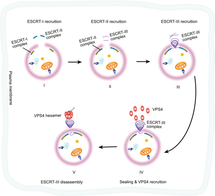
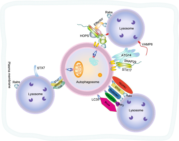
Verification
Claims the paper makes
The claims and statistics of this paper haven’t been checked yet.
Run the check — free, no account needed. It reads the open-access text and checks the sources it cites.
Claims other papers make about this one
Each is checked against this paper’s own text: does it say what the citing paper says it does?
In the citing paper
“Studies suggest the dyshomeostasis of lipid transport35,36 and/or the dysfunction of autophagy may play a role in CE dysregulation37,38.”
From GRAMD1B is a regulator of lipid homeostasis, autophagic flux and phosphorylated tau (2025) · cites this paper as [37]
In the citing paper
“Aster proteins, as well as lipids and lipid droplets, can also be regulated by autophagy37,38.”
From GRAMD1B is a regulator of lipid homeostasis, autophagic flux and phosphorylated tau (2025) · cites this paper as [37]
In the citing paper
“Given the degradation of both lipids and tau protein is regulated by autophagy37,38”
From GRAMD1B is a regulator of lipid homeostasis, autophagic flux and phosphorylated tau (2025) · cites this paper as [37]
In the citing paper
“CE accumulation could be due to the dyshomeostasis of lipid transport35,36 and/or the dysfunction of autophagy to degrade CE37,38.”
From GRAMD1B is a regulator of lipid homeostasis, autophagic flux and phosphorylated tau (2025) · cites this paper as [37]
In the citing paper
“Growing evidence demonstrates that dysfunctions in autophagy, lipid metabolism, and tau clearance are interconnected37,38”
From GRAMD1B is a regulator of lipid homeostasis, autophagic flux and phosphorylated tau (2025) · cites this paper as [37]
Self-check
Percentages, sums and reported differences re-computed from the paper’s own numbers.
The self-check needs the paper’s full text.
Citations
Cites
The Role of Atg Proteins in Autophagosome Formation
Mizushima, N., Yoshimori, T., & Ohsumi, Y. (2011) · Annual Review of Cell and Developmental Biology · doi:10.1146/annurev-cellbio-092910-154005 · Cited by 2,978
Biological Functions of Autophagy Genes: A Disease Perspective
Levine, B., & Kroemer, G. (2019) · Cell · doi:10.1016/j.cell.2018.09.048 · Cited by 2,832
Phosphoinositides in cell regulation and membrane dynamics
Di Paolo, G., & De Camilli, P. (2006) · Nature · doi:10.1038/nature05185 · Cited by 2,790
Molecular anatomy of a trafficking organelle
Takamori, S., Holt, M., Stenius, K., Lemke, E. A., Grønborg, M., Riedel, D., Urlaub, H., Schenck, S., Brügger, B., Ringler, P., et al. (2006) · Cell · doi:10.1016/j.cell.2006.10.030 · Cited by 2,373
Nutrient-dependent mTORC1 Association with the ULK1–Atg13–FIP200 Complex Required for Autophagy
Hosokawa, N., Hara, T., Kaizuka, T., Kishi, C., Takamura, A., Miura, Y., Iemura, S.-I., Natsume, T., Takehana, K., Yamada, N., et al. (2009) · Molecular Biology of the Cell · doi:10.1091/mbc.e08-12-1248 · Cited by 2,076
Network organization of the human autophagy system
Behrends, C., Sowa, M. E., Gygi, S. P., & Harper, J. W. (2010) · Nature · doi:10.1038/nature09204 · Cited by 1,554
The Atg12-Atg5 Conjugate Has a Novel E3-like Activity for Protein Lipidation in Autophagy
Hanada, T., Noda, N. N., Satomi, Y., Ichimura, Y., Fujioka, Y., Takao, T., Inagaki, F., & Ohsumi, Y. (2007) · Journal of Biological Chemistry · doi:10.1074/jbc.c700195200 · Cited by 1,123 · cited 1×
“PI3P recruits the ATG16L1ATG5∼ATG12 complex through WIPI2 (WD repeat domain phosphoinositide-interacting protein 2) for conjugating PE to LC3 (microtubule-associated proteins 1A/1B light chain 3) (Hanada et al., 2007; Fujita et al., 2008).”
BackgroundPlasma membrane contributes to the formation of pre-autophagosomal structures
Ravikumar, B., Moreau, K., Jahreiss, L., Puri, C., & Rubinsztein, D. C. (2010) · Nature Cell Biology · doi:10.1038/ncb2078 · Cited by 902
Formation Process of Autophagosome Is Traced with Apg8/Aut7p in Yeast
Kirisako, T., Baba, M., Ishihara, N., Miyazawa, K., Ohsumi, M., Yoshimori, T., Noda, T., & Ohsumi, Y. (1999) · The Journal of Cell Biology · doi:10.1083/jcb.147.2.435 · Cited by 881
Molecular mechanism of multivesicular body biogenesis by ESCRT complexes
Wollert, T., & Hurley, J. H. (2010) · Nature · doi:10.1038/nature08849 · Cited by 804
Research works printed but not linked: 176
- Abada A, Levin-Zaidman S, Porat Z, Dadosh T, Elazar Z (2017) SNARE priming is essential for maturation of autophagosomes but not for their formation. Proc Natl Acad Sci USA 114:12749–12754 · DOI
- Asao H, Sasaki Y, Arita T, Tanaka N, Endo K, Kasai H, Takeshita T, Endo Y, Fujita T, Sugamura K (1997) Hrs is associated with STAM, a signal-transducing adaptor molecule. Its suppressive effect on cytokine-induced cell growth. J Biol Chem 272:32785–32791 · DOI
- Axe EL, Walker SA, Manifava M, Chandra P, Roderick HL, Habermann A, Griffiths G, Ktistakis NT (2008) Autophagosome formation from membrane compartments enriched in phosphatidylinositol 3-phosphate and dynamically connected to the endoplasmic reticulum. J Cell Biol 182:685–701 · DOI · Paper page
- Baba T, Toth DJ, Sengupta N, Kim YJ, Balla T (2019) Phosphatidylinositol 4,5-bisphosphate controls Rab7 and PLEKHM1 membrane cycling during autophagosome-lysosome fusion. EMBO J 38:e100312 · DOI
- Babst M, Katzmann DJ, Estepa-Sabal EJ, Meerloo T, Emr SD (2002a) Escrt-III: an endosome-associated heterooligomeric protein complex required for mvb sorting. Dev Cell 3:271–282 · DOI
- Babst M, Katzmann DJ, Snyder WB, Wendland B, Emr SD (2002b) Endosome-associated complex, ESCRT-II, recruits transport machinery for protein sorting at the multivesicular body. Dev Cell 3:283–289 · DOI
- Baskaran S, Ragusa MJ, Boura E, Hurley JH (2012) Two-site recognition of phosphatidylinositol 3-phosphate by PROPPINs in autophagy. Mol Cell 47:339–348 · DOI
- Bas L, Papinski D, Licheva M, Torggler R, Rohringer S, Schuschnig M, Kraft C (2018) Reconstitution reveals Ykt6 as the autophagosomal SNARE in autophagosome–vacuole fusion. J Cell Biol 217:3656–3669 · DOI · Paper page
- Bean BD, Dziurdzik SK, Kolehmainen KL, Fowler CM, Kwong WK, Grad LI, Davey M, Schluter C, Conibear E (2018) Competitive organelle-specific adaptors recruit Vps13 to membrane contact sites. J Cell Biol 217:3593–3607 · DOI
- Besprozvannaya M, Dickson E, Li H, Ginburg KS, Bers DM, Auwerx J, Nunnari J (2018) GRAM domain proteins specialize functionally distinct ER-PM contact sites in human cells. Elife 7:e31019 · DOI · Paper page
- Bian X, Zhang Z, Xiong Q, De Camilli P, Lin C (2019) A programmable DNA-origami platform for studying lipid transfer between bilayers. Nat Chem Biol 15:830–837 · DOI
- Bielli A, Haney CJ, Gabreski G, Watkins SC, Bannykh SI, Aridor M (2005) Regulation of Sar1 NH2 terminus by GTP binding and hydrolysis promotes membrane deformation to control COPII vesicle fission. The Journal of cell biology 171:919–924 · DOI
- Buchkovich NJ, Henne WM, Tang S, Emr SD (2013) Essential N-terminal insertion motif anchors the ESCRT-III filament during MVB vesicle formation. Dev Cell 27:201–214 · DOI
- Caillat C, Macheboeuf P, Wu Y, McCarthy AA, Boeri-Erba E, Effantin G, Gottlinger HG, Weissenhorn W, Renesto P (2015) Asymmetric ring structure of Vps4 required for ESCRT-III disassembly. Nat Commun 6:8781 · DOI
- Carlsson SR, Simonsen A (2015) Membrane dynamics in autophagosome biogenesis. J Cell Sci 128:193–205
- Carroll B, Mohd-Naim N, Maximiano F, Frasa MA, McCormack J, Finelli M, Thoresen SB, Perdios L, Daigaku R, Francis RE et al (2013) The TBC/RabGAP Armus coordinates Rac1 and Rab7 functions during autophagy. Dev Cell 25:15–28 · DOI
- Chang C, Young LN, Morris KL, von Bülow S, Schöneberg J, Yamamoto-Imoto H, Oe Y, Yamamoto K, Nakamura S, Stjepanovic G (2019) Bidirectional control of autophagy by BECN1 BARA domain dynamics. Mol Cell 73(339–353):e336
- Chan EY, Longatti A, McKnight NC, Tooze SA (2009) Kinase-inactivated ULK proteins inhibit autophagy via their conserved C-terminal domains using an Atg13-independent mechanism. Mol Cell Biol 29:157–171 · DOI · Paper page
- Chen D, Fan W, Lu Y, Ding X, Chen S, Zhong Q (2012) A mammalian autophagosome maturation mechanism mediated by TECPR1 and the Atg12-Atg5 conjugate. Mol Cell 45:629–641 · DOI
- Chiaruttini N, Redondo-Morata L, Colom A, Humbert F, Lenz M, Scheuring S, Roux A (2015) Relaxation of loaded ESCRT-III spiral springs drives membrane deformation. Cell 163:866–879 · DOI
- Chowdhury S, Otomo C, Leitner A, Ohashi K, Aebersold R, Lander GC, Otomo T (2018) Insights into autophagosome biogenesis from structural and biochemical analyses of the ATG2A-WIPI4 complex. Proc Natl Acad Sci 115:E9792–E9801 · DOI
- Chung T (2019) How phosphoinositides shape autophagy in plant cells. Plant Sci 281:146–158 · DOI
- Cudjoe EK Jr, Saleh T, Hawkridge AM, Gewirtz DA (2017) Proteomics insights into autophagy. Proteomics 17:1700022 · DOI
- Daum G, Vance JE (1997) Import of lipids into mitochondria. Prog Lipid Res 36:103–130 · DOI
- de la Ballina LR, Munson MJ, Simonsen A (2020) Lipids and lipid-binding proteins in selective autophagy. J Mol Biol 432:135–159 · DOI
- de Kroon AI, Dolis D, Mayer A, Lill R, de Kruijff B (1997) Phospholipid composition of highly purified mitochondrial outer membranes of rat liver and Neurospora crassa. Is cardiolipin present in the mitochondrial outer membrane? Biochim Biophys Acta (BBA) 1325:108–116 · DOI
- Delorme-Axford E, Klionsky DJ (2018) Transcriptional and post-transcriptional regulation of autophagy in the yeast Saccharomyces cerevisiae. J Biol Chem 293:5396–5403 · DOI
- Diao J, Ishitsuka Y, Lee H, Joo C, Su Z, Syed S, Shin YK, Yoon TY, Ha T (2012) A single vesicle-vesicle fusion assay for in vitro studies of SNAREs and accessory proteins. Nat Protoc 7:921–934 · DOI
- Diao J, Liu R, Rong Y, Zhao M, Zhang J, Lai Y, Zhou Q, Wilz LM, Li J, Vivona S et al (2015) ATG14 promotes membrane tethering and fusion of autophagosomes to endolysosomes. Nature 520:563–566 · DOI
- Dikic I, Elazar Z (2018) Mechanism and medical implications of mammalian autophagy. Nat Rev Mol Cell Biol 19:349–364 · DOI · Paper page
- Ding X, Jiang X, Tian R, Zhao P, Li L, Wang X, Chen S, Zhu Y, Mei M, Bao S et al (2019) RAB2 regulates the formation of autophagosome and autolysosome in mammalian cells. Autophagy 15:1774–1786 · DOI
- Dove SK, Dong K, Kobayashi T, Williams FK, Michell RH (2009) Phosphatidylinositol 3,5-bisphosphate and Fab1p/PIKfyve underPPIn endo-lysosome function. Biochem J 419:1–13 · DOI
- Dudley LJ, Cabodevilla AG, Makar AN, Sztacho M, Michelberger T, Marsh JA, Houston DR, Martens S, Jiang X, Gammoh N (2019) Intrinsic lipid binding activity of ATG16L1 supports efficient membrane anchoring and autophagy. EMBO J 38:e100554 · DOI
- Ebner P, Poetsch I, Deszcz L, Hoffmann T, Zuber J, Ikeda F (2018) The IAP family member BRUCE regulates autophagosome–lysosome fusion. Nat Commun 9:1–15 · DOI
- Feng Q, Luo Y, Zhang XN, Yang XF, Hong XY, Sun DS, Li XC, Hu Y, Li XG, Zhang JF et al (2020) MAPT/Tau accumulation represses autophagy flux by disrupting IST1-regulated ESCRT-III complex formation: a vicious cycle in Alzheimer neurodegeneration. Autophagy 16:641–658 · DOI
- Fujioka Y, Noda NN, Nakatogawa H, Ohsumi Y, Inagaki F (2010) Dimeric coiled-coil structure of Saccharomyces cerevisiae Atg16 and its functional significance in autophagy. J Biol Chem 285:1508–1515 · DOI
- Fujioka Y, Alam JM, Noshiro D, Mouri K, Ando T, Okada Y, May AI, Knorr RL, Suzuki K, Ohsumi Y et al (2020) Phase separation organizes the site of autophagosome formation. Nature 578:301–305 · DOI
- Fujita N, Itoh T, Omori H, Fukuda M, Noda T, Yoshimori T (2008) The Atg16L complex specifies the site of LC3 lipidation for membrane biogenesis in autophagy. Mol Biol Cell 19:2092–2100 · DOI
- Gatica D, Lahiri V, Klionsky DJ (2018) Cargo recognition and degradation by selective autophagy. Nat Cell Biol 20:233–242 · DOI · Paper page
- Gatta AT, Carlton JG (2019) The ESCRT-machinery: closing holes and expanding roles. Curr Opin Cell Biol 59:121–132 · DOI
- Ge L, Schekman R (2014) The ER-Golgi intermediate compartment feeds the phagophore membrane. Autophagy 10:170–172 · DOI
- Ge L, Melville D, Zhang M, Schekman R (2013) The ER–Golgi intermediate compartment is a key membrane source for the LC3 lipidation step of autophagosome biogenesis. Elife 2:e00947 · DOI
- Ge L, Zhang M, Schekman R (2014) Phosphatidylinositol 3-kinase and COPII generate LC3 lipidation vesicles from the ER-Golgi intermediate compartment. elife 3:e04135 · DOI
- Ge L, Zhang M, Kenny SJ, Liu D, Maeda M, Saito K, Mathur A, Xu K, Schekman R (2017) Remodeling of ER-exit sites initiates a membrane supply pathway for autophagosome biogenesis. EMBO Rep 18:1586–1603 · DOI
- Gómez-Sánchez R, Rose J, Guimarães R, Mari M, Papinski D, Rieter E, Geerts WJ, Hardenberg R, Kraft C, Ungermann C (2018) Atg9 establishes Atg2-dependent contact sites between the endoplasmic reticulum and phagophores. J Cell Biol 217:2743–2763 · DOI
- Graef M (2020) Recent advances in the understanding of autophagosome biogenesis. F1000Res 9. · DOI
- Graef M, Friedman JR, Graham C, Babu M, Nunnari J (2013) ER exit sites are physical and functional core autophagosome biogenesis components. Mol Biol Cell 24:2918–2931 · DOI
- Hailey DW, Rambold AS, Satpute-Krishnan P, Mitra K, Sougrat R, Kim PK, Lippincott-Schwartz J (2010) Mitochondria supply membranes for autophagosome biogenesis during starvation. Cell 141:656–667 · DOI
- Hasegawa J, Iwamoto R, Otomo T, Nezu A, Hamasaki M, Yoshimori T (2016) Autophagosome-lysosome fusion in neurons requires INPP5E, a protein associated with Joubert syndrome. EMBO J 35:1853–1867 · DOI
- Hayashi-Nishino M, Fujita N, Noda T, Yamaguchi A, Yoshimori T, Yamamoto A (2009) A subdomain of the endoplasmic reticulum forms a cradle for autophagosome formation. Nat Cell Biol 11:1433–1437 · DOI
- He S, Ni D, Ma B, Lee J-H, Zhang T, Ghozalli I, Pirooz SD, Zhao Z, Bharatham N, Li B (2013) PtdIns (3) P-bound UVRAG coordinates Golgi–ER retrograde and Atg9 transport by differential interactions with the ER tether and the beclin 1 complex. Nat Cell Biol 15:1206–1219 · DOI
- Hollenstein DM, Kraft C (2020) Autophagosomes are formed at a distinct cellular structure. Curr Opin Cell Biol 65:50–57 · DOI
- Ho CY, Alghamdi TA, Botelho RJ (2012) Phosphatidylinositol-3,5-bisphosphate: no longer the poor PIP2. Traffic 13:1–8 · DOI
- Huang X, Sun S, Wang X, Fan F, Zhou Q, Lu S, Cao Y, Wang QW, Dong MQ, Yao J et al (2019) Mechanistic insights into the SNARE complex disassembly. Sci Adv 5:eaau8164 · DOI
- Hurley JH, Young LN (2017) Mechanisms of autophagy initiation. Annu Rev Biochem 86:225–244 · DOI
- Ichimura Y, Kirisako T, Takao T, Satomi Y, Shimonishi Y, Ishihara N, Mizushima N, Tanida I, Kominami E, Ohsumi M et al (2000) A ubiquitin-like system mediates protein lipidation. Nature 408:488–492 · DOI
- Ishihara N, Hamasaki M, Yokota S, Suzuki K, Kamada Y, Kihara A, Yoshimori T, Noda T, Ohsumi Y (2001) Autophagosome requires specific early Sec proteins for its formation and NSF/SNARE for vacuolar fusion. Mol Biol Cell 12:3690–3702 · DOI
- Itakura E, Kishi-Itakura C, Mizushima N (2012) The hairpin-type tail-anchored SNARE syntaxin 17 targets to autophagosomes for fusion with endosomes/lysosomes. Cell 151:1256–1269 · DOI
- Jang DJ, Lee JA (2016) The roles of phosphoinositides in mammalian autophagy. Arch Pharm Res 39:1129–1136 · DOI
- Jeynov B, Lay D, Schmidt F, Tahirovic S, Just WW (2006) Phosphoinositide synthesis and degradation in isolated rat liver peroxisomes. FEBS Lett 580:5917–5924 · DOI
- Jiang P, Nishimura T, Sakamaki Y, Itakura E, Hatta T, Natsume T, Mizushima N (2014) The HOPS complex mediates autophagosome-lysosome fusion through interaction with syntaxin 17. Mol Biol Cell 25:1327–1337 · DOI · Paper page
- Johansen T, Lamark T (2020) Selective autophagy: ATG8 family proteins, LIR motifs and cargo receptors. J Mol Biol 432:80–103 · DOI
- Jung CH, Jun CB, Ro SH, Kim YM, Otto NM, Cao J, Kundu M, Kim DH (2009) ULK-Atg13-FIP200 complexes mediate mTOR signaling to the autophagy machinery. Mol Biol Cell 20:1992–2003 · DOI · Paper page
- Jun Y, Wickner W (2007) Assays of vacuole fusion resolve the stages of docking, lipid mixing, and content mixing. Proc Natl Acad Sci 104:13010–13015 · DOI
- Kabeya Y, Mizushima N, Ueno T, Yamamoto A, Kirisako T, Noda T, Kominami E, Ohsumi Y, Yoshimori T (2000) LC3, a mammalian homologue of yeast Apg8p, is localized in autophagosome membranes after processing. EMBO J 19:5720–5728 · DOI · Paper page
- Karanasios E, Stapleton E, Manifava M, Kaizuka T, Mizushima N, Walker SA, Ktistakis NT (2013) Dynamic association of the ULK1 complex with omegasomes during autophagy induction. J Cell Sci 126:5224–5238
- Katzmann DJ, Babst M, Emr SD (2001) Ubiquitin-dependent sorting into the multivesicular body pathway requires the function of a conserved endosomal protein sorting complex, ESCRT-I. Cell 106:145–155 · DOI
- Knaevelsrud H, Soreng K, Raiborg C, Haberg K, Rasmuson F, Brech A, Liestol K, Rusten TE, Stenmark H, Neufeld TP et al (2013) Membrane remodeling by the PX-BAR protein SNX18 promotes autophagosome formation. J Cell Biol 202:331–349 · DOI
- Komada M, Kitamura N (1995) Growth factor-induced tyrosine phosphorylation of Hrs, a novel 115-kilodalton protein with a structurally conserved putative zinc finger domain. Mol Cell Biol 15:6213–6221 · DOI · Paper page
- Kostelansky MS, Schluter C, Tam YY, Lee S, Ghirlando R, Beach B, Conibear E, Hurley JH (2007) Molecular architecture and functional model of the complete yeast ESCRT-I heterotetramer. Cell 129:485–498 · DOI · Paper page
- Kriegenburg F, Ungermann C, Reggiori F (2018) Coordination of autophagosome–lysosome fusion by ATG8 family members. Curr Biol 28:R512–R518 · DOI
- Kriegenburg F, Bas L, Gao J, Ungermann C, Kraft C (2019) The multi-functional SNARE protein Ykt6 in autophagosomal fusion processes. Cell Cycle 18:639–651 · DOI
- Ktistakis NT (2019) Who plays the ferryman: ATG2 channels lipids into the forming autophagosome. J Cell Biol 218:1767 · DOI
- Kumar N, Leonzino M, Hancock-Cerutti W, Horenkamp FA, Li P, Lees JA, Wheeler H, Reinisch KM, De Camilli P (2018) VPS13A and VPS13C are lipid transport proteins differentially localized at ER contact sites. J Cell Biol 217:3625–3639 · DOI
- Kyoung M, Srivastava A, Zhang Y, Diao J, Vrljic M, Grob P, Nogales E, Chu S, Brunger AT (2011) In vitro system capable of differentiating fast Ca2+-triggered content mixing from lipid exchange for mechanistic studies of neurotransmitter release. Proc Natl Acad Sci 108:E304–E313 · DOI · Paper page
- Kyoung M, Zhang Y, Diao J, Chu S, Brunger AT (2013) Studying calcium-triggered vesicle fusion in a single vesicle-vesicle content and lipid-mixing system. Nat Protoc 8:1–16 · DOI
- Lai LTF, Ye H, Zhang W, Jiang L, Lau WCY (2019) Structural biology and electron microscopy of the autophagy molecular machinery. Cells 8:1627 · DOI
- Laraia L, Friese A, Corkery DP, Konstantinidis G, Erwin N, Hofer W, Karatas H, Klewer L, Brockmeyer A, Metz M (2019) The cholesterol transfer protein GRAMD1A regulates autophagosome biogenesis. Nat Chem Biol 15:710–720 · DOI
- Lemus L, Ribas JL, Sikorska N, Goder V (2016) An ER-localized SNARE protein is exported in specific COPII vesicles for autophagosome biogenesis. Cell Rep 14:1710–1722 · DOI
- Liu X, Seven AB, Camacho M, Esser V, Xu J, Trimbuch T, Quade B, Su L, Ma C, Rosenmund C et al (2016) Functional synergy between the Munc13 C-terminal C1 and C2 domains. Elife 5:e13696 · DOI
- Liu X, Seven AB, Xu J, Esser V, Su L, Ma C, Rizo J (2017) Simultaneous lipid and content mixing assays for in vitro reconstitution studies of synaptic vesicle fusion. Nat Protoc 12:2014–2028 · DOI
- Li L, Zhong Q (2016) Autophagosome-lysosome fusion: PIs to the rescue. EMBO J 35:1845–1847 · DOI
- Lystad AH, Simonsen A (2016) Phosphoinositide-binding proteins in autophagy. FEBS Lett 590:2454–2468 · DOI
- Lystad AH, Carlsson SR, Laura R, Kauffman KJ, Nag S, Yoshimori T, Melia TJ, Simonsen A (2019) Distinct functions of ATG16L1 isoforms in membrane binding and LC3B lipidation in autophagy-related processes. Nat Cell Biol 21:372–383 · DOI
- Maeda S, Otomo C, Otomo T (2019) The autophagic membrane tether ATG2A transfers lipids between membranes. Elife 8:e45777 · DOI
- Matsui T, Jiang P, Nakano S, Sakamaki Y, Yamamoto H, Mizushima N (2018) Autophagosomal YKT6 is required for fusion with lysosomes independently of syntaxin 17. J Cell Biol 217:2633–2645 · DOI · Paper page
- Matsushita M, Suzuki NN, Obara K, Fujioka Y, Ohsumi Y, Inagaki F (2007) Structure of Atg5.Atg16, a complex essential for autophagy. J Biol Chem 282:6763–6772 · DOI
- Ma C, Su L, Seven AB, Xu Y, Rizo J (2013) Reconstitution of the vital functions of Munc18 and Munc13 in neurotransmitter release. Science 339:421–425 · DOI
- Ma M, Liu J-J, Li Y, Huang Y, Ta N, Chen Y, Fu H, Ye M-D, Ding Y, Huang W (2017) Cryo-EM structure and biochemical analysis reveal the basis of the functional difference between human PI3KC3-C1 and-C2. Cell Res 27:989–1001 · DOI
- McEwan DG, Popovic D, Gubas A, Terawaki S, Suzuki H, Stadel D, Coxon FP, Miranda de Stegmann D, Bhogaraju S, Maddi K et al (2015) PLEKHM1 regulates autophagosome-lysosome fusion through HOPS complex and LC3/GABARAP proteins. Mol Cell 57:39–54 · DOI · Paper page
- Melia TJ, Lystad AH, Simonsen A (2020) Autophagosome biogenesis: from membrane growth to closure. J Cell Biol 219:e202002085 · DOI
- Mercer TJ, Gubas A, Tooze SA (2018) A molecular perspective of mammalian autophagosome biogenesis. J Biol Chem 293:5386–5395 · DOI
- Miao G, Zhang Y, Chen D, Zhang H (2020) The ER-localized transmembrane protein TMEM39A/SUSR2 regulates autophagy by controlling the trafficking of the PtdIns(4)P phosphatase SAC1. Mol Cell 77(618–632):e615
- Mizushima N (2007) Autophagy: process and function. Genes Dev 21:2861–2873 · DOI
- Moreau K, Ravikumar B, Renna M, Puri C, Rubinsztein DC (2011) Autophagosome precursor maturation requires homotypic fusion. Cell 146:303–317 · DOI
- Muñoz-Braceras S, Calvo R, Escalante R (2015) TipC and the chorea-acanthocytosis protein VPS13A regulate autophagy in Dictyostelium and human HeLa cells. Autophagy 11:918–927 · DOI
- Nair U, Jotwani A, Geng J, Gammoh N, Richerson D, Yen W-L, Griffith J, Nag S, Wang K, Moss T (2011) SNARE proteins are required for macroautophagy. Cell 146:290–302 · DOI
- Naito T, Ercan B, Krshnan L, Triebl A, Koh DHZ, Wei F-Y, Tomizawa K, Torta FT, Wenk MR, Saheki Y (2019) Movement of accessible plasma membrane cholesterol by the GRAMD1 lipid transfer protein complex. eLife 8:e51401 · DOI · Paper page
- Nakamura S, Yoshimori T (2017) New insights into autophagosome–lysosome fusion. J Cell Sci 130:1209–1216 · DOI
- Nakatogawa H (2020) Mechanisms governing autophagosome biogenesis. Nat Rev Mol Cell Biol · DOI
- Nakatogawa H, Ichimura Y, Ohsumi Y (2007) Atg8, a ubiquitin-like protein required for autophagosome formation, mediates membrane tethering and hemifusion. Cell 130:165–178 · DOI
- Nascimbeni AC, Codogno P, Morel E (2017) Phosphatidylinositol-3-phosphate in the regulation of autophagy membrane dynamics. FEBS J 284:1267–1278 · DOI
- Nath S, Dancourt J, Shteyn V, Puente G, Fong WM, Nag S, Bewersdorf J, Yamamoto A, Antonny B, Melia TJ (2014) Lipidation of the LC3/GABARAP family of autophagy proteins relies on a membrane-curvature-sensing domain in Atg3. Nat Cell Biol 16:415–424 · DOI
- Nishimura T, Tooze SA (2020) Emerging roles of ATG proteins and membrane lipids in autophagosome formation. Cell Discov 6:32 · DOI
- Odorizzi G, Babst M, Emr SD (1998) Fab1p PtdIns(3)P 5-kinase function essential for protein sorting in the multivesicular body. Cell 95:847–858 · DOI · Paper page
- Ogawa M, Yoshikawa Y, Kobayashi T, Mimuro H, Fukumatsu M, Kiga K, Piao Z, Ashida H, Yoshida M, Kakuta S (2011) A Tecpr1-dependent selective autophagy pathway targets bacterial pathogens. Cell Host Microbe 9:376–389 · DOI
- Omari S, Makareeva E, Roberts-Pilgrim A, Mirigian L, Jarnik M, Ott C, Lippincott-Schwartz J, Leikin S (2018) Noncanonical autophagy at ER exit sites regulates procollagen turnover. Proc Natl Acad Sci 115:E10099–E10108 · DOI
- Osawa T, Noda NN (2019) Atg2: A novel phospholipid transfer protein that mediates de novo autophagosome biogenesis. Protein Sci 28:1005–1012 · DOI
- Osawa T, Alam JM, Noda NN (2019a) Membrane-binding domains in autophagy. Chem Phys Lipids 218:1–9 · DOI
- Osawa T, Ishii Y, Noda NN (2019b) Human ATG2B possesses a lipid transfer activity which is accelerated by negatively charged lipids and WIPI4. Genes Cells 25:65 · DOI
- Osawa T, Kotani T, Kawaoka T, Hirata E, Suzuki K, Nakatogawa H, Ohsumi Y, Noda NN (2019c) Atg2 mediates direct lipid transfer between membranes for autophagosome formation. Nat Struct Mol Biol 26:281–288 · DOI
- Otomo T, Maeda S (2019) ATG2A transfers lipids between membranes in vitro. Autophagy 15:2031–2032 · DOI
- Otomo T, Chowdhury S, Lander GC (2018) The rod-shaped ATG2A-WIPI4 complex tethers membranes in vitro. Contact 1:2515256418819936 · DOI
- Palamiuc L, Ravi A, Emerling BM (2020) Phosphoinositides in autophagy: current roles and future insights. FEBS J 287:222–238 · DOI
- Pankiv S, Clausen TH, Lamark T, Brech A, Bruun JA, Outzen H, Overvatn A, Bjorkoy G, Johansen T (2007) p62/SQSTM1 binds directly to Atg8/LC3 to facilitate degradation of ubiquitinated protein aggregates by autophagy. J Biol Chem 282:24131–24145 · DOI · Paper page
- Petiot A, Ogier-Denis E, Blommaart EF, Meijer AJ, Codogno P (2000) Distinct classes of phosphatidylinositol 3′-kinases are involved in signaling pathways that control macroautophagy in HT-29 cells. J Biol Chem 275:992–998 · DOI
- Polson HE, de Lartigue J, Rigden DJ, Reedijk M, Urbé S, Clague MJ, Tooze SA (2010) Mammalian Atg18 (WIPI2) localizes to omegasome-anchored phagophores and positively regulates LC3 lipidation. Autophagy 6:506–522 · DOI · Paper page
- Puri C, Renna M, Bento CF, Moreau K, Rubinsztein DC (2013) Diverse autophagosome membrane sources coalesce in recycling endosomes. Cell 154:1285–1299 · DOI
- Ragusa MJ, Stanley RE, Hurley JH (2012) Architecture of the Atg17 complex as a scaffold for autophagosome biogenesis. Cell 151:1501–1512 · DOI · Paper page
- Raymond CK, Howald-Stevenson I, Vater CA, Stevens TH (1992) Morphological classification of the yeast vacuolar protein sorting mutants: evidence for a prevacuolar compartment in class E vps mutants. Mol Biol Cell 3:1389–1402 · DOI
- Reggiori F, Ungermann C (2017) Autophagosome maturation and fusion. J Mol Biol 429:486–496 · DOI
- Romanov J, Walczak M, Ibiricu I, Schuchner S, Ogris E, Kraft C, Martens S (2012) Mechanism and functions of membrane binding by the Atg5-Atg12/Atg16 complex during autophagosome formation. EMBO J 31:4304–4317 · DOI
- Rong Y, Liu M, Ma L, Du W, Zhang H, Tian Y, Cao Z, Li Y, Ren H, Zhang C et al (2012) Clathrin and phosphatidylinositol-4,5-bisphosphate regulate autophagic lysosome reformation. Nat Cell Biol 14:924–934 · DOI · Paper page
- Russell RC, Tian Y, Yuan H, Park HW, Chang YY, Kim J, Kim H, Neufeld TP, Dillin A, Guan KL (2013) ULK1 induces autophagy by phosphorylating Beclin-1 and activating VPS34 lipid kinase. Nat Cell Biol 15:741–750 · DOI · Paper page
- Rusten TE, Stenmark H (2009) How do ESCRT proteins control autophagy? J Cell Sci 122:2179–2183 · DOI
- Schoneberg J, Lee IH, Iwasa JH, Hurley JH (2017) Reverse-topology membrane scission by the ESCRT proteins. Nat Rev Mol Cell Biol 18:5–17 · DOI
- Schoneberg J, Pavlin MR, Yan S, Righini M, Lee IH, Carlson LA, Bahrami AH, Goldman DH, Ren X, Hummer G et al (2018) ATP-dependent force generation and membrane scission by ESCRT-III and Vps4. Science 362:1423–1428 · DOI
- Schütter M, Giavalisco P, Brodesser S, Graef M (2020) Local fatty acid channeling into phospholipid synthesis drives phagophore expansion during autophagy. Cell 180(135–149):e114
- Schu PV, Takegawa K, Fry MJ, Stack JH, Waterfield MD, Emr SD (1993) Phosphatidylinositol 3-kinase encoded by yeast VPS34 gene essential for protein sorting. Science 260:88–91 · DOI · Paper page
- Shatz O, Holland P, Elazar Z, Simonsen A (2016) Complex relations between phospholipids, autophagy, and neutral lipids. Trends Biochem Sci 41:907–923 · DOI
- Shibutani ST, Yoshimori T (2014) A current perspective of autophagosome biogenesis. Cell Res 24:58–68 · DOI
- Shima T, Kirisako H, Nakatogawa H (2019) COPII vesicles contribute to autophagosomal membranes. J Cell Biol 218:1503–1510 · DOI
- Shintani T, Suzuki K, Kamada Y, Noda T, Ohsumi Y (2001) Apg2p functions in autophagosome formation on the perivacuolar structure. J Biol Chem 276:30452–30460 · DOI
- Sitarska E, Xu J, Park S, Liu X, Quade B, Stepien K, Sugita K, Brautigam CA, Sugita S, Rizo J (2017) Autoinhibition of Munc18-1 modulates synaptobrevin binding and helps to enable Munc13-dependent regulation of membrane fusion. Elife 6:e24278 · DOI
- Slessareva JE, Routt SM, Temple B, Bankaitis VA, Dohlman HG (2006) Activation of the phosphatidylinositol 3-kinase Vps34 by a G protein α subunit at the endosome. Cell 126:191–203 · DOI
- Soreng K, Munson MJ, Lamb CA, Bjorndal GT, Pankiv S, Carlsson SR, Tooze SA, Simonsen A (2018) SNX18 regulates ATG9A trafficking from recycling endosomes by recruiting Dynamin-2. EMBO Rep 19:e44837 · DOI
- Stadel D, Millarte V, Tillmann KD, Huber J, Tamin-Yecheskel BC, Akutsu M, Demishtein A, Ben-Zeev B, Anikster Y, Perez F et al (2015) TECPR2 cooperates with LC3C to regulate COPII-dependent ER export. Mol cell 60:89–104 · DOI · Paper page
- Stroupe C, Collins KM, Fratti RA, Wickner W (2006) Purification of active HOPS complex reveals its affinities for phosphoinositides and the SNARE Vam7p. EMBO J 25:1579–1589 · DOI
- Sun Q, Fan W, Chen K, Ding X, Chen S, Zhong Q (2008) Identification of Barkor as a mammalian autophagy-specific factor for Beclin 1 and class III phosphatidylinositol 3-kinase. Proc Natl Acad Sci 105:19211–19216 · DOI · Paper page
- Sun Q, Zhang J, Fan W, Wong KN, Ding X, Chen S, Zhong Q (2011) The RUN domain of rubicon is important for hVps34 binding, lipid kinase inhibition, and autophagy suppression. J Biol Chem 286:185–191 · DOI · Paper page
- Suzuki H, Osawa T, Fujioka Y, Noda NN (2017) Structural biology of the core autophagy machinery. Curr Opin Struct Biol 43:10–17 · DOI
- Takahashi Y, Coppola D, Matsushita N, Cualing HD, Sun M, Sato Y, Liang C, Jung JU, Cheng JQ, Mul JJ (2007) Bif-1 interacts with Beclin 1 through UVRAG and regulates autophagy and tumorigenesis. Nat Cell Biol 9:1142–1151 · DOI
- Takahashi Y, Meyerkord CL, Hori T, Runkle K, Fox TE, Kester M, Loughran TP, Wang HG (2011) Bif-1 regulates Atg9 trafficking by mediating the fission of Golgi membranes during autophagy. Autophagy 7:61–73 · DOI
- Takahashi Y, He H, Tang Z, Hattori T, Liu Y, Young MM, Serfass JM, Chen L, Gebru M, Chen C et al (2018) An autophagy assay reveals the ESCRT-III component CHMP2A as a regulator of phagophore closure. Nat Commun 9:2855 · DOI · Paper page
- Takahashi Y, Liang X, Hattori T, Tang Z, He H, Chen H, Liu X, Abraham T, Imamura-Kawasawa Y, Buchkovich NJ et al (2019) VPS37A directs ESCRT recruitment for phagophore closure. J Cell Biol 218:3336–3354 · DOI
- Tang Z, Takahashi Y, He H, Hattori T, Chen C, Liang X, Chen H, Young MM, Wang HG (2019) TOM40 targets Atg2 to mitochondria-associated ER membranes for phagophore expansion. Cell Rep 28(1744–1757):e1745
- Thorburn A (2018) Autophagy and disease. J Biol Chem 293:5425–5430 · DOI
- Tong J, Manik MK, Im YJ (2018) Structural basis of sterol recognition and nonvesicular transport by lipid transfer proteins anchored at membrane contact sites. Proc Natl Acad Sci 115:E856–E865 · DOI
- Tsukada M, Ohsumi Y (1993) Isolation and characterization of autophagy-defective mutants of Saccharomyces cerevisiae. FEBS Lett 333:169–174 · DOI
- Valverde DP, Yu S, Boggavarapu V, Kumar N, Lees JA, Walz T, Reinisch KM, Melia TJ (2019) ATG2 transports lipids to promote autophagosome biogenesis. J Cell Biol 218:1787–1798 · DOI
- Velikkakath AK, Nishimura T, Oita E, Ishihara N, Mizushima N (2012) Mammalian Atg2 proteins are essential for autophagosome formation and important for regulation of size and distribution of lipid droplets. Mol Biol Cell 23:896–909 · DOI
- Wang K, Yang Z, Liu X, Mao K, Nair U, Klionsky DJ (2012) Phosphatidylinositol 4-kinases are required for autophagic membrane trafficking. J Biol Chem 287:37964–37972 · DOI
- Wang Z, Miao G, Xue X, Guo X, Yuan C, Wang Z, Zhang G, Chen Y, Feng D, Hu J (2016) The Vici syndrome protein EPG5 is a Rab7 effector that determines the fusion specificity of autophagosomes with late endosomes/lysosomes. Mol Cell 63:781–795 · DOI · Paper page
- Watanabe Y, Kobayashi T, Yamamoto H, Hoshida H, Akada R, Inagaki F, Ohsumi Y, Noda NN (2012) Structure-based analyses reveal distinct binding sites for Atg2 and phosphoinositides in Atg18. J Biol Chem 287:31681–31690 · DOI
- Weber T, Zemelman BV, McNew JA, Westermann B, Gmachl M, Parlati F, Söllner TH, Rothman JE (1998) SNAREpins: minimal machinery for membrane fusion. Cell 92:759–772 · DOI
- Wetzel L, Blanchard S, Rama S, Beier V, Kaufmann A, Wollert T (2020) TECPR1 promotes aggrephagy by direct recruitment of LC3C autophagosomes to lysosomes. Nat Commun 11:1–16 · DOI
- Wherrett JR, Huterer S (1972) Enrichment of bis-(monoacylglyceryl) phosphate in lysosomes from rat liver. J Biol Chem 247:4114–4120 · DOI
- White KI, Zhao M, Choi UB, Pfuetzner RA, Brunger AT (2018) Structural principles of SNARE complex recognition by the AAA+ protein NSF. Elife 7:e38888 · DOI
- Wickner W, Rizo J (2017) A cascade of multiple proteins and lipids catalyzes membrane fusion. Mol Biol Cell 28:707–711 · DOI
- Xie Z, Nair U, Klionsky DJ (2008) Atg8 controls phagophore expansion during autophagosome formation. Mol Biol Cell 19:3290–3298 · DOI · Paper page
- Xu Z, Yang L, Xu S, Zhang Z, Cao Y (2015) The receptor proteins: pivotal roles in selective autophagy. Acta Biochim Biophys Sin 47:571–580 · DOI
- Xu J, Camacho M, Xu Y, Esser V, Liu X, Trimbuch T, Pan YZ, Ma C, Tomchick DR, Rosenmund C et al (2017) Mechanistic insights into neurotransmitter release and presynaptic plasticity from the crystal structure of Munc13-1 C1C2BMUN. Elife 6:e22567 · DOI · Paper page
- Yamamoto H, Kakuta S, Watanabe TM, Kitamura A, Sekito T, Kondo-Kakuta C, Ichikawa R, Kinjo M, Ohsumi Y (2012) Atg9 vesicles are an important membrane source during early steps of autophagosome formation. J Cell Biol 198:219–233 · DOI · Paper page
- Ylä-Anttila P, Vihinen H, Jokitalo E, Eskelinen E-L (2009) 3D tomography reveals connections between the phagophore and endoplasmic reticulum. Autophagy 5:1180–1185 · DOI
- Yorikawa C, Shibata H, Waguri S, Hatta K, Horii M, Katoh K, Kobayashi T, Uchiyama Y, Maki M (2005) Human CHMP6, a myristoylated ESCRT-III protein, interacts directly with an ESCRT-II component EAP20 and regulates endosomal cargo sorting. Biochem J 387:17–26 · DOI
- Yu Z-Q, Ni T, Hong B, Wang H-Y, Jiang F-J, Zou S, Chen Y, Zheng X-L, Klionsky DJ, Liang Y (2012) Dual roles of Atg8− PE deconjugation by Atg4 in autophagy. Autophagy 8:883–892 · DOI
- Yu H, Rathore SS, Lopez JA, Davis EM, James DE, Martin JL, Shen J (2013) Comparative studies of Munc18c and Munc18-1 reveal conserved and divergent mechanisms of Sec1/Munc18 proteins. Proc Natl Acad Sci USA 110:E3271–3280 · DOI
- Yu L, Chen Y, Tooze SA (2018) Autophagy pathway: cellular and molecular mechanisms. Autophagy 14:207–215 · DOI
- Zambrano F, Fleischer S, Fleischer B (1975) Lipid composition of the Golgi apparatus of rat kidney and liver in comparison with other subcellular organelles. Biochim Biophys Acta (BBA) 380:357–369 · DOI
- Zhang X, Wang L, Ireland SC, Ahat E, Li J, Bekier ME, Zhang Z, Wang Y (2019) GORASP2/GRASP55 collaborates with the PtdIns3K UVRAG complex to facilitate autophagosome-lysosome fusion. Autophagy 15:1787–1800 · DOI
- Zhang A, Meng Y, Li Q, Liang Y (2020) The ESCRT complex negatively regulates Erg6 degradation under specific glucose restriction conditions. Traffic · DOI
- Zhao YG, Zhang H (2019) Autophagosome maturation: an epic journey from the ER to lysosomes. J Cell Biol 218:757–770 · DOI
- Zhao M, Wu S, Zhou Q, Vivona S, Cipriano DJ, Cheng Y, Brunger AT (2015) Mechanistic insights into the recycling machine of the SNARE complex. Nature 518:61–67 · DOI
- Zhou F, Wu Z, Zhao M, Murtazina R, Cai J, Zhang A, Li R, Sun D, Li W, Zhao L et al (2019) Rab5-dependent autophagosome closure by ESCRT. J Cell Biol 218:1908–1927 · DOI
- Zinser E, Daum G (1995) Isolation and biochemical characterization of organelles from the yeast, Saccharomyces cerevisiae. Yeast 11:493–536 · DOI
- Zucchi PC, Zick M (2011) Membrane fusion catalyzed by a Rab, SNAREs, and SNARE chaperones is accompanied by enhanced permeability to small molecules and by lysis. Mol Biol Cell 22:4635–4646 · DOI
Cited by
Sorted by citations
GRAMD1B is a regulator of lipid homeostasis, autophagic flux and phosphorylated tau
Acosta Ingram, D., Turkes, E., Kim, T. Y., Vo, S., Sweeney, N., Bonte, M.-A., Rutherford, R., Julian, D. L., Pan, M., Marsh, J., et al. (2025) · Nature Communications · doi:10.1038/s41467-025-58585-w · Cited by 12
Metadata
- Authors
- Li, L., Tong, M., Fu, Y., Chen, F., Zhang, S., Chen, H., Ma, X., Li, D., Liu, X., & Zhong, Q.
- Venue
- Protein & Cell
- Publisher
- Oxford University Press (OUP)
- Published
- 2020-11-05
- Volume
- 12
- Issue
- 7
- Pages
- 520-544
- ISSN
- 1674-800X
- 1674-8018
- PMC
- PMC8225772
- Cited by
- 88
- References
- 16
- Language
- en
About these checks
Registry, retraction, Cites and statistics results are computed, not judged, with one exception: a printed reference that no index lists is read once by a model before it counts as a citation error. Claims are extracted by a model and kept only when their quote appears verbatim in the text; each cited source is then checked, with a model’s help, for whether it says what the claim relies on. Those verdicts can be wrong — read the claim’s quote and the cited source before relying on one.
Statistics and arithmetic are recomputed, not judged.
Confidence bands are shown only for fields calibrated on our annotated test set. None is calibrated yet, so no band is shown.
What counts as a claim ›How we measure confidence ›How we check this ›
Are you an author, or do you see a problem with this page? Request a review
