

The DOI matches its Crossref record; 70 of the 70 references it cites were found in a registry or an index.
Yolk sac macrophage progenitors traffic to the embryo during defined stages of development
Abstract
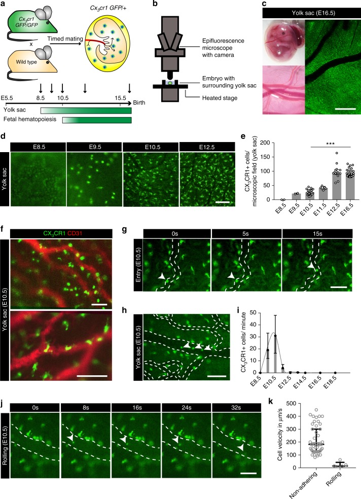
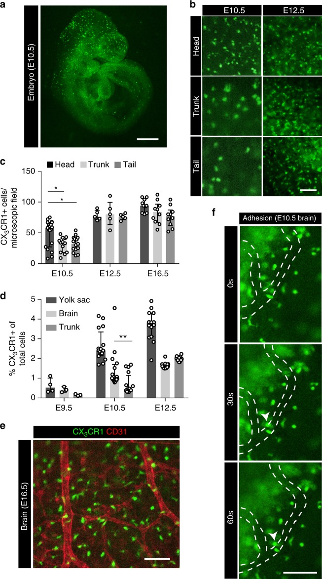
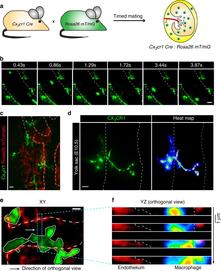
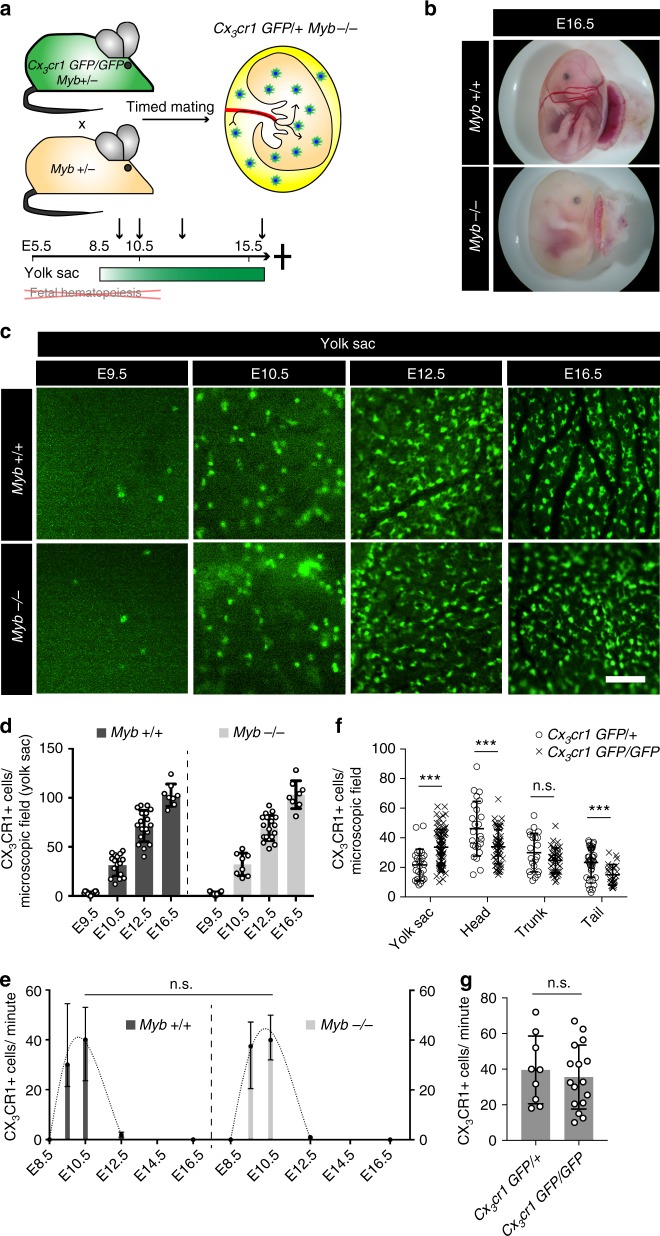
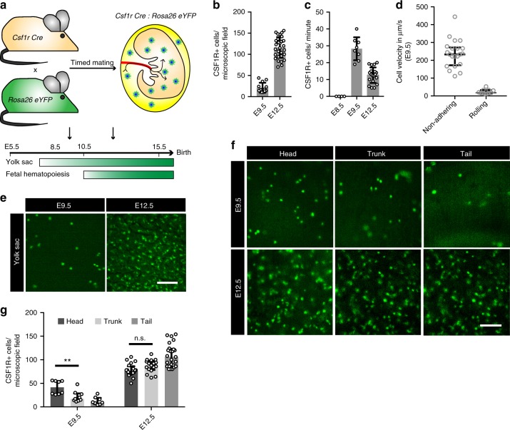
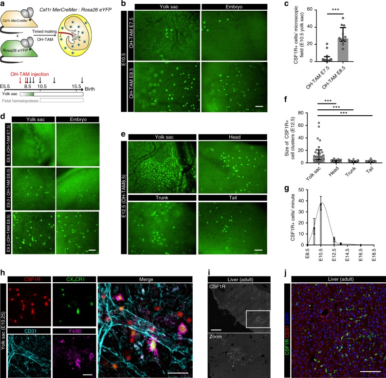
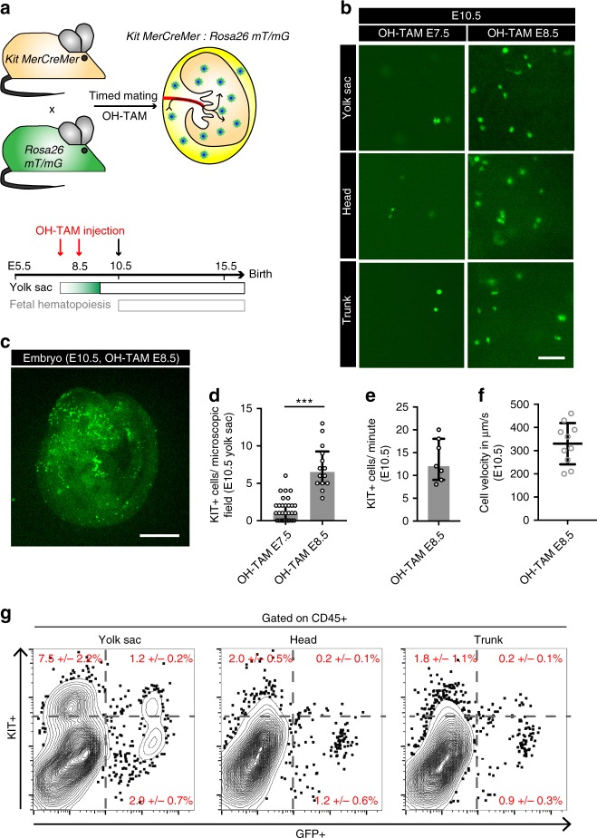
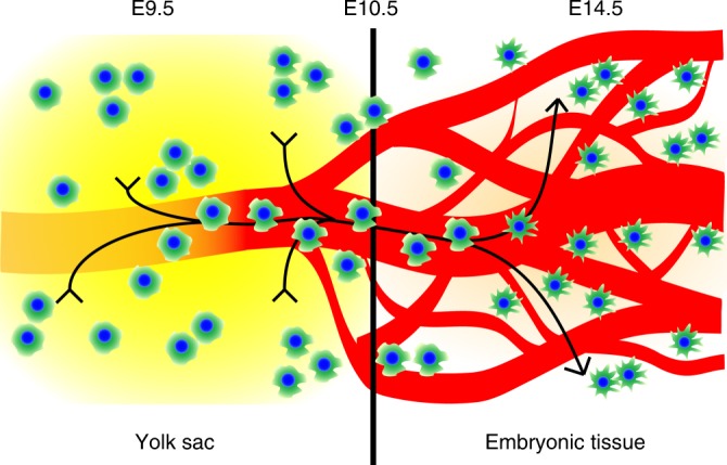
Tissue macrophages in many adult organs originate from yolk sac (YS) progenitors, which invade the developing embryo and persist by means of local self-renewal. However, the route and characteristics of YS macrophage trafficking during embryogenesis are incompletely understood. Here we show the early migration dynamics of YS-derived macrophage progenitors in vivo using fate mapping and intravital microscopy. From embryonic day 8.5 (E8.5) CX 3 CR1+ pre-macrophages are present in the mouse YS where they rapidly proliferate and gain access to the bloodstream to migrate towards the embryo. Trafficking of pre-macrophages and their progenitors from the YS to tissues peaks around E10.5, dramatically decreases towards E12.5 and is no longer evident from E14.5 onwards. Thus, YS progenitors use the vascular system during a restricted time window of embryogenesis to invade the growing fetus. These findings close an important gap in our understanding of the development of the innate immune system.
Overview
The claims and statistics of this paper haven’t been checked yet.
Run the check — free, no account needed. It reads the open-access text and checks the sources it cites.
Integrity signals
Registry
Cites
Statistics
Figures
Verification
Claims the paper makes
The claims and statistics of this paper haven’t been checked yet.
Run the check — free, no account needed. It reads the open-access text and checks the sources it cites.
Claims other papers make about this one
Each is checked against this paper’s own text: does it say what the citing paper says it does?
In the citing paper
“Microglia are yolk sac-derived macrophages residing in the parenchyma of brain and spinal cord, where they interact with neurons and other glial.”
From Engrafted parenchymal brain macrophages differ from microglia in transcriptome, chromatin landscape and response to challenge (2018) · cites this paper as [1]
Self-check
Percentages, sums and reported differences re-computed from the paper’s own numbers.
The self-check needs the paper’s full text.
Citations
Cites
Fiji: an open-source platform for biological-image analysis
Schindelin, J., Arganda-Carreras, I., Frise, E., Kaynig, V., Longair, M., Pietzsch, T., Preibisch, S., Rueden, C., Saalfeld, S., Schmid, B., et al. (2012) · Nature Methods · doi:10.1038/nmeth.2019 · Cited by 66,890
A robust and high-throughput Cre reporting and characterization system for the whole mouse brain
Madisen, L., Zwingman, T. A., Sunkin, S. M., Oh, S. W., Zariwala, H. A., Gu, H., Ng, L. L., Palmiter, R. D., Hawrylycz, M. J., Jones, A. R., et al. (2010) · Nature Neuroscience · doi:10.1038/nn.2467 · Cited by 6,907
Fate Mapping Analysis Reveals That Adult Microglia Derive from Primitive Macrophages
Ginhoux, F., Greter, M., Leboeuf, M., Nandi, S., See, P., Gokhan, S., Mehler, M. F., Conway, S. J., Ng, L. G., Stanley, E. R., et al. (2010) · Science · doi:10.1126/science.1194637 · Cited by 5,175
Tumor-Associated Macrophages: From Mechanisms to Therapy
Noy, R., & Pollard, J. W. (2014) · Immunity · doi:10.1016/j.immuni.2014.06.010 · Cited by 4,089
A global double-fluorescent Cre reporter mouse
Muzumdar, M. D., Tasic, B., Miyamichi, K., Li, L., & Luo, L. (2007) · genesis · doi:10.1002/dvg.20335 · Cited by 3,491
Fate Mapping Reveals Origins and Dynamics of Monocytes and Tissue Macrophages under Homeostasis
Yona, S., Kim, K.-W., Wolf, Y., Mildner, A., Varol, D., Breker, M., Strauss-Ayali, D., Viukov, S., Guilliams, M., Misharin, A., et al. (2013) · Immunity · doi:10.1016/j.immuni.2012.12.001 · Cited by 3,245
Self-citationCre reporter strains produced by targeted insertion of EYFP and ECFP into the ROSA26 locus
Srinivas, S., Watanabe, T., Lin, C.-S., William, C. M., Tanabe, Y., Jessell, T. M., & Costantini, F. (2001) · BMC Developmental Biology · doi:10.1186/1471-213x-1-4 · Cited by 3,121
CCL2 recruits inflammatory monocytes to facilitate breast-tumour metastasis
Qian, B.-Z., Li, J., Zhang, H., Kitamura, T., Zhang, J., Campion, L. R., Kaiser, E. A., Snyder, L. A., & Pollard, J. W. (2011) · Nature · doi:10.1038/nature10138 · Cited by 2,874
Jung, S., Aliberti, J., Graemmel, P., Sunshine, M. J., Kreutzberg, G. W., Sher, A., & Littman, D. R. (2000) · Molecular and Cellular Biology · doi:10.1128/mcb.20.11.4106-4114.2000 · Cited by 2,632
A Lineage of Myeloid Cells Independent of Myb and Hematopoietic Stem Cells
Schulz, C., Perdiguero, E. G., Chorro, L., Szabo-Rogers, H., Cagnard, N., Kierdorf, K., Prinz, M., Wu, B., Jacobsen, S. E. W., Pollard, J. W., et al. (2012) · Science · doi:10.1126/science.1219179 · Cited by 2,578
Self-citation
Cited by
Microglia states and nomenclature: A field at its crossroads
Paolicelli, R. C., Sierra, A., Stevens, B., Tremblay, M.-E., Aguzzi, A., Ajami, B., Amit, I., Audinat, E., Bechmann, I., Bennett, M., et al. (2022) · Neuron · doi:10.1016/j.neuron.2022.10.020 · Cited by 1,973
Microglia in neurodegenerative diseases: mechanism and potential therapeutic targets
Gao, C., Jiang, J., Tan, Y., & Chen, S. (2023) · Signal Transduction and Targeted Therapy · doi:10.1038/s41392-023-01588-0 · Cited by 1,616
Microglia Biology: One Century of Evolving Concepts
Prinz, M., Jung, S., & Priller, J. (2019) · Cell · doi:10.1016/j.cell.2019.08.053 · Cited by 1,525
Shemer, A., Grozovski, J., Tay, T. L., Tao, J., Volaski, A., Süß, P., Ardura-Fabregat, A., Gross-Vered, M., Kim, J.-S., David, E., et al. (2018) · Nature Communications · doi:10.1038/s41467-018-07548-5 · Cited by 1,052
Targeting tumor-associated macrophages in cancer
Pathria, P., Louis, T. L., & Varner, J. A. (2019) · Trends in Immunology · doi:10.1016/j.it.2019.02.003 · Cited by 933
Tissue-specific macrophages: how they develop and choreograph tissue biology
Mass, E., Nimmerjahn, F., Kierdorf, K., & Schlitzer, A. (2023) · Nature Reviews Immunology · doi:10.1038/s41577-023-00848-y · Cited by 621
Self-citationMicroglia and Central Nervous System–Associated Macrophages—From Origin to Disease Modulation
Prinz, M., Masuda, T., Wheeler, M. A., & Quintana, F. J. (2021) · Annual Review of Immunology · doi:10.1146/annurev-immunol-093019-110159 · Cited by 584
Decoding human fetal liver haematopoiesis
Popescu, D.-M., Botting, R. A., Stephenson, E., Green, K., Webb, S., Jardine, L., Calderbank, E. F., Polanski, K., Goh, I., Efremova, M., et al. (2019) · Nature · doi:10.1038/s41586-019-1652-y · Cited by 606
Dick, S. A., Wong, A., Hamidzada, H., Nejat, S., Nechanitzky, R., Vohra, S., Mueller, B., Zaman, R., Kantores, C., Aronoff, L., et al. (2022) · Science Immunology · doi:10.1126/sciimmunol.abf7777 · Cited by 538
Determinants of resident tissue macrophage identity and function
Blériot, C., Chakarov, S., & Ginhoux, F. (2020) · Immunity · doi:10.1016/j.immuni.2020.05.014 · Cited by 507
Macrophages at CNS interfaces: Ontogeny and function in health and disease
Kierdorf, K., Masuda, T., Jordão, M. J. C., & Prinz, M. (2019) · Nature Reviews Neuroscience · doi:10.1038/s41583-019-0201-x · Cited by 416
Bordt, E. A., Ceasrine, A. M., & Bilbo, S. D. (2020) · Glia · doi:10.1002/glia.23753 · Cited by 125
Inflammation within the neurovascular unit: Focus on microglia for stroke injury and recovery
Eldahshan, W., Fagan, S. C., & Ergul, A. (2019) · Pharmacological Research · doi:10.1016/j.phrs.2019.104349 · Cited by 104
Bordeleau, M., Carrier, M., Luheshi, G. N., & Tremblay, M.-È. (2019) · Seminars in Cell & Developmental Biology · doi:10.1016/j.semcdb.2019.06.001 · Cited by 75
The niche matters: Origin, function and fate of CNS-associated macrophages during health and disease
Dalmau Gasull, A., Glavan, M., Samawar, S. K. R., Kapupara, K., Kelk, J., Rubio, M., Fumagalli, S., Sorokin, L., Vivien, D., & Prinz, M. (2024) · Acta Neuropathologica · doi:10.1007/s00401-023-02676-9 · Cited by 60
Mast cell ontogeny: From fetal development to life‐long health and disease
Chia, S. L., Kapoor, S., Carvalho, C., Bajénoff, M., & Gentek, R. (2023) · Immunological Reviews · doi:10.1111/imr.13191 · Cited by 60
Mechanisms of myeloid cell entry to the healthy and diseased central nervous system
Amann, L., Masuda, T., & Prinz, M. (2023) · Nature Immunology · doi:10.1038/s41590-022-01415-8 · Cited by 50
Fetal‐derived immune cells at the roots of lifelong pathophysiology
Mass, E., & Gentek, R. (2021) · Frontiers in Cell and Developmental Biology · doi:10.3389/fcell.2021.648313 · Cited by 47
Self-citationMicroglia: Activity‐dependent regulators of neural circuits
Durán Laforet, V., & Schafer, D. P. (2024) · Annals of the New York Academy of Sciences · doi:10.1111/nyas.15105 · Cited by 32
“10 Onceinthe CNS,microgliabegintocolonizethe brain, 11 where they adopt a unique resident microglia molecular signature 12 and become poised to play critical roles in shaping the development FIGURE 1 (Continued) tumor necrosis factor alpha; TREM2, triggering receptor expressed on myeloid cells 2;…”
Bartalska, K., Hübschmann, V., Korkut-Demirbaş, M., Cubero, R. J. A., Venturino, A., Rössler, K., Czech, T., & Siegert, S. (2022) · iScience · doi:10.1016/j.isci.2022.104580 · Cited by 22
Common principles of macrophage biology in blood–tissue barriers
Hiraga, S., & Masuda, T. (2024) · Clinical and Experimental Neuroimmunology · doi:10.1111/cen3.12812 · Cited by 2
Metadata
- Authors
- Stremmel, C., Schuchert, R., Wagner, F., Thaler, R., Weinberger, T., Pick, R., Mass, E., Ishikawa-Ankerhold, H. C., Margraf, A., Hutter, S., Vagnozzi, R., Klapproth, S., Frampton, J., Yona, S., Scheiermann, C., Molkentin, J. D., Jeschke, U., Moser, M., Sperandio, M., Massberg, S., Geissmann, F., & Schulz, C.
- Venue
- Nature Communications
- Published
- 2018-01-08
- Volume
- 9
- Issue
- 1
- ISSN
- 2041-1723
- PMC
- PMC5758709
- Cited by
- 278
- References
- 70
- Language
- en
- Update notices
- correction (2018-09-07)
- Licence
- creativecommons.org/licenses/by/4.0 (tdm)
- creativecommons.org/licenses/by/4.0 (vor)
About these checks
Registry, retraction, Cites and statistics results are computed, not judged, with one exception: a printed reference that no index lists is read once by a model before it counts as a citation error. Claims are extracted by a model and kept only when their quote appears verbatim in the text; each cited source is then checked, with a model’s help, for whether it says what the claim relies on. Those verdicts can be wrong — read the claim’s quote and the cited source before relying on one.
Statistics and arithmetic are recomputed, not judged.
Confidence bands are shown only for fields calibrated on our annotated test set. None is calibrated yet, so no band is shown.
What counts as a claim ›How we measure confidence ›How we check this ›
Are you an author, or do you see a problem with this page? Request a review
Data sources
Bibliographic data: Semantic Scholar, OpenAlex, Crossref.
Contains information from the Semantic Scholar Academic Graph Datasets, made available under the ODC Attribution License (ODC-BY 1.0).
Semantic Scholar’s data is described in: Kinney, R., et al. (2023). The Semantic Scholar Open Data Platform. arXiv:2301.10140.
