

The DOI matches its Crossref record; 116 of the 116 references it cites were found in a registry or an index; 4 of the 111 sources its claims cite were checked for support.
Federated learning enables big data for rare cancer boundary detection
Open accessAbstract
Although machine learning (ML) has shown promise across disciplines, out-of-sample generalizability is concerning. This is currently addressed by sharing multi-site data, but such centralization is challenging/infeasible to scale due to various limitations. Federated ML (FL) provides an alternative paradigm for accurate and generalizable ML, by only sharing numerical model updates. Here we present the largest FL study to-date, involving data from 71 sites across 6 continents, to generate an automatic tumor boundary detector for the rare disease of glioblastoma, reporting the largest such dataset in the literature ( n = 6, 314). We demonstrate a 33% delineation improvement for the surgically targetable tumor, and 23% for the complete tumor extent, over a publicly trained model. We anticipate our study to: 1) enable more healthcare studies informed by large diverse data, ensuring meaningful results for rare diseases and underrepresented populations, 2) facilitate further analyses for glioblastoma by releasing our consensus model, and 3) demonstrate the FL effectiveness at such scale and task-complexity as a paradigm shift for multi-site collaborations, alleviating the need for data-sharing.
Overview
AI snapshot
Written from the paper’s full text.
TL;DR
This paper reports the largest real-world federated learning study to date for glioblastoma boundary detection, using data from 6,314 patients across 71 sites on six continents. The final consensus model improved tumor sub-compartment delineation over a publicly trained initial model and showed that federated learning can enable large, diverse rare-disease datasets without sharing patient data.
Key findings
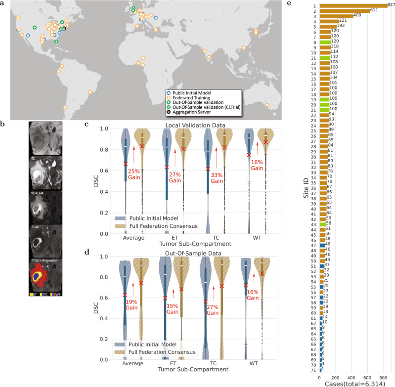
- The final consensus model improved over the public initial model on collaborators’ local validation data by 27% for enhancing tumor (ET), 33% for tumor core (TC), and 16% for whole tumor (WT); on complete out-of-sample data, improvements were 15% for ET, 27% for TC, and 16% for WT. The abstract reports a 33% delineation improvement for the surgically targetable tumor and 23% for the complete tumor extent over a publicly trained model.
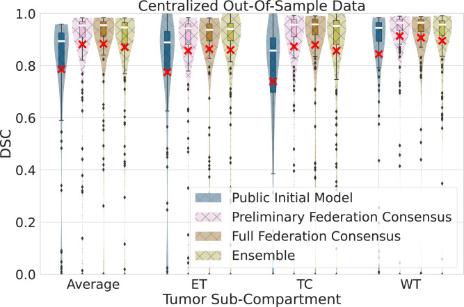
- Data size alone did not linearly predict success: public initial vs. preliminary consensus model showed significant improvements on centralized out-of-sample data (p < 7 × 10⁻¹⁸), while preliminary vs. final consensus model differences were insignificant (p > 0.067).
- The final consensus model was always superior or insignificantly different from an ensemble of local models from the four largest contributing sites (>200 cases each) across all sub-compartments (pAverage = 0.1, pET = 0.5, pTC = 0.2, pWT = 0.06).
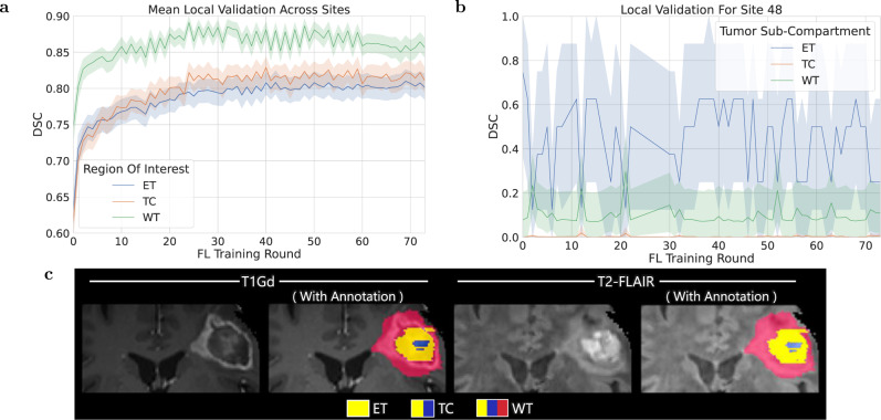
- Federated learning was robust to data quality issues: Site 48 had erroneous annotations (n = 46) and significantly lower local validation scores (pET < 3 × 10⁻¹², pTC < 3 × 10⁻¹², pWT < 3 × 10⁻¹²), but the global consensus model performance was not adversely affected.
Methods
- Design: staged real-world federated learning federation using a central aggregation server, starting from a public initial model (231 cases from 16 sites), then a preliminary consensus model (2,471 cases from 35 sites), and finally a final consensus model (6,314 cases from 71 sites).
- Data: multi-parametric MRI (T1, T1Gd, T2, T2-FLAIR) from adult-type diffuse glioma/glioblastoma patients; 55 independent collaborating sites contributed 6,083 cases, with 49 sites in training (5,493 cases) and 6 out-of-sample sites (590 cases; centralized out-of-sample subset n = 332). Each site held out 20% of cases as local validation.
- Analysis: federated averaging (FedAvg) of model updates, 3D-ResUNet architecture, Dice similarity coefficient (DSC) for ET, TC, and WT, Wilcoxon signed-rank tests, and 73 federated rounds.
Limitations
- The paper states there is no automated mechanism to assess inputted data quality from collaborators, which could result in models trained on sub-optimal data; it also states that only data quality issues largely affecting the global validation score could be identified during training, while subtler issues were found only after training.
- The paper states it used a single off-the-shelf neural network architecture, whereas model ensembles have been shown to perform better for this task, and it remains unexplored how such a strategy would work in a federated study.
- The paper states that the federation required significant manual coordination and orchestration, with no tools available to automate it, and that security/privacy threats remain; evident from the text, the study relied on a network of trusted sites, manual inspection of local and global validation scores, and TLS plus trusted execution environments considered sufficient for its threat model.
Model-written summary. Check it against the paper before relying on it.
Integrity signals
Registry
Cites
Statistics
Figures
Verification
Claims the paper makes
38 Not checked yet · 30 Own result · 19 Own interpretation · 16 Own method · 12 No basis given · 2 Partly supported · accuracy not yet measured
The cited sources support only part of this claim — read each source’s verdict below.
In the paper
“This functionality follows the harmonized data preprocessing protocol defined by the BraTS challenge35–38, as described below. This would allow accounting for inter-site acquisition protocol variations, e.g., 3D vs. 2D axial plane acquisitions.”
§ Federated learning enables big data for rare cancer boundary detection > Methods > Harmonized data preprocessing
What the sources say
- Supportedcites [35] Menze, B. H. et al. The multimodal brain tumor image segmentation benchmark (brats). IEEE Trans. Med. Imaging 34, 1993–2024 (2014). source
In Section C (Image datasets), the paper describes both 2D and 3D acquisitions and says the images were co-registered and resampled to a standardized resolution and orientation to homogenize the data. Section E (Limitations) also notes resolution differences between patients and centers, while acknowledging that variability from acquisition protocols remained. (LLM, conf=0.91)
- Partly supportedcites [36] Bakas, S. et al. Advancing the cancer genome atlas glioma MRI collections with expert segmentation labels and radiomic features. Sci. data 4, 1–13 (2017). sourceExperimental — accuracy not yet measured
The Data Records section says BraTS’15 organizers skull-stripped scans, co-registered them to a common template, and resampled them to 1 mm³ resolution. Methods > Data collection documents scans from multiple institutions with differing acquisition parameters. Our copy does not specifically say that this preprocessing accounts for 3D versus 2D axial acquisitions, so that example goes beyond what it establishes. (LLM, conf=0.82)
- Not checked yetcites [37] Bakas, S. et al. Identifying the best machine learning algorithms for brain tumor segmentation, progression assessment, and overall survival prediction in the brats challenge. arXiv preprint arXiv:1811.02629 (2018).
- Not checked yetcites [38] Baid, U. et al. The rsna-asnr-miccai brats 2021 benchmark on brain tumor segmentation and radiogenomic classification. arXiv preprint arXiv:2107.02314 (2021).
hedgeddirect- Asserted: passes — The authors put it forward as true.
- Checkable: passes — It can be checked against evidence.
- Load-bearing: passes — The paper’s conclusions depend on it.
- Based: passes — The paper gives a reason why it holds.
- Supported
The paper states this without a source or data behind it here, so there is nothing to check it against. The quote shows it is in the paper.
In the paper
“To summarize the model performance with a single collective score, we then calculate the average DSC (across all 3 tumor sub-compartments per case, and then across all cases of all sites) as equal to 0.66.”
§ Federated learning enables big data for rare cancer boundary detection > Results > Increased data can improve performance
plain- Asserted: passes — The authors put it forward as true.
- Checkable: passes — It can be checked against evidence.
- Load-bearing: passes — The paper’s conclusions depend on it.
- Based: passes — The paper gives a reason why it holds.
None of the sources cited for this claim have been checked yet.
In the paper
“Notably, the largest and most significant improvement (33%, p < 7 × 10−60) is noted for the TC sub-compartment, which is surgically actionable and not a trivial sub-compartment to delineate accurately43,44.”
§ Federated learning enables big data for rare cancer boundary detection > Results > FL benefits the more challenging tasks
What the sources say
- Not checked yetcites [43] Beiko, J. et al. Idh1 mutant malignant astrocytomas are more amenable to surgical resection and have a survival benefit associated with maximal surgical resection. Neuro-oncology 16, 81–91 (2014). source
- Not checked yetcites [44] Olson, J. J. Congress of neurological surgeons systematic review and evidence-based guidelines for the treatment of adults with progressive glioblastoma update: introduction and methods. J. Neuro-oncol 158, 133–137 (2022). source
plaindirect- Asserted: passes — The authors put it forward as true.
- Checkable: passes — It can be checked against evidence.
- Load-bearing: passes — The paper’s conclusions depend on it.
- Based: passes — The paper gives a reason why it holds.
A result the paper reports from its own data — it cites no other source for it, so there is none to check. The quote shows it is in the paper.
In the paper
“As noted in our results, due to the lack of such generalizable solutions, we were only able to identify quality issues after the model training.”
§ Federated learning enables big data for rare cancer boundary detection > Discussion
plaindirect- Asserted: passes — The authors put it forward as true.
- Checkable: passes — It can be checked against evidence.
- Load-bearing: unclear — It is unclear whether the paper’s conclusions depend on it.
- Based: passes — The paper gives a reason why it holds.
A result the paper reports from its own data — it cites no other source for it, so there is none to check. The quote shows it is in the paper.
In the paper
“Notably, this describes the largest and most diverse dataset of glioblastoma patients ever considered in the literature.”
- Also stated in: § Discussion · plain
“Notably, this extensive global footprint of the collaborating sites in this study also yields the largest dataset ever reported in the literature assessing this rare disease.”
§ Federated learning enables big data for rare cancer boundary detection > Introduction
plainn = 6,314- Asserted: passes — The authors put it forward as true.
- Checkable: passes — It can be checked against evidence.
- Load-bearing: passes — The paper’s conclusions depend on it.
- Based: passes — The paper gives a reason why it holds.
- Also stated in: § Discussion · plain
A result the paper reports from its own data — it cites no other source for it, so there is none to check. The quote shows it is in the paper.
In the paper
“The findings of this evaluation indicate that the final consensus model performance is always superior or insignificantly different (pAverage = 0.1, pET = 0.5, pTC = 0.2, pWT = 0.06, Wilcoxon signed-rank test) to the ensemble of the local models of these four largest contributing collaborators, for all tumor sub-compartments (Fig. 2).”
§ Federated learning enables big data for rare cancer boundary detection > Results > Data size alone may not predict success
hedged- Asserted: passes — The authors put it forward as true.
- Checkable: passes — It can be checked against evidence.
- Load-bearing: passes — The paper’s conclusions depend on it.
- Based: passes — The paper gives a reason why it holds.
- The authors state this as an assumption: The independent models and their ensemble from the four largest sites could have inherited some of the public initial model's data diversity because they initiated training from it, which could justify the observed insignificant differences.
- Rests on an assumption: The centralized out-of-sample subset of 332 cases is representative of the complete out-of-sample data population of 590 cases.
A result the paper reports from its own data — it cites no other source for it, so there is none to check. The quote shows it is in the paper.
In the paper
“Our findings indicate that the benefits of FL are more pronounced for the more challenging sub-compartments, i.e., larger performance improvements for ET and TC compared to WT (Fig. 1c).”
§ Federated learning enables big data for rare cancer boundary detection > Results > FL benefits the more challenging tasks
hedgeddirect- Asserted: passes — The authors put it forward as true.
- Checkable: passes — It can be checked against evidence.
- Load-bearing: passes — The paper’s conclusions depend on it.
- Based: passes — The paper gives a reason why it holds.
A result the paper reports from its own data — it cites no other source for it, so there is none to check. The quote shows it is in the paper.
In the paper
“Here we present the largest FL study to-date, involving data from 71 sites across 6 continents, to generate an automatic tumor boundary detector for the rare disease of glioblastoma, reporting the largest such dataset in the literature (n = 6, 314).”
- Also stated in: § Introduction · plain
“This study describes the largest to-date global FL effort to develop an accurate and generalizable ML model for detecting glioblastoma sub-compartment boundaries, based on data from 6314 glioblastoma patients from 71 geographically distinct sites, across six continents (Fig. 1a).”
- Also stated in: § Discussion · plain
“In this study, we have described the largest real-world FL effort to-date utilizing data of 6314 glioblastoma patients from 71 geographically unique sites spread across 6 continents, to develop an accurate and generalizable ML model for detecting glioblastoma sub-compartment boundaries.”
§ Federated learning enables big data for rare cancer boundary detection > Abstract
plainn = 6,314- Asserted: passes — The authors put it forward as true.
- Checkable: passes — It can be checked against evidence.
- Load-bearing: passes — The paper’s conclusions depend on it.
- Based: passes — The paper gives a reason why it holds.
- Also stated in: § Introduction · plain
A result the paper reports from its own data — it cites no other source for it, so there is none to check. The quote shows it is in the paper.
In the paper
“When the federation began, the public initial model was evaluated against the local validation data of all sites, resulting in an average (across all cases of all sites) DSC per sub-compartment, of DSCET = 0.63, DSCTC = 0.62, DSCWT = 0.75.”
§ Federated learning enables big data for rare cancer boundary detection > Results > Increased data can improve performance
plain- Asserted: passes — The authors put it forward as true.
- Checkable: passes — It can be checked against evidence.
- Load-bearing: passes — The paper’s conclusions depend on it.
- Based: passes — The paper gives a reason why it holds.
A result the paper reports from its own data — it cites no other source for it, so there is none to check. The quote shows it is in the paper.
In the paper
“Data quality issues relating to erroneous reference annotations (with potential negative downstream effects on output predictions) were identified by monitoring the global consensus model performance during training.”
§ Federated learning enables big data for rare cancer boundary detection > Results > FL is robust to data quality issues
plain- Asserted: passes — The authors put it forward as true.
- Checkable: passes — It can be checked against evidence.
- Load-bearing: passes — The paper’s conclusions depend on it.
- Based: passes — The paper gives a reason why it holds.
- Probably not a substantial claim. 49
- It may state two separate claims as one. 46
- The quote only partly states this claim. 1
The cited sources support only part of this claim — read each source’s verdict below.
In the paper
“Following the precise definition of our problem statement35–38, ensuring strict compliance with the preprocessing and annotation protocol for the generation of reference standards was vital for the model to learn correct information during training.”
§ Federated learning enables big data for rare cancer boundary detection > Discussion
What the sources say
- Partly supportedcites [35] Menze, B. H. et al. The multimodal brain tumor image segmentation benchmark (brats). IEEE Trans. Med. Imaging 34, 1993–2024 (2014). sourceExperimental — accuracy not yet measured
The paper’s “Expert annotation of tumor structures” section describes a defined annotation protocol, and “Lessons learned” strongly recommends homogenized preprocessing. It does not say strict compliance with both protocols was vital for a model to learn correctly; “Lessons learned” instead says many algorithms can overcome slight inconsistencies or errors in training data. Page numbers are not available in our copy. (LLM, conf=0.83)
- Partly supportedcites [36] Bakas, S. et al. Advancing the cancer genome atlas glioma MRI collections with expert segmentation labels and radiomic features. Sci. data 4, 1–13 (2017). sourceExperimental — accuracy not yet measured
The Methods (Pre-processing) and Technical Validation (Manual correction) sections describe defined image-processing steps and expert-reviewed corrections to segmentation labels. They do not state that strict compliance with those protocols was vital for a model to learn correct information during training; that appears to be the citing authors’ inference. (LLM, conf=0.82)
- Not checked yetcites [37] Bakas, S. et al. Identifying the best machine learning algorithms for brain tumor segmentation, progression assessment, and overall survival prediction in the brats challenge. arXiv preprint arXiv:1811.02629 (2018).
- Not checked yetcites [38] Baid, U. et al. The rsna-asnr-miccai brats 2021 benchmark on brain tumor segmentation and radiogenomic classification. arXiv preprint arXiv:2107.02314 (2021).
plaindirect- Asserted: passes — The authors put it forward as true.
- Checkable: passes — It can be checked against evidence.
- Load-bearing: passes — The paper’s conclusions depend on it.
- Based: passes — The paper gives a reason why it holds.
Probably not a substantial claim.
- Partly supported
The paper states this without a source or data behind it here, so there is nothing to check it against. The quote shows it is in the paper.
In the paper
“However, the decision of using a singlet or a triplet model should also rely on computational cost considerations, as triplets will be three times more expensive than singlets during model inference.”
§ Federated learning enables big data for rare cancer boundary detection > Results > Optimal model selection is non-trivial
plaindirect- Asserted: passes — The authors put it forward as true.
- Checkable: passes — It can be checked against evidence.
- Load-bearing: passes — The paper’s conclusions depend on it.
- Based: fails — The paper may give no reason why it holds.
Probably not a substantial claim. The paper may give no reason why it holds.
A result the paper reports from its own data — it cites no other source for it, so there is none to check. The quote shows it is in the paper.
In the paper
“Specifically, we noted major significant (p < 7 × 10⁻¹⁸, Wilcoxon signed-rank test) performance improvements between the public initial model and the preliminary consensus model, as opposed to the insignificant (p > 0.067, Wilcoxon signed-rank test) ones between the preliminary and the final consensus model, as quantified in the centralized out-of-sample data for all sub-compartments and their ave”
§ Federated learning enables big data for rare cancer boundary detection > Results > Data size alone may not predict success
plain- Asserted: passes — The authors put it forward as true.
- Checkable: passes — It can be checked against evidence.
- Load-bearing: passes — The paper’s conclusions depend on it.
- Based: passes — The paper gives a reason why it holds.
It may state two separate claims as one.
- Rests on an assumption: The centralized out-of-sample subset of 332 cases is representative of the complete out-of-sample data population of 590 cases.
A result the paper reports from its own data — it cites no other source for it, so there is none to check. The quote shows it is in the paper.
In the paper
“The curves of Fig. 3a indicate that the global consensus model continues to consistently gain knowledge from the federation as a whole during training, highlighting robustness to such data quality issues.”
§ Federated learning enables big data for rare cancer boundary detection > Discussion
hedgeddirectn = 46- Asserted: passes — The authors put it forward as true.
- Checkable: passes — It can be checked against evidence.
- Load-bearing: passes — The paper’s conclusions depend on it.
- Based: passes — The paper gives a reason why it holds.
It may state two separate claims as one.
A result the paper reports from its own data — it cites no other source for it, so there is none to check. The quote shows it is in the paper.
In the paper
“However, only data quality issues that largely affected the global validation score could be identified and corrected during training. Those with more subtle effects in the global validation score were only identified after the completion of the model training by looking for relatively low local validation scores of the consensus model across collaborating sites.”
§ Federated learning enables big data for rare cancer boundary detection > Results > FL is robust to data quality issues
hedgeddirect- Asserted: passes — The authors put it forward as true.
- Checkable: passes — It can be checked against evidence.
- Load-bearing: passes — The paper’s conclusions depend on it.
- Based: passes — The paper gives a reason why it holds.
It may state two separate claims as one.
A result the paper reports from its own data — it cites no other source for it, so there is none to check. The quote shows it is in the paper.
In the paper
“To further evaluate the potential generalizability improvements of the final consensus model on unseen data, we compared it with the public initial model against the complete out-of-sample data and noted significant performance improvements of 15% (pET < 1 × 10⁻⁵), 27% (pTC < 1 × 10⁻¹⁶), and 16% (pWT < 1 × 10⁻⁷), for ET, TC, and WT, respectively (Fig. 1d).”
§ Federated learning enables big data for rare cancer boundary detection > Results > Increased data can improve performance
plaindirect- Asserted: passes — The authors put it forward as true.
- Checkable: passes — It can be checked against evidence.
- Load-bearing: passes — The paper’s conclusions depend on it.
- Based: passes — The paper gives a reason why it holds.
It may state two separate claims as one.
A result the paper reports from its own data — it cites no other source for it, so there is none to check. The quote shows it is in the paper.
In the paper
“Only small differences are observed between the singlet and triplet models, and these differences diminish as the sub-compartment size increases.”
§ Federated learning enables big data for rare cancer boundary detection > Results > Optimal model selection is non-trivial
plaincorrelational- Asserted: passes — The authors put it forward as true.
- Checkable: passes — It can be checked against evidence.
- Load-bearing: unclear — It is unclear whether the paper’s conclusions depend on it.
- Based: passes — The paper gives a reason why it holds.
It may state two separate claims as one.
A result the paper reports from its own data — it cites no other source for it, so there is none to check. The quote shows it is in the paper.
In the paper
“Comparing the means of singlet and triplet, the larger (and only significant) performance improvement difference compared to the public initial model is noted for the ET sub-compartment (improved by < 3%, pET = 0.02), followed by TC (improved by < 1.4%, pTC = 0.09), and then lastly WT (improved by < 1.1%, pWT = 0.2) (Tables S1 and S2).”
§ Federated learning enables big data for rare cancer boundary detection > Results > Optimal model selection is non-trivial
plaindirect- Asserted: passes — The authors put it forward as true.
- Checkable: passes — It can be checked against evidence.
- Load-bearing: passes — The paper’s conclusions depend on it.
- Based: passes — The paper gives a reason why it holds.
It may state two separate claims as one.
A result the paper reports from its own data — it cites no other source for it, so there is none to check. The quote shows it is in the paper.
In the paper
“Following model training across all sites, the final consensus model garnered significant performance improvements against the collaborators’ local validation data of 27% (pET < 1 × 10−36), 33% (pTC < 1 × 10−59), and 16% (pWT < 1 × 10⁻²¹), for ET, TC, and WT, respectively (Fig. 1c).”
§ Federated learning enables big data for rare cancer boundary detection > Results > Increased data can improve performance
plaindirect- Asserted: passes — The authors put it forward as true.
- Checkable: passes — It can be checked against evidence.
- Load-bearing: passes — The paper’s conclusions depend on it.
- Based: passes — The paper gives a reason why it holds.
It may state two separate claims as one.
The paper states this without a source or data behind it here, so there is nothing to check it against. The quote shows it is in the paper.
In the paper
“This happened even though the final consensus model was trained on over twice the number of cases (and included 2 of the largest contributing sites—Sites 1 and 4) when compared to the preliminary consensus model.”
§ Federated learning enables big data for rare cancer boundary detection > Discussion
plain- Asserted: passes — The authors put it forward as true.
- Checkable: passes — It can be checked against evidence.
- Load-bearing: unclear — It is unclear whether the paper’s conclusions depend on it.
- Based: passes — The paper gives a reason why it holds.
It may state two separate claims as one.
A result the paper reports from its own data — it cites no other source for it, so there is none to check. The quote shows it is in the paper.
In the paper
“The data from all 55 collaborating sites followed a male:female ratio of 1.47:1 with ages ranging between 7 and 94 years.”
§ Federated learning enables big data for rare cancer boundary detection > Methods > Data
plainn = 6,083- Asserted: passes — The authors put it forward as true.
- Checkable: passes — It can be checked against evidence.
- Load-bearing: fails — The paper’s conclusions may not depend on it.
- Based: passes — The paper gives a reason why it holds.
Probably not a substantial claim. The paper’s conclusions may not depend on it. It may state two separate claims as one.
A result the paper reports from its own data — it cites no other source for it, so there is none to check. The quote shows it is in the paper.
In the paper
“In terms of hardware requirements at each site, 88% used a dedicated workstation for their local workload, and the remaining 12% used either a containerized form of the FeTS tool or a virtual machine.”
§ Federated learning enables big data for rare cancer boundary detection > Discussion
plain- Asserted: passes — The authors put it forward as true.
- Checkable: passes — It can be checked against evidence.
- Load-bearing: fails — The paper’s conclusions may not depend on it.
- Based: passes — The paper gives a reason why it holds.
Probably not a substantial claim. The paper’s conclusions may not depend on it. It may state two separate claims as one.
The paper states this without a source or data behind it here, so there is nothing to check it against. The quote shows it is in the paper.
In the paper
“55 sites participated as independent collaborators in the study defining a dataset of 6083 cases.”
§ Federated learning enables big data for rare cancer boundary detection > Methods > Data
plainn = 6,083- Asserted: passes — The authors put it forward as true.
- Checkable: passes — It can be checked against evidence.
- Load-bearing: passes — The paper’s conclusions depend on it.
- Based: passes — The paper gives a reason why it holds.
It may state two separate claims as one.
A result the paper reports from its own data — it cites no other source for it, so there is none to check. The quote shows it is in the paper.
In the paper
“Furthermore, 84% of the sites appreciated the user-friendly interface of the provided tool and its associated complete functionality (beyond its FL backend), and indicated their intention to use it and recommend it for projects and data analysis pipelines beyond the scope of this study.”
§ Federated learning enables big data for rare cancer boundary detection > Discussion
plain- Asserted: passes — The authors put it forward as true.
- Checkable: passes — It can be checked against evidence.
- Load-bearing: unclear — It is unclear whether the paper’s conclusions depend on it.
- Based: passes — The paper gives a reason why it holds.
It may state two separate claims as one.
A result the paper reports from its own data — it cites no other source for it, so there is none to check. The quote shows it is in the paper.
In the paper
“We demonstrate a 33% delineation improvement for the surgically targetable tumor, and 23% for the complete tumor extent, over a publicly trained model.”
§ Federated learning enables big data for rare cancer boundary detection > Abstract
boosteddirectn = 6- Asserted: passes — The authors put it forward as true.
- Checkable: passes — It can be checked against evidence.
- Load-bearing: passes — The paper’s conclusions depend on it.
- Based: passes — The paper gives a reason why it holds.
It may state two separate claims as one.
A result the paper reports from its own data — it cites no other source for it, so there is none to check. The quote shows it is in the paper.
In the paper
“The out-of-sample data totaled 590 cases intentionally held out of the federation, with the intention of validating the consensus model in completely unseen cases.”
§ Federated learning enables big data for rare cancer boundary detection > Methods > Data
plainn = 590- Asserted: passes — The authors put it forward as true.
- Checkable: passes — It can be checked against evidence.
- Load-bearing: passes — The paper’s conclusions depend on it.
- Based: passes — The paper gives a reason why it holds.
It may state two separate claims as one.
A result the paper reports from its own data — it cites no other source for it, so there is none to check. The quote shows it is in the paper.
In the paper
“To generate the reference standard labels for their local data, 86% of the collaborating sites indicated that they used either the FeTS Tool39 (i.e., the tool developed for this study), CaPTk51, or ITK-SNAP52, whereas the remaining 14% used either 3D-Slicer53, the BraTS toolkit54, or something else.”
§ Federated learning enables big data for rare cancer boundary detection > Discussion
hedged- Asserted: passes — The authors put it forward as true.
- Checkable: passes — It can be checked against evidence.
- Load-bearing: fails — The paper’s conclusions may not depend on it.
- Based: passes — The paper gives a reason why it holds.
Probably not a substantial claim. The paper’s conclusions may not depend on it.
A result the paper reports from its own data — it cites no other source for it, so there is none to check. The quote shows it is in the paper.
In the paper
“According to the provided feedback, 96% of the sites found the comprehensive documentation on preprocessing and data curation essential and thought that lack of such documentation could have resulted in inconsistent annotations.”
§ Federated learning enables big data for rare cancer boundary detection > Discussion
plainconditional- Asserted: passes — The authors put it forward as true.
- Checkable: passes — It can be checked against evidence.
- Load-bearing: fails — The paper’s conclusions may not depend on it.
- Based: passes — The paper gives a reason why it holds.
Probably not a substantial claim. The paper’s conclusions may not depend on it. It may state two separate claims as one.
The paper states this without a source or data behind it here, so there is nothing to check it against. The quote shows it is in the paper.
In the paper
“Moreover, the instantiation of the federation involved a significant amount of coordination between each site and considering the limited real-world FL studies at the time, there were no tools available to automate such coordination and orchestration.”
§ Federated learning enables big data for rare cancer boundary detection > Discussion
plain- Asserted: passes — The authors put it forward as true.
- Checkable: passes — It can be checked against evidence.
- Load-bearing: fails — The paper’s conclusions may not depend on it.
- Based: passes — The paper gives a reason why it holds.
Probably not a substantial claim. The paper’s conclusions may not depend on it. It may state two separate claims as one.
The paper states this without a source or data behind it here, so there is nothing to check it against. The quote shows it is in the paper.
In the paper
“Following screening for the availability of the four required mpMRI scans with sufficient signal-to-noise ratio judged by visual observation, a subset of 362 cases from the original trial data were included in this study.”
§ Federated learning enables big data for rare cancer boundary detection > Methods > Data
plainn = 362- Asserted: passes — The authors put it forward as true.
- Checkable: passes — It can be checked against evidence.
- Load-bearing: passes — The paper’s conclusions depend on it.
- Based: passes — The paper gives a reason why it holds.
Probably not a substantial claim.
The paper states this without a source or data behind it here, so there is nothing to check it against. The quote shows it is in the paper.
In the paper
“The MRI scanners used for data acquisition were from multiple vendors (i.e., Siemens, GE, Philips, Hitachi, Toshiba), with magnetic field strength ranging from 1T to 3T.”
§ Federated learning enables big data for rare cancer boundary detection > Methods > Data
plainn = 6,083- Asserted: passes — The authors put it forward as true.
- Checkable: passes — It can be checked against evidence.
- Load-bearing: passes — The paper’s conclusions depend on it.
- Based: passes — The paper gives a reason why it holds.
Probably not a substantial claim. It may state two separate claims as one.
A result the paper reports from its own data — it cites no other source for it, so there is none to check. The quote shows it is in the paper.
In the paper
“The collaborative network of the present study spans 6 continents (Fig. 1), with data from 71 geographically distinct sites.”
§ Federated learning enables big data for rare cancer boundary detection > Methods > The Federation
plain- Asserted: passes — The authors put it forward as true.
- Checkable: passes — It can be checked against evidence.
- Load-bearing: passes — The paper’s conclusions depend on it.
- Based: passes — The paper gives a reason why it holds.
It may state two separate claims as one.
A result the paper reports from its own data — it cites no other source for it, so there is none to check. The quote shows it is in the paper.
In the paper
“Additionally, 92% found the documentation relating to establishing secure connectivity to the aggregation server easy to follow and essential to expedite reviews by the related groups.”
§ Federated learning enables big data for rare cancer boundary detection > Discussion
plain- Asserted: passes — The authors put it forward as true.
- Checkable: passes — It can be checked against evidence.
- Load-bearing: fails — The paper’s conclusions may not depend on it.
- Based: passes — The paper gives a reason why it holds.
Probably not a substantial claim. The paper’s conclusions may not depend on it.
The paper states this without a source or data behind it here, so there is nothing to check it against. The quote shows it is in the paper.
In the paper
“higher throughput (equal to the 4.48 × speedup improvement since the batch size used is equal to 1)”
§ Federated learning enables big data for rare cancer boundary detection > Methods > Model runtime in low-resource settings
plaindirect- Asserted: passes — The authors put it forward as true.
- Checkable: passes — It can be checked against evidence.
- Load-bearing: unclear — It is unclear whether the paper’s conclusions depend on it.
- Based: passes — The paper gives a reason why it holds.
It may state two separate claims as one.
A conclusion the paper draws from its own results — it cites no other source for it, so there is none to check. The quote shows it is in the paper.
In the paper
“The conclusion of this finding reinforces the importance of using large and diverse data for generalizable models to ultimately drive patient care.”
§ Federated learning enables big data for rare cancer boundary detection > Results > Increased data can improve performance
plaindirect- Asserted: passes — The authors put it forward as true.
- Checkable: fails — It may be too vague to check against evidence.
- Load-bearing: passes — The paper’s conclusions depend on it.
- Based: passes — The paper gives a reason why it holds.
Probably not a substantial claim. It may be too vague to check against evidence. It may state two separate claims as one.
A conclusion the paper draws from its own results — it cites no other source for it, so there is none to check. The quote shows it is in the paper.
In the paper
“This finding supports the importance of robustness at a global scale.”
§ Federated learning enables big data for rare cancer boundary detection > Results > FL is robust to data quality issues
plain- Asserted: passes — The authors put it forward as true.
- Checkable: passes — It can be checked against evidence.
- Load-bearing: passes — The paper’s conclusions depend on it.
- Based: passes — The paper gives a reason why it holds.
It may state two separate claims as one. The quote only partly states this claim.
The paper states this without a source or data behind it here, so there is nothing to check it against. The quote shows it is in the paper.
In the paper
“Though these findings are an indication that the inclusion of more data alone may not lead to better performance, it is worth noting that these four largest sites used for the independent model training represent comprehensive cancer centers (compared to hospitals in community settings) with affiliated sophisticated labs focusing on brain tumor research, and hence were familiar with the intrica”
§ Federated learning enables big data for rare cancer boundary detection > Discussion
plain- Asserted: passes — The authors put it forward as true.
- Checkable: passes — It can be checked against evidence.
- Load-bearing: fails — The paper’s conclusions may not depend on it.
- Based: passes — The paper gives a reason why it holds.
Probably not a substantial claim. The paper’s conclusions may not depend on it. It may state two separate claims as one.
What the paper did — it cites no source for this, so there is none to check. The quote shows it is in the paper.
In the paper
“To quantitatively evaluate the performance of the trained models, 20% of the total cases contributed by each participating site were excluded from the model training process and used as “local validation data”.”
§ Federated learning enables big data for rare cancer boundary detection > Results
plainn = 6,314- Asserted: passes — The authors put it forward as true.
- Checkable: passes — It can be checked against evidence.
- Load-bearing: passes — The paper’s conclusions depend on it.
- Based: passes — The paper gives a reason why it holds.
Probably not a substantial claim.
None of the sources cited for this claim have been checked yet.
In the paper
“Federated ML (FL) provides an alternative paradigm for accurate and generalizable ML, by only sharing numerical model updates.”
- Also stated in: § Introduction · plain
“In contrast to this centralized paradigm, “federated learning” (FL) describes a paradigm where models are trained by only sharing model parameter updates from decentralized data (i.e., each site retains its data locally)10,11,14–16, without sacrificing performance when compared to CL-trained models11,15,17–21.”
§ Federated learning enables big data for rare cancer boundary detection > Abstract
What the sources say
- Not checked yetcites [10] Rieke, N. et al. The future of digital health with federated learning. NPJ Digit. Med. 3, 1–7 (2020). source
- Not checked yetcites [11] Sheller, M. J. et al. Federated learning in medicine: facilitating multi-institutional collaborations without sharing patient data. Sci. Rep. 10, 1–12 (2020). source
- Not checked yetcites [14] McMahan, B., Moore, E., Ramage, D., Hampson, S. & y Arcas, B. A. Communication-efficient learning of deep networks from decentralized data. In Artificial Intelligence and Statistics (eds Singh, A. & Zhu, J.) 1273–1282 (PMLR, 2017).
- Not checked yetcites [15] Sheller, M. J., Reina, G. A., Edwards, B., Martin, J. & Bakas, S.Multi-institutional deep learning modeling without sharing patient data: a feasibility study on brain tumor segmentation. In International MICCAI Brainlesion Workshop (eds Crimi, A. et al.) 92–104 (Springer, 2018). source
- Not checked yetcites [16] Dayan, I. et al. Federated learning for predicting clinical outcomes in patients with covid-19. Nat. Med. 27, 1735–1743 (2021). source
- Not checked yetcites [17] Chang, K. et al. Distributed deep learning networks among institutions for medical imaging. J. Am. Med. Inform. Assoc. 25, 945–954 (2018). source
- Not checked yetcites [18] Nilsson, A., Smith, S., Ulm, G., Gustavsson, E. & Jirstrand, M. A performance evaluation of federated learning algorithms. In Proceedings of the Second Workshop on Distributed Infrastructures for Deep Learning, 1–8 (Association for Computing Machinery, New York, 2018). source
- Not checked yetcites [19] Sarma, K. V. et al. Federated learning improves site performance in multicenter deep learning without data sharing. J. Am. Med. Inform. Assoc. 28, 1259–1264 (2021). source
- Not checked yetcites [20] Shen, C. et al. Multi-task federated learning for heterogeneous pancreas segmentation. In Clinical Image-Based Procedures, Distributed and Collaborative Learning, Artificial Intelligence for Combating COVID-19 and Secure and Privacy-Preserving Machine Learning (eds Laura, C. O. et al.) 101–110 (Springer, 2021). source
- Not checked yetcites [21] Yang, D. et al. Federated semi-supervised learning for covid region segmentation in chest ct using multi-national data from China, Italy, Japan. Med. Image Anal. 70, 101992 (2021). source
plaindirect- Asserted: passes — The authors put it forward as true.
- Checkable: passes — It can be checked against evidence.
- Load-bearing: passes — The paper’s conclusions depend on it.
- Based: passes — The paper gives a reason why it holds.
Probably not a substantial claim.
- Also stated in: § Introduction · plain
What the paper did — it cites no source for this, so there is none to check. The quote shows it is in the paper.
In the paper
“To facilitate further evaluation without burdening the collaborating sites, a subset (n = 332) of these cases was aggregated to serve as a “centralized out-of-sample” dataset.”
§ Federated learning enables big data for rare cancer boundary detection > Results
plainn = 332- Asserted: passes — The authors put it forward as true.
- Checkable: passes — It can be checked against evidence.
- Load-bearing: passes — The paper’s conclusions depend on it.
- Based: passes — The paper gives a reason why it holds.
Probably not a substantial claim.
- Rests on an assumption: The centralized out-of-sample subset of 332 cases is representative of the complete out-of-sample data population of 590 cases.
None of the sources cited for this claim have been checked yet.
In the paper
“While glioblastoma is the most common malignant primary brain tumor26–28, it is still classified as a “rare” disease, as its incidence rate (i.e., 3/100,000 people) is substantially lower than the rare disease definition rate (i.e., <10/100,000 people)24.”
§ Federated learning enables big data for rare cancer boundary detection > Introduction
What the sources say
- Not checked yetcites [26] Brennan, C. W. et al. The somatic genomic landscape of glioblastoma. Cell 155, 462–477 (2013). source
- Not checked yetcites [27] Verhaak, R. G. et al. Integrated genomic analysis identifies clinically relevant subtypes of glioblastoma characterized by abnormalities in pdgfra, idh1, egfr, and nf1. Cancer Cell 17, 98–110 (2010). source
- Not checked yetcites [28] Sottoriva, A. et al. Intratumor heterogeneity in human glioblastoma reflects cancer evolutionary dynamics. Proc. Natl Acad. Sci. USA 110, 4009–4014 (2013). source
- Not checked yetcites [24] Griggs, R. C. et al. Clinical research for rare disease: opportunities, challenges, and solutions. Mol. Genet. Metab. 96, 20–26 (2009). source
plain- Asserted: passes — The authors put it forward as true.
- Checkable: passes — It can be checked against evidence.
- Load-bearing: passes — The paper’s conclusions depend on it.
- Based: passes — The paper gives a reason why it holds.
It may state two separate claims as one.
What the paper did — it cites no source for this, so there is none to check. The quote shows it is in the paper.
In the paper
“To further evaluate the generalizability of the models in unseen data, 6 sites were not involved in any of the training stages to represent an unseen “out-of-sample” data population of 590 cases.”
§ Federated learning enables big data for rare cancer boundary detection > Results
plainn = 590- Asserted: passes — The authors put it forward as true.
- Checkable: passes — It can be checked against evidence.
- Load-bearing: passes — The paper’s conclusions depend on it.
- Based: passes — The paper gives a reason why it holds.
Probably not a substantial claim.
None of the sources cited for this claim have been checked yet.
In the paper
“Further noting that the preliminary federation model was already within the intra- and inter-rater variability range for this use-case (20% and 28%, respectively)47, any further improvements for the full federation consensus model would be expected to be minimal35–38.”
§ Federated learning enables big data for rare cancer boundary detection > Discussion
What the sources say
- Not checked yetcites [47] Mazzara, G. P., Velthuizen, R. P., Pearlman, J. L., Greenberg, H. M. & Wagner, H. Brain tumor target volume determination for radiation treatment planning through automated MRI segmentation. Int. J. Radiat. Oncol.* Biol.* Phys. 59, 300–312 (2004). source
- Not checked yetcites [35] Menze, B. H. et al. The multimodal brain tumor image segmentation benchmark (brats). IEEE Trans. Med. Imaging 34, 1993–2024 (2014). source
- Not checked yetcites [36] Bakas, S. et al. Advancing the cancer genome atlas glioma MRI collections with expert segmentation labels and radiomic features. Sci. data 4, 1–13 (2017). source
- Not checked yetcites [37] Bakas, S. et al. Identifying the best machine learning algorithms for brain tumor segmentation, progression assessment, and overall survival prediction in the brats challenge. arXiv preprint arXiv:1811.02629 (2018).
- Not checked yetcites [38] Baid, U. et al. The rsna-asnr-miccai brats 2021 benchmark on brain tumor segmentation and radiogenomic classification. arXiv preprint arXiv:2107.02314 (2021).
hedged- Asserted: passes — The authors put it forward as true.
- Checkable: passes — It can be checked against evidence.
- Load-bearing: passes — The paper’s conclusions depend on it.
- Based: passes — The paper gives a reason why it holds.
It may state two separate claims as one.
A conclusion the paper draws from its own results — it cites no other source for it, so there is none to check. The quote shows it is in the paper.
In the paper
“These local validation scores (Fig. 3b) indicate that the model is not gaining knowledge from these local data, and their comparison with the average scores across the federation (Fig. 3a) indicates that the global consensus model performance is not adversely affected.”
§ Federated learning enables big data for rare cancer boundary detection > Results > FL is robust to data quality issues
hedged- Asserted: passes — The authors put it forward as true.
- Checkable: passes — It can be checked against evidence.
- Load-bearing: passes — The paper’s conclusions depend on it.
- Based: passes — The paper gives a reason why it holds.
It may state two separate claims as one.
None of the sources cited for this claim have been checked yet.
In the paper
“The extended global footprint and the task complexity are what sets this study apart from current literature, since it dealt with a multi-parametric multi-class problem with reference standards that require expert clinicians following an involved manual annotation protocol, rather than simply recording a categorical entry from medical records16,32.”
- Also stated in: § Discussion · plain
“In comparison with the limited existing real-world FL studies16,32, our use-case is larger in scale and substantially more complex, since it (1) addresses a multi-parametric multi-class problem, with reference standards that require expert collaborating clinicians to follow an involved manual annotation protocol, rather than simply recording a categorical entry from medical records, and (2) requ”
§ Federated learning enables big data for rare cancer boundary detection > Introduction
What the sources say
- Not checked yetcites [16] Dayan, I. et al. Federated learning for predicting clinical outcomes in patients with covid-19. Nat. Med. 27, 1735–1743 (2021). source
- Not checked yetcites [32] Roth, H. R. et al. Federated learning for breast density classification: a real-world implementation. In Domain Adaptation and Representation Transfer, and Distributed and Collaborative Learning (eds Albarqouni, S. et al.) 181–191 (Springer, 2020).
plainn = 6,314- Asserted: passes — The authors put it forward as true.
- Checkable: passes — It can be checked against evidence.
- Load-bearing: passes — The paper’s conclusions depend on it.
- Based: passes — The paper gives a reason why it holds.
It may state two separate claims as one.
- Also stated in: § Discussion · plain
A conclusion the paper draws from its own results — it cites no other source for it, so there is none to check. The quote shows it is in the paper.
In the paper
“This means that single sites cannot collect large and diverse datasets to train robust and generalizable ML models, necessitating collaboration between geographically distinct sites.”
§ Federated learning enables big data for rare cancer boundary detection > Introduction
plaindirect- Asserted: passes — The authors put it forward as true.
- Checkable: passes — It can be checked against evidence.
- Load-bearing: passes — The paper’s conclusions depend on it.
- Based: passes — The paper gives a reason why it holds.
It may state two separate claims as one.
None of the sources cited for this claim have been checked yet.
In the paper
“These findings are aligned with literature observations (on the same use-case)48, where a DL model49 trained on 641 glioblastoma cases from 8 sites produced higher quality predictions on average than those created as reference standard labels by radiology expert operators.”
§ Federated learning enables big data for rare cancer boundary detection > Discussion
What the sources say
- Not checked yetcites [48] Mitchell, J. R. et al. Deep neural network to locate and segment brain tumors outperformed the expert technicians who created the training data. J. Med. Imaging 7, 055501 (2020). source
- Not checked yetcites [49] Kamnitsas, K. et al. Efficient multi-scale 3d CNN with fully connected CRF for accurate brain lesion segmentation. Med. Image Anal. 36, 61–78 (2017). source
plaindirectn = 641- Asserted: passes — The authors put it forward as true.
- Checkable: passes — It can be checked against evidence.
- Load-bearing: fails — The paper’s conclusions may not depend on it.
- Based: passes — The paper gives a reason why it holds.
Probably not a substantial claim. The paper’s conclusions may not depend on it.
None of the sources cited for this claim have been checked yet.
In the paper
“Additionally, we used a single off-the-shelf neural network architecture for training, but it has been shown that model ensembles perform better for the task at hand35–38, and it remains to be explored how such a strategy could be explored in a federated study.”
§ Federated learning enables big data for rare cancer boundary detection > Discussion
What the sources say
- Not checked yetcites [35] Menze, B. H. et al. The multimodal brain tumor image segmentation benchmark (brats). IEEE Trans. Med. Imaging 34, 1993–2024 (2014). source
- Not checked yetcites [36] Bakas, S. et al. Advancing the cancer genome atlas glioma MRI collections with expert segmentation labels and radiomic features. Sci. data 4, 1–13 (2017). source
- Not checked yetcites [37] Bakas, S. et al. Identifying the best machine learning algorithms for brain tumor segmentation, progression assessment, and overall survival prediction in the brats challenge. arXiv preprint arXiv:1811.02629 (2018).
- Not checked yetcites [38] Baid, U. et al. The rsna-asnr-miccai brats 2021 benchmark on brain tumor segmentation and radiogenomic classification. arXiv preprint arXiv:2107.02314 (2021).
boosteddirect- Asserted: passes — The authors put it forward as true.
- Checkable: passes — It can be checked against evidence.
- Load-bearing: fails — The paper’s conclusions may not depend on it.
- Based: passes — The paper gives a reason why it holds.
Probably not a substantial claim. The paper’s conclusions may not depend on it.
What the paper did — it cites no source for this, so there is none to check. The quote shows it is in the paper.
In the paper
“The study and results presented in this manuscript comply with all relevant ethical regulations and follow appropriate ethical standards in conducting research and writing the manuscript, following all applicable laws and regulations regarding the treatment of human subjects.”
§ Federated learning enables big data for rare cancer boundary detection > Methods
plain- Asserted: passes — The authors put it forward as true.
- Checkable: passes — It can be checked against evidence.
- Load-bearing: fails — The paper’s conclusions may not depend on it.
- Based: passes — The paper gives a reason why it holds.
Probably not a substantial claim. The paper’s conclusions may not depend on it. It may state two separate claims as one.
What the paper did — it cites no source for this, so there is none to check. The quote shows it is in the paper.
In the paper
“Our study was based on a collaborative network of trusted sites, where authentication was based on personal communication across collaborating sites and the combination of TLS and TEEs were considered sufficient.”
§ Federated learning enables big data for rare cancer boundary detection > Discussion
plain- Asserted: passes — The authors put it forward as true.
- Checkable: passes — It can be checked against evidence.
- Load-bearing: fails — The paper’s conclusions may not depend on it.
- Based: fails — The paper may give no reason why it holds.
Probably not a substantial claim. The paper’s conclusions may not depend on it. The paper may give no reason why it holds. It may state two separate claims as one.
- Rests on an assumption: The collaborative network consists of trusted sites.
- The authors state this as an assumption: The combination of TLS and TEEs is sufficient to address the security and privacy concerns for this study.
A conclusion the paper draws from its own results — it cites no other source for it, so there is none to check. The quote shows it is in the paper.
In the paper
“Thus, FL can offer an alternative to CL, potentially creating a paradigm shift that alleviates the need for data sharing, and hence increase access to geographically distinct collaborators, thereby increasing the size and diversity of data used to train ML models.”
§ Federated learning enables big data for rare cancer boundary detection > Introduction
hedgedconditional- Asserted: passes — The authors put it forward as true.
- Checkable: passes — It can be checked against evidence.
- Load-bearing: passes — The paper’s conclusions depend on it.
- Based: passes — The paper gives a reason why it holds.
It may state two separate claims as one.
None of the sources cited for this claim have been checked yet.
In the paper
“Homomorphic encryption58, secure multiparty compute59, and trusted execution environments (TEEs)60,61 allow for collaborative computations to be performed with untrusted parties while maintaining confidentiality of the inputs to the computation.”
§ Federated learning enables big data for rare cancer boundary detection > Discussion
What the sources say
- Not checked yetcites [58] Gentry, C. Fully homomorphic encryption using ideal lattices. In Proc. 41st Annual ACM Symposium on Theory of Computing, 169–178 (Association for Computing Machinery, New York, 2009). source
- Not checked yetcites [59] Yao, A. C.Protocols for secure computations. In 23rd Annual Symposium on Foundations of Computer Science (SFCS 1982), 160–164 (IEEE, 1982). source
- Not checked yetcites [60] Sabt, M., Achemlal, M. & Bouabdallah, A. Trusted execution environment: what it is, and what it is not. In 2015 IEEE Trustcom/BigDataSE/ISPA Vol. 1, 57–64 (IEEE, 2015). source
- Not checked yetcites [61] Schneider, M., Masti, R. J., Shinde, S., Capkun, S. & Perez, R.Sok: Hardware-supported trusted execution environments. arXiv preprint arXiv:2205.12742 (2022).
plaindirect- Asserted: passes — The authors put it forward as true.
- Checkable: passes — It can be checked against evidence.
- Load-bearing: fails — The paper’s conclusions may not depend on it.
- Based: passes — The paper gives a reason why it holds.
Probably not a substantial claim. The paper’s conclusions may not depend on it.
In the paper
“One of the most common types of MRI noise is based on the inhomogeneity of the magnetic field83.”
§ Federated learning enables big data for rare cancer boundary detection > Methods > Harmonized data preprocessing
What the sources say
- Not checked yetcites [83] Song, S., Zheng, Y. & He, Y. A review of methods for bias correction in medical images. Biomed. Eng. Rev. 1, 2375–9151 (2017). source
plaindirect- Asserted: passes — The authors put it forward as true.
- Checkable: passes — It can be checked against evidence.
- Load-bearing: fails — The paper’s conclusions may not depend on it.
- Based: passes — The paper gives a reason why it holds.
Probably not a substantial claim. The paper’s conclusions may not depend on it.
What the paper did — it cites no source for this, so there is none to check. The quote shows it is in the paper.
In the paper
“These specific 6 out-of-sample sites (Site IDs: 8, 11, 19, 20, 21, 43) were allocated based on their availability, i.e., they have indicated expected delayed participation rendering them optimal for model generalizability validation.”
§ Federated learning enables big data for rare cancer boundary detection > Methods > Data
plaindirect- Asserted: passes — The authors put it forward as true.
- Checkable: passes — It can be checked against evidence.
- Load-bearing: passes — The paper’s conclusions depend on it.
- Based: passes — The paper gives a reason why it holds.
It may state two separate claims as one.
- Rests on an assumption: The 6 out-of-sample sites are representative enough for model generalizability validation.
In the paper
“The public initial model was used to initialize the FL training, instead of a randomly generated initialization, as starting from a pre-trained model leads to faster convergence41.”
§ Federated learning enables big data for rare cancer boundary detection > Methods > Data
What the sources say
- Not checked yetcites [41] Young, J. C. & Suryadibrata, A. Applicability of various pre-trained deep convolutional neural networks for pneumonia classification based on x-ray images. Int. J. Adv. Trends Comput. Sci. Eng. 9, 2649–2654 (2020). source
plaindirect- Asserted: passes — The authors put it forward as true.
- Checkable: passes — It can be checked against evidence.
- Load-bearing: passes — The paper’s conclusions depend on it.
- Based: passes — The paper gives a reason why it holds.
Probably not a substantial claim.
A conclusion the paper draws from its own results — it cites no other source for it, so there is none to check. The quote shows it is in the paper.
In the paper
“Since glioblastoma boundary detection is critical for treatment planning and the requisite first step for further quantitative analyses, the models generated during this study have the potential to make a far-reaching clinical impact.”
§ Federated learning enables big data for rare cancer boundary detection > Discussion
hedgedconditionaln = 6,314- Asserted: passes — The authors put it forward as true.
- Checkable: fails — It may be too vague to check against evidence.
- Load-bearing: passes — The paper’s conclusions depend on it.
- Based: passes — The paper gives a reason why it holds.
Probably not a substantial claim. It may be too vague to check against evidence. It may state two separate claims as one.
None of the sources cited for this claim have been checked yet.
In the paper
“These threats include attempted extraction of training data information from intermediate and final models, model theft, and submission of poison model updates with the goal of introducing unwanted model behavior (including incentivizing the model to memorize more information about the training data in support of subsequent extraction, i.e., leakage).”
§ Federated learning enables big data for rare cancer boundary detection > Discussion
What the sources say
- Not checked yetcites [55] Kairouz, P. et al. Advances and open problems in federated learning. Found. Trends® in Mach. Learn. 14, 1–210 (2021). source
- Not checked yetcites [56] Nasr, M., Shokri, R. & Houmansadr, A. Comprehensive privacy analysis of deep learning: passive and active white-box inference attacks against centralized and federated learning. In 2019 IEEE Symposium on Security and Privacy (SP), 739–753 (IEEE, 2019). source
- Not checked yetcites [57] Lam, M., Wei, G.-Y., Brooks, D., Reddi, V. J. & Mitzenmacher, M. Gradient disaggregation: breaking privacy in federated learning by reconstructing the user participant matrix. In International Conference on Machine Learning, 5959–5968 (PMLR, 2021).
plain- Asserted: passes — The authors put it forward as true.
- Checkable: passes — It can be checked against evidence.
- Load-bearing: fails — The paper’s conclusions may not depend on it.
- Based: passes — The paper gives a reason why it holds.
Probably not a substantial claim. The paper’s conclusions may not depend on it. It may state two separate claims as one.
A conclusion the paper draws from its own results — it cites no other source for it, so there is none to check. The quote shows it is in the paper.
In the paper
“Each of these technologies comes with its own benefits in terms of security and/or privacy, as well as costs and limitations, such as increased computational complexity, associated hardware requirements and/or reduced quality of computational output”
§ Federated learning enables big data for rare cancer boundary detection > Discussion
plaincorrelational- Asserted: passes — The authors put it forward as true.
- Checkable: passes — It can be checked against evidence.
- Load-bearing: fails — The paper’s conclusions may not depend on it.
- Based: passes — The paper gives a reason why it holds.
Probably not a substantial claim. The paper’s conclusions may not depend on it.
What the paper did — it cites no source for this, so there is none to check. The quote shows it is in the paper.
In the paper
“Use of the private retrospective data collection of each collaborating site has been approved by their respective institutional review board, where informed consent from all participants was also obtained and stored.”
§ Federated learning enables big data for rare cancer boundary detection > Methods
plain- Asserted: passes — The authors put it forward as true.
- Checkable: passes — It can be checked against evidence.
- Load-bearing: passes — The paper’s conclusions depend on it.
- Based: passes — The paper gives a reason why it holds.
Probably not a substantial claim. It may state two separate claims as one.
In the paper
“Finally, assurance that remote computations are executed with integrity can be designed for with the use of hardware-based trust provided by TEEs, as well as with some software-based integrity checking65.”
§ Federated learning enables big data for rare cancer boundary detection > Discussion
What the sources say
- Not checked yetcites [65] Tramer, F. & Boneh, D. Slalom: fast, verifiable and private execution of neural networks in trusted hardware. arXiv preprint arXiv:1806.03287 (2018).
plaindirect- Asserted: passes — The authors put it forward as true.
- Checkable: passes — It can be checked against evidence.
- Load-bearing: fails — The paper’s conclusions may not depend on it.
- Based: passes — The paper gives a reason why it holds.
Probably not a substantial claim. The paper’s conclusions may not depend on it. It may state two separate claims as one.
What the paper did — it cites no source for this, so there is none to check. The quote shows it is in the paper.
In the paper
“Although our study has the potential to become the baseline upon which future ML research studies will be done, there is no automated mechanism to assess inputted data quality from collaborators, which could result in models trained using sub-optimal data.”
§ Federated learning enables big data for rare cancer boundary detection > Discussion
hedgedconditional- Asserted: passes — The authors put it forward as true.
- Checkable: passes — It can be checked against evidence.
- Load-bearing: fails — The paper’s conclusions may not depend on it.
- Based: passes — The paper gives a reason why it holds.
Probably not a substantial claim. The paper’s conclusions may not depend on it. It may state two separate claims as one.
None of the sources cited for this claim have been checked yet.
In the paper
“FL has tremendous potential in healthcare22,23, particularly towards addressing health disparities, under-served populations, and “rare” diseases24, by enabling ML models to gain knowledge from ample and diverse data that would otherwise not be available.”
§ Federated learning enables big data for rare cancer boundary detection > Introduction
What the sources say
- Not checked yetcites [22] De Fauw, J. et al. Clinically applicable deep learning for diagnosis and referral in retinal disease. Nat. Med. 24, 1342–1350 (2018). source
- Not checked yetcites [23] Hannun, A. Y. et al. Cardiologist-level arrhythmia detection and classification in ambulatory electrocardiograms using a deep neural network. Nat. Med. 25, 65–69 (2019). source
- Not checked yetcites [24] Griggs, R. C. et al. Clinical research for rare disease: opportunities, challenges, and solutions. Mol. Genet. Metab. 96, 20–26 (2009). source
plaindirect- Asserted: passes — The authors put it forward as true.
- Checkable: passes — It can be checked against evidence.
- Load-bearing: passes — The paper’s conclusions depend on it.
- Based: passes — The paper gives a reason why it holds.
Probably not a substantial claim. It may state two separate claims as one.
What the paper did — it cites no source for this, so there is none to check. The quote shows it is in the paper.
In the paper
“The only exclusion criterion was for T1-FLAIR scans that were intentionally excluded to avoid mixing varying tissue appearance due to the type of sequence, across native T1-weighted scans.”
§ Federated learning enables big data for rare cancer boundary detection > Methods > Data
plaindirect- Asserted: passes — The authors put it forward as true.
- Checkable: passes — It can be checked against evidence.
- Load-bearing: fails — The paper’s conclusions may not depend on it.
- Based: passes — The paper gives a reason why it holds.
Probably not a substantial claim. The paper’s conclusions may not depend on it.
What the paper did — it cites no source for this, so there is none to check. The quote shows it is in the paper.
In the paper
“From all 55 collaborating sites, 49 were chosen to be part of the training phase, and 6 sites were categorized as “out-of-sample”, i.e., none of these were part of the training stage.”
§ Federated learning enables big data for rare cancer boundary detection > Methods > Data
plain- Asserted: passes — The authors put it forward as true.
- Checkable: passes — It can be checked against evidence.
- Load-bearing: passes — The paper’s conclusions depend on it.
- Based: passes — The paper gives a reason why it holds.
Probably not a substantial claim. It may state two separate claims as one.
What the paper did — it cites no source for this, so there is none to check. The quote shows it is in the paper.
In the paper
“The eligibility of collaborating sites to participate in the federation was determined based on data availability, and approval by their respective institutional review board.”
§ Federated learning enables big data for rare cancer boundary detection > Methods > Data
plaindirect- Asserted: passes — The authors put it forward as true.
- Checkable: passes — It can be checked against evidence.
- Load-bearing: fails — The paper’s conclusions may not depend on it.
- Based: passes — The paper gives a reason why it holds.
Probably not a substantial claim. The paper’s conclusions may not depend on it. It may state two separate claims as one.
None of the sources cited for this claim have been checked yet.
In the paper
“Differentially private training algorithms62–64 allow for mitigation of information leakage from both the collaborator model updates and the global consensus aggregated models.”
§ Federated learning enables big data for rare cancer boundary detection > Discussion
What the sources say
- Not checked yetcites [62] Dwork, C. Differential privacy: a survey of results. In International Conference on Theory and Applications of Models of Computation 1–19 (Springer, 2008). source
- Not checked yetcites [63] Wei, K. et al. Federated learning with differential privacy: algorithms and performance analysis. IEEE Trans. Inf. Forensics Secur. 15, 3454–3469 (2020). source
- Not checked yetcites [64] Adnan, M., Kalra, S., Cresswell, J. C., Taylor, G. W. & Tizhoosh, H. R. Federated learning and differential privacy for medical image analysis. Sci. Rep. 12, 1–10 (2022). source
plaindirect- Asserted: passes — The authors put it forward as true.
- Checkable: passes — It can be checked against evidence.
- Load-bearing: fails — The paper’s conclusions may not depend on it.
- Based: passes — The paper gives a reason why it holds.
Probably not a substantial claim. The paper’s conclusions may not depend on it.
In the paper
“For this step we utilized the Brain Mask Generator (BrainMaGe)87, which has been explicitly developed to address brain scans in presence of diffuse glioma and considers brain shape as a prior, hence being agnostic to the sequence/modality input.”
§ Federated learning enables big data for rare cancer boundary detection > Methods > Harmonized data preprocessing
What the sources say
- Not checked yetcites [87] Thakur, S. et al. Brain extraction on MRI scans in presence of diffuse glioma: Multi-institutional performance evaluation of deep learning methods and robust modality-agnostic training. NeuroImage 220, 117081 (2020). source
plain- Asserted: passes — The authors put it forward as true.
- Checkable: passes — It can be checked against evidence.
- Load-bearing: fails — The paper’s conclusions may not depend on it.
- Based: passes — The paper gives a reason why it holds.
Probably not a substantial claim. The paper’s conclusions may not depend on it. It may state two separate claims as one.
None of the sources cited for this claim have been checked yet.
In the paper
“The BraTS challenge35–38, seeking methodological advancements in the domain of neuro-oncology, has been providing the community with (i) the largest publicly available and manually-curated mpMRI dataset of diffuse glioma patients (an example of which is illustrated in Fig. 1b), and (ii) a harmonized preprocessing pipeline51,70,71 to handle differences in inter-site acquisition protocols.”
§ Federated learning enables big data for rare cancer boundary detection > Methods > Data
What the sources say
- Not checked yetcites [35] Menze, B. H. et al. The multimodal brain tumor image segmentation benchmark (brats). IEEE Trans. Med. Imaging 34, 1993–2024 (2014). source
- Not checked yetcites [36] Bakas, S. et al. Advancing the cancer genome atlas glioma MRI collections with expert segmentation labels and radiomic features. Sci. data 4, 1–13 (2017). source
- Not checked yetcites [37] Bakas, S. et al. Identifying the best machine learning algorithms for brain tumor segmentation, progression assessment, and overall survival prediction in the brats challenge. arXiv preprint arXiv:1811.02629 (2018).
- Not checked yetcites [38] Baid, U. et al. The rsna-asnr-miccai brats 2021 benchmark on brain tumor segmentation and radiogenomic classification. arXiv preprint arXiv:2107.02314 (2021).
- Not checked yetcites [51] Davatzikos, C. et al. Cancer imaging phenomics toolkit: quantitative imaging analytics for precision diagnostics and predictive modeling of clinical outcome. J. Med. Imaging 5, 011018 (2018). source
- Not checked yetcites [70] Rathore, S. et al. Brain cancer imaging phenomics toolkit (brain-captk): an interactive platform for quantitative analysis of glioblastoma. In International MICCAI Brainlesion Workshop (eds Crimi, A. et al.) 133–145 (Springer, 2017). source
- Not checked yetcites [71] Pati, S. et al. The cancer imaging phenomics toolkit (captk): technical overview. In International MICCAI Brainlesion Workshop (eds Crimi, A. & Bakas, S.) 380–394 (Springer, 2019). source
plain- Asserted: passes — The authors put it forward as true.
- Checkable: passes — It can be checked against evidence.
- Load-bearing: fails — The paper’s conclusions may not depend on it.
- Based: passes — The paper gives a reason why it holds.
Probably not a substantial claim. The paper’s conclusions may not depend on it. It may state two separate claims as one.
What the paper did — it cites no source for this, so there is none to check. The quote shows it is in the paper.
In the paper
“Cases with any of these sequences missing were not included in the study.”
§ Federated learning enables big data for rare cancer boundary detection > Methods > Data
plain- Asserted: passes — The authors put it forward as true.
- Checkable: passes — It can be checked against evidence.
- Load-bearing: fails — The paper’s conclusions may not depend on it.
- Based: passes — The paper gives a reason why it holds.
Probably not a substantial claim. The paper’s conclusions may not depend on it.
What the paper did — it cites no source for this, so there is none to check. The quote shows it is in the paper.
In the paper
“To avoid any potential data leakage, we reduced the size of the complete BraTS dataset by removing cases acquired by these specific sites, resulting in a dataset of 231 cases from 16 international sites, with varying contributing cases across sites (Fig. 1e).”
§ Federated learning enables big data for rare cancer boundary detection > Methods > Data
plaindirectn = 231- Asserted: passes — The authors put it forward as true.
- Checkable: passes — It can be checked against evidence.
- Load-bearing: passes — The paper’s conclusions depend on it.
- Based: passes — The paper gives a reason why it holds.
Probably not a substantial claim.
None of the sources cited for this claim have been checked yet.
In the paper
“The data considered in this study described patient populations with adult-type diffuse glioma30, and specifically displaying the radiological features of glioblastoma, scanned with mpMRI to characterize the anatomical tissue structure25.”
§ Federated learning enables big data for rare cancer boundary detection > Methods > Data
What the sources say
- Not checked yetcites [30] Louis, D. N. et al. The 2021 WHO classification of tumors of the central nervous system: a summary. Neuro-oncology 23, 1231–1251 (2021). source
- Not checked yetcites [25] Shukla, G. et al. Advanced magnetic resonance imaging in glioblastoma: a review. Chin. Clin. Oncol. 6, 40 (2017). source
plain- Asserted: passes — The authors put it forward as true.
- Checkable: passes — It can be checked against evidence.
- Load-bearing: passes — The paper’s conclusions depend on it.
- Based: passes — The paper gives a reason why it holds.
It may state two separate claims as one.
None of the sources cited for this claim have been checked yet.
In the paper
“Based on our previously conducted performance evaluation for this use-case11, we chose to perform aggregation of all collaborator updates in the present study, using the federated averaging (FedAvg) approach14, i.e., average of collaborator’s model updates weighted according to collaborator’s contributing data.”
§ Federated learning enables big data for rare cancer boundary detection > Methods > The Federation
What the sources say
- Not checked yetcites [11] Sheller, M. J. et al. Federated learning in medicine: facilitating multi-institutional collaborations without sharing patient data. Sci. Rep. 10, 1–12 (2020). source
- Not checked yetcites [14] McMahan, B., Moore, E., Ramage, D., Hampson, S. & y Arcas, B. A. Communication-efficient learning of deep networks from decentralized data. In Artificial Intelligence and Statistics (eds Singh, A. & Zhu, J.) 1273–1282 (PMLR, 2017).
plain- Asserted: passes — The authors put it forward as true.
- Checkable: passes — It can be checked against evidence.
- Load-bearing: passes — The paper’s conclusions depend on it.
- Based: passes — The paper gives a reason why it holds.
Probably not a substantial claim.
A conclusion the paper draws from its own results — it cites no other source for it, so there is none to check. The quote shows it is in the paper.
In the paper
“We have demonstrated the utility of an FL workflow to develop an accurate and generalizable ML model for detecting glioblastoma sub-compartment boundaries, a finding which is of particular relevance for neurosurgical and radiotherapy planning in patients with this disease.”
§ Federated learning enables big data for rare cancer boundary detection > Discussion
boosted- Asserted: passes — The authors put it forward as true.
- Checkable: fails — It may be too vague to check against evidence.
- Load-bearing: passes — The paper’s conclusions depend on it.
- Based: passes — The paper gives a reason why it holds.
Probably not a substantial claim. It may be too vague to check against evidence.
The paper states this without a source or data behind it here, so there is nothing to check it against. The quote shows it is in the paper.
In the paper
“As this study is the first at this scale and task complexity, we have compiled a set of governance insights from our experience that can serve as considerations for future successful FL studies.”
§ Federated learning enables big data for rare cancer boundary detection > Discussion
plain- Asserted: passes — The authors put it forward as true.
- Checkable: passes — It can be checked against evidence.
- Load-bearing: passes — The paper’s conclusions depend on it.
- Based: fails — The paper may give no reason why it holds.
Probably not a substantial claim. The paper may give no reason why it holds.
What the paper did — it cites no source for this, so there is none to check. The quote shows it is in the paper.
In the paper
“The performance of every model was compared against the local validation data cohort on every federated round.”
§ Federated learning enables big data for rare cancer boundary detection > Methods > Harmonized data preprocessing
plain- Asserted: passes — The authors put it forward as true.
- Checkable: passes — It can be checked against evidence.
- Load-bearing: fails — The paper’s conclusions may not depend on it.
- Based: passes — The paper gives a reason why it holds.
Probably not a substantial claim. The paper’s conclusions may not depend on it.
None of the sources cited for this claim have been checked yet.
In the paper
“Additionally, we performed all operations on the aggregator on secure hardware (TEE112), in order to increase the trust by all parties in the confidentiality of the model updates being computed and shared, as well as to increase the confidence in the integrity of the computations being performed113.”
§ Federated learning enables big data for rare cancer boundary detection > Methods > The Federation
What the sources say
- Not checked yetcites [112] Kaissis, G. A., Makowski, M. R., Rückert, D. & Braren, R. F. Secure, privacy-preserving and federated machine learning in medical imaging. Nat. Mach. Intell. 2, 305–311 (2020). source
- Not checked yetcites [113] Ekberg, J.-E., Kostiainen, K. & Asokan, N. The untapped potential of trusted execution environments on mobile devices. IEEE Secur. Priv. 12, 29–37 (2014). source
plaindirect- Asserted: passes — The authors put it forward as true.
- Checkable: passes — It can be checked against evidence.
- Load-bearing: unclear — It is unclear whether the paper’s conclusions depend on it.
- Based: passes — The paper gives a reason why it holds.
It may state two separate claims as one.
None of the sources cited for this claim have been checked yet.
In the paper
“These insights differ from previous literature that describes studies that were smaller in scale and involved simpler tasks16,32.”
§ Federated learning enables big data for rare cancer boundary detection > Discussion
What the sources say
- Not checked yetcites [16] Dayan, I. et al. Federated learning for predicting clinical outcomes in patients with covid-19. Nat. Med. 27, 1735–1743 (2021). source
- Not checked yetcites [32] Roth, H. R. et al. Federated learning for breast density classification: a real-world implementation. In Domain Adaptation and Representation Transfer, and Distributed and Collaborative Learning (eds Albarqouni, S. et al.) 181–191 (Springer, 2020).
plain- Asserted: passes — The authors put it forward as true.
- Checkable: passes — It can be checked against evidence.
- Load-bearing: fails — The paper’s conclusions may not depend on it.
- Based: passes — The paper gives a reason why it holds.
Probably not a substantial claim. The paper’s conclusions may not depend on it.
None of the sources cited for this claim have been checked yet.
In the paper
“which is typically done in academic research projects35–38”
- Also stated in: § Model runtime in low-resource settings · plain
“as typically done in academic research projects35–38”
§ Federated learning enables big data for rare cancer boundary detection > Methods > Model runtime in low-resource settings
What the sources say
- Not checked yetcites [35] Menze, B. H. et al. The multimodal brain tumor image segmentation benchmark (brats). IEEE Trans. Med. Imaging 34, 1993–2024 (2014). source
- Not checked yetcites [36] Bakas, S. et al. Advancing the cancer genome atlas glioma MRI collections with expert segmentation labels and radiomic features. Sci. data 4, 1–13 (2017). source
- Not checked yetcites [37] Bakas, S. et al. Identifying the best machine learning algorithms for brain tumor segmentation, progression assessment, and overall survival prediction in the brats challenge. arXiv preprint arXiv:1811.02629 (2018).
- Not checked yetcites [38] Baid, U. et al. The rsna-asnr-miccai brats 2021 benchmark on brain tumor segmentation and radiogenomic classification. arXiv preprint arXiv:2107.02314 (2021).
plain- Asserted: passes — The authors put it forward as true.
- Checkable: passes — It can be checked against evidence.
- Load-bearing: fails — The paper’s conclusions may not depend on it.
- Based: passes — The paper gives a reason why it holds.
Probably not a substantial claim. The paper’s conclusions may not depend on it.
- Also stated in: § Model runtime in low-resource settings · plain
In the paper
“In this study, all the network communications during the FL model training process were based on TLS111, to mitigate potential exposure of information during transit.”
§ Federated learning enables big data for rare cancer boundary detection > Methods > The Federation
What the sources say
- Not checked yetcites [111] Knauth, T. et al. Integrating remote attestation with transport layer security. arXiv preprint arXiv:1801.05863 (2018).
plaindirect- Asserted: passes — The authors put it forward as true.
- Checkable: passes — It can be checked against evidence.
- Load-bearing: fails — The paper’s conclusions may not depend on it.
- Based: passes — The paper gives a reason why it holds.
Probably not a substantial claim. The paper’s conclusions may not depend on it.
A conclusion the paper draws from its own results — it cites no other source for it, so there is none to check. The quote shows it is in the paper.
In the paper
“These security discussions highlighted the benefit of the platform being open-source, making security code reviews easier.”
§ Federated learning enables big data for rare cancer boundary detection > Discussion
plaindirect- Asserted: passes — The authors put it forward as true.
- Checkable: passes — It can be checked against evidence.
- Load-bearing: fails — The paper’s conclusions may not depend on it.
- Based: passes — The paper gives a reason why it holds.
Probably not a substantial claim. The paper’s conclusions may not depend on it.
What the paper did — it cites no source for this, so there is none to check. The quote shows it is in the paper.
In the paper
“Once the data were preprocessed, training and validation cohorts were created randomly in a 4:1 ratio, and the splits were preserved during the entire duration of the FL training to prevent data leakage.”
§ Federated learning enables big data for rare cancer boundary detection > Methods > Harmonized data preprocessing
plaindirect- Asserted: passes — The authors put it forward as true.
- Checkable: passes — It can be checked against evidence.
- Load-bearing: passes — The paper’s conclusions depend on it.
- Based: passes — The paper gives a reason why it holds.
Probably not a substantial claim. It may state two separate claims as one.
None of the sources cited for this claim have been checked yet.
In the paper
“Additionally, we demonstrated the feasibility of TEEs60,61 for federated training by running the aggregator workload on the secure enclaves of Intel’s Secure Guard Extensions (SGX) hardware (Intel® Xeon® E-2286M vPro 8-Core 2.4-5.0GHz Turbo), which ensured the confidentiality of the updates being aggregated and the integrity of the consensus model.”
§ Federated learning enables big data for rare cancer boundary detection > Methods > The Federation
What the sources say
- Not checked yetcites [60] Sabt, M., Achemlal, M. & Bouabdallah, A. Trusted execution environment: what it is, and what it is not. In 2015 IEEE Trustcom/BigDataSE/ISPA Vol. 1, 57–64 (IEEE, 2015). source
- Not checked yetcites [61] Schneider, M., Masti, R. J., Shinde, S., Capkun, S. & Perez, R.Sok: Hardware-supported trusted execution environments. arXiv preprint arXiv:2205.12742 (2022).
boosteddirect- Asserted: passes — The authors put it forward as true.
- Checkable: passes — It can be checked against evidence.
- Load-bearing: unclear — It is unclear whether the paper’s conclusions depend on it.
- Based: passes — The paper gives a reason why it holds.
It may state two separate claims as one.
What the paper did — it cites no source for this, so there is none to check. The quote shows it is in the paper.
In the paper
“After not observing any meaningful changes since round 42, we stopped the training after a total of 73 federated rounds.”
§ Federated learning enables big data for rare cancer boundary detection > Methods > The Federation
plaindirect- Asserted: passes — The authors put it forward as true.
- Checkable: passes — It can be checked against evidence.
- Load-bearing: fails — The paper’s conclusions may not depend on it.
- Based: passes — The paper gives a reason why it holds.
Probably not a substantial claim. The paper’s conclusions may not depend on it. It may state two separate claims as one.
A conclusion the paper draws from its own results — it cites no other source for it, so there is none to check. The quote shows it is in the paper.
In the paper
“In general, debugging issues with the inputted local data and annotations is more difficult during FL due to the level of coordination and/or privacy issues involved, since the data are always retained at the collaborating site.”
§ Federated learning enables big data for rare cancer boundary detection > Discussion
plaindirect- Asserted: passes — The authors put it forward as true.
- Checkable: passes — It can be checked against evidence.
- Load-bearing: fails — The paper’s conclusions may not depend on it.
- Based: passes — The paper gives a reason why it holds.
Probably not a substantial claim. The paper’s conclusions may not depend on it.
The authors state this as an assumption.
The independent models and their ensemble from the four largest sites could have inherited some of the public initial model's data diversity because they initiated training from it, which could justify the observed insignificant differences.
“The underlying assumption for these results is that since each of these collaborators initiated their training from the public initial model (which included diverse data from 16 sites), their independent models and their ensemble could have inherited some of the initial model’s data diversity, which could justify the observed insignificant differences (Fig. 2 and Supplementary Fig. 3).”
The paper relies on this without support.
The centralized out-of-sample subset of 332 cases is representative of the complete out-of-sample data population of 590 cases.
“To facilitate further evaluation without burdening the collaborating sites, a subset (n = 332) of these cases was aggregated to serve as a “centralized out-of-sample” dataset.”
The paper relies on this without support.
The collaborative network consists of trusted sites.
“Our study was based on a collaborative network of trusted sites, where authentication was based on personal communication across collaborating sites and the combination of TLS and TEEs were considered sufficient.”
The authors state this as an assumption.
The combination of TLS and TEEs is sufficient to address the security and privacy concerns for this study.
“Our study was based on a collaborative network of trusted sites, where authentication was based on personal communication across collaborating sites and the combination of TLS and TEEs were considered sufficient.”
The paper relies on this without support.
The 6 out-of-sample sites are representative enough for model generalizability validation.
“These specific 6 out-of-sample sites (Site IDs: 8, 11, 19, 20, 21, 43) were allocated based on their availability, i.e., they have indicated expected delayed participation rendering them optimal for model generalizability validation.”
The paper relies on this without support.
Manual refinements by neuroradiology experts following the annotation protocol produce accurate reference standard labels.
“It was communicated to all participating sites to leverage the annotations generated using the automated mechanism as a baseline on which manual refinements were needed by neuroradiology experts, following a consistently communicated annotation protocol.”
The paper relies on this without support.
Clinical environments typically have constrained computational resources, such as limited availability of specialized hardware (e.g., DL acceleration cards) and increased memory, which affect the runtime performance of deep learning inference workloads.
“Clinical environments typically have constrained computational resources, such as the availability of specialized hardware (e.g., DL acceleration cards) and increased memory, which affect the runtime performance of DL inference workloads.”
Claims other papers make about this one
Each is checked against this paper’s own text: does it say what the citing paper says it does?
In the citing paper
“It is even more acutely important for rare cancers, where individual sites may only treat a small number of such patients, and multisite data integration is necessary to establish meaningful scientific or clinical insights (161).”
From Artificial Intelligence in Oncology: Current Landscape, Challenges, and Future Directions (2024) · cites this paper as [161]
In the citing paper
“A study by Pati and colleagues (161) also used FL across 71 sites to predict tumor boundaries within multiparametric MRI scans of glioblastoma patients.”
From Artificial Intelligence in Oncology: Current Landscape, Challenges, and Future Directions (2024) · cites this paper as [161]
In the citing paper
“This is the standard, default approach used in most commercial and academic AI efforts to date.”
From Artificial Intelligence in Oncology: Current Landscape, Challenges, and Future Directions (2024) · cites this paper as [161]
Self-check
Percentages, sums and reported differences re-computed from the paper’s own numbers.
Each contradiction between two passages listed here was confirmed by an independent check.
Rated major: it would touch a result — a value a result rests on, reported and recomputed p-values on opposite sides of the significance level, two passages that conflict with no sign which is right, or a cited work retracted before this paper was published.
In the paper
§ Federated learning enables big data for rare cancer boundary detection > Abstract
“We demonstrate a 33% delineation improvement for the surgically targetable tumor, and 23% for the complete tumor extent, over a publicly trained model.”
§ Federated learning enables big data for rare cancer boundary detection > Results > Increased data can improve performance
“we compared it with the public initial model against the complete out-of-sample data and noted significant performance improvements of 15% (pET < 1 × 10⁻⁵), 27% (pTC < 1 × 10⁻¹⁶), and 16% (pWT < 1 × 10⁻⁷), for ET, TC, and WT, respectively”
Rated minor: it would not touch a result — a value no result rests on, a p-value that stays on the same side of the significance level, or a conflict that is clerical, resolved elsewhere in the paper, or outside its own findings.
In the paper
§ Federated learning enables big data for rare cancer boundary detection > Methods > Data
“a subset consisting of 332 cases (including the multi-site clinical data provided by ACR) from this out-of-sample data was aggregated, to serve as the “centralized out-of-sample” dataset.”
§ Federated learning enables big data for rare cancer boundary detection > Results > Data size alone may not predict success
“Generalizable Dice similarity coefficient (DSC) evaluation on ‘centralized’ out-of-sample data (n = 154 biologically independent cases)”
Rated minor: it would not touch a result — a value no result rests on, a p-value that stays on the same side of the significance level, or a conflict that is clerical, resolved elsewhere in the paper, or outside its own findings.
In the paper
§ Federated learning enables big data for rare cancer boundary detection > Methods > Data
“The out-of-sample data totaled 590 cases intentionally held out of the federation, with the intention of validating the consensus model in completely unseen cases.”
§ Federated learning enables big data for rare cancer boundary detection > Introduction
“on the complete out-of-sample data (in d with n = 518 biologically independent cases)”
Citations
Cites
Deep Residual Learning for Image Recognition
He, K., Zhang, X., Ren, S., & Sun, J. (2016) · 2016 IEEE Conference on Computer Vision and Pattern Recognition (CVPR) · doi:10.1109/cvpr.2016.90 · Cited by 235,474
U-Net: Convolutional Networks for Biomedical Image Segmentation
Ronneberger, O., Fischer, P., & Brox, T. (2015) · Lecture notes in computer science · doi:10.1007/978-3-319-24574-4_28 · Cited by 99,404
Adam: A Method for Stochastic Optimization
Kingma, D. P., & Ba, J. (2014) · arXiv · doi:10.48550/arxiv.1412.6980 · Cited by 98,946
PyTorch: An Imperative Style, High-Performance Deep Learning Library
Paszke, A., Gross, S., Massa, F., Lerer, A., Bradbury, J., Chanan, G., Killeen, T., Lin, Z., Gimelshein, N., Antiga, L., et al. (2019) · arXiv · doi:10.48550/arxiv.1912.01703 · Cited by 31,473
Radiotherapy Plus Concomitant and Adjuvant Temozolomide for Glioblastoma
Stupp, R., Mason, W. P., van den Bent, M. J., Weller, M., Fisher, B., Taphoorn, M. J. B., Belanger, K., Brandes, A. A., Marosi, C., Bogdahn, U., et al. (2005) · New England Journal of Medicine · doi:10.1056/nejmoa043330 · Cited by 21,807
The FAIR Guiding Principles for scientific data management and stewardship
Wilkinson, M. D., Dumontier, M., Aalbersberg, I. J., Appleton, G., Axton, M., Baak, A., Blomberg, N., Boiten, J.-W., da Silva Santos, L. B., Bourne, P. E., et al. (2016) · Scientific Data · doi:10.1038/sdata.2016.18 · Cited by 18,423
The 2021 WHO classification of tumors of the central nervous system: a summary
Louis, D. N., Perry, A., Wesseling, P., Brat, D. J., Cree, I. A., Figarella-Branger, D., Hawkins, C., Ng, H. K., Pfister, S. M., Reifenberger, G., et al. (2021) · Neuro-Oncology · doi:10.1093/neuonc/noab106 · Cited by 12,273
Ostrom, Q. T., Gittleman, H. R., Farah, P., Ondracek, A., Chen, Y., Wolinsky, Y., Stroup, N. E., Kruchko, C., & Barnholtz‐Sloan, J. S. (2013) · Neuro-Oncology · doi:10.1093/neuonc/not151 · Cited by 12,063
nnU-Net: a self-configuring method for deep learning-based biomedical image segmentation
Isensee, F., Jaeger, P. F., Kohl, S. A. A., Petersen, J., & Maier-Hein, K. H. (2021) · Nature Methods · doi:10.1038/s41592-020-01008-z · Cited by 10,477
Self-citationYushkevich, P. A., Piven, J., Hazlett, H. C., Smith, R. G., Ho, S., Gee, J. C., & Gerig, G. (2006) · NeuroImage · doi:10.1016/j.neuroimage.2006.01.015 · Cited by 8,868
Research works printed but not linked: 10
- McMahan, B., Moore, E., Ramage, D., Hampson, S. & y Arcas, B. A. Communication-efficient learning of deep networks from decentralized data. In Artificial Intelligence and Statistics (eds Singh, A. & Zhu, J.) 1273–1282 (PMLR, 2017).
- Bakas, S. et al. Identifying the best machine learning algorithms for brain tumor segmentation, progression assessment, and overall survival prediction in the brats challenge. arXiv preprint arXiv:1811.02629 (2018). · arXiv
- Raghu, M., Zhang, C., Kleinberg, J. & Bengio, S. Transfusion: Understanding transfer learning for medical imaging. Proceedings of the 33rd International Conference on Neural Information Processing Systems 32, 3347–3357 (Association for Computing Machinery, 2019).
- Marsland, S. Novelty detection in learning systems. Neural Comput. Surv. 3, 157–195 (2003).
- Lam, M., Wei, G.-Y., Brooks, D., Reddi, V. J. & Mitzenmacher, M. Gradient disaggregation: breaking privacy in federated learning by reconstructing the user participant matrix. In International Conference on Machine Learning, 5959–5968 (PMLR, 2021).
- Mustra, M., Delac, K. & Grgic, M. Overview of the dicom standard. In 2008 50th International Symposium ELMAR Vol. 1, 39–44 (IEEE, 2008).
- Cox, R. et al. A (sort of) new image data format standard: Nifti-1. In: Proc. 10th Annual Meeting of the Organization for Human Brain Mapping 22 (Wiley, 2004).
- Barrodale, I. L1 approximation and the analysis of data. J. R. Stat. Soc.: Ser. C (Appl. Stat.) 17, 51–57 (1968).
- Rodriguez, A. et al. Lower numerical precision deep learning inference and training. Intel White Paper 3, 1–19 (2018).
- Lin, D., Talathi, S. & Annapureddy, S. Fixed point quantization of deep convolutional networks. In International Conference on Machine Learning (eds Balcan, M. F. & Weinberger, K. Q.) 2849–2858 (PMLR, 2016).
Cited by
Sorted by citations
Items 301–390 of 390
Prognostic Models for BCS : Ideals Meet Reality
Xia, D., & Han, G. (2025) · Liver International · doi:10.1111/liv.70317
Ye, L. P., Hua, F., Xu, G., Li, L., & Yang, Y. (2025) · SSRN Electronic Journal · doi:10.2139/ssrn.5415442
Federated Learning in the Healthcare Industry
Mondal, R. (2025) · Studies in systems, decision and control · doi:10.1007/978-3-031-99270-4_2
Collaborative Health Intelligence
Soni, N., Chowdhury, A., & Bansal, H. (2025) · Advances in computational intelligence and robotics book series · doi:10.4018/979-8-3373-3306-9.ch005
Singh, K. P. B., Lee, M. M., Young, M. G., et al. (2025) · Neuroradiology · doi:10.1007/s00234-025-03766-z
Kopřivová, T., Dostál, M., Jůza, T., Vybíhal, V., Ovesná, P., Kozubek, M., & Keřkovský, M. (2025) · Journal of Neuroimaging · doi:10.1111/jon.70103
Phillips, V., & Woodwal, P. (2025) · AI and Ethics · doi:10.1007/s43681-025-00875-6
Understanding machine learning for patient education
Shams, W., Tantray, J., Parveen, H., Nikumbh, P., & Prajapati, B. G. (2025) · Elsevier eBooks · doi:10.1016/b978-0-443-43823-3.00013-3
Federated learning based approaches for edge-enabled networking in healthcare
Kone, S. S., Mulla, R. A., & Swain, C. K. (2025) · Elsevier eBooks · doi:10.1016/b978-0-443-33441-2.00011-2
Farhat, M., Dagher, S. A., Ozkara, B. B., Ozcan, B. B., Karabacak, M., Chen, M., Yedavalli, V. S., & Wintermark, M. (2025) · The Neuroradiology Journal · doi:10.1177/19714009251407175
Commentary on “Towards practical federated learning and evaluation for medical prediction models”
Yang, W.-J. (2025) · International Journal of Medical Informatics · doi:10.1016/j.ijmedinf.2025.106249
Peng, S., Chang, X., Luo, Y., Tang, H. Q., Yang, M., Zhong, C., Deng, C., Hao, S., Jin, Y., Li, M., et al. (2025) · npj Precision Oncology · doi:10.1038/s41698-025-01238-4
Simulating Federated Learning for Enhanced Breast Cancer Detection in Ultrasound Images
Derecho, J., Caro, J. D. L., & Solano, G. A. (2026) · Lecture notes in networks and systems · doi:10.1007/978-3-032-10827-2_31
Federated and Continual Learning of AI Models from Routine Clinical Data Under Privacy Constraints
Kollerathu, V. A., Bhombore, V. V., Ramesh, R., Tejani, A., Wolf, M., & Valadez, G. H. (2026) · Lecture notes in computer science · doi:10.1007/978-3-032-13961-0_11
Linardos, A., Willem, T., Buyx, A., Hossain, S., Jung, T., Makris, D., & Bakas, S. (2026) · AI & Society · doi:10.1007/s00146-025-02817-6
Irureta, J., Imaz, J., Lojo, A., Fernandez-Marques, J., Perona, I., & González, M. (2026) · Knowledge-Based Systems · doi:10.1016/j.knosys.2026.115334
Gradient-Refined Federated Learning on Head-Tail Imbalanced Data
Wang, Y., Zhang, H., Xu, C., Gu, L., Zhang, J., Zeng, T., & Gao, Z. (2026) · IEEE Transactions on Neural Networks and Learning Systems · doi:10.1109/tnnls.2026.3653211
MedKRG: A Knowledge and Retrieval-Guided Framework for Electronic Health Record Synthesis
Lu, F., & Duan, J. (2025) · IEEE International Conference on Bioinformatics and Biomedicine (BIBM) · doi:10.1109/bibm66473.2025.11356281
Sehgal, T., Joshi, T., Chowdhary, R., Goyal, O., Kalra, S., Goyal, R., Taranikanti, V., Vuthaluru, A. R., & Goyal, M. K. (2026) · World Journal of Gastrointestinal Oncology · doi:10.4251/wjgo.v18.i2.115974
Wang, Y., Fan, J., Bao, Z., Zhang, S., Liu, L., Yang, M., & Church, G. M. (2026) · Frontiers in Blockchain · doi:10.3389/fbloc.2025.1631217 · Cited by 1
Devi, G., & Maram, B. (2025) · International Conference on Internet of Everything, Microwave Engineering, Communication and Networks (IEMECON) · doi:10.1109/iemecon69302.2025.11365720
Lee, R. S. M., LaBella, D., Zhang, J., Magudia, K. A., & Calabrese, E. (2026) · American Journal of Neuroradiology · doi:10.3174/ajnr.a9217
Self-citationWang, X., Xiong, D., Cui, S., Duan, B., Hung, Y., Jing, H. E., Ding, G., Tang, Y., & (15087726), Q. W. (2026) · Discover Oncology · doi:10.1007/s12672-026-04864-x · Cited by 3
Latent-Space Ensemble Synthesis of Missing Brain Tumor MRI Modalities for BraTS Challenge
Lathulerie, A. C., Abramova, V., Díaz, M. R., Estrada, U. M. L.-T., Yalçın, C., Hamadache, R. E., Lisazo, C., Casamitjana, A., Oliver, A., & Lladó, X. (2026) · Lecture notes in computer science · doi:10.1007/978-3-032-16370-7_3
A survey: Federated learning in healthcare
Gayen, A., Das, R., Mondal, A., Singha, S., & Jana, A. (2026) · Computers & Electrical Engineering · doi:10.1016/j.compeleceng.2026.111195 · Cited by 3
Multi-Institutional Annotated Multiparametric MRI Dataset of Pediatric High-Grade Gliomas
Kazerooni, A. F., Jiang, Z., Gandhi, D., et al. (2026) · Radiology Artificial Intelligence · doi:10.1148/ryai.250902 · Cited by 1
Self-citationInvisible signals, visible dilemmas: The ethical frontiers of liquid biopsy in precision oncology
Aryal, B., Yamakuchi, M., & Hashiguchi, T. (2026) · Critical Reviews in Oncology/Hematology · doi:10.1016/j.critrevonc.2026.105351 · Cited by 1
Zhu, D., Wei, S., & Zhang, X. (2026) · Computers · doi:10.3390/computers15050286
Open neuroinformatics infrastructure ecosystem for federated multisite studiesPreprint
Wang, M., Bhagwat, N., Cremonesi, F., Dugré, M., Pfarr, J., d’Angremont, E., Dai, A., Jahanpour, A., Urchs, S. G. W., Cansiz, S., et al. (2026) · bioRxiv (Cold Spring Harbor Laboratory) · doi:10.64898/2026.04.30.721944
Federated AI and the Metaverse
Muralidhar, L. B., Swapna, H. R., Madhu, S., Sultana, P. H. R., & Sheeba, K. P. (2026) · Advances in computational intelligence and robotics book series · doi:10.4018/979-8-3373-8889-2.ch007
Li, L., & Li, J. (2026) · Scientific Reports · doi:10.1038/s41598-026-51183-w
Yu, Z., Liu, Y. (., & Chen, Q. (2026) · Engineering Applications of Artificial Intelligence · doi:10.1016/j.engappai.2026.115017 · Cited by 1
Artificial Intelligence in Rare Diseases: Workflow-Integrated Precision Kidney Care
Thongprayoon, C., Pesce, F., & Cheungpasitporn, W. (2026) · Clinics and Practice · doi:10.3390/clinpract16060101
Guha, R., Wang, W., Price, E., Bhhatarai, B., Hassan, M., DiFranzo, A., Keefer, C. E., Woody, N. A., Broccatelli, F., Winiwarter, S., et al. (2026) · Journal of Medicinal Chemistry · doi:10.1021/acs.jmedchem.5c03681
Sankeawthong, S., Cao, X., Zhao, S., & Zhang, Y. (2026) · Computer Networks · doi:10.1016/j.comnet.2026.112430
Taigonyrov, A., Rai, H. M., Jamwal, P. K., Pal, A., & Razaque, A. (2026) · Archives of Computational Methods in Engineering · doi:10.1007/s11831-026-10657-w
Ruba, T. R. R., & Prakasam, P. (2026) · The Egyptian Journal of Radiology and Nuclear Medicine · doi:10.1186/s43055-026-01779-z
Closing the Loop in Precision Oncology: A Digital Twin‐Driven Paradigm for Dynamic Decision‐Making
Zhu, J., Yang, M., Ouyang, C., & Zhang, L. (2026) · iMetaMed · doi:10.1002/imm3.70044 · Cited by 1
Decentralized federated learning with credit-driven incentive mechanism for wind power prediction
Rong, J., Cheng, Y., Li, H., Jiang, F., & Yang, Y. (2026) · Applied Soft Computing · doi:10.1016/j.asoc.2026.115768
Wang, B., & Hua, X. (2026) · Cluster Computing · doi:10.1007/s10586-026-06141-6
Canberk, Ş., & Nosé, V. (2026) · Advances in Anatomic Pathology · doi:10.1097/pap.0000000000000544
da Silva, R. R. T., & Alencar, L. (2026) · Peer-to-Peer Networking and Applications · doi:10.1007/s12083-026-02265-z
Algoritmos nómadas: el fin del aislamiento de las enfermedades raras
Lopez, A. D. F. (2026) · doi:10.64628/aao.f3yshmpmw
Hua, L., Zhao, C., Yuan, Z., et al. (2026) · Frontiers in Medicine · doi:10.3389/fmed.2026.1801351
Hussain, A., & Hussain, A. (2026) · AI in Precision Oncology · doi:10.1177/2993091x261468502
Dietrich, N., & Stubbert, B. (2026) · Clinical Imaging · doi:10.1016/j.clinimag.2026.110895
Federated Computing in Orthopaedic Surgery
Hill, B. G., Nyul, T. E., Moschetti, W. E., & Schilling, P. L. (2026) · Journal of Bone and Joint Surgery · doi:10.2106/jbjs.25.01034 · Cited by 1
Effects of Data Heterogeneity and Differential Privacy on Federated Learning
Meßmer, L.-M., Reich, C., Ziekow, H., & Abdeslam, D. O. (2026) · Lecture notes in networks and systems · doi:10.1007/978-3-032-24804-6_30
A Fuzzy Logic Approach to Big Data Analysis
Bakhtiyarov, B., Jabiyeva, A. J., & Karimova, M. (2026) · Springer tracts in additive manufacturing · doi:10.1007/978-3-032-21869-8_16
Language-Guided Segmentation of Medical Images: A Review of Foundation Models
Qamar, S. (2026) · Bioengineering · doi:10.3390/bioengineering13070803 · Cited by 1
Federated causal discovery in medicine: trends, opportunities, and challenges
Rocchi, N., Scutari, M., Zanga, A., Nagarajan, R., & Stella, F. (2026) · Frontiers in Digital Health · doi:10.3389/fdgth.2026.1846020
Togunwa, T. O., Olayiwola, T., Oludele, H. D., Fatade, O. E., Adepoju, A., Adeyemi, A. T., Haglund, M. M., Kolls, B. J., Ogbole, G. I., & Ukachukwu, A.-E. K. (2026) · Journal of Imaging Informatics in Medicine · doi:10.1007/s10278-026-02121-9
Wang, Z., Liu, B., Balluet, M., et al. (2026) · doi:10.1109/prmvai70103.2026.11605872
Deep Learning Architectures for Multimodal Brain Tumor Classification
Yadav, P., & Kumari, J. (2026) · Advances in computational intelligence and robotics book series · doi:10.4018/979-8-3373-9650-7.ch004
Wu, H., Yin, X., Cheng, J., Zhang, J., Zheng, L., Zhang, L., Wu, F., Zhao, Q., Yang, J., & Wang, H. (2026) · Insights into Imaging · doi:10.1186/s13244-026-02359-5
Periyasamy, T. S., Sekar, N., & Lakshmanan, H. (2026) · Journal of Alzheimer s Disease · doi:10.1177/13872877261471812
Wu, G., Yang, F., & Wu, Q. (2026) · Advances in Medical Sciences · doi:10.1016/j.advms.2026.07.003
A systematic literature review on federated learning approaches in healthcare
Kaur, A., & Kukreja, V. (2026) · Multimedia Tools and Applications · doi:10.1007/s11042-026-21840-3
Sowan, B. I., & Massad, B. (2026) · Informatics in Medicine Unlocked · doi:10.1016/j.imu.2026.101794
Zhou, Y., Kanesan, J., Chow, C., & Xu, C. (2026) · Computerized Medical Imaging and Graphics · doi:10.1016/j.compmedimag.2026.102807 · Cited by 1
A Review of Neuroproteomics in Neurological Disorders: The Use of Machine Learning and Deep Learning
Mahendran, G., & Kirupaharan, P. (2026) · Sci · doi:10.3390/sci8080207 · Cited by 3
FLORA-KD: Efficient Communication in FL for Multi-organ Segmentation
Seo, Y., Jo, H.-K., & Huh, E. (2026) · Communications in computer and information science · doi:10.1007/978-981-92-2597-2_13
Learning Medication-Safety Lessons across Hospitals while Every Record Stays Home
Youssef, S. B., Trabelsi, A., Boudiaf, K., & Jebali, N. (2026) · Archives of Pharmacy Practice · doi:10.51847/jirwvkx4uy
Russu, E.-A., Ilinca, R., Popa, L. G., Dumitriu, A. S., Păunica, S., Giurcăneanu, C., & Albu, C.-C. (2026) · Pharmaceuticals · doi:10.3390/ph19081292
Obstacles and Trials in Treating Primary Brain Tumors
Popescu, Ş. O., Popa, D. C., Alexandru, O., & Dricu, A. (2026) · International Journal of Molecular Sciences · doi:10.3390/ijms27167390
Machine Learning in Mitochondrial Mutation Detection and Pathogenicity Assessment
Kesavardhini, K., Saranraj, P., Dayana, S., Indhu, S., & Gayathri, K. V. (2026) · Advances in computational intelligence and robotics book series · doi:10.4018/979-8-2600-0161-5.ch007
Integration of artificial intelligence and multi-omics for precision medicine
Marei, H. E. S., Cenciarelli, C., & Vagni, D. (2026) · Functional & Integrative Genomics · doi:10.1007/s10142-026-02024-6
Anwer, M., Bhaliya, K., & Wei, M. Q. (2026) · Translational Oncology · doi:10.1016/j.tranon.2026.103006
Federated learning for multi-institutional AI in healthcare via digital pathology
Soleimani, R., Azimi, M., & Talebi, N. (2026) · Biomedizinische Technik/Biomedical Engineering · doi:10.1515/bmt-2025-0489 · Cited by 2
Digital Twins in Oncology: From Multimodal Data Integration to Precision Clinical Practice
Vairavan, A., Bhattacharjee, R., & Raghavan, B. (2026) · Indian journal of radiology and imaging - new series/Indian journal of radiology and imaging/Indian Journal of Radiology & Imaging · doi:10.1055/s-0046-1827804
Dupont, C., & Martin, J. (2022) · Journal of Health Informatics and Digital Systems · doi:10.68159/b452442230
A Conceptual Framework for Federated Learning in Acute Kidney Injury Prediction
Hernandez, M., & Vega, C. (2022) · Journal of Artificial Intelligence for Healthcare Systems · doi:10.68159/t935635935
Petrov, I., & Smirnov, A. (2026) · Journal of Artificial Intelligence for Healthcare Systems · doi:10.68159/s417811021
Bianchi, L., Rossi, M., & Romano, G. (2026) · Journal of Artificial Intelligence for Healthcare Systems · doi:10.68159/b293037132
Mahfouz, K., Abdelaziz, R. A., Adel, S., Mostafa, D., Nabil, T., & Saad, R. (2025) · Journal of Artificial Intelligence for Healthcare Systems · doi:10.68159/i028497975
O’Brien, K., Murphy, L., & Doyle, S. (2026) · Journal of Artificial Intelligence for Healthcare Systems · doi:10.68159/m372713091
Berger, T., Pini, L., Salvalaggio, A., Cauzzo, S., Klein, C., Lolich, M., Pïcca, A., Ramankulov, D., Rocca, M. A., Rudà, R., et al. (2026) · European Journal of Neurology · doi:10.1111/ene.70708 · Cited by 1
Smet, W. D., Dam, L. V., & Janssens, P. (2024) · Journal of Medical Sciences and Interdisciplinary Research · doi:10.51847/fmq0cjpj7g
MEDAL: Sequential adapter learning for privacy-preserving multicenter clinical language models
Bakr, A., García-Agúndez, A., Atkison, T., Rudrapatna, V. A., & Gupta, T. (2026) · Journal of Biomedical Informatics · doi:10.1016/j.jbi.2026.105101 · Cited by 1
Hu, S., Cai, L., He, H., Yang, S., Jia, H., & Wu, X. (2026) · Computer Methods in Biomechanics and Biomedical Engineering Imaging & Visualization · doi:10.1080/21681163.2026.2723012
Kim, D., Kim, J., Seo, S., Kim, Y. K., Choi, J., Yoon, H., & Lee, J. S. (2026) · Neurocomputing · doi:10.1016/j.neucom.2026.135151
Baianonie, H., & Alahdab, F. (2026) · Health Policy and Technology · doi:10.1016/j.hlpt.2026.101321
Privacy Preserving Collaborative AI for Osteosarcoma Classification Using Federated Learning
Iqbal, M. A., Chung, V., Anaissi, A., Shahzadi, H., Alyatimi, A., & Chen, H. (2026) · Lecture notes in networks and systems · doi:10.1007/978-981-92-0971-2_35
Wang, Z.-Y., Liu, N., & Chen, M. (2026) · Frontiers in Molecular Biosciences · doi:10.3389/fmolb.2026.1875788
Patient-governed agentic health data exchange
Drew, S., Tang, G., Saad, Z., Zhou, J., Chen, Y., & Wang, F. (2026) · npj Health Systems · doi:10.1038/s44401-026-00155-3
Rahman, H., Mahmood, T., & Raza, A. (2026) · Journal of Health Informatics and Digital Systems · doi:10.68159/z947012713
Rasyak, M. R., & Pardamean, B. (2026) · International Conference on Information Management and Technology (ICIMTech) · doi:10.1109/icimtech202671078.2026.11698983
Zaffarullah, Z. R. A., Gudala, V. M., Shanthi, J. M., Srivastava, S., Srivastava, S., & Kumar, S. (2026) · International Conference on Secure Information Systems and Technologies (ICSIST) · doi:10.1109/icsist70215.2026.11706080
When Average Calibration Fails: Site-Conditional Federated Conformal Risk Control
Shahid, N. F. (2026) · arXiv · doi:10.48550/arxiv.2606.20115
Metadata
- Authors
- Pati, S., Baid, U., Edwards, B., Sheller, M., Wang, S.-H., Reina, G. A., Foley, P., Gruzdev, A., Karkada, D., Davatzikos, C., Sako, C., Ghodasara, S., Bilello, M., Mohan, S., Vollmuth, P., Brugnara, G., Preetha, C. J., Sahm, F., Maier-Hein, K., Zenk, M., Bendszus, M., Wick, W., Calabrese, E., Rudie, J., Villanueva-Meyer, J., Cha, S., Ingalhalikar, M., Jadhav, M., Pandey, U., Saini, J., Garrett, J., Larson, M., Jeraj, R., Currie, S., Frood, R., Fatania, K., Huang, R. Y., Chang, K., Balaña, C., Capellades, J., Puig, J., Trenkler, J., Pichler, J., Necker, G., Haunschmidt, A., Meckel, S., Shukla, G., Liem, S., Alexander, G. S., Lombardo, J., Palmer, J. D., Flanders, A. E., Dicker, A. P., Sair, H. I., Jones, C. K., Venkataraman, A., Jiang, M., So, T. Y., Chen, C., Heng, P. A., Dou, Q., Kozubek, M., Lux, F., Michálek, J., Matula, P., Keřkovský, M., Kopřivová, T., Dostál, M., Vybíhal, V., Vogelbaum, M. A., Mitchell, J. R., Farinhas, J., Maldjian, J. A., Yogananda, C. G. B., Pinho, M. C., Reddy, D., Holcomb, J., Wagner, B. C., Ellingson, B. M., Cloughesy, T. F., Raymond, C., Oughourlian, T., Hagiwara, A., Wang, C., To, M.-S., Bhardwaj, S., Chong, C., Agzarian, M., Falcão, A. X., Martins, S. B., Teixeira, B. C. A., Sprenger, F., Menotti, D., Lucio, D. R., LaMontagne, P., Marcus, D., Wiestler, B., Kofler, F., Ezhov, I., Metz, M., Jain, R., Lee, M., Lui, Y. W., McKinley, R., Slotboom, J., Radojewski, P., Meier, R., Wiest, R., Murcia, D., Fu, E., Haas, R., Thompson, J., Ormond, D. R., Badve, C., Sloan, A. E., Vadmal, V., Waite, K., Colen, R. R., Pei, L., Ak, M., Srinivasan, A., Bapuraj, J. R., Rao, A., Wang, N., Yoshiaki, O., Moritani, T., Turk, S., Lee, J., Prabhudesai, S., Morón, F., Mandel, J., Kamnitsas, K., Glocker, B., Dixon, L. V. M., Williams, M., Zampakis, P., Panagiotopoulos, V., Tsiganos, P., Alexiou, S., Haliassos, I., Zacharaki, E. I., Moustakas, K., Kalogeropoulou, C., Kardamakis, D. M., Choi, Y. S., Lee, S.-K., Chang, J. H., Ahn, S. S., Luo, B., Poisson, L., Wen, N., Tiwari, P., Verma, R., Bareja, R., Yadav, I., Chen, J., Kumar, N., Smits, M., van der Voort, S. R., Alafandi, A., Incekara, F., Wijnenga, M. M. J., Kapsas, G., Gahrmann, R., Schouten, J. W., Dubbink, H. J., Vincent, A. J. P. E., van den Bent, M. J., French, P. J., Klein, S., Yuan, Y., Sharma, S., Tseng, T.-C., Adabi, S., Niclou, S. P., Keunen, O., Hau, A.-C., Vallières, M., Fortin, D., Lepage, M., Landman, B., Ramadass, K., Xu, K., Chotai, S., Chambless, L. B., Mistry, A., Thompson, R. C., Gusev, Y., Bhuvaneshwar, K., Sayah, A., Bencheqroun, C., Belouali, A., Madhavan, S., Booth, T. C., Chelliah, A., Modat, M., Shuaib, H., Dragos, C., Abayazeed, A., Kolodziej, K., Hill, M., Abbassy, A., Gamal, S., Mekhaimar, M., Qayati, M., Reyes, M., Park, J. E., Yun, J., Kim, H. S., Mahajan, A., Muzi, M., Benson, S., Beets-Tan, R. G. H., Teuwen, J., Herrera-Trujillo, A., Trujillo, M., Escobar, W., Abello, A., Bernal, J., Gómez, J., Choi, J., Baek, S., Kim, Y., Ismael, H., Allen, B., Buatti, J. M., Kotrotsou, A., Li, H., Weiss, T., Weller, M., Bink, A., Pouymayou, B., Shaykh, H. F., Saltz, J., Prasanna, P., Shrestha, S., Mani, K. M., Payne, D., Kurc, T., Pelaez, E., Franco-Maldonado, H., Loayza, F., Quevedo, S., Guevara, P., Torche, E., Mendoza, C., Vera, F., Ríos, E., López, E., Velastin, S. A., Ogbole, G., Soneye, M., Oyekunle, D., Odafe-Oyibotha, O., Osobu, B., Shu’aibu, M., Dorcas, A., Dako, F., Simpson, A. L., Hamghalam, M., Peoples, J. J., Hu, R., Tran, A., Cutler, D., Moraes, F. Y., Boss, M. A., Gimpel, J., Veettil, D. K., Schmidt, K., Bialecki, B., Marella, S., Price, C., Cimino, L., Apgar, C., Shah, P., Menze, B., Barnholtz-Sloan, J. S., Martin, J., & Bakas, S.
- Affiliations
- Technical University of Munich (DE)
- University of Pennsylvania (US)
- Intel (United States) (US)
- University Hospital Heidelberg (DE)
- German Cancer Research Center (DE)
- University of California, San Francisco (US)
- Symbiosis International University (IN)
- National Institute of Mental Health and Neurosciences (IN)
- University of Wisconsin–Madison (US)
- Leeds Teaching Hospitals NHS Trust (GB)
- Brigham and Women's Hospital (US)
- Massachusetts General Hospital (US)
- Parc de Salut (ES)
- Institut d'Investigació Biomèdica de Girona (ES)
- Kepler Universitätsklinikum (AT)
- Christiana Care Health System (US)
- Thomas Jefferson University (US)
- University of Maryland, Baltimore (US)
- The Ohio State University Comprehensive Cancer Center – Arthur G. James Cancer Hospital and Richard J. Solove Research Institute (US)
- Johns Hopkins University (US)
- Chinese University of Hong Kong (HK)
- Masaryk University (CZ)
- Moffitt Cancer Center (US)
- University of Alberta (CA)
- The University of Texas Southwestern Medical Center (US)
- University of California, Los Angeles (US)
- Flinders University (AU)
- Flinders Medical Centre (AU)
- Baylor College of Medicine (US)
- Universidade Estadual de Campinas (UNICAMP) (BR)
- Federal Institute of São Paulo (BR)
- Hospital de Clínicas Universidade Federal do Paraná (BR)
- Universidade Federal do Paraná (BR)
- Washington University in St. Louis (US)
- TUM Klinikum (DE)
- New York University (US)
- University of Bern (CH)
- University of Colorado Anschutz (US)
- University Hospitals of Cleveland (US)
- University Hospitals Seidman Cancer Center (US)
- Case Western Reserve University (US)
- National Institutes of Health (US)
- The University of Texas MD Anderson Cancer Center (US)
- University of Pittsburgh Medical Center (US)
- University of Pittsburgh (US)
- University of Michigan (US)
- University of Oxford (GB)
- Imperial College London (GB)
- Imperial College Healthcare NHS Trust (GB)
- University of Patras (GR)
- Yonsei University (KR)
- Henry Ford Health (US)
- Alberta Machine Intelligence Institute (CA)
- Erasmus MC (NL)
- Erasmus MC Cancer Institute (NL)
- Icahn School of Medicine at Mount Sinai (US)
- Luxembourg Institute of Health (LU)
- Université de Sherbrooke (CA)
- Vanderbilt University (US)
- Vanderbilt University Medical Center (US)
- Georgetown University (US)
- MedStar Georgetown University Hospital (US)
- King's College London (GB)
- Queen's University (CA)
- Stoke Mandeville Hospital (GB)
- Cairo University (EG)
- Asan Medical Center (KR)
- Clatterbridge Cancer Centre NHS Foundation Trust (GB)
- University of Washington (US)
- The Netherlands Cancer Institute (NL)
- Universidad del Valle (CO)
- University of Iowa (US)
- University of Zurich (CH)
- University of Alabama at Birmingham (US)
- Stony Brook University (US)
- Sociedad de Lucha Contra el Cáncer del Ecuador (EC)
- Escuela Superior Politecnica del Litoral (EC)
- University of Concepción (CL)
- University College Hospital, Ibadan (NG)
- Muhammad Abdullahi Wase Teaching Hospital (NG)
- Qazvin Islamic Azad University (IR)
- American College of Radiology (US)
- Corresponding author
- Bakas, Spyridon
- ORCID
- Pati, Sarthak
- Baid, Ujjwal
- Edwards, Brandon
- Sheller, Micah
- Reina, G. Anthony
- Foley, Patrick
- Karkada, Deepthi
- Davatzikos, Christos
- Sako, Chiharu
- Ghodasara, Satyam
- Bilello, Michel
- Mohan, Suyash
- Vollmuth, Philipp
- Brugnara, Gianluca
- Preetha, Chandrakanth J.
- Sahm, Felix
- Maier-Hein, Klaus
- Zenk, Maximilian
- Bendszus, Martin
- Wick, Wolfgang
- Calabrese, Evan
- Rudie, Jeffrey
- Villanueva-Meyer, Javier
- Cha, Soonmee
- Ingalhalikar, Madhura
- Jadhav, Manali
- Pandey, Umang
- Saini, Jitender
- Garrett, John
- Jeraj, Robert
- Currie, Stuart
- Frood, Russell
- Fatania, Kavi
- Huang, Raymond Y.
- Chang, Ken
- Balaña, Carmen
- Capellades, Jaume
- Puig, Josep
- Trenkler, Johannes
- Pichler, Josef
- Necker, Georg
- Haunschmidt, Andreas
- Meckel, Stephan
- Shukla, Gaurav
- Alexander, Gregory S.
- Palmer, Joshua D.
- Flanders, Adam E.
- Dicker, Adam P.
- Sair, Haris I.
- Jones, Craig K.
- Venkataraman, Archana
- Jiang, Meirui
- So, Tiffany Y.
- Chen, Cheng
- Heng, Pheng Ann
- Dou, Qi
- Kozubek, Michal
- Lux, Filip
- Michálek, Jan
- Matula, Petr
- Keřkovský, Miloš
- Kopřivová, Tereza
- Dostál, Marek
- Vybíhal, Václav
- Vogelbaum, Michael A.
- Mitchell, J. Ross
- Farinhas, Joaquim
- Maldjian, Joseph A.
- Yogananda, Chandan Ganesh Bangalore
- Pinho, Marco C.
- Reddy, Divya
- Wagner, Benjamin C.
- Ellingson, Benjamin M.
- Cloughesy, Timothy F.
- Raymond, Catalina
- Oughourlian, Talia
- Hagiwara, Akifumi
- Wang, Chencai
- To, Minh-Son
- Agzarian, Marc
- Falcão, Alexandre Xavier
- Martins, Samuel B.
- Teixeira, Bernardo C. A.
- Sprenger, Flávia
- Menotti, David
- Lucio, Diego R.
- LaMontagne, Pamela
- Marcus, Daniel
- Wiestler, Benedikt
- Kofler, Florian
- Ezhov, Ivan
- Metz, Marie
- Jain, Rajan
- Lee, Matthew
- Lui, Yvonne W.
- McKinley, Richard
- Slotboom, Johannes
- Radojewski, Piotr
- Wiest, Roland
- Murcia, Derrick
- Fu, Eric
- Thompson, John
- Ormond, David Ryan
- Badve, Chaitra
- Sloan, Andrew E.
- Waite, Kristin
- Colen, Rivka R.
- Pei, Linmin
- Ak, Murat
- Srinivasan, Ashok
- Bapuraj, J. Rajiv
- Rao, Arvind
- Wang, Nicholas
- Moritani, Toshio
- Lee, Joonsang
- Prabhudesai, Snehal
- Morón, Fanny
- Mandel, Jacob
- Kamnitsas, Konstantinos
- Glocker, Ben
- Dixon, Luke V. M.
- Williams, Matthew
- Zampakis, Peter
- Panagiotopoulos, Vasileios
- Tsiganos, Panagiotis
- Haliassos, Ilias
- Zacharaki, Evangelia I.
- Moustakas, Konstantinos
- Kalogeropoulou, Christina
- Kardamakis, Dimitrios M.
- Choi, Yoon Seong
- Lee, Seung-Koo
- Chang, Jong Hee
- Ahn, Sung Soo
- Poisson, Laila
- Wen, Ning
- Tiwari, Pallavi
- Verma, Ruchika
- Bareja, Rohan
- Yadav, Ipsa
- Chen, Jonathan
- Kumar, Neeraj
- Smits, Marion
- Incekara, Fatih
- Wijnenga, Maarten M. J.
- Kapsas, Georgios
- Gahrmann, Renske
- Schouten, Joost W.
- Dubbink, Hendrikus J.
- Vincent, Arnaud J. P. E.
- van den Bent, Martin J.
- French, Pim J.
- Klein, Stefan
- Yuan, Yading
- Sharma, Sonam
- Adabi, Saba
- Niclou, Simone P.
- Keunen, Olivier
- Hau, Ann-Christin
- Vallières, Martin
- Fortin, David
- Lepage, Martin
- Landman, Bennett
- Ramadass, Karthik
- Xu, Kaiwen
- Chotai, Silky
- Chambless, Lola B.
- Mistry, Akshitkumar
- Gusev, Yuriy
- Bhuvaneshwar, Krithika
- Sayah, Anousheh
- Belouali, Anas
- Madhavan, Subha
- Booth, Thomas C.
- Chelliah, Alysha
- Shuaib, Haris
- Dragos, Carmen
- Mekhaimar, Mahmoud
- Qayati, Mohamed
- Reyes, Mauricio
- Park, Ji Eun
- Yun, Jihye
- Kim, Ho Sung
- Mahajan, Abhishek
- Muzi, Mark
- Benson, Sean
- Teuwen, Jonas
- Herrera-Trujillo, Alejandro
- Escobar, William
- Bernal, Jose
- Choi, Joseph
- Baek, Stephen
- Kim, Yusung
- Allen, Bryan
- Buatti, John M.
- Kotrotsou, Aikaterini
- Li, Hongwei
- Weiss, Tobias
- Weller, Michael
- Bink, Andrea
- Pouymayou, Bertrand
- Saltz, Joel
- Prasanna, Prateek
- Shrestha, Sampurna
- Mani, Kartik M.
- Payne, David
- Kurc, Tahsin
- Pelaez, Enrique
- Franco-Maldonado, Heydy
- Loayza, Francis
- Quevedo, Sebastian
- Guevara, Pamela
- Torche, Esteban
- Mendoza, Cristobal
- Vera, Franco
- Ríos, Elvis
- López, Eduardo
- Ogbole, Godwin
- Oyekunle, Dotun
- Dako, Farouk
- Simpson, Amber L.
- Hamghalam, Mohammad
- Peoples, Jacob J.
- Hu, Ricky
- Tran, Anh
- Cutler, Danielle
- Moraes, Fabio Y.
- Boss, Michael A.
- Gimpel, James
- Veettil, Deepak Kattil
- Schmidt, Kendall
- Bialecki, Brian
- Cimino, Lisa
- Shah, Prashant
- Menze, Bjoern
- Barnholtz-Sloan, Jill S.
- Martin, Jason
- Bakas, Spyridon
- Venue
- Nature Communications
- Published
- 2022-12-05
- Volume
- 13
- Issue
- 1
- Pages
- 7346-7346
- ISSN
- 2041-1723 (ISSN-L)
- PMC
- PMC9722782
- OpenAlex
- W4310852955
- Semantic Scholar
- 6c783c7aa7d6832fae4dd54ed523b8cc84d2833a
- Cited by
- 394
- References
- 115
- Citations by year
- 2026: 111 · 2025: 114 · 2024: 99 · 2023: 62 · 2022: 8
- FWCI
- 23.29
- Type
- journal article
- Language
- en
- Update notices
- correction (2023-01-26)
- Open access
- gold
- doi.org
- Locations
- Nature Communications (publishedVersion, cc-by)
- PubMed Central (publishedVersion, cc-by)
- PubMed (publishedVersion)
- Research Publications (Maastricht University) (publishedVersion, cc-by)
- Pure Amsterdam UMC (publishedVersion, other-oa)
- EUR Research Repository (Erasmus University Rotterdam) (publishedVersion, cc-by)
- Zurich Open Repository and Archive (University of Zurich) (acceptedVersion, cc-by)
- YUHSpace (Yonsei University Medical Library) (submittedVersion, cc-by-nc-nd)
- Open Repository and Bibliography (University of Luxembourg) (submittedVersion, cc-by)
- Digital Commons-TMC (Texas Medical Center) (submittedVersion)
- Digital Commons@Becker (Washington University School of Medicine) (submittedVersion, cc-by)
- EUR Research Repository (Erasmus University Rotterdam) (submittedVersion, other-oa)
- DOAJ (DOAJ: Directory of Open Access Journals) (submittedVersion, cc-by-sa)
- eScholarship (California Digital Library) (submittedVersion, cc-by)
- The HKU Scholars Hub (University of Hong Kong) (submittedVersion)
- Research Portal (King's College London) (submittedVersion, cc-by)
- mediaTUM – the media and publications repository of the Technical University Munich (Technical University Munich) (submittedVersion)
- Publication Server of Goethe University Frankfurt am Main (Goethe University Frankfurt) (submittedVersion, cc-by)
- Repositori digital de la UPF (Universitat Pompeu Fabra) (submittedVersion, cc-by)
- Zurich Open Repository and Archive (University of Zurich) (submittedVersion, cc-by)
- Zurich Open Repository and Archive (University of Zurich)
- Funders
- National Science Foundation (2040462, 2040532)
- U.S. Department of Defense (200025, U10CA180820, W81XWH, R50CA211270, W81XWH-18, U10CA180794)
- V Foundation for Cancer Research
- Intel Corporation
- Brigham and Women's Hospital
- Massachusetts General Hospital
- Radiological Society of North America (RR2011)
- American College of Radiology Imaging Network (U10CA180820, CA079778, CA080098)
- University of Pennsylvania
- Ohio State University
- Varian Medical Systems (UL1TR001433)
- Washington University in St. Louis
- Henry Ford Health System
- Canadian Institute for Advanced Research
- Johns Hopkins University
- University of Pittsburgh
- Case Western Reserve University
- Thomas Jefferson University
- Case Comprehensive Cancer Center, Case Western Reserve University
- School of Medicine, Case Western Reserve University
- Deutsches Krebsforschungszentrum
- University of Patras
- University of Bern
- Moffitt Cancer Center
- Wellcome Trust
- Alberta Machine Intelligence Institute
- Ohio State University Comprehensive Cancer Center – Arthur G. James Cancer Hospital and Richard J. Solove Research Institute
- UK Research and Innovation
- Inselspital, Universitätsspital Bern
- Kepler Universitätsklinikum
- National Institute of Mental Health and Neurosciences
- Agenția Națională pentru Cercetare și Dezvoltare
- University of Alberta
- Cancer Research UK (28832, 9RO1/1403, C19942/A28832)
- Imperial College London
- University of Oxford
- European Commission (316679, 757173, HORIZON2020, FP7- PEOPLE-2012-ITN)
- Deutsche Forschungsgemeinschaft (SFB824, SFB 1389, 404521405)
- Schweizerischer Nationalfonds zur Förderung der Wissenschaftlichen Forschung (203148, 140958)
- Flinders University
- Fundação de Amparo à Pesquisa do Estado de São Paulo (2014/, FB0008, 2014/12236- 1)
- Fonds National de la Recherche Luxembourg (C20/BM/14646004/GLASSLUX)
- Yonsei University
- Ministerstvo Zdravotnictví Ceské Republiky (NU21‐08‐00359)
- Conselho Nacional de Desenvolvimento Científico e Tecnológico (303808/2018‐7, FB0008, 2014/12236-1)
- KWF Kankerbestrijding (EMCR 2015–7859)
- Chinese University of Hong Kong
- Shanghai Jiao Tong University
- Technische Universität München
- Centre d'Imagerie BioMédicale
- Universidade Federal do Paraná
- Masarykova Univerzita
- Lékařská fakulta, Masarykova univerzita
- Deutschen Konsortium für Translationale Krebsforschung
- Ruijin Hospital
- Universitätsklinikum Heidelberg
- Agencia Nacional de Investigación y Desarrollo (BASAL/FB0008, FB0008, FB210017)
- National Institutes of Health (UL1TR001409, UL1TR001433, 5u24ca215109-05, 3u10ca079778-14s1, W81XWH, SFB 824, 1s10od023495-01, RSG-16-005-01, CA180868, CA180820, CA180794, NCI:U24CA215109)
- University of California, Los Angeles
- University of Texas MD Anderson Cancer Center
- Medical Center, University of Pittsburgh
- Perelman School of Medicine, University of Pennsylvania
- Anschutz Medical Campus, University of Colorado
- Whiting School of Engineering, Johns Hopkins University
- NYU Grossman School of Medicine
- Athinoula A. Martinos Center for Biomedical Imaging, Massachusetts General Hospital
- Medical Research Council (MR/W021684/1, C19942/A28832, 2096671)
- Engineering and Physical Sciences Research Council (203148/ Z/16/Z)
- Yonsei University College of Medicine
- School of Medicine, Shanghai Jiao Tong University
- National Institute of Mental Health
- National Cancer Institute (U01CA079778, U01CA248226, P30 CA-51008, U10-CA-21661, U10CA180820, U10CA037422, CA 079778, U10CA080098, U01CA176110, CA 080098, CA180820, CA180822, CA180794, U01CA080098, U10CA180868, U10CA37422, R37CA214955, R01CA233888, U10 CA079778, U10CA021661, U10CA180794, U24CA189523, U24CA215109,, U01CA242871, R01CA082500, CA180868, P50CA211015, P30CA051008, R01CA269948, R50CA211270, R01CA270027, U10CA180822)
- National Institute of Neurological Disorders and Stroke (R01NS042645, NINDS:R01NS042645)
- National Institute of Biomedical Imaging and Bioengineering (R21EB030209)
- U.S. National Library of Medicine (R01LM013151)
- National Center for Advancing Translational Sciences
- David Geffen School of Medicine, University of California, Los Angeles
- FP7 People: Marie-Curie Actions (FP7‐PEOPLE‐2012‐ITN, 2012-ITN)
- CHIST-ERA
- DOD Peer Reviewed Cancer Research Program (W81XWH-18-1-0404)
- Topics
- Physical Sciences › Computer Science › Artificial Intelligence › Privacy-Preserving Technologies in Data
- Physical Sciences › Computer Science › Computer Vision and Pattern Recognition › Brain Tumor Detection and Classification
- Health Sciences › Medicine › Health Informatics › Artificial Intelligence in Healthcare and Education
- Keywords
- federated learning, glioblastoma, tumor boundary detection, multi-site collaboration, rare tumor, data privacy, consensus model
- MeSH terms
- Machine Learning, Big Data, Glioblastoma, Humans, Information Dissemination, Rare Diseases
About these checks
Registry, retraction, Cites and statistics results are computed, not judged, with one exception: a printed reference that no index lists is read once by a model before it counts as a citation error. Claims are extracted by a model and kept only when their quote appears verbatim in the text; each cited source is then checked, with a model’s help, for whether it says what the claim relies on. Those verdicts can be wrong — read the claim’s quote and the cited source before relying on one.
A language model drew the claims from the paper on 2026-10-07, under version 3 of the claim definition (model deepseek-v4.1-flash, prompt extract-v3).
A second model labelled each claim on 2026-10-07 (model jev-1.13-20260917, definition revision gold-guideline-v0.2).
Cited sources are checked with a language model’s help (gpt-6-sol).
Statistics and arithmetic are recomputed, not judged.
Confidence bands are shown only for fields calibrated on our annotated test set. None is calibrated yet, so no band is shown.
What counts as a claim ›How we measure confidence ›How we check this ›
Are you an author, or do you see a problem with this page? Request a review
