

The DOI matches its Crossref record; 64 of the 64 references it cites were found in a registry or an index.
Extracellular microRNAs in human circulation are associated with miRISC complexes that are accessible to anti-AGO2 antibody and can bind target mimic oligonucleotides
Abstract
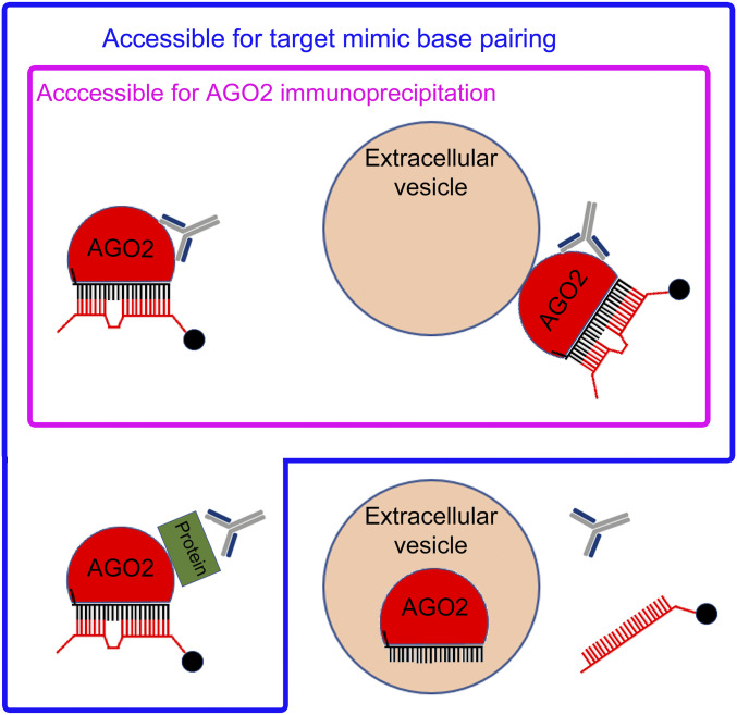
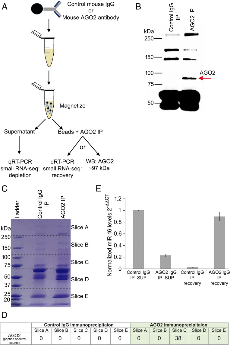
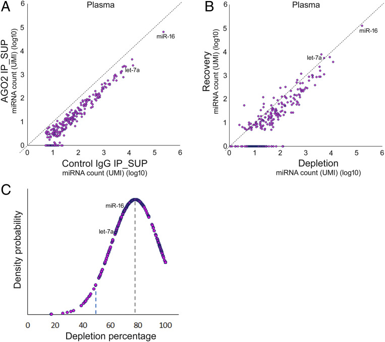
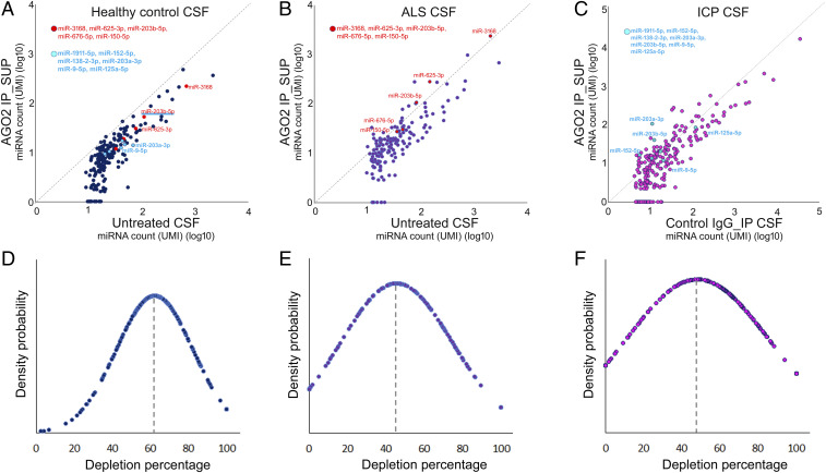
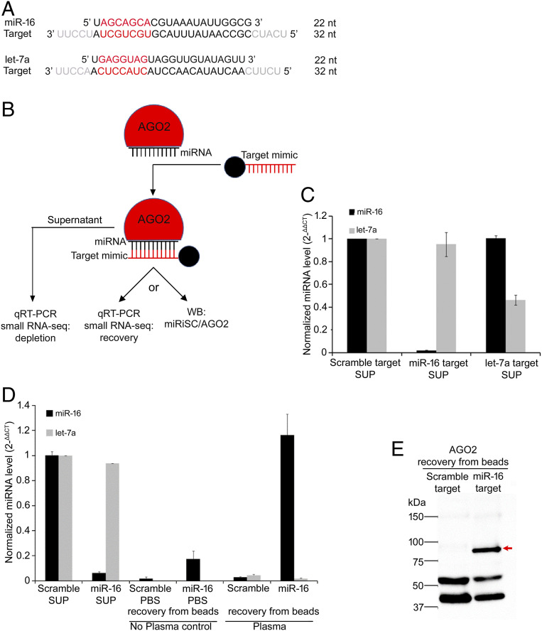
Significance: MicroRNAs (miRNAs) are abundant inside cells, where they regulate gene expression posttranscriptionally, but are also found in circulation as remarkably stable extracellular constituents of body fluids such as human blood plasma and cerebrospinal fluid. We observed that these circulating miRNAs are predominantly associated with free Argonaut 2 (AGO2) complexes that are accessible to immunoprecipitation by AGO2 antibodies and are able to base-pair with complementary target RNAs, suggesting that, under normal physiological conditions, circulating miRNAs are largely contained in functional miRNA-mediated silencing complexes (miRISCs). However, we observe that, under certain pathological conditions, more miRNAs become associated with complexes other than free AGO2/miRISC, suggesting that the assortment of circulating miRNAs into distinct complexes offers a potential new dimension to miRNA-based diagnostics.
Overview
The claims and statistics of this paper haven’t been checked yet.
Run the check — free, no account needed. It reads the open-access text and checks the sources it cites.
Integrity signals
Registry
Cites
Statistics
Figures
Verification
Claims the paper makes
The claims and statistics of this paper haven’t been checked yet.
Run the check — free, no account needed. It reads the open-access text and checks the sources it cites.
Claims other papers make about this one
Each is checked against this paper’s own text: does it say what the citing paper says it does?
The cited source says what the claim attributes to it.
In the citing paper
“Extracellular AGO-bound miRNAs are present within biofluids including plasma (Arroyo et al., 2011; Turchinovich et al., 2011), probably released from diverse tissues (Turchinovich and Burwinkel, 2012), and their altered composition may have diagnostic value (Geekiyanage et al., 2020).”
From Brain cell-specific origin of circulating microRNA biomarkers in experimental temporal lobe epilepsy (2023) · cites this paper as Geekiyanage et al., 2020
Self-check
Percentages, sums and reported differences re-computed from the paper’s own numbers.
The self-check needs the paper’s full text.
Citations
Cites
Moderated estimation of fold change and dispersion for RNA-seq data with DESeq2
Love, M. I., Huber, W., & Anders, S. (2014) · Genome Biology · doi:10.1186/s13059-014-0550-8 · Cited by 95,106
Bartel (2004) · doi:10.1016/s0092-8674(04)00045-5 · Cited by 34,714
Valadi, H., Ekström, K., Bossios, A., Sjöstrand, M., Lee, J. J., & Lötvall, J. O. (2007) · Nature Cell Biology · doi:10.1038/ncb1596 · Cited by 12,834
Mantovani, A., Allavena, P., Sica, A., & Balkwill, F. (2008) · Nature · doi:10.1038/nature07205 · Cited by 11,399
Circulating microRNAs as stable blood-based markers for cancer detection
Mitchell, P. S., Parkin, R. K., Kroh, E. M., Fritz, B. R., Wyman, S. K., Pogosova-Agadjanyan, E. L., Peterson, A., Noteboom, J., O'Briant, K. C., Allen, A., et al. (2008) · Proceedings of the National Academy of Sciences · doi:10.1073/pnas.0804549105 · Cited by 7,969
MicroRNAs: small RNAs with a big role in gene regulation
He, L., & Hannon, G. J. (2004) · Nature Reviews Genetics · doi:10.1038/nrg1379 · Cited by 7,118
Skog, J., Würdinger, T., van Rijn, S., Meijer, D. H., Gainche, L., Curry, W. T., Carter, B. S., Krichevsky, A. M., & Breakefield, X. O. (2008) · Nature Cell Biology · doi:10.1038/ncb1800 · Cited by 5,001
Chen, X., Ba, Y., Ma, L., Cai, X., Yin, Y., Wang, K., Guo, J., Zhang, Y., Chen, J., Guo, X., et al. (2008) · Cell Research · doi:10.1038/cr.2008.282 · Cited by 4,560
Arroyo, J. D., Chevillet, J. R., Kroh, E. M., Ruf, I. K., Pritchard, C. C., Gibson, D. F., Mitchell, P. S., Bennett, C. F., Pogosova-Agadjanyan, E. L., Stirewalt, D. L., et al. (2011) · Proceedings of the National Academy of Sciences · doi:10.1073/pnas.1019055108 · Cited by 3,294
MicroRNAs are transported in plasma and delivered to recipient cells by high-density lipoproteins
Vickers, K. C., Palmisano, B. T., Shoucri, B. M., Shamburek, R. D., & Remaley, A. T. (2011) · Nature Cell Biology · doi:10.1038/ncb2210 · Cited by 2,741
Intercellular transfer of the oncogenic receptor EGFRvIII by microvesicles derived from tumour cells
Al-Nedawi, K., Meehan, B., Micallef, J., Lhotak, V., May, L., Guha, A., & Rak, J. (2008) · Nature Cell Biology · doi:10.1038/ncb1725 · Cited by 2,034
Characterization of extracellular circulating microRNA
Turchinovich, A., Weiz, L., Langheinz, A., & Burwinkel, B. (2011) · Nucleic Acids Research · doi:10.1093/nar/gkr254 · Cited by 1,941
Unidirectional transfer of microRNA-loaded exosomes from T cells to antigen-presenting cells
Mittelbrunn, M., Gutiérrez-Vázquez, C., Villarroya-Beltri, C., González, S., Sánchez-Cabo, F., González, M. Á., Bernad, A., & Sánchez-Madrid, F. (2011) · Nature Communications · doi:10.1038/ncomms1285 · Cited by 1,769
Adipose-derived circulating miRNAs regulate gene expression in other tissues
Thomou, T., Mori, M. A., Dreyfuss, J. M., Konishi, M., Sakaguchi, M., Wolfrum, C., Rao, T. N., Winnay, J. N., Garcia-Martin, R., Grinspoon, S. K., et al. (2017) · Nature · doi:10.1038/nature21365 · Cited by 1,539
Delivery of MicroRNA-126 by Apoptotic Bodies Induces CXCL12-Dependent Vascular Protection
Zernecke, A., Bidzhekov, K., Noels, H., Shagdarsuren, E., Gan, L., Denecke, B., Hristov, M., Köppel, T., Jahantigh, M. N., Lutgens, E., et al. (2009) · Science Signaling · doi:10.1126/scisignal.2000610 · Cited by 1,334
MicroRNA Pathways in Flies and Worms
Ambros, V. (2003) · Cell · doi:10.1016/s0092-8674(03)00428-8 · Cited by 1,217
Self-citationSecreted Monocytic miR-150 Enhances Targeted Endothelial Cell Migration
Zhang, Y., Liu, D., Chen, X., Li, J., Li, L., Bian, Z., Sun, F., Lu, J., Yin, Y., Cai, X., et al. (2010) · Molecular Cell · doi:10.1016/j.molcel.2010.06.010 · Cited by 1,195
Ying, W., Riopel, M., Bandyopadhyay, G., Dong, Y., Birmingham, A., Seo, J. B., Ofrecio, J. M., Wollam, J., Hernandez-Carretero, A., Fu, W., et al. (2017) · Cell · doi:10.1016/j.cell.2017.08.035 · Cited by 1,191
Quantitative and stoichiometric analysis of the microRNA content of exosomes
Chevillet, J. R., Kang, Q., Ruf, I. K., Briggs, H., Vojtech, L. N., Hughes, S. M., Cheng, H. H., Arroyo, J. D., Meredith, E. K., Gallichotte, E. N., et al. (2014) · Proceedings of the National Academy of Sciences · doi:10.1073/pnas.1408301111 · Cited by 1,148
Antibody mediated in vivo delivery of small interfering RNAs via cell-surface receptors
Song, E., Zhu, P., Lee, S., Chowdhury, D., Kussman, S., Dykxhoorn, D. M., Feng, Y., Palliser, D., Weiner, D. B., Shankar, P., et al. (2005) · Nature Biotechnology · doi:10.1038/nbt1101 · Cited by 1,027
Export of microRNAs and microRNA-protective protein by mammalian cells
Wang, K., Zhang, S., Weber, J., Baxter, D., & Galas, D. J. (2010) · Nucleic Acids Research · doi:10.1093/nar/gkq601 · Cited by 1,002
Gibbings, D. J., Ciaudo, C., Erhardt, M., & Voinnet, O. (2009) · Nature Cell Biology · doi:10.1038/ncb1929 · Cited by 991
Schiffelers, R. M., Ansari, A., Xu, J., Zhou, Q., Tang, Q., Storm, G., Molema, G., Lu, P. Y., Scaria, P. V., & Woodle, M. C. (2004) · Nucleic Acids Research · doi:10.1093/nar/gnh140 · Cited by 904
The Crystal Structure of Human Argonaute2
Schirle, N. T., & MacRae, I. J. (2012) · Science · doi:10.1126/science.1221551 · Cited by 696
The Structure of Human Argonaute-2 in Complex with miR-20a
Elkayam, E., Kuhn, C.-D., Tocilj, A., Haase, A. D., Greene, E. M., Hannon, G. J., & Joshua-Tor, L. (2012) · Cell · doi:10.1016/j.cell.2012.05.017 · Cited by 613
Structural basis for microRNA targeting
Schirle, N. T., Sheu‐Gruttadauria, J., & MacRae, I. J. (2014) · Science · doi:10.1126/science.1258040 · Cited by 582
Ohshima, K., Inoue, K., Fujiwara, A., Hatakeyama, K., Kanto, K., Watanabe, Y., Muramatsu, K., Fukuda, Y., Ogura, S.-I., Yamaguchi, K., et al. (2010) · PLoS ONE · doi:10.1371/journal.pone.0013247 · Cited by 577
Jin, X., Chen, Y., Chen, H., Fei, S., Chen, D., Cai, X., Liu, L., Lin, B., Su, H., Zhao, L., et al. (2017) · Clinical Cancer Research · doi:10.1158/1078-0432.ccr-17-0577 · Cited by 558
Endogenous miRNA and Target Concentrations Determine Susceptibility to Potential ceRNA Competition
Bosson, A. D., Zamudio, J. R., & Sharp, P. A. (2014) · Molecular Cell · doi:10.1016/j.molcel.2014.09.018 · Cited by 458
Comparison of isolation methods of exosomes and exosomal RNA from cell culture medium and serum
Tang, Y.-T., Huang, Y.-Y., Zheng, L., Qin, S.-H., Xu, X.-P., An, T.-X., Xu, Y., Wu, Y.-S., Hu, X.-M., Ping, B.-H., et al. (2017) · International Journal of Molecular Medicine · doi:10.3892/ijmm.2017.3080 · Cited by 455
KRAS-MEK Signaling Controls Ago2 Sorting into Exosomes
McKenzie, A. J., Hoshino, D., Hong, N. H., Cha, D. J., Franklin, J. L., Coffey, R. J., Patton, J. G., & Weaver, A. M. (2016) · Cell Reports · doi:10.1016/j.celrep.2016.03.085 · Cited by 423
Exosome-mediated transfer of miR-10b promotes cell invasion in breast cancer
Singh, R., Pochampally, R. R., Watabe, K., Lü, Z., & Mo, Y. (2014) · Molecular Cancer · doi:10.1186/1476-4598-13-256 · Cited by 408
Argonaute Divides Its RNA Guide into Domains with Distinct Functions and RNA-Binding Properties
Wee, L. M., Flores-Jasso, C. F., Salomon, W. E., & Zamore, P. D. (2012) · Cell · doi:10.1016/j.cell.2012.10.036 · Cited by 401
Williams, Z., Ben-Dov, I. Z., Elias, R., Mihailovic, A., Brown, M., Rosenwaks, Z., & Tuschl, T. (2013) · Proceedings of the National Academy of Sciences · doi:10.1073/pnas.1214046110 · Cited by 339
Cutler, R. G., Pedersen, W. A., Camandola, S., Rothstein, J. D., & Mattson, M. P. (2002) · Annals of Neurology · doi:10.1002/ana.10312 · Cited by 323
Blood serum miRNA: Non-invasive biomarkers for Alzheimer's disease
Geekiyanage, H., Jicha, G. A., Nelson, P. T., & Chan, C. (2011) · Experimental Neurology · doi:10.1016/j.expneurol.2011.11.026 · Cited by 299
Self-citationSalomon, W. E., Jolly, S. M., Moore, M. J., Zamore, P. D., & Serebrov, V. (2015) · Cell · doi:10.1016/j.cell.2015.06.029 · Cited by 270
A Dynamic Search Process Underlies MicroRNA Targeting
Chandradoss, S. D., Schirle, N. T., Szczepaniak, M., MacRae, I. J., & Joo, C. (2015) · Cell · doi:10.1016/j.cell.2015.06.032 · Cited by 259
Geekiyanage, H., & Chan, C. (2011) · The Journal of Neuroscience · doi:10.1523/jneurosci.3883-11.2011 · Cited by 254
Self-citationDEBrowser: interactive differential expression analysis and visualization tool for count data
Küçükural, A., Yukselen, O., Özata, D. M., Moore, M. J., & Garber, M. G. (2019) · BMC Genomics · doi:10.1186/s12864-018-5362-x · Cited by 248
Gene regulation. Structural basis for microRNA targeting.
Schirle, N. T., Sheu‐Gruttadauria, J., & MacRae, I. J. (2014) · Science · doi:10.2210/pdb4w5n/pdb · Cited by 226
Human Argonaute 2 Has diverse reaction pathways on Target RNAs
Jo, M. H., Shin, S., Jung, S.-R., Kim, E., Song, J.-J., & Hohng, S. (2015) · Molecular Cell · doi:10.1016/j.molcel.2015.04.027 · Cited by 207
RNA-based recognition and targeting: sowing the seeds of specificity
Gorski, S. A., Vogel, J., & Doudna, J. A. (2017) · Nature Reviews Molecular Cell Biology · doi:10.1038/nrm.2016.174 · Cited by 197
Ward, J. A., Kanchagar, C., Veksler‐Lublinsky, I., Lee, R. C., McGill, M. R., Jaeschke, H., Curry, S. C., & Ambros, V. (2014) · Proceedings of the National Academy of Sciences · doi:10.1073/pnas.1412608111 · Cited by 177
Self-citationExtracellular miRNA: A Collision of Two Paradigms
Turchinovich, A. A., Tonevitsky, A., & Burwinkel, B. (2016) · Trends in Biochemical Sciences · doi:10.1016/j.tibs.2016.08.004 · Cited by 168
Horman, S. R., Janas, M. M., Litterst, C. M., Wang, B., MacRae, I. J., Sever, M. J., Morrissey, D. V., Graves, P. R., Luo, B., Umesalma, S., et al. (2013) · Molecular Cell · doi:10.1016/j.molcel.2013.03.015 · Cited by 164
Systemic dysregulation of TDP-43 binding microRNAs in amyotrophic lateral sclerosis
Freischmidt, A., Müller, K., Ludolph, A. C., & Weishaupt, J. H. (2013) · Acta Neuropathologica Communications · doi:10.1186/2051-5960-1-42 · Cited by 159
Human plasma and serum extracellular small RNA reference profiles and their clinical utility
Max, K. E. A., Bertram, K., Akat, K. M., Bogardus, K. A., Li, J., Morozov, P. S., Ben‐Dov, I. Z., Li, X., Weiss, Z. R., Azizian, A., et al. (2018) · Proceedings of the National Academy of Sciences · doi:10.1073/pnas.1714397115 · Cited by 158
Raoof, R., Jimenez-Mateos, E. M., Bauer, S., Tackenberg, B., Rosenow, F., Lang, J., Onugoren, M. D., Hamer, H., Huchtemann, T., Körtvélyessy, P., et al. (2017) · Scientific Reports · doi:10.1038/s41598-017-02969-6 · Cited by 109
MicroRNAs as Biomarkers in Amyotrophic Lateral Sclerosis
Ricci, C., Marzocchi, C., & Battistini, S. (2018) · Cells · doi:10.3390/cells7110219 · Cited by 107
Waller, R., Wyles, M., Heath, P. R., Kazoka, M., Wollff, H., Shaw, P. J., & Kirby, J. (2018) · Frontiers in Neuroscience · doi:10.3389/fnins.2017.00731 · Cited by 101
Isolation and Identification of miRNAs in exosomes derived from serum of colon cancer patients
Zhao, L., Yu, J., Wang, J., Li, H., Che, J., & Cao, B. (2017) · Journal of Cancer · doi:10.7150/jca.18026 · Cited by 83
Benigni, M., Ricci, C., Jones, A. R., Giannini, F., Al‐Chalabi, A., & Battistini, S. (2016) · NeuroMolecular Medicine · doi:10.1007/s12017-016-8396-8 · Cited by 78
A novel serum microRNA signature to screen esophageal squamous cell carcinoma
Huang, Z., Zhang, L., Zhu, D., Shan, X., Zhou, X., Qi, L., Wu, L., Zhu, J., Cheng, W., Zhang, H., et al. (2017) · Cancer Medicine · doi:10.1002/cam4.973 · Cited by 74 · cited 2×
“Circulating miRNAs have been investigated as potential diagnostic markers for numerous diseases, including cancer (11, 12), liver toxicity (14), and neurodegenerative disorders (10, 13).”
BackgroundCompetitive Endogenous RNAs Cannot Alter MicroRNA Function In Vivo
Broderick, J. A., & Zamore, P. D. (2014) · Molecular Cell · doi:10.1016/j.molcel.2014.05.023 · Cited by 70
Circulating Exosomal miR-141-3p and miR-375 in Metastatic Progression of Rectal Cancer
Meltzer, S., Bjørnetrø, T., Lyckander, L. G., Flatmark, K., Dueland, S., Samiappan, R., Johansen, C., Kalanxhi, E., Ree, A. H., & Redalen, K. R. (2019) · Translational Oncology · doi:10.1016/j.tranon.2019.04.014 · Cited by 70
Optimizing exosomal RNA isolation for RNA-Seq analyses of archival sera specimens
Prendergast, E. N., Fonseca, M. A. D. S., Dezem, F. S., Lester, J., Karlan, B. Y., Noushmehr, H., Lin, X., & Lawrenson, K. (2018) · PLoS ONE · doi:10.1371/journal.pone.0196913 · Cited by 59
Prud'homme, G., Glinka, Y., Lichner, Z., & Yousef, G. (2016) · OncoTarget · doi:10.18632/oncotarget.10929 · Cited by 49
Differentially expressed miRNAs in circulating exosomes between atrial fibrillation and sinus rhythm
Wang, S., Min, J., Yu, Y., Yin, L., Wang, Q., Shen, H. M., Yang, J. A., Zhang, P., Xiao, J., & Wang, Z. (2019) · Journal of Thoracic Disease · doi:10.21037/jtd.2019.09.50 · Cited by 45
An efficient and sensitive method for preparing cDNA libraries from scarce biological samples
Sterling, C. H., Veksler‐Lublinsky, I., & Ambros, V. (2014) · Nucleic Acids Research · doi:10.1093/nar/gku637 · Cited by 24
Self-citationTanrıverdi, K., Küçükural, A., Mikhalev, E., et al. (2016) · Analytical Biochemistry · doi:10.1016/j.ab.2016.02.019 · Cited by 24
Argonaute Pull-Down and RISC Analysis Using 2’-O-Methylated Oligonucleotides Affinity Matrices
Jannot, G., Vasquez-Rifo, A., & Simard, M. J. (2011) · Methods in molecular biology · doi:10.1007/978-1-61779-046-1_16 · Cited by 21
Teruel‐Montoya, R., Luengo‐Gil, G., Vallejo, F., Yuste, J. E., Bohdan, N., García‐Barberá, N., Espín, S., Martı́nez, C., Espı́n, J. C., Vicente, V., et al. (2019) · Scientific Reports · doi:10.1038/s41598-019-40453-5 · Cited by 18
Li, F., Huang, J., Liu, J., Xu, W., & Yuan, Z. (2019) · Cancer Cell International · doi:10.1186/s12935-019-0918-7 · Cited by 8
Cited by
Advances in the development of new biomarkers for Alzheimer’s disease
Klyucherev, T. O., Olszewski, P., Shalimova, A. A., Chubarev, V. N., Tarasov, V. V., Attwood, M. M., Syvänen, S., & Schiöth, H. B. (2022) · Translational Neurodegeneration · doi:10.1186/s40035-022-00296-z · Cited by 241
Garcia, G., Pinto, S., Cunha, M., Fernandes, A., Koistinaho, J., & Brites, D. (2021) · Cells · doi:10.3390/cells10092424 · Cited by 39
Brain cell-specific origin of circulating microRNA biomarkers in experimental temporal lobe epilepsy
Brindley, E., Heiland, M., Mooney, C., Diviney, M., Mamad, O., Hill, T. D. M., Yan, Y., Venø, M. T., Reschke, C. R., Batool, A., et al. (2023) · Frontiers in Molecular Neuroscience · doi:10.3389/fnmol.2023.1230942 · Cited by 21
Metadata
- Authors
- Geekiyanage, H., Rayatpisheh, S., Wohlschlegel, J. A., Brown, R., & Ambros, V.
- Affiliations
- Department of Molecular Medicine, University of Massachusetts Medical School, Worcester, MA 01605;
- Department of Biological Chemistry, Geffen School of Medicine, University of California, Los Angeles, CA 90095;
- Department of Neurology, University of Massachusetts Medical School, Worcester, MA 01605
- Venue
- Proceedings of the National Academy of Sciences
- Publisher
- National Academy of Sciences
- Published
- 2020-09-14
- Volume
- 117
- Issue
- 39
- Pages
- 24213-24223
- ISSN
- 0027-8424
- 1091-6490
- PMC
- PMC7533700
- Cited by
- 148
- References
- 64
- Language
- en
- Funders
- HHS | NIH | National Institute of General Medical Sciences (R01GM034028, R01GM088365, GM089778)
About these checks
Registry, retraction, Cites and statistics results are computed, not judged, with one exception: a printed reference that no index lists is read once by a model before it counts as a citation error. Claims are extracted by a model and kept only when their quote appears verbatim in the text; each cited source is then checked, with a model’s help, for whether it says what the claim relies on. Those verdicts can be wrong — read the claim’s quote and the cited source before relying on one.
Statistics and arithmetic are recomputed, not judged.
Confidence bands are shown only for fields calibrated on our annotated test set. None is calibrated yet, so no band is shown.
What counts as a claim ›How we measure confidence ›How we check this ›
Are you an author, or do you see a problem with this page? Request a review
Data sources
Bibliographic data: Semantic Scholar, OpenAlex, Crossref.
Contains information from the Semantic Scholar Academic Graph Datasets, made available under the ODC Attribution License (ODC-BY 1.0).
Semantic Scholar’s data is described in: Kinney, R., et al. (2023). The Semantic Scholar Open Data Platform. arXiv:2301.10140.
