

The DOI matches its Crossref record; 82 of the 82 references it cites were found in a registry or an index; 34 of the 56 sources its claims cite were checked for support.
Regulation of beta-amyloid production in neurons by astrocyte-derived cholesterol
Open accessPublished version of a preprint
Abstract
Significance: The accumulation of amyloid β (Aβ) in the brain appears to be a necessary event in the pathogenesis of Alzheimer’s disease (AD). However, processes linked to the endogenous regulation of Aβ production are still not completely understood. Here, the authors show that Aβ accumulation in neurons is tightly regulated by cholesterol synthesis and apoE transport from astrocytes. The study provides a molecular context for understanding the endogenous regulation of Aβ accumulation and why it correlates with AD. The tight regulation suggests that Aβ may perform an important cellular function. A complete understanding of the mechanism is likely necessary to predict whether the selective removal of Aβ has potential for a therapeutic benefit.
Overview
AI snapshot
Written from the paper’s full text.
TL;DR
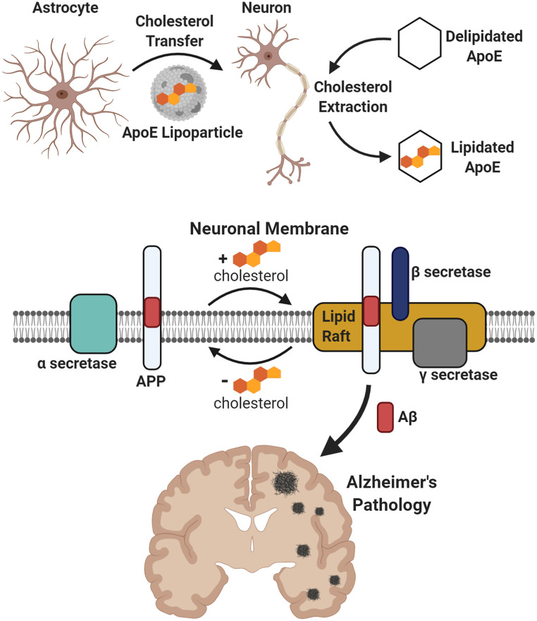
Astrocyte-derived cholesterol, transported to neurons by apoE, regulates APP localization to GM1 lipid clusters and thereby shifts APP processing between nonamyloidogenic sAPP-α and amyloidogenic Aβ. Deleting astrocyte SREBP2 in 3xTg-AD mice nearly eliminates insoluble Aβ and plaques, linking astrocyte cholesterol, apoE, and amyloid pathology in one pathway.
Key findings
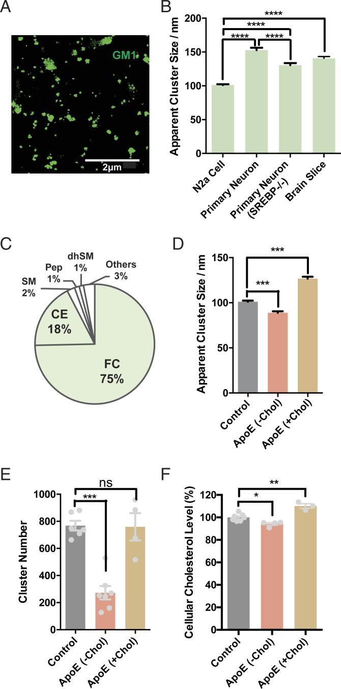
- dSTORM imaging showed GM1 domains averaged ∼141 nm in WT mouse cortical tissue, ∼150 nm in primary neurons, and ∼100 nm in N2a cells; astrocyte SREBP2 knockout reduced primary neuron cluster size to 130 nm, and apoE +FBS increased N2a cluster diameter from 100 to 130 nm.
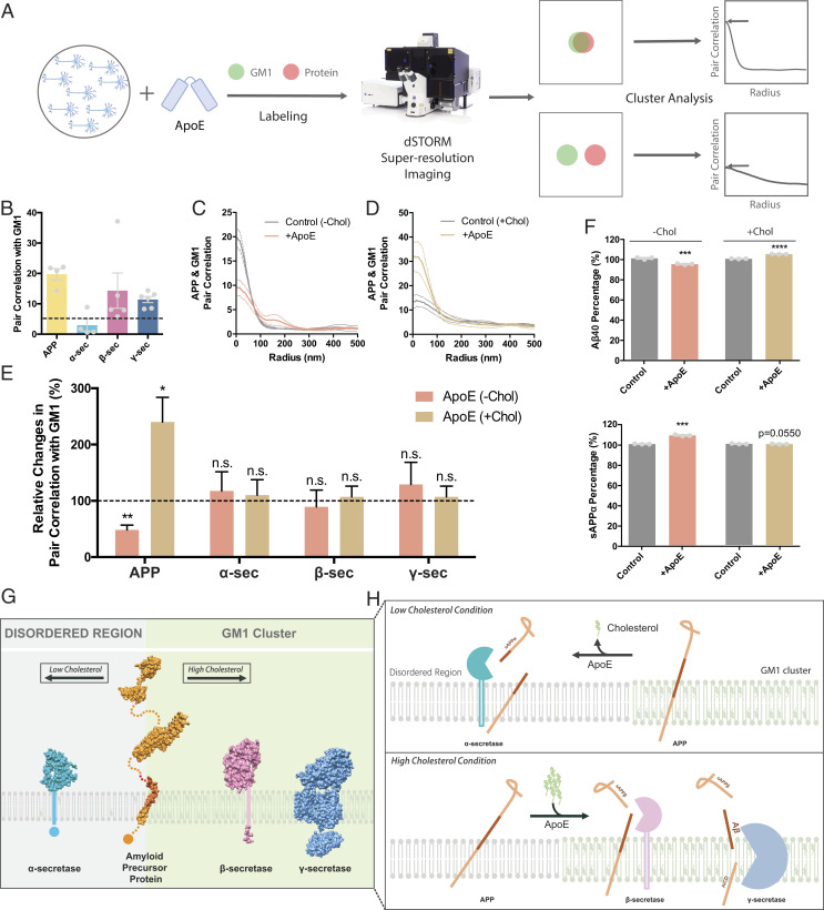
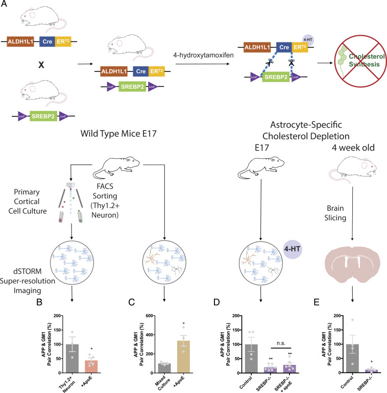
- APP, β-secretase, and γ-secretase associated with GM1 clusters (pair correlation 10–15), while α-secretase did not (<3); apoE cholesterol loading increased APP–GM1 association almost twofold versus efflux, and in primary cultures apoE decreased APP association >twofold in purified neurons but increased it 2.5-fold when astrocytes were present.
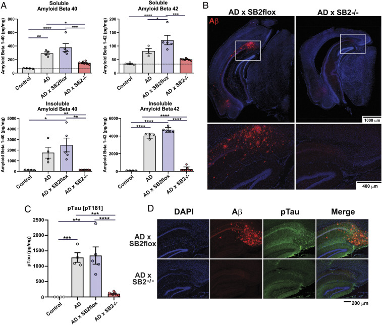
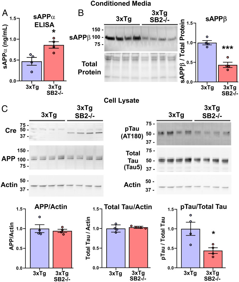
- In 3xTg-AD × SREBP2fl/fl GFAP-Cre mice at 60 wk, soluble Aβ40 and Aβ42 were reduced twofold, insoluble Aβ40 and Aβ42 were almost entirely eliminated, amyloid plaques were absent, and pTau at T181 was eliminated, with total APP, GFAP, and ApoE unchanged.
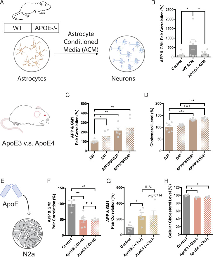
- ApoE4 knock-in brains showed increased APP–GM1 association versus apoE3, but purified apoE3 and apoE4 did not differ significantly in loading or unloading cholesterol in N2a cells.
Methods
- Superresolution dSTORM imaging in N2a cells, primary cortical neurons, mixed neuron/astrocyte cultures, and mouse brain slices; GM1 lipids were labeled with fluorescent CTxB and APP/secretases with antibodies, followed by pair-correlation and cluster analysis.
- Mouse genetics included astrocyte-specific SREBP2 knockout via ALDH1L1-Cre or GFAP-Cre, crosses with 3xTg-AD and APOE-KI/APP/PS1 mice, and measurements by ELISA for Aβ40/42, sAPP-α, and pTau, plus Western blot, lipidomics mass spectrometry, and live-cell cholesterol assays.
- Cell culture used embryonic day 17 cortical neurons, FACS sorting with Thy1.2, astrocyte-conditioned media from WT or APOE−/− astrocytes, and apoE3/apoE4 treatments with or without FBS/cholesterol.
Limitations
- Stated: all astrocyte-targeting Cre lines also delete to some degree in neural progenitor cells, and the exact mechanisms by which astrocyte cholesterol influences neuronal tau phosphorylation require future investigations.
- Evident: most mechanistic evidence comes from cultured cells and mouse models, with no human tissue or clinical data; the in vivo apoE4 versus apoE3 cholesterol trend was not statistically significant, and purified apoE3/apoE4 showed no significant functional difference in cholesterol transport.
- Evident: reduced brain tissue volume was observed in AD × SB2−/− animals, similar to prior astrocyte-specific SB2−/− brains, which could complicate interpretation of pathology endpoints.
Model-written summary. Check it against the paper before relying on it.
Integrity signals
Registry
Cites
Statistics
Figures
Verification
Claims the paper makes
48 Own result · 35 Own interpretation · 18 Partly supported · 9 Supported · 6 No full text of the sources · 5 Not checked yet · 2 Sources not found · 2 Own method · 1 Supported so far · accuracy not yet measured
Every source the paper cites for this claim was checked and supports it.
In the paper
“Importantly, apoE is not present in FBS (44).”
§ Regulation of beta-amyloid production in neurons by astrocyte-derived cholesterol > Results > Characterization of Astrocyte-Derived Cholesterol on Neuronal Cluster Formation.
What the sources say
- Supportedcites [44] V. P. Varma., Ocular fluid as a replacement for serum in cell cryopreservation media. PLoS One 10, e0131291 (2015). 26135924 PMC4489643 source
Table 2 in the Results section (Proteomic analysis of BuOF and FBS) lists apolipoprotein E as absent in FBS, directly supporting the cited statement. (LLM, conf=0.94)
plain- Asserted: passes — The authors put it forward as true.
- Checkable: passes — It can be checked against evidence.
- Load-bearing: passes — The paper’s conclusions depend on it.
- Based: passes — The paper gives a reason why it holds.
- Supported
The cited sources support only part of this claim — read each source’s verdict below.
In the paper
“SREBP2 is the master transcriptional regulator of the majority of enzymes involved in cholesterol synthesis.”
§ Regulation of beta-amyloid production in neurons by astrocyte-derived cholesterol > Results > The Role of Astrocyte Cholesterol in Regulating APP Processing in Cultured Primary Neurons.
What the sources say
- Partly supportedcites [39] X. Hua., SREBP-2, a second basic-helix-loop-helix-leucine zipper protein that stimulates transcription by binding to a sterol regulatory element. Proc. Natl. Acad. Sci. U.S.A. 90, 11603– 11607 (1993). 7903453 PMC48032 sourceExperimental — accuracy not yet measured
The Abstract (p. 11603) identifies SREBP-2 as a transcription factor and reports that it activates reporters containing a sterol regulatory element also found in the HMG-CoA synthase promoter. It does not establish that SREBP-2 regulates the majority of cholesterol-synthesis enzymes or that it is their master regulator. (LLM, conf=0.86)
plaindirect- Asserted: passes — The authors put it forward as true.
- Checkable: passes — It can be checked against evidence.
- Load-bearing: passes — The paper’s conclusions depend on it.
- Based: passes — The paper gives a reason why it holds.
- Partly supported
The cited sources support only part of this claim — read each source’s verdict below.
In the paper
“ApoE can both load and unload cholesterol from cells, including neurons (41–43) (see also SI Appendix).”
§ Regulation of beta-amyloid production in neurons by astrocyte-derived cholesterol > Results > Characterization of Astrocyte-Derived Cholesterol on Neuronal Cluster Formation.
What the sources say
- Not checked — no full text of the sourcecites [41] J. Luo, H. Yang, B.-L. Song, Mechanisms and regulation of cholesterol homeostasis. Nat. Rev. Mol. Cell Biol. 21, 225– 245 (2020). 31848472 source
- Partly supportedcites [42] J. Chen, X. Zhang, H. Kusumo, L. G. Costa, M. Guizzetti, Cholesterol efflux is differentially regulated in neurons and astrocytes: Implications for brain cholesterol homeostasis. Biochim. Biophys. Acta 1831, 263– 275 (2013). 23010475 PMC3534809 sourceExperimental — accuracy not yet measured
The Results (§3.1; page unavailable) show that apoE3 increased cholesterol efflux from neurons, supporting the unloading part. The Introduction (§1; page unavailable) describes apoE-containing lipoproteins interacting with neuronal receptors but says whether their effects involve cholesterol uptake by neurons remains controversial; it does not establish the loading part as stated. (LLM, conf=0.84)
- Not checked yetcites [43] H. Hayashi, R. B. Campenot, D. E. Vance, J. E. Vance, Apolipoprotein E-containing lipoproteins protect neurons from apoptosis via a signaling pathway involving low-density lipoprotein receptor-related protein-1. J. Neurosci. 27, 1933– 1941 (2007). 17314289 PMC6673537 source
Checked before — its earlier verdict is held back until a new check runs.
plaindirect- Asserted: passes — The authors put it forward as true.
- Checkable: passes — It can be checked against evidence.
- Load-bearing: passes — The paper’s conclusions depend on it.
- Based: passes — The paper gives a reason why it holds.
Every source the paper cites for this claim was checked and supports it.
In the paper
“Strikingly, both pathways are finely regulated by cholesterol (6) (SI Appendix, Fig. S1B).”
§ Regulation of beta-amyloid production in neurons by astrocyte-derived cholesterol > Abstract
What the sources say
- Supportedcites [6] M.-P. Marzolo, G. Bu, Lipoprotein receptors and cholesterol in APP trafficking and proteolytic processing, implications for Alzheimer’s disease. Semin. Cell Dev. Biol. 20, 191– 200 (2009). 19041409 PMC2691858 source
Section 4.1 reports that cholesterol levels regulate amyloidogenic secretase activity and that cholesterol-reducing drugs increase nonamyloidogenic sAPPα secretion while decreasing Aβ formation. Together, these passages support the claim that cholesterol regulates both APP processing pathways. (LLM, conf=0.91)
boosteddirect- Asserted: passes — The authors put it forward as true.
- Checkable: passes — It can be checked against evidence.
- Load-bearing: passes — The paper’s conclusions depend on it.
- Based: passes — The paper gives a reason why it holds.
- Supported
Every source the paper cites for this claim was checked and supports it.
In the paper
“Mice with SREBP2 knocked out of astrocytes have reduced the production of cholesterol in the astrocytes (47) (SI Appendix, Fig. S4A).”
§ Regulation of beta-amyloid production in neurons by astrocyte-derived cholesterol > Results > The Role of Astrocyte Cholesterol in Regulating APP Processing in Cultured Primary Neurons.
What the sources say
- Supportedcites [47] H. A. Ferris., Loss of astrocyte cholesterol synthesis disrupts neuronal function and alters whole-body metabolism. Proc. Natl. Acad. Sci. U.S.A. 114, 1189– 1194 (2017). 28096339 PMC5293102 source
The Discussion states that the authors inactivated SREBP2 in astrocytes specifically to reduce astroglial cholesterol synthesis. The section on normal brain development says the knockout mice show a loss of cholesterol synthesis in astrocytes, partially compensated by increased cholesterol production in neurons. (LLM, conf=0.93)
plaindirect- Asserted: passes — The authors put it forward as true.
- Checkable: passes — It can be checked against evidence.
- Load-bearing: passes — The paper’s conclusions depend on it.
- Based: passes — The paper gives a reason why it holds.
- Supported
The cited sources support only part of this claim — read each source’s verdict below.
In the paper
“Cholesterol is high in AD brains (1, 6).”
§ Regulation of beta-amyloid production in neurons by astrocyte-derived cholesterol > Results > The In Vivo Role of Astrocyte Cholesterol on Aβ and Plaque Formation in Mouse Brain.
What the sources say
- Not checked — no full text of the sourcecites [1] C. Haass, D. J. Selkoe, Cellular processing of β-amyloid precursor protein and the genesis of amyloid β-peptide. Cell 75, 1039– 1042 (1993). 8261505 source
- Partly supportedcites [6] M.-P. Marzolo, G. Bu, Lipoprotein receptors and cholesterol in APP trafficking and proteolytic processing, implications for Alzheimer’s disease. Semin. Cell Dev. Biol. 20, 191– 200 (2009). 19041409 PMC2691858 sourceExperimental — accuracy not yet measured
Section 4 reports that one study suggested altered cholesterol homeostasis in AD brains, resulting in higher total cholesterol levels. The claim presents that qualified finding as an established general fact. (LLM, conf=0.84)
plain- Asserted: passes — The authors put it forward as true.
- Checkable: passes — It can be checked against evidence.
- Load-bearing: unclear — It is unclear whether the paper’s conclusions depend on it.
- Based: passes — The paper gives a reason why it holds.
The cited sources support only part of this claim — read each source’s verdict below.
In the paper
“Neurons appear incapable of increasing cholesterol synthesis to compensate for astrocyte deficiency in any appreciable way (48).”
§ Regulation of beta-amyloid production in neurons by astrocyte-derived cholesterol > Results > The Role of Astrocyte Cholesterol in Regulating APP Processing in Cultured Primary Neurons.
What the sources say
- Partly supportedcites [48] C.-C. Liu, C.-C. Liu, T. Kanekiyo, H. Xu, G. Bu, Apolipoprotein E and Alzheimer disease: Risk, mechanisms and therapy. Nat. Rev. Neurol. 9, 106– 118 (2013). 23296339 PMC3726719 sourceExperimental — accuracy not yet measured
The Introduction (page unavailable) says astrocytes supply cholesterol to neurons, and the Lipid metabolism section (page unavailable) suggests reduced delivery may impair neuronal health. Neither section says whether neurons can increase their own cholesterol synthesis when astrocyte supply is deficient, so the claim’s central assertion is unsupported. (LLM, conf=0.75)
hedged- Asserted: passes — The authors put it forward as true.
- Checkable: passes — It can be checked against evidence.
- Load-bearing: passes — The paper’s conclusions depend on it.
- Based: passes — The paper gives a reason why it holds.
- Partly supported
Every source the paper cites for this claim was checked and supports it.
In the paper
“APP/PS1 AD mice overexpressing a truncated active form of SREBP2 display an accelerated Aβ burden with aging (60).”
§ Regulation of beta-amyloid production in neurons by astrocyte-derived cholesterol > Discussion
What the sources say
- Supportedcites [60] E. Barbero-Camps, A. Fernández, L. Martínez, J. C. Fernández-Checa, A. Colell, APP/PS1 mice overexpressing SREBP-2 exhibit combined Aβ accumulation and tau pathology underlying Alzheimer’s disease. Hum. Mol. Genet. 22, 3460– 3476 (2013). 23648430 PMC3736868 source
The Introduction states that the authors generated APP/PS1 mice constitutively overexpressing an active truncated form of SREBP-2. In the Results section on Aβ accumulation, the triple-transgenic mice showed Aβ deposits and elevated Aβ(1–42) at 4 months, increasing at 7 and 10 months; comparable changes in APP/PS1 mice were first detectable at 7 months. (LLM, conf=0.94)
plaindirect- Asserted: passes — The authors put it forward as true.
- Checkable: passes — It can be checked against evidence.
- Load-bearing: unclear — It is unclear whether the paper’s conclusions depend on it.
- Based: passes — The paper gives a reason why it holds.
- Supported
The cited sources support only part of this claim — read each source’s verdict below.
In the paper
“In addition, microglia, which are also able to produce apoE (47) and are not impacted by the Cre, are unable to compensate for a lack of astrocyte cholesterol synthesis.”
§ Regulation of beta-amyloid production in neurons by astrocyte-derived cholesterol > Discussion
What the sources say
- Partly supportedcites [47] H. A. Ferris., Loss of astrocyte cholesterol synthesis disrupts neuronal function and alters whole-body metabolism. Proc. Natl. Acad. Sci. U.S.A. 114, 1189– 1194 (2017). 28096339 PMC5293102 sourceExperimental — accuracy not yet measured
The Discussion (page unavailable) reports persistent brain and behavioral defects after astrocyte SREBP2 deletion, despite partial compensation by neurons. It does not establish that microglia were unaffected by Cre or test whether they could compensate; its compensation finding concerns neurons, not microglia. (LLM, conf=0.84)
plain- Asserted: passes — The authors put it forward as true.
- Checkable: passes — It can be checked against evidence.
- Load-bearing: passes — The paper’s conclusions depend on it.
- Based: passes — The paper gives a reason why it holds.
- Partly supported
The cited sources support only part of this claim — read each source’s verdict below.
In the paper
“The conclusion is further supported by the existence of a cholesterol binding site in the APP protein (61).”
§ Regulation of beta-amyloid production in neurons by astrocyte-derived cholesterol > Discussion
What the sources say
- Partly supportedcites [61] P. J. Barrett,. The amyloid precursor protein has a flexible transmembrane domain and binds cholesterol. Science 336, 1168– 1171 (2012). 22654059 PMC3528355 sourceExperimental — accuracy not yet measured
The paper identifies a cholesterol-binding site in C99, a fragment of APP, and reports cholesterol binding by that fragment. It says full-length APP is *likely* to bind cholesterol with similar avidity; the unqualified claim about the full-length protein goes beyond what was directly shown. (LLM, conf=0.82)
plain- Asserted: passes — The authors put it forward as true.
- Checkable: passes — It can be checked against evidence.
- Load-bearing: unclear — It is unclear whether the paper’s conclusions depend on it.
- Based: passes — The paper gives a reason why it holds.
- Partly supported
- It may state two separate claims as one. 35
- Probably not a substantial claim. 16
The sources with a verdict support this claim; the rest weren’t checked yet, have no full text we could read, or their verdicts aren’t rated yet.
In the paper
“In cellular membranes, cholesterol regulates the formation of lipid clusters (also known as lipid rafts) and the affinity of proteins to lipid clusters (7), including β-secretase and γ-secretase (8–10).”
§ Regulation of beta-amyloid production in neurons by astrocyte-derived cholesterol > Abstract
What the sources say
- Not checked — no full text of the sourcecites [7] I. Levental, D. Lingwood, M. Grzybek, U. Coskun, K. Simons, Palmitoylation regulates raft affinity for the majority of integral raft proteins. Proc. Natl. Acad. Sci. U.S.A. 107, 22050– 22054 (2010). 21131568 PMC3009825 source
- Supportedcites [8] K. S. Vetrivel, G. Thinakaran, Membrane rafts in Alzheimer’s disease beta-amyloid production. Biochim. Biophys. Acta 1801, 860– 867 (2010). 20303415 PMC2886169 source
Section 5.1 identifies cholesterol as a main constituent of lipid rafts and says cholesterol depletion disrupts raft integrity and displaces secretases from raft domains. It specifically discusses raft-associated BACE1 (β-secretase) and γ-secretase processing, supporting the part of the sentence attributed to this source. (LLM, conf=0.90)
- Not checked — no full text of the sourcecites [9] W. Araki, Post-translational regulation of the β-secretase BACE1. Brain Res. Bull. 126, 170– 177 (2016). 27086128 source
- Supportedcites [10] R. Bhattacharyya, C. Barren, D. M. Kovacs, Palmitoylation of amyloid precursor protein regulates amyloidogenic processing in lipid rafts. J. Neurosci. 33, 11169– 11183 (2013). 23825420 PMC3718372 source
The Introduction and Discussion state that BACE1 (β-secretase) and γ-secretase components are found in lipid rafts. In the citing sentence, this source supports the secretase example; the separate claim that cholesterol regulates raft formation and protein affinity is attached to citation (7), not this source. (LLM, conf=0.91)
plaindirect- Asserted: passes — The authors put it forward as true.
- Checkable: passes — It can be checked against evidence.
- Load-bearing: passes — The paper’s conclusions depend on it.
- Based: passes — The paper gives a reason why it holds.
It may state two separate claims as one.
The cited sources support only part of this claim — read each source’s verdict below.
In the paper
“These GM1 domains are separate from phosphatidylinositol 4,5 bisphosphate domains, which are polyunsaturated and cholesterol independent (18, 32, 33).”
§ Regulation of beta-amyloid production in neurons by astrocyte-derived cholesterol > Results > Characterization of Astrocyte-Derived Cholesterol on Neuronal Cluster Formation.
What the sources say
- Supportedcites [18] E. N. Petersen, H.-W. Chung, A. Nayebosadri, S. B. Hansen, Kinetic disruption of lipid rafts is a mechanosensor for phospholipase D. Nat. Commun. 7, 13873 (2016). 27976674 PMC5171650 source
In Results (“Live-cell imaging reveals lipid raft assembly and disruption”), the paper describes PIP2 domains as separate from GM1-containing rafts and reports that their size and distribution changed little after cholesterol depletion, supporting cholesterol independence. The same section states that PIP2 is polyunsaturated. Page numbers are not provided in our copy. (LLM, conf=0.93)
- Partly supportedcites [32] G. van den Bogaart., Membrane protein sequestering by ionic protein-lipid interactions. Nature 479, 552– 555 (2011). 22020284 PMC3409895 sourceExperimental — accuracy not yet measured
In the passage on reconstituted vesicles (section labeled Abstract; page unavailable), the paper reports that PIP2 domains did not depend on cholesterol. It also distinguishes PIP2-mediated clusters from rafts, but does not identify GM1 domains or establish that the two are separate. The passages describe membranes without saturated lipids, not PIP2 domains as polyunsaturated. (LLM, conf=0.86)
- Partly supportedcites [33] J. Wang, D. A. Richards, Segregation of PIP2 and PIP3 into distinct nanoscale regions within the plasma membrane. Biol. Open 1, 857– 862 (2012). 23213479 PMC3507238 sourceExperimental — accuracy not yet measured
The Results and Discussion report that PIP2 is enriched in segregated membrane domains. The paper distinguishes PIP2 domains from PIP3 domains, but does not establish their separation from GM1 domains or describe PIP2 domains as polyunsaturated and cholesterol independent. (LLM, conf=0.78)
plain- Asserted: passes — The authors put it forward as true.
- Checkable: passes — It can be checked against evidence.
- Load-bearing: fails — The paper’s conclusions may not depend on it.
- Based: passes — The paper gives a reason why it holds.
Probably not a substantial claim. The paper’s conclusions may not depend on it. It may state two separate claims as one.
- Supported
The cited sources support only part of this claim — read each source’s verdict below.
In the paper
“Neurons are believed to be the major source of Aβ in normal and AD brains (22, 23).”
§ Regulation of beta-amyloid production in neurons by astrocyte-derived cholesterol > Abstract
What the sources say
- Partly supportedcites [22] J. Zhao., β-secretase processing of the β-amyloid precursor protein in transgenic mice is efficient in neurons but inefficient in astrocytes. J. Biol. Chem. 271, 31407– 31411 (1996). 8940150 sourceExperimental — accuracy not yet measured
The Abstract says the transgenic-mouse results provide strong in vivo evidence that neurons, rather than astrocytes, are the major source of Aβ in the brain. It does not establish this specifically for normal and Alzheimer’s disease human brains; that part extends beyond the studied mice. (LLM, conf=0.84)
- Not checked — no full text of the sourcecites [23] M. E. Calhoun., Neuronal overexpression of mutant amyloid precursor protein results in prominent deposition of cerebrovascular amyloid. Proc. Natl. Acad. Sci. U.S.A. 96, 14088– 14093 (1999). 10570203 PMC24195 source
hedged- Asserted: passes — The authors put it forward as true.
- Checkable: passes — It can be checked against evidence.
- Load-bearing: passes — The paper’s conclusions depend on it.
- Based: passes — The paper gives a reason why it holds.
Probably not a substantial claim.
- Partly supported
The cited sources support only part of this claim — read each source’s verdict below.
In the paper
“Instead, astrocytes make cholesterol and transport it to neurons with apolipoprotein E (apoE) (25–27).”
§ Regulation of beta-amyloid production in neurons by astrocyte-derived cholesterol > Abstract
What the sources say
- Supportedcites [25] J. M. Dietschy, S. D. Turley, Thematic review series: Brain lipids. Cholesterol metabolism in the central nervous system during early development and in the mature animal. J. Lipid Res. 45, 1375– 1397 (2004). 15254070 source
The section “Cholesterol Movement Among the Cells Within the CNS” states that, in the mature CNS, astrocytes synthesize and secrete apoE-associated cholesterol that neuronal axons take up. This supports the attributed paraphrase. (LLM, conf=0.93)
- Partly supportedcites [26] D. W. Russell, R. W. Halford, D. M. O. Ramirez, R. Shah, T. Kotti, Cholesterol 24-hydroxylase: An enzyme of cholesterol turnover in the brain. Annu. Rev. Biochem. 78, 1017– 1040 (2009). 19489738 PMC2837268 sourceExperimental — accuracy not yet measured
The Origin of Brain Cholesterol section says the brain synthesizes its cholesterol, and Cholesterol Turnover and Learning describes a proposed role for ApoE in brain cholesterol metabolism. The Cell-Type-Specific Expression Pattern section mentions a proposed role for support cells in intercellular cholesterol transfer, but our copy does not say that astrocytes make cholesterol and deliver it to neurons using ApoE. The retrieved passages provide no page numbers. (LLM, conf=0.79)
- Not checked — no full text of the sourcecites [27] J. E. Vance, H. Hayashi, Formation and function of apolipoprotein E-containing lipoproteins in the nervous system. Biochim. Biophys. Acta 1801, 806– 818 (2010). 20170744 source
plaindirect- Asserted: passes — The authors put it forward as true.
- Checkable: passes — It can be checked against evidence.
- Load-bearing: passes — The paper’s conclusions depend on it.
- Based: passes — The paper gives a reason why it holds.
It may state two separate claims as one.
- Supported
The cited sources support only part of this claim — read each source’s verdict below.
In the paper
“GM1 lipids reside in cholesterol-dependent lipid clusters and bind cholera toxin B (CTxB) with high affinity (30, 31).”
§ Regulation of beta-amyloid production in neurons by astrocyte-derived cholesterol > Results > Characterization of Astrocyte-Derived Cholesterol on Neuronal Cluster Formation.
What the sources say
- Not checked — no full text of the sourcecites [30] D. Lingwood, K. Simons, Lipid rafts as a membrane-organizing principle. Science 327, 46– 50 (2010). 20044567 source
- Partly supportedcites [31] E. Sezgin, I. Levental, S. Mayor, C. Eggeling, The mystery of membrane organization: Composition, regulation and roles of lipid rafts. Nat. Rev. Mol. Cell Biol. 18, 361– 374 (2017). 28356571 PMC5500228 sourceExperimental — accuracy not yet measured
The Nature and composition of lipid rafts section (page unavailable) says ganglioside lipids form cholesterol-rich domains in model membranes, and Probes selective to membrane domains identifies cholera toxin as a ganglioside-binding ligand. The copy does not establish that GM1 generally resides in cholesterol-dependent clusters or specify high-affinity binding by cholera toxin B; it also cautions that cholera toxin can induce clustering of its binding partners. (LLM, conf=0.85)
plaincorrelational- Asserted: passes — The authors put it forward as true.
- Checkable: passes — It can be checked against evidence.
- Load-bearing: passes — The paper’s conclusions depend on it.
- Based: passes — The paper gives a reason why it holds.
Probably not a substantial claim. It may state two separate claims as one.
Every source the paper cites for this claim was checked and supports it.
In the paper
“The pair correlation (a unitless number) ranged from 10 to 15 for APP, β-secretase, and γ-secretase, while the pair correlation for α-secretase was <3, a value typical for little or no pair correlation, a result also consistent with previous experiments in DRMs (15).”
§ Regulation of beta-amyloid production in neurons by astrocyte-derived cholesterol > Results > Superresolution Imaging of Amyloid-Processing Proteins in Lipid Clusters.
What the sources say
- Supportedcites [15] R. Ehehalt, P. Keller, C. Haass, C. Thiele, K. Simons, Amyloidogenic processing of the Alzheimer β-amyloid precursor protein depends on lipid rafts. J. Cell Biol. 160, 113– 123 (2003). 12515826 PMC2172747 source
The citation is attached to the statement that the pair-correlation result is consistent with previous DRM experiments, not to the numerical pair-correlation values. In Results (‘Antibody induced cross-linking increases association to detergent-resistant membranes’; page unavailable), the paper reports DRM association of APP and BACE1; its Discussion also reports raft association of γ-secretase. It does not report pair-correlation measurements. (LLM, conf=0.86)
plaincorrelational- Asserted: passes — The authors put it forward as true.
- Checkable: passes — It can be checked against evidence.
- Load-bearing: passes — The paper’s conclusions depend on it.
- Based: passes — The paper gives a reason why it holds.
It may state two separate claims as one.
- Supported
The cited sources support only part of this claim — read each source’s verdict below.
In the paper
“Interestingly, apoE, specifically the e4 subtype (apoE4), is the strongest genetic risk factor associated with sporadic AD (28, 29).”
§ Regulation of beta-amyloid production in neurons by astrocyte-derived cholesterol > Abstract
What the sources say
- Partly supportedcites [28] W. J. Strittmatter., Apolipoprotein E: High-avidity binding to β-amyloid and increased frequency of type 4 allele in late-onset familial Alzheimer disease. Proc. Natl. Acad. Sci. U.S.A. 90, 1977– 1981 (1993). 8446617 PMC46003 sourceExperimental — accuracy not yet measured
The Abstract and allele-association passage report an association between APOE-e4 and late-onset familial Alzheimer disease. The paper says a separate report will address sporadic Alzheimer disease; it does not establish that apoE4 is the strongest genetic risk factor for sporadic disease. Page numbers are not supplied in the passages. (LLM, conf=0.88)
- Not checked — no full text of the sourcecites [29] Corder, E. H.. Gene dose of apolipoprotein E type 4 allele and the risk of Alzheimer’s disease in late onset families. Science 261, 921– 923 (1993). 8346443 source
plaincorrelational- Asserted: passes — The authors put it forward as true.
- Checkable: passes — It can be checked against evidence.
- Load-bearing: passes — The paper’s conclusions depend on it.
- Based: passes — The paper gives a reason why it holds.
Probably not a substantial claim.
- Partly supported
The cited sources support only part of this claim — read each source’s verdict below.
In the paper
“and is consistent with cholesterol unloading decreasing Aβ formation in cell culture (63) and on the plasma membrane (64).”
§ Regulation of beta-amyloid production in neurons by astrocyte-derived cholesterol > Discussion
What the sources say
- Supportedcites [63] O. Sano., ABCG1 and ABCG4 suppress γ-secretase activity and amyloid β production. PLoS One 11, e0155400 (2016). 27196068 PMC4872999 source
The Discussion reports that MβCD extracted cholesterol from cells and decreased Aβ secretion; the authors attribute this effect to cholesterol removal from the plasma membrane. This supports the plasma-membrane part of the attributed claim, although the paper distinguishes that mechanism from the cholesterol redistribution it proposes for ABCG1 and ABCG4. (LLM, conf=0.86)
- Could not verifycites [64] A. A. Escamilla-Ayala., Super-resolution microscopy reveals majorly mono- and dimeric presenilin1/γ-secretase at the cell surface. eLife 9, e56679 (2020). 32631487 PMC7340497 sourceExperimental — accuracy not yet measured
The Introduction discusses cholesterol associated with γ-secretase, and the Results examine γ-secretase at the plasma membrane, but neither reports that cholesterol unloading decreases Aβ formation there. Searches for cholesterol depletion and cholesterol-related Aβ production found no passage addressing that effect. The cell-culture portion is attached to a different citation. (LLM, conf=0.76)
plaindirect- Asserted: passes — The authors put it forward as true.
- Checkable: passes — It can be checked against evidence.
- Load-bearing: passes — The paper’s conclusions depend on it.
- Based: passes — The paper gives a reason why it holds.
It may state two separate claims as one.
- Supported
Every source the paper cites for this claim was checked and supports it.
In the paper
“Many channels are palmitoylated (72) and may respond to apoE and GM1 lipid domain function similar to APP.”
§ Regulation of beta-amyloid production in neurons by astrocyte-derived cholesterol > Discussion
What the sources say
- Supportedcites [72] M. J. Shipston, Ion channel regulation by protein palmitoylation. J. Biol. Chem. 286, 8709– 8716 (2011). 21216969 PMC3058972 source
The Palmitoylation Site Prediction section reports experimentally identified palmitoylation sites in ion channels and predicts that more than 50% of human channel subunits may be palmitoylated. Citation (72) is attached to “Many channels are palmitoylated,” not to the subsequent speculation about apoE, GM1, or APP. (LLM, conf=0.91)
hedgedconditional- Asserted: passes — The authors put it forward as true.
- Checkable: passes — It can be checked against evidence.
- Load-bearing: fails — The paper’s conclusions may not depend on it.
- Based: passes — The paper gives a reason why it holds.
Probably not a substantial claim. The paper’s conclusions may not depend on it.
- Supported
The cited sources support only part of this claim — read each source’s verdict below.
In the paper
“we recently demonstrated that deletion of astrocyte apoE in a mouse model of tauopathy mitigates pTau pathology, neurodegeneration, and synapse loss, though total tau levels remained unchanged (65).”
§ Regulation of beta-amyloid production in neurons by astrocyte-derived cholesterol > Discussion
What the sources say
- Partly supportedcites [65] C. Wang., Selective removal of astrocytic APOE4 strongly protects against tau-mediated neurodegeneration and decreases synaptic phagocytosis by microglia. Neuron 109, 1657– 1674.e7 (2021). 33831349 PMC8141024 sourceExperimental — accuracy not yet measured
The Results sections report that removing astrocytic APOE4 in P301S tau mice reduced neurodegeneration and synapse loss, while total human tau levels showed no significant change. The claim omits important limits: reduced pTau pathology was most clearly demonstrated in females, and removing astrocytic APOE3 did not provide comparable protection against neurodegeneration. (LLM, conf=0.88)
boosteddirect- Asserted: passes — The authors put it forward as true.
- Checkable: passes — It can be checked against evidence.
- Load-bearing: unclear — It is unclear whether the paper’s conclusions depend on it.
- Based: passes — The paper gives a reason why it holds.
It may state two separate claims as one.
- Partly supported
The cited sources support only part of this claim — read each source’s verdict below.
In the paper
“In separate studies, we saw that cholesterol loading with apoE caused the potassium channel TREK-1 to traffic to lipid clusters similar to APP (73, 74)—an effect that was reversed by mechanical force (75).”
§ Regulation of beta-amyloid production in neurons by astrocyte-derived cholesterol > Discussion
What the sources say
- Not checked — no full text of the sourcecites [73] A. Nayebosadri, E. N. Petersen, C. Cabanos, S. B. Hansen, A membrane thickness sensor in TREK-1 channels transduces mechanical force. SSRN [Preprint] (2018). https://papers.ssrn.com/sol3/papers.cfm?abstract_id=3155650. Accessed 3 August 2021. source
- Partly supportedcites [74] E. N. Petersen, M. A. Pavel, H. Wang, S. B. Hansen, Disruption of palmitate-mediated localization; a shared pathway of force and anesthetic activation of TREK-1 channels. Biochim. Biophys. Acta Biomembr. 1862, 183091 (2020). 31672538 PMC6907892 sourceExperimental — accuracy not yet measured
Citation (75) is attached to the mechanical-force reversal. In “Lipid domains and cholesterol-induced palmitoyl de-clustering” (page unavailable), the paper says shear force moves PLD2 out of GM1 lipid domains and links that movement to TREK-1 activation. It does not say force reverses apoE-induced trafficking of TREK-1 itself; the apoE-loading claim is attached to citations (73, 74), not this one. (LLM, conf=0.85)
- Not checked yetcites [75] E. N. Petersen,. Phospholipase D transduces force to TREK-1 channels in a biological membrane. bioRxiv [Preprint] (2019). https://www.biorxiv.org/content/10.1101/758896v1. Accessed 3 August 2021.
plaindirect- Asserted: passes — The authors put it forward as true.
- Checkable: passes — It can be checked against evidence.
- Load-bearing: unclear — It is unclear whether the paper’s conclusions depend on it.
- Based: passes — The paper gives a reason why it holds.
It may state two separate claims as one.
The cited sources support only part of this claim — read each source’s verdict below.
In the paper
“suggesting a difference in relative affinity or additional factors that facilitate its release from GM1 domains [e.g., hydrophobic mismatch (71)].”
§ Regulation of beta-amyloid production in neurons by astrocyte-derived cholesterol > Discussion
What the sources say
- Partly supportedcites [71] D. Milovanovic., Hydrophobic mismatch sorts SNARE proteins into distinct membrane domains. Nat. Commun. 6, 5984 (2015). 25635869 PMC4313621 sourceExperimental — accuracy not yet measured
The Results (Segregation of syntaxins 1 and 4 by hydrophobic mismatch; page not provided) show that hydrophobic mismatch contributes to sorting SNARE proteins into separate membrane clusters, supporting it as an example of a sorting factor. The paper does not show that hydrophobic mismatch facilitates β- or γ-secretase release from GM1 domains. (LLM, conf=0.85)
hedgeddirect- Asserted: passes — The authors put it forward as true.
- Checkable: passes — It can be checked against evidence.
- Load-bearing: fails — The paper’s conclusions may not depend on it.
- Based: passes — The paper gives a reason why it holds.
Probably not a substantial claim. The paper’s conclusions may not depend on it.
- Partly supported
The cited sources support only part of this claim — read each source’s verdict below.
In the paper
“Cholesterol loading into glial cells is known to cluster inflammatory proteins and cause inflammation (69)”
§ Regulation of beta-amyloid production in neurons by astrocyte-derived cholesterol > Discussion
What the sources say
- Partly supportedcites [69] Y. I. Miller, J. M. Navia-pelaez, M. Corr, T. L. Yaksh, Lipid rafts in glial cells: Role in neuroinflammation and pain processing. J. Lipid Res. 61, 655– 666 (2020). 31862695 PMC7193960 sourceExperimental — accuracy not yet measured
The Inflammaraft section describes cholesterol-rich lipid rafts in glial cells consolidating inflammatory signaling proteins and serving as platforms for inflammatory signaling. The Cholesterol efflux section reports increased raft cholesterol and TLR4 clustering after LPS stimulation. Neither passage establishes that cholesterol loading into glial cells alone causes the clustering or inflammation; the paper describes these changes in response to inflammatory stimuli. (LLM, conf=0.84)
boosteddirect- Asserted: passes — The authors put it forward as true.
- Checkable: passes — It can be checked against evidence.
- Load-bearing: unclear — It is unclear whether the paper’s conclusions depend on it.
- Based: passes — The paper gives a reason why it holds.
It may state two separate claims as one.
- Partly supported
Every source the paper cites for this claim was checked and supports it.
In the paper
“Notably, a homozygous carrier of the APOE Christchurch mutation was reported to be resistant to cognitive decline and pTau accumulation, even in the presence of a presenilin 1 mutation and robust amyloid plaque deposition (67).”
§ Regulation of beta-amyloid production in neurons by astrocyte-derived cholesterol > Discussion
What the sources say
- Supportedcites [67] J. F. Arboleda-Velasquez., Resistance to autosomal dominant Alzheimer’s disease in an APOE3 Christchurch homozygote: A case report. Nat. Med. 25, 1680– 1683 (2019). 31686034 PMC6898984 source
The paper reports that a woman with the PSEN1 E280A mutation and two copies of APOE3 Christchurch remained cognitively unimpaired until her seventies, decades beyond the typical onset. Its neuroimaging findings show unusually high amyloid plaque burden but relatively limited tau/tangle burden (Abstract; page not specified). This supports resistance to, not complete absence of, cognitive decline and tau accumulation. (LLM, conf=0.90)
plaincorrelationalSingle case- Asserted: passes — The authors put it forward as true.
- Checkable: passes — It can be checked against evidence.
- Load-bearing: fails — The paper’s conclusions may not depend on it.
- Based: passes — The paper gives a reason why it holds.
Probably not a substantial claim. The paper’s conclusions may not depend on it.
- Supported
The cited sources support only part of this claim — read each source’s verdict below.
In the paper
“The trend toward increased levels of cholesterol in apoE4 KI mice and the increased APP/GM1 clustering are consistent with the increased cholesterol in familial AD patients, which leads to the clustering of APP with lipid rafts (14) and the increased Aβ production seen in neurons expressing apoE4 isoforms (68).”
§ Regulation of beta-amyloid production in neurons by astrocyte-derived cholesterol > Discussion
What the sources say
- Partly supportedcites [14] Y. Y. Cho, O.-H. Kwon, M. K. Park, T.-W. Kim, S. Chung, Elevated cellular cholesterol in familial Alzheimer’s presenilin 1 mutation is associated with lipid raft localization of β-amyloid precursor protein. PLoS One 14, e0210535 (2019). 30682043 PMC6347419 sourceExperimental — accuracy not yet measured
Split into 2 atoms — 0 confirmed, 2 partially supported, 0 disputed, 0 misattributed, 0 unverifiable. Expand the claim to see per-atom verdicts.
- Partly supportedcites [68] S. Ye., Apolipoprotein (apo) E4 enhances amyloid β peptide production in cultured neuronal cells: apoE structure as a potential therapeutic target. Proc. Natl. Acad. Sci. U.S.A. 102, 18700– 18705 (2005). 16344478 PMC1311738 sourceExperimental — accuracy not yet measured
Split into 2 atoms — 0 confirmed, 2 partially supported, 0 disputed, 0 misattributed, 0 unverifiable. Expand the claim to see per-atom verdicts.
plaindirect- Asserted: passes — The authors put it forward as true.
- Checkable: passes — It can be checked against evidence.
- Load-bearing: unclear — It is unclear whether the paper’s conclusions depend on it.
- Based: passes — The paper gives a reason why it holds.
It may state two separate claims as one.
- Partly supported
Every source the paper cites for this claim was checked and supports it.
In the paper
“A previous study found gene expression–controlled Aβ production (70).”
§ Regulation of beta-amyloid production in neurons by astrocyte-derived cholesterol > Discussion
What the sources say
- Supportedcites [70] Y.-W. A. Huang, B. Zhou, M. Wernig, T. C. Südhof, ApoE2, ApoE3, and ApoE4 differentially stimulate APP transcription and Aβ secretion. Cell 168, 427– 441.e21 (2017). 28111074 PMC5310835 source
The Results section reports that suppressing App promoter activity reduced APP mRNA and protein levels and decreased Aβ40 and Aβ42 secretion in mouse neuron/glia cultures. It concludes that this signaling pathway controls Aβ production in human and mouse neurons. No page number is available in our copy. (LLM, conf=0.92)
plaindirect- Asserted: passes — The authors put it forward as true.
- Checkable: passes — It can be checked against evidence.
- Load-bearing: fails — The paper’s conclusions may not depend on it.
- Based: passes — The paper gives a reason why it holds.
Probably not a substantial claim. The paper’s conclusions may not depend on it.
- Supported
A result the paper reports from its own data — it cites no other source for it, so there is none to check. The quote shows it is in the paper.
In the paper
“In agreement with both of these hypotheses, phosphorylation of tau at the key T181 residue, but not total tau, is eliminated in vivo in the AD × SB2−/− hippocampus and reduced in mixed cultures in vitro (Figs. 4 C and D and 5C and SI Appendix, Fig. S6A).”
§ Regulation of beta-amyloid production in neurons by astrocyte-derived cholesterol > Results > The In Vivo Role of Astrocyte Cholesterol on Aβ and Plaque Formation in Mouse Brain.
plaindirect- Asserted: passes — The authors put it forward as true.
- Checkable: passes — It can be checked against evidence.
- Load-bearing: passes — The paper’s conclusions depend on it.
- Based: passes — The paper gives a reason why it holds.
It may state two separate claims as one.
A result the paper reports from its own data — it cites no other source for it, so there is none to check. The quote shows it is in the paper.
In the paper
“After 1 h incubation with apoE (−Chol), PLD activity significantly increased, confirming cluster disruption (SI Appendix, Fig. S2F).”
§ Regulation of beta-amyloid production in neurons by astrocyte-derived cholesterol > Results > Characterization of Astrocyte-Derived Cholesterol on Neuronal Cluster Formation.
boosteddirect- Asserted: passes — The authors put it forward as true.
- Checkable: passes — It can be checked against evidence.
- Load-bearing: passes — The paper’s conclusions depend on it.
- Based: passes — The paper gives a reason why it holds.
It may state two separate claims as one.
A result the paper reports from its own data — it cites no other source for it, so there is none to check. The quote shows it is in the paper.
In the paper
“An enzyme-linked immunosorbent assay (ELISA) confirmed a shift from Aβ40 to sAPP-α production under low-cholesterol condition and increased Aβ40 generation in high-membrane cholesterol (Fig. 2F).”
§ Regulation of beta-amyloid production in neurons by astrocyte-derived cholesterol > Results > Cellular Regulation of APP Exposure to α- and β-Secretase by apoE.
plaindirect- Asserted: passes — The authors put it forward as true.
- Checkable: passes — It can be checked against evidence.
- Load-bearing: passes — The paper’s conclusions depend on it.
- Based: passes — The paper gives a reason why it holds.
It may state two separate claims as one.
A result the paper reports from its own data — it cites no other source for it, so there is none to check. The quote shows it is in the paper.
In the paper
“Compared with control neurons without ACM treatment, WT ACM significantly increased APP’s association with GM1 clusters, while APOE−/− ACM did not show any effect (Fig. 6B).”
§ Regulation of beta-amyloid production in neurons by astrocyte-derived cholesterol > Results > The Role of Astrocytic apoE in Regulating Membrane Cholesterol.
plaindirect- Asserted: passes — The authors put it forward as true.
- Checkable: passes — It can be checked against evidence.
- Load-bearing: passes — The paper’s conclusions depend on it.
- Based: passes — The paper gives a reason why it holds.
It may state two separate claims as one.
A result the paper reports from its own data — it cites no other source for it, so there is none to check. The quote shows it is in the paper.
In the paper
“Similarly, when exposed to high cholesterol, apoE3 and apoE4 increase APP’s cluster association with no significant difference between isoforms (Fig. 6G).”
§ Regulation of beta-amyloid production in neurons by astrocyte-derived cholesterol > Results > Isoform Specificity of ApoE.
plaindirect- Asserted: passes — The authors put it forward as true.
- Checkable: passes — It can be checked against evidence.
- Load-bearing: passes — The paper’s conclusions depend on it.
- Based: passes — The paper gives a reason why it holds.
It may state two separate claims as one.
A result the paper reports from its own data — it cites no other source for it, so there is none to check. The quote shows it is in the paper.
In the paper
“Under low-cholesterol condition, apoE extracts cholesterol from the cells, decreasing APP’s GM1 clustering.”
§ Regulation of beta-amyloid production in neurons by astrocyte-derived cholesterol > Results > The Role of Astrocytic apoE in Regulating Membrane Cholesterol.
plaindirect- Asserted: passes — The authors put it forward as true.
- Checkable: passes — It can be checked against evidence.
- Load-bearing: passes — The paper’s conclusions depend on it.
- Based: passes — The paper gives a reason why it holds.
It may state two separate claims as one.
A result the paper reports from its own data — it cites no other source for it, so there is none to check. The quote shows it is in the paper.
In the paper
“Since astrocytes regulate cholesterol levels and cluster formation in neurons (Fig. 1), we hypothesized that astrocytes could directly control Aβ production in neuronal membranes.”
§ Regulation of beta-amyloid production in neurons by astrocyte-derived cholesterol > Results > The Role of Astrocyte Cholesterol in Regulating APP Processing in Cultured Primary Neurons.
plaindirect- Asserted: passes — The authors put it forward as true.
- Checkable: passes — It can be checked against evidence.
- Load-bearing: passes — The paper’s conclusions depend on it.
- Based: fails — The paper may give no reason why it holds.
Probably not a substantial claim. The paper may give no reason why it holds. It may state two separate claims as one.
A result the paper reports from its own data — it cites no other source for it, so there is none to check. The quote shows it is in the paper.
In the paper
“The rise and fall of Aβ peptide with cholesterol is striking (Fig. 4 A and B)”
§ Regulation of beta-amyloid production in neurons by astrocyte-derived cholesterol > Discussion
plaincorrelational- Asserted: passes — The authors put it forward as true.
- Checkable: passes — It can be checked against evidence.
- Load-bearing: passes — The paper’s conclusions depend on it.
- Based: passes — The paper gives a reason why it holds.
Probably not a substantial claim.
A result the paper reports from its own data — it cites no other source for it, so there is none to check. The quote shows it is in the paper.
In the paper
“Adding recombinant, cholesterol-containing apoE enriches neuronal membrane cholesterol levels, while delipidated apoE reduces membrane cholesterol.”
§ Regulation of beta-amyloid production in neurons by astrocyte-derived cholesterol > Discussion
plaindirect- Asserted: passes — The authors put it forward as true.
- Checkable: passes — It can be checked against evidence.
- Load-bearing: passes — The paper’s conclusions depend on it.
- Based: passes — The paper gives a reason why it holds.
It may state two separate claims as one.
A result the paper reports from its own data — it cites no other source for it, so there is none to check. The quote shows it is in the paper.
In the paper
“Though not statistically significant, the E4F brains have a slightly higher cholesterol level than E3F brains (Fig. 6D).”
§ Regulation of beta-amyloid production in neurons by astrocyte-derived cholesterol > Results > Isoform Specificity of ApoE.
plain- Asserted: passes — The authors put it forward as true.
- Checkable: passes — It can be checked against evidence.
- Load-bearing: fails — The paper’s conclusions may not depend on it.
- Based: passes — The paper gives a reason why it holds.
Probably not a substantial claim. The paper’s conclusions may not depend on it. It may state two separate claims as one.
A result the paper reports from its own data — it cites no other source for it, so there is none to check. The quote shows it is in the paper.
In the paper
“Here, we show that astrocyte-derived cholesterol controls Aβ accumulation in vivo and links apoE, Aβ, and plaque formation to a single molecular pathway.”
§ Regulation of beta-amyloid production in neurons by astrocyte-derived cholesterol > Abstract
boosteddirect- Asserted: passes — The authors put it forward as true.
- Checkable: passes — It can be checked against evidence.
- Load-bearing: passes — The paper’s conclusions depend on it.
- Based: passes — The paper gives a reason why it holds.
It may state two separate claims as one.
A result the paper reports from its own data — it cites no other source for it, so there is none to check. The quote shows it is in the paper.
In the paper
“In contrast, α-secretase is not associated with GM1 domains (i.e., lacks pair correlation with CTxB) (Fig. 2B).”
§ Regulation of beta-amyloid production in neurons by astrocyte-derived cholesterol > Results > Superresolution Imaging of Amyloid-Processing Proteins in Lipid Clusters.
plain- Asserted: passes — The authors put it forward as true.
- Checkable: passes — It can be checked against evidence.
- Load-bearing: passes — The paper’s conclusions depend on it.
- Based: passes — The paper gives a reason why it holds.
It may state two separate claims as one.
A result the paper reports from its own data — it cites no other source for it, so there is none to check. The quote shows it is in the paper.
In the paper
“At the molecular level, cholesterol loading regulates two functions: 1) the localization of APP to GM1 domains and 2) the total number of GM1 domains (Fig. 1 D and E).”
§ Regulation of beta-amyloid production in neurons by astrocyte-derived cholesterol > Discussion
plaindirect- Asserted: passes — The authors put it forward as true.
- Checkable: passes — It can be checked against evidence.
- Load-bearing: passes — The paper’s conclusions depend on it.
- Based: passes — The paper gives a reason why it holds.
It may state two separate claims as one.
A result the paper reports from its own data — it cites no other source for it, so there is none to check. The quote shows it is in the paper.
In the paper
“We saw dramatic changes in Aβ production without any appreciable change to APP expression levels (Fig. 5).”
§ Regulation of beta-amyloid production in neurons by astrocyte-derived cholesterol > Discussion
plain- Asserted: passes — The authors put it forward as true.
- Checkable: passes — It can be checked against evidence.
- Load-bearing: passes — The paper’s conclusions depend on it.
- Based: passes — The paper gives a reason why it holds.
It may state two separate claims as one.
A conclusion the paper draws from its own results — it cites no other source for it, so there is none to check. The quote shows it is in the paper.
In the paper
“In a control experiment, FBS alone (+Chol with no apoE added) did not increase APP trafficking to lipid clusters (Fig. 2 C and D and SI Appendix, Fig. S2 G and H), confirming that apoE is a specific and necessary mediator of cholesterol transport to the neuron.”
§ Regulation of beta-amyloid production in neurons by astrocyte-derived cholesterol > Results > Cellular Regulation of APP Exposure to α- and β-Secretase by apoE.
boosteddirect- Asserted: passes — The authors put it forward as true.
- Checkable: passes — It can be checked against evidence.
- Load-bearing: passes — The paper’s conclusions depend on it.
- Based: passes — The paper gives a reason why it holds.
It may state two separate claims as one.
In the paper
“α-secretase does not reside in lipid clusters; rather, α-secretase is thought to reside in a region made up of disordered polyunsaturated lipids (11).”
§ Regulation of beta-amyloid production in neurons by astrocyte-derived cholesterol > Abstract
What the sources say
- Not checked — no full text of the sourcecites [11] J. M. Cordy, N. M. Hooper, A. J. Turner, The involvement of lipid rafts in Alzheimer’s disease. Mol. Membr. Biol. 23, 111– 122 (2006). 16611586 source
hedged- Asserted: passes — The authors put it forward as true.
- Checkable: passes — It can be checked against evidence.
- Load-bearing: fails — The paper’s conclusions may not depend on it.
- Based: passes — The paper gives a reason why it holds.
Probably not a substantial claim. The paper’s conclusions may not depend on it. It may state two separate claims as one.
A conclusion the paper draws from its own results — it cites no other source for it, so there is none to check. The quote shows it is in the paper.
In the paper
“These results suggest a functional role of apoE in extracting/loading cellular cholesterol and modulate lipid clusters in cellular membranes.”
§ Regulation of beta-amyloid production in neurons by astrocyte-derived cholesterol > Results > Characterization of Astrocyte-Derived Cholesterol on Neuronal Cluster Formation.
hedgedconditional- Asserted: passes — The authors put it forward as true.
- Checkable: passes — It can be checked against evidence.
- Load-bearing: passes — The paper’s conclusions depend on it.
- Based: passes — The paper gives a reason why it holds.
It may state two separate claims as one.
A conclusion the paper draws from its own results — it cites no other source for it, so there is none to check. The quote shows it is in the paper.
In the paper
“Cholesterol is synthesized in astrocytes and shuttled to neurons in apoE lipoprotein particles.”
§ Regulation of beta-amyloid production in neurons by astrocyte-derived cholesterol > Discussion
plaindirect- Asserted: passes — The authors put it forward as true.
- Checkable: passes — It can be checked against evidence.
- Load-bearing: passes — The paper’s conclusions depend on it.
- Based: passes — The paper gives a reason why it holds.
It may state two separate claims as one.
A conclusion the paper draws from its own results — it cites no other source for it, so there is none to check. The quote shows it is in the paper.
In the paper
“Rather than targeting a receptor, it targets GM1 domains and sets the threshold for APP processing by altering GM1 domain function.”
§ Regulation of beta-amyloid production in neurons by astrocyte-derived cholesterol > Discussion
plaindirect- Asserted: passes — The authors put it forward as true.
- Checkable: passes — It can be checked against evidence.
- Load-bearing: passes — The paper’s conclusions depend on it.
- Based: passes — The paper gives a reason why it holds.
It may state two separate claims as one.
A conclusion the paper draws from its own results — it cites no other source for it, so there is none to check. The quote shows it is in the paper.
In the paper
“This suggests that apoE delivers cholesterol from the astrocytes to the neurons and increases GM1 domain affinity for APP.”
§ Regulation of beta-amyloid production in neurons by astrocyte-derived cholesterol > Results > The Role of Astrocyte Cholesterol in Regulating APP Processing in Cultured Primary Neurons.
hedgeddirect- Asserted: passes — The authors put it forward as true.
- Checkable: passes — It can be checked against evidence.
- Load-bearing: passes — The paper’s conclusions depend on it.
- Based: passes — The paper gives a reason why it holds.
It may state two separate claims as one.
A conclusion the paper draws from its own results — it cites no other source for it, so there is none to check. The quote shows it is in the paper.
In the paper
“When neuronal membranes are loaded with cholesterol, APP increasingly interacts with β- and γ-secretases generating Aβ peptides, resulting in brain plaque formation over time.”
§ Regulation of beta-amyloid production in neurons by astrocyte-derived cholesterol > Discussion
plaindirect- Asserted: passes — The authors put it forward as true.
- Checkable: passes — It can be checked against evidence.
- Load-bearing: passes — The paper’s conclusions depend on it.
- Based: passes — The paper gives a reason why it holds.
It may state two separate claims as one.
A conclusion the paper draws from its own results — it cites no other source for it, so there is none to check. The quote shows it is in the paper.
In the paper
“During high cholesterol, apoE moves APP to β- and γ-secretase, which produce Aβ peptide (Fig. 2 H, Bottom).”
§ Regulation of beta-amyloid production in neurons by astrocyte-derived cholesterol > Results > Cellular Regulation of APP Exposure to α- and β-Secretase by apoE.
plaindirect- Asserted: passes — The authors put it forward as true.
- Checkable: passes — It can be checked against evidence.
- Load-bearing: passes — The paper’s conclusions depend on it.
- Based: passes — The paper gives a reason why it holds.
It may state two separate claims as one.
None of the sources cited for this claim have been checked yet.
In the paper
“but β- and γ-secretases are also palmitoylated and remain localized to GM1 lipids”
§ Regulation of beta-amyloid production in neurons by astrocyte-derived cholesterol > Discussion
plaincorrelational- Asserted: passes — The authors put it forward as true.
- Checkable: passes — It can be checked against evidence.
- Load-bearing: unclear — It is unclear whether the paper’s conclusions depend on it.
- Based: passes — The paper gives a reason why it holds.
It may state two separate claims as one.
What the paper did — it cites no source for this, so there is none to check. The quote shows it is in the paper.
In the paper
“Both antibodies were previously validated; see SI Appendix, Extended Methods for a complete description of all the antibodies and their validation used for imaging.”
§ Regulation of beta-amyloid production in neurons by astrocyte-derived cholesterol > Materials and Methods > dSTORM.
plain- Asserted: passes — The authors put it forward as true.
- Checkable: passes — It can be checked against evidence.
- Load-bearing: fails — The paper’s conclusions may not depend on it.
- Based: passes — The paper gives a reason why it holds.
Probably not a substantial claim. The paper’s conclusions may not depend on it.
In the paper
“Palmitoylation drives the cluster association of the majority of integral cluster proteins (7)”
§ Regulation of beta-amyloid production in neurons by astrocyte-derived cholesterol > Discussion
What the sources say
- Not checked — no full text of the sourcecites [7] I. Levental, D. Lingwood, M. Grzybek, U. Coskun, K. Simons, Palmitoylation regulates raft affinity for the majority of integral raft proteins. Proc. Natl. Acad. Sci. U.S.A. 107, 22050– 22054 (2010). 21131568 PMC3009825 source
plaindirect- Asserted: passes — The authors put it forward as true.
- Checkable: passes — It can be checked against evidence.
- Load-bearing: fails — The paper’s conclusions may not depend on it.
- Based: passes — The paper gives a reason why it holds.
Probably not a substantial claim. The paper’s conclusions may not depend on it.
What the paper did — it cites no source for this, so there is none to check. The quote shows it is in the paper.
In the paper
“All astrocyte-targeting Cre lines also delete to some degree in neural progenitor cells.”
§ Regulation of beta-amyloid production in neurons by astrocyte-derived cholesterol > Discussion
plaindirect- Asserted: passes — The authors put it forward as true.
- Checkable: passes — It can be checked against evidence.
- Load-bearing: passes — The paper’s conclusions depend on it.
- Based: fails — The paper may give no reason why it holds.
Probably not a substantial claim. The paper may give no reason why it holds.
A conclusion the paper draws from its own results — it cites no other source for it, so there is none to check. The quote shows it is in the paper.
In the paper
“Thus, the reduced levels of pTau we observe when astrocyte SREBP2 is ablated are likely driven by both reduced cholesterol levels as well as the alleviation of amyloid burden”
§ Regulation of beta-amyloid production in neurons by astrocyte-derived cholesterol > Discussion
hedgedconditional- Asserted: passes — The authors put it forward as true.
- Checkable: passes — It can be checked against evidence.
- Load-bearing: passes — The paper’s conclusions depend on it.
- Based: passes — The paper gives a reason why it holds.
It may state two separate claims as one.
In the paper
“Interestingly, statins had no effect on neuronal pTau levels in the presence of proteasome inhibitors, suggesting that high levels of cholesterol in neurons may slow the intracellular clearance of tau protein.”
§ Regulation of beta-amyloid production in neurons by astrocyte-derived cholesterol > Discussion
What the sources say
- Not matchedcites [51] R. van der Kant., Cholesterol metabolism is a druggable axis that independently regulates tau and amyloid-β in iPSC-derived Alzheimer’s disease neurons. Cell Stem Cell 24, 363– 375.e9 (2019). 30686764 PMC6414424
Couldn’t be matched to a paper in Crossref, so it can’t be checked.
hedgedconditional- Asserted: passes — The authors put it forward as true.
- Checkable: passes — It can be checked against evidence.
- Load-bearing: unclear — It is unclear whether the paper’s conclusions depend on it.
- Based: passes — The paper gives a reason why it holds.
It may state two separate claims as one.
- Not matched
In the paper
“In a large study of Alzheimer’s pathology, APOE variants were shown to have strong effects on tau tangle burden, with APOE4/E4 carriers most adversely affected (66).”
§ Regulation of beta-amyloid production in neurons by astrocyte-derived cholesterol > Discussion
What the sources say
- Not checked yetcites [66] E. M. Reiman.; Alzheimer’s Disease Genetics Consortium, Exceptionally low likelihood of Alzheimer’s dementia in APOE2 homozygotes from a 5,000-person neuropathological study. Nat. Commun. 11, 667 (2020). 32015339 PMC6997393
boostedcorrelational- Asserted: passes — The authors put it forward as true.
- Checkable: passes — It can be checked against evidence.
- Load-bearing: fails — The paper’s conclusions may not depend on it.
- Based: passes — The paper gives a reason why it holds.
Probably not a substantial claim. The paper’s conclusions may not depend on it. It may state two separate claims as one.
The paper relies on this without support.
ApoE is the major cholesterol transport protein in the brain.
“Although apoE is the major cholesterol transport protein in the brain, there are other proteins that may also contribute significantly to cholesterol transportation in the central nervous system.”
The paper relies on this without support.
The APOE knock-in mouse model, in which human APOE ε3 and ε4 replace the endogenous murine Apoe locus, faithfully models human apoE3/apoE4 isoform-specific effects in vivo.
“we analyzed the brain tissue from APOE knock in (KI) mice, in which the human APOE variants (ε3 and ε4) replace the endogenous murine Apoe locus (termed E3F and E4F mice individually).”
The paper relies on this without support.
Brain tissue cholesterol level is a valid proxy for neuronal membrane cholesterol relevant to APP-GM1 clustering.
“We also measured the tissue cholesterol level of the brains and observed a similar trend, as we found in APP-GM1 pair correlation.”
The paper relies on this without support.
Purified apoE3 and apoE4 proteins added to N2a cells model astrocyte-secreted apoE cholesterol trafficking to neurons.
“We incubated N2a cells with purified apoE proteins with or without cholesterol exposure (Fig. 6E).”
Claims other papers make about this one
Each is checked against this paper’s own text: does it say what the citing paper says it does?
The cited source says what the claim attributes to it.
In the citing paper
“Outside of lipid rafts, APP is processed at the PM by α-secretase and -secretases producing sAPP-α (non-amyloidogenic pathway) [38–40].”
From Inhibition of ACAT as a Therapeutic Target for Alzheimer's Disease Is Independent of ApoE4 Lipidation (2023) · cites this paper as [40]
The cited source says what the claim attributes to it.
In the citing paper
“Recent data suggest that the amount of cholesterol in neuronal lipid rafts is regulated by astrocytic apoE-lipoproteins promoting or limiting APP processing by β- and γ-secretases, and hence the amount of Aβ production [40].”
From Inhibition of ACAT as a Therapeutic Target for Alzheimer's Disease Is Independent of ApoE4 Lipidation (2023) · cites this paper as [40]
Self-check
Percentages, sums and reported differences re-computed from the paper’s own numbers.
Each contradiction between two passages listed here was confirmed by an independent check.
Rated major: it would touch a result — a value a result rests on, reported and recomputed p-values on opposite sides of the significance level, two passages that conflict with no sign which is right, or a cited work retracted before this paper was published.
In the paper
§ Regulation of beta-amyloid production in neurons by astrocyte-derived cholesterol > Results > The Role of Astrocyte Cholesterol in Regulating APP Processing in Cultured Primary Neurons.
“Neurons appear incapable of increasing cholesterol synthesis to compensate for astrocyte deficiency in any appreciable way (48).”
§ Regulation of beta-amyloid production in neurons by astrocyte-derived cholesterol > Discussion
“we have previously demonstrated that overall cholesterol synthesis by neurons increases to compensate for the loss of astrocyte cholesterol (47).”
Rated minor: it would not touch a result — a value no result rests on, a p-value that stays on the same side of the significance level, or a conflict that is clerical, resolved elsewhere in the paper, or outside its own findings.
In the paper
§ Regulation of beta-amyloid production in neurons by astrocyte-derived cholesterol > Materials and Methods > Statistics.
“For the multiple comparison test, significance was calculated using an ordinary one-way ANOVA with Dunnett’s multiple comparisons test.”
§ Regulation of beta-amyloid production in neurons by astrocyte-derived cholesterol > Results > The In Vivo Role of Astrocyte Cholesterol on Aβ and Plaque Formation in Mouse Brain.
“Data are expressed as mean ± SEM, n = 4 to 7, one-way ANOVA with Tukey’s post hoc analysis, *P < 0.05, **P < 0.01, ***P < 0.001, and ****P < 0.0001.”
Citations
Cites
Corder, E. H., Saunders, A. M., Strittmatter, W. J., Schmechel, D. E., Gaskell, P. C., Small, G. W., Roses, A. D., Haines, J. L., & Pericak-Vance, M. A. (1993) · Science · doi:10.1126/science.8346443 · Cited by 9,429
Imaging Intracellular Fluorescent Proteins at Nanometer Resolution
Betzig, E., Patterson, G. H., Sougrat, R., Lindwasser, O. W., Olenych, S. G., Bonifacino, J. S., Davidson, M. W., Lippincott‐Schwartz, J., & Hess, H. F. (2006) · Science · doi:10.1126/science.1127344 · Cited by 8,941
The amyloid hypothesis of Alzheimer's disease at 25 years
Selkoe, D. J., & Hardy, J. (2016) · EMBO Molecular Medicine · doi:10.15252/emmm.201606210 · Cited by 6,164
Sleep Drives Metabolite Clearance from the Adult Brain
Xie, L., Kang, H., Xu, Q., Chen, M. J., Liao, Y., Thiyagarajan, M., O’Donnell, J., Christensen, D. J., Nicholson, C., Iliff, J. J., et al. (2013) · Science · doi:10.1126/science.1241224 · Cited by 5,411
Lipid rafts as a membrane-organizing principle
Lingwood, D., & Simons, K. (2010) · Science · doi:10.1126/science.1174621 · Cited by 4,417
Strittmatter, W. J., Saunders, A. M., Schmechel, D., Pericak-Vance, M., Enghild, J., Salvesen, G. S., & Roses, A. D. (1993) · Proceedings of the National Academy of Sciences · doi:10.1073/pnas.90.5.1977 · Cited by 4,268
Triple-Transgenic Model of Alzheimer’s Disease with Plaques and Tangles
Oddo, S., Caccamo, A., Shepherd, J. D., Murphy, M. P., Golde, T. E., Kayed, R., Metherate, R., Mattson, M. P., Akbari, Y., & LaFerla, F. M. (2003) · Neuron · doi:10.1016/s0896-6273(03)00434-3 · Cited by 4,126
Evidence for the Formation of Symmetric and Asymmetric DLPC-DAPC Lipid Bilayer Domains
Ritter, M., Schmidt, S., Jakab, M., Paulmichl, M., & Henderson, R. M. (2013) · Cellular Physiology and Biochemistry · doi:10.1159/000350122 · Cited by 3,923
Ultra-High Resolution Imaging by Fluorescence Photoactivation Localization Microscopy
Hess, S. T., Girirajan, T. P. K., & Mason, M. D. (2006) · Biophysical Journal · doi:10.1529/biophysj.106.091116 · Cited by 3,684
Apolipoprotein E and Alzheimer disease: risk, mechanisms and therapy
Liu, C.-C., Kanekiyo, T., Xu, H., & Bu, G. (2013) · Nature Reviews Neurology · doi:10.1038/nrneurol.2012.263 · Cited by 3,459
Cited by
Sorted by citations
Items 301–302 of 302
Muñoz‐Estrada, J., Jiang, Y., & Meyer, J. G. (2026) · bioRxiv (Cold Spring Harbor Laboratory) · doi:10.64898/2026.10.20.760505
He, C. C. J., Kishi, N., Sanosaka, T., Kohyama, J., & Okano, H. (2026) · Neuroscience Research · doi:10.1016/j.neures.2026.105145
Metadata
- Authors
- Wang, H., Kulas, J. A., Wang, C., Holtzman, D. M., Ferris, H. A., & Hansen, S. B.
- Affiliations
- Scripps Research Institute (US)
- University of Virginia (US)
- Washington University in St. Louis (US)
- Corresponding author
- Ferris, Heather A.
- Hansen, Scott B.
- Venue
- Proceedings of the National Academy of Sciences
- Publisher
- National Academy of Sciences
- Published
- 2021-08-12
- Volume
- 118
- Issue
- 33
- ISSN
- 0027-8424 (ISSN-L)
- 1091-6490
- PMC
- PMC8379952
- OpenAlex
- W3103328760
- Semantic Scholar
- 5a42b23f7b9e9c76ec8aa6f32bf87f6bb51613ce
- Cited by
- 301
- References
- 79
- Citations by year
- 2026: 32 · 2025: 55 · 2024: 61 · 2023: 76 · 2022: 68 · 2021: 8
- FWCI
- 19.39
- Type
- journal article
- Language
- en
- Open access
- hybrid
- doi.org
- Licence
- Locations
- Proceedings of the National Academy of Sciences (publishedVersion, cc-by-nc-nd)
- PubMed Central (publishedVersion, cc-by-nc-nd)
- PubMed (publishedVersion)
- Digital Commons@Becker (Washington University School of Medicine) (submittedVersion, cc-by-nc-nd)
- Funders
- HHS | NIH | NIH Office of the Director (DP2NS087943)
- HHS | NIH | National Institute of Neurological Disorders and Stroke (R01NS112534)
- HHS | NIH | National Institute of Diabetes and Digestive and Kidney Diseases (K08DK097293, T32DK764627)
- HHS | NIH | National Institute on Aging (NS090934, AG047644)
- Topics
- Life Sciences › Neuroscience › Cellular and Molecular Neuroscience › Alzheimer's disease research and treatments
- Life Sciences › Neuroscience › Neurology › Neuroinflammation and Neurodegeneration Mechanisms
- Life Sciences › Biochemistry, Genetics and Molecular Biology › Biochemistry › Cholesterol and Lipid Metabolism
- Keywords
- Alzheimer's disease, amyloid-beta, apolipoprotein E, amyloid precursor protein, lipid clusters, cholesterol biosynthesis, secretases, alpha-secretase, soluble APP alpha, astrocyte-neuron interaction, amyloid plaques, tau protein
- MeSH terms
- Animals, Apolipoproteins E, Astrocytes, Brain, Cell Membrane, Cholesterol, Gene Expression Regulation, Humans, Neurons, Amyloid beta-Peptides, Mice, Knockout, Protein Isoforms, Mice, Sterol Regulatory Element Binding Protein 2, Amyloid Precursor Protein Secretases, HEK293 Cells
About these checks
Registry, retraction, Cites and statistics results are computed, not judged, with one exception: a printed reference that no index lists is read once by a model before it counts as a citation error. Claims are extracted by a model and kept only when their quote appears verbatim in the text; each cited source is then checked, with a model’s help, for whether it says what the claim relies on. Those verdicts can be wrong — read the claim’s quote and the cited source before relying on one.
A language model drew the claims from the paper on 2026-11-09, under version 3 of the claim definition (model deepseek-v4.1-flash, prompt extract-v3).
A second model labelled each claim on 2026-11-09 (model jev-1.13-20260917, definition revision gold-guideline-v0.2).
Cited sources are checked with a language model’s help (gpt-6-sol).
Statistics and arithmetic are recomputed, not judged.
Confidence bands are shown only for fields calibrated on our annotated test set. None is calibrated yet, so no band is shown.
What counts as a claim ›How we measure confidence ›How we check this ›
Are you an author, or do you see a problem with this page? Request a review
