Cited by 149

The DOI matches its Crossref record; 2 of the 2 references it cites were found in a registry or an index.

The Human Papillomavirus 16 E6 Protein Binds to Fas-associated Death Domain and Protects Cells from Fas-triggered Apoptosis

Filippova, Maria, Parkhurst, Lindsey, Duerksen-Hughes, Penelope J.·2004·Journal of Biological Chemistry

Overview

The claims and statistics of this paper haven’t been checked yet.

Run the check — free, no account needed. It reads the open-access text and checks the sources it cites.

A few free checks a day · papers already verified open instantly · results are public

Integrity signals

Registry

Crossref · 10.1074/jbc.m401172200

Cites

2 of 2 references verified0 citation errors · 0 not checkable

Statistics

Figures

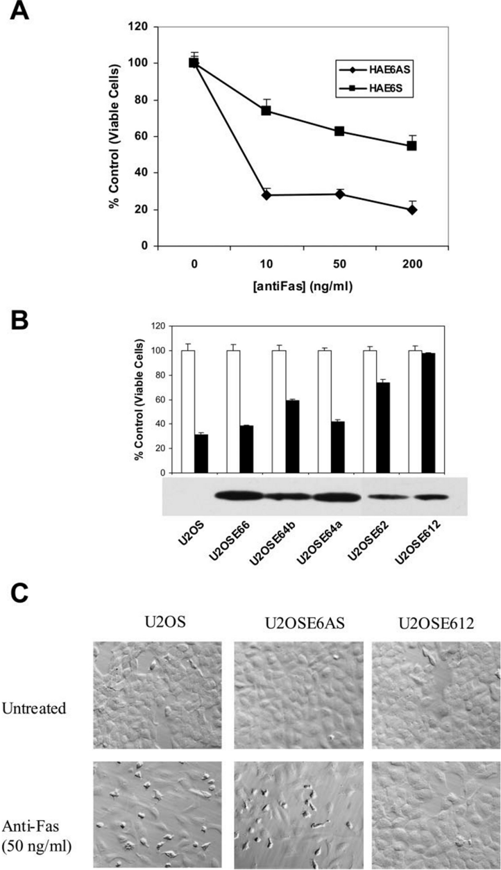
FIG. 1 HPV16E6protects cells from anti-Fas. A , U2OS cells were transfected with either HA-E6 AS or HA-E6 S. Pools of stably transfected cells were then treated with the indicated concentrations of anti-Fas in the presence of cycloheximide (5 𝛍 g/ml). Following a 16-h incubation, cell viability was measured by the MTT assay. Measurements were made in triplicate, and the error bars represent the standard deviation. B , U2OS cells were transfected with HA-E6, and stable clones were selected and characterized. The parental U2OS cells and E6-expressing clones were each untreated ( open bars ) or treated ( black bars ) with 50 ng/ml anti-Fas for 16 h, and the resulting cell death was measured by the MTT assay. Measurements were made in triplicate, and the error bars represent the means ± S.D. The lower panel depicts an immunoprecipitation of the HA-E6 expressed by each clone, using anti-HA antibodies. C , U2OS cells ( top left and bottom left ), U2OSE6AS cells ( top middle and bottom middle ), and U2OSE612 cells ( top right and bottom right ) were either untreated ( top panel ) or treated with 𝛂 -Fas (50 ng/ml) ( bottom panel ) for 12 h.
FIG. 1 HPV16E6protects cells from anti-Fas. A , U2OS cells were transfected with either HA-E6 AS or HA-E6 S. Pools of stably transfected cells were then treated with the indicated concentrations of anti-Fas in the presence of cycloheximide (5 𝛍 g/ml). Following a 16-h incubation, cell viability was measured by the MTT assay. Measurements were made in triplicate, and the error bars represent the standard deviation. B , U2OS cells were transfected with HA-E6, and stable clones were selected and characterized. The parental U2OS cells and E6-expressing clones were each untreated ( open bars ) or treated ( black bars ) with 50 ng/ml anti-Fas for 16 h, and the resulting cell death was measured by the MTT assay. Measurements were made in triplicate, and the error bars represent the means ± S.D. The lower panel depicts an immunoprecipitation of the HA-E6 expressed by each clone, using anti-HA antibodies. C , U2OS cells ( top left and bottom left ), U2OSE6AS cells ( top middle and bottom middle ), and U2OSE612 cells ( top right and bottom right ) were either untreated ( top panel ) or treated with 𝛂 -Fas (50 ng/ml) ( bottom panel ) for 12 h.CC-BY · From the PDF
Image 2
CC-BY · From the PDF
Image 3
CC-BY · From the PDF
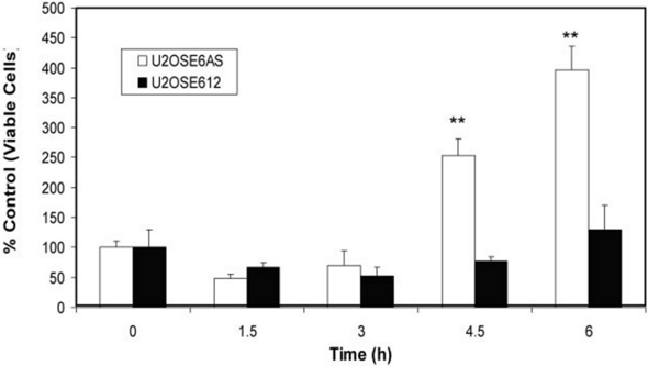
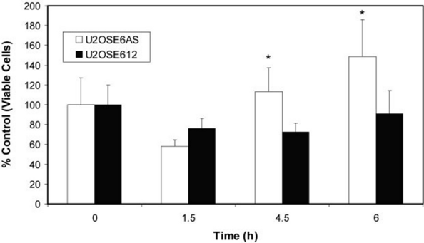
FIG. 3 Caspase 3 and 8 activation is suppressed in HA-E6-expressing cells. A , U2OSE6AS (coding for the antisense version of E6) and U2OSE612 (coding for the sense version of E6) cells were treated with anti-Fas (50 ng/ml) in the presence of cycloheximide (5 𝛍 g/ml) for the times indicated and then lysed. Lysates were analyzed for caspase 3 activity using Ac-DEVDp NA as the substrate in the presence and absence of the caspase 3 inhibitor Ac-DEVD-CHO. The activity in wells containing the inhibitor was subtracted from that in wells lacking the inhibitor and then normalized for the amount of protein added to each well. Activity is expressed as the percentage of caspase activity in untreated U2OSE6AS cells. Each time point was measured in triplicate, and error bars represent the means ± S.D. The Student ' s one-tailed t test was used to determine statistical significance, with * ( B ) representing a > 0.95 level of confidence and ** ( A ) representing a > 0.99 level of confidence. B, U2OSE6AS and U2OSE612 cells were treated and lysed as described in A and then analyzed for caspase 8 activity using IETDp NA as the substrate in the presence and absence of the caspase 8 inhibitor Ac-IETDCHO. Data were analyzed as described in A .
FIG. 3 Caspase 3 and 8 activation is suppressed in HA-E6-expressing cells. A , U2OSE6AS (coding for the antisense version of E6) and U2OSE612 (coding for the sense version of E6) cells were treated with anti-Fas (50 ng/ml) in the presence of cycloheximide (5 𝛍 g/ml) for the times indicated and then lysed. Lysates were analyzed for caspase 3 activity using Ac-DEVDp NA as the substrate in the presence and absence of the caspase 3 inhibitor Ac-DEVD-CHO. The activity in wells containing the inhibitor was subtracted from that in wells lacking the inhibitor and then normalized for the amount of protein added to each well. Activity is expressed as the percentage of caspase activity in untreated U2OSE6AS cells. Each time point was measured in triplicate, and error bars represent the means ± S.D. The Student ' s one-tailed t test was used to determine statistical significance, with * ( B ) representing a > 0.95 level of confidence and ** ( A ) representing a > 0.99 level of confidence. B, U2OSE6AS and U2OSE612 cells were treated and lysed as described in A and then analyzed for caspase 8 activity using IETDp NA as the substrate in the presence and absence of the caspase 8 inhibitor Ac-IETDCHO. Data were analyzed as described in A .CC-BY · From the PDF
Image 5
CC-BY · From the PDF
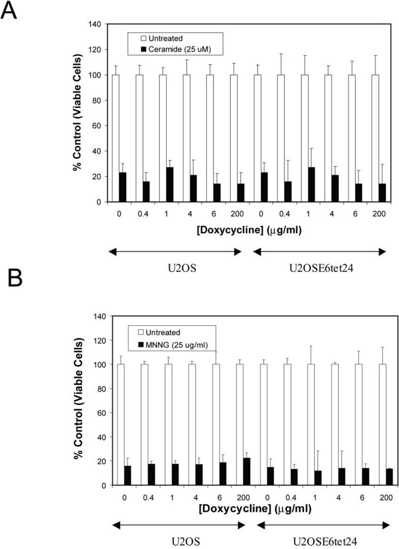
FIG. 4 E6 does not interfere with the mitochondrial apoptotic pathway. Untransfected U2OS cells or cells stably transfected with HA-E6 under the control of the tet-responsive element were cultured in the presence of the indicated concentration of doxycycline and treated with either ceramide (25 𝛍 M) ( A ) or MNNG (25 𝛍 g/ml) ( B ) for 24 or 16 h, respectively. Cell viability was measured using the MTT assay. Each measurement was done in triplicate, and error bars indicate the means ± S.D.
FIG. 4 E6 does not interfere with the mitochondrial apoptotic pathway. Untransfected U2OS cells or cells stably transfected with HA-E6 under the control of the tet-responsive element were cultured in the presence of the indicated concentration of doxycycline and treated with either ceramide (25 𝛍 M) ( A ) or MNNG (25 𝛍 g/ml) ( B ) for 24 or 16 h, respectively. Cell viability was measured using the MTT assay. Each measurement was done in triplicate, and error bars indicate the means ± S.D.CC-BY · From the PDF
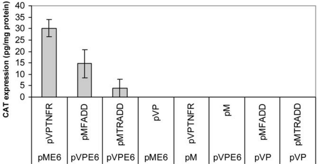
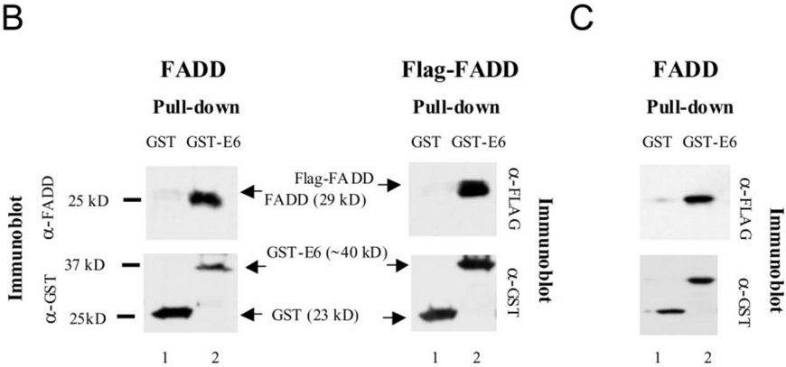
FIG. 5 HPV 16 E6 binds to FADD. A , mammalian two-hybrid assay. Sequences encoding E6 and the death domain of TNF R1, FADD, and TRADD were cloned into the bait or prey plasmids of a mammalian two-hybrid assay kit (Clontech). The indicated combinations of plasmids were then transfected into U2OS cells, along with a reporter plasmid coding for CAT, and expression of the CAT gene was measured colorimetrically by using a CAT-ELISA kit. Each of the three experimental values was measured in three independent experiments, with two measurements taken for each experiment. The error bars show the 90% confidence intervals. B , in vitro pull-down assays using cell lysates. GST-E6 beads were used to pull down FADD, which was detected with anti-FADD antibodies ( left set ) or FLAG-FADD, which was detected with anti-FLAG antibodies ( right set ). Top panels , lysates were prepared from U2OS cells transiently transfected with either pcDNA3 FADD ( left ) or pFLAG-FADD ( right ) and then incubated with glutathione beads complexed with either GST ( lane 1 ) or GST-E6 ( lane 2 ). Beads were washed, and bound proteins were eluted with SDS prior to separation by SDS-PAGE. FADD was detected by immunoblot using polyclonal anti-FADD and FLAG-FADD with monoclonal anti-FLAG. Bottom panels , the same membrane shown in the top panel was reblotted using monoclonal anti-GST. C , in vitro pull-down assay using transcription/translation-produced FADD. The experimental design was as described above, with the exception that the source of FADD was full-length FADD protein expressed using the T7 TNT Quick Coupled Transcription/Translation System (10 𝛍 l of reticulocyte lysate) rather than cell lysates.
FIG. 5 HPV 16 E6 binds to FADD. A , mammalian two-hybrid assay. Sequences encoding E6 and the death domain of TNF R1, FADD, and TRADD were cloned into the bait or prey plasmids of a mammalian two-hybrid assay kit (Clontech). The indicated combinations of plasmids were then transfected into U2OS cells, along with a reporter plasmid coding for CAT, and expression of the CAT gene was measured colorimetrically by using a CAT-ELISA kit. Each of the three experimental values was measured in three independent experiments, with two measurements taken for each experiment. The error bars show the 90% confidence intervals. B , in vitro pull-down assays using cell lysates. GST-E6 beads were used to pull down FADD, which was detected with anti-FADD antibodies ( left set ) or FLAG-FADD, which was detected with anti-FLAG antibodies ( right set ). Top panels , lysates were prepared from U2OS cells transiently transfected with either pcDNA3 FADD ( left ) or pFLAG-FADD ( right ) and then incubated with glutathione beads complexed with either GST ( lane 1 ) or GST-E6 ( lane 2 ). Beads were washed, and bound proteins were eluted with SDS prior to separation by SDS-PAGE. FADD was detected by immunoblot using polyclonal anti-FADD and FLAG-FADD with monoclonal anti-FLAG. Bottom panels , the same membrane shown in the top panel was reblotted using monoclonal anti-GST. C , in vitro pull-down assay using transcription/translation-produced FADD. The experimental design was as described above, with the exception that the source of FADD was full-length FADD protein expressed using the T7 TNT Quick Coupled Transcription/Translation System (10 𝛍 l of reticulocyte lysate) rather than cell lysates.CC-BY · From the PDF
Image 8
CC-BY · From the PDF
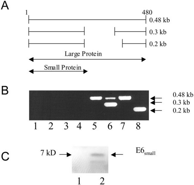
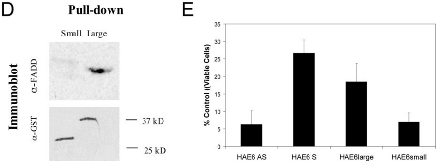
FIG. 6 HPV 16 E6large, but not HPV 16 E6small, binds to FADD and protects U2OS cells from apoptosis induced by Fas. A , diagram of the transcripts and proteins produced from the HPV 16 gene. Three transcripts are produced with lengths of 0.48, 0.3, and 0.2 kb. The large transcript produces the large, full-length protein, and both of the smaller transcripts (0.3 and 0.2 kb) code for the small E6 protein. B , cells transfected with HA-E6 express transcripts coding for the small, the large, or both proteins. U2OS cells were transfected with constructs coding for antisense E6 ( lanes 1 and 5 ), the unmodified full-length gene ( lanes 2 and 6 ), only the large version of E6 ( lanes 3 and 7 ), or only the small E6 version ( lanes 4 and 8 ) and the messages produced detected by PCR. Primers corresponded to the 5 ′ and 3 ′ ends of E6, and no reverse transcriptase was included in the reactions corresponding to lanes 1 -4 . C , the small E6 protein is produced in transfected cells. Lysates were prepared from U2OS cells ( lane 1 ) or from U2OS cells transiently transfected with a construct coding for the small version of E6 (HA-E6 small) and immunoprecipitated using beads cross-linked to antibodies directed against HA. Following elution from the beads, separation by SDS-PAGE, and transfer to a membrane, HA-E6small was visualized by immunoblotting. D , HPV 16 E6small does not bind to FADD. Bacterially expressed and purified D1 FADD, lacking amino acids 24 -62, was incubated with glutathione beads bound to either GST E6 small or GST E6large , as indicated. Following elution from the beads, separation by SDS-PAGE, and transfer to a membrane, the membrane was blotted with anti-FADD. The bottom panel shows the same membrane reblotted with anti-GST antibodies. E , HPV 16 E6large , but not HPV 16 E6small , protects U2OS cells from apoptosis induced by Fas. U2OS cells were stably transfected with either HA-E6AS, HA-E6 S, HA-E6 large , or HA-E6 small . Pools of stably transfected cells were then treated with 100 ng/ml anti-Fas in the presence of cycloheximide (5 𝛍 g/ml). Following a 16-h incubation, cell viability was measured by the MTT assay. The error bars represent the means ± S.D. of the average of five repeats.
FIG. 6 HPV 16 E6large, but not HPV 16 E6small, binds to FADD and protects U2OS cells from apoptosis induced by Fas. A , diagram of the transcripts and proteins produced from the HPV 16 gene. Three transcripts are produced with lengths of 0.48, 0.3, and 0.2 kb. The large transcript produces the large, full-length protein, and both of the smaller transcripts (0.3 and 0.2 kb) code for the small E6 protein. B , cells transfected with HA-E6 express transcripts coding for the small, the large, or both proteins. U2OS cells were transfected with constructs coding for antisense E6 ( lanes 1 and 5 ), the unmodified full-length gene ( lanes 2 and 6 ), only the large version of E6 ( lanes 3 and 7 ), or only the small E6 version ( lanes 4 and 8 ) and the messages produced detected by PCR. Primers corresponded to the 5 ′ and 3 ′ ends of E6, and no reverse transcriptase was included in the reactions corresponding to lanes 1 -4 . C , the small E6 protein is produced in transfected cells. Lysates were prepared from U2OS cells ( lane 1 ) or from U2OS cells transiently transfected with a construct coding for the small version of E6 (HA-E6 small) and immunoprecipitated using beads cross-linked to antibodies directed against HA. Following elution from the beads, separation by SDS-PAGE, and transfer to a membrane, HA-E6small was visualized by immunoblotting. D , HPV 16 E6small does not bind to FADD. Bacterially expressed and purified D1 FADD, lacking amino acids 24 -62, was incubated with glutathione beads bound to either GST E6 small or GST E6large , as indicated. Following elution from the beads, separation by SDS-PAGE, and transfer to a membrane, the membrane was blotted with anti-FADD. The bottom panel shows the same membrane reblotted with anti-GST antibodies. E , HPV 16 E6large , but not HPV 16 E6small , protects U2OS cells from apoptosis induced by Fas. U2OS cells were stably transfected with either HA-E6AS, HA-E6 S, HA-E6 large , or HA-E6 small . Pools of stably transfected cells were then treated with 100 ng/ml anti-Fas in the presence of cycloheximide (5 𝛍 g/ml). Following a 16-h incubation, cell viability was measured by the MTT assay. The error bars represent the means ± S.D. of the average of five repeats.CC-BY · From the PDF
Image 10
CC-BY · From the PDF
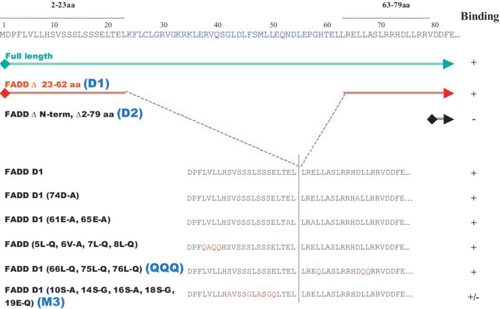
FIG. 7 Schematic representation of the FADD DED mutants used for mapping and summary of the results. The five single and multiple point mutation mutants are all derived from the D1 (lacking amino acids ( aa ) 23 -62) deletion mutant.
FIG. 7 Schematic representation of the FADD DED mutants used for mapping and summary of the results. The five single and multiple point mutation mutants are all derived from the D1 (lacking amino acids ( aa ) 23 -62) deletion mutant.CC-BY · From the PDF
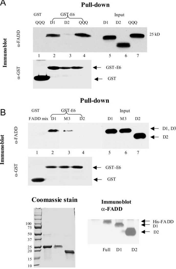
D1 M3 D2
D1 M3 D2CC-BY · From the PDF
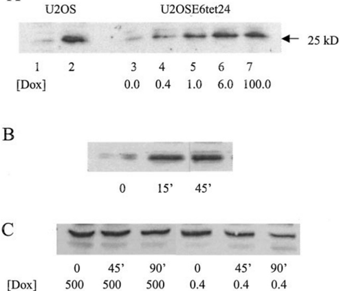
FIG. 9 HPV 16 E6 accelerates the degradation of FADD. A , E6 decreases cellular levels of FADD in a dose-dependent manner. U2OS cells were either untransfected ( lane 1 ) or transfected with pcDNA-FADD ( lane 2 ). U2OSE6tet24 cells ( lanes 3 -7 ) transfected with FADD were incubated in the presence of the indicated concentration of doxycycline. Lysates were prepared, subjected to SDS-PAGE, and transferred to a membrane and then immunoblotted with antibodies directed against FADD. B , proteosome inhibition increases cellular levels of FADD. U2OSE6tet24 cells, in the absence of dox, were transfected with pcDNA FADD. Forty eight hours post-transfection, MG 132 (50 𝛍 M) was applied to the cells, and the cells were lysed 0, 15, or 45 min post-MG 132 treatment. The FADD content of the lysates was then analyzed by immunoblot. C , HPV 16 E6 accelerates FADD degradation. U2OSE6tet24 cells, in the presence of either 500 or 0.4 ng/ml dox, were transfected with pcDNA FADD. Twenty four hours post-transfection, cells were treated with cycloheximide (5 𝛍 g/ml) and lysed at 0, 45, or 90 min post-cycloheximide treatment. Lysates were then analyzed for their FADD content by immunoblot. The initial amounts of FADD protein in the 1st and 4th lanes were equivalent.
FIG. 9 HPV 16 E6 accelerates the degradation of FADD. A , E6 decreases cellular levels of FADD in a dose-dependent manner. U2OS cells were either untransfected ( lane 1 ) or transfected with pcDNA-FADD ( lane 2 ). U2OSE6tet24 cells ( lanes 3 -7 ) transfected with FADD were incubated in the presence of the indicated concentration of doxycycline. Lysates were prepared, subjected to SDS-PAGE, and transferred to a membrane and then immunoblotted with antibodies directed against FADD. B , proteosome inhibition increases cellular levels of FADD. U2OSE6tet24 cells, in the absence of dox, were transfected with pcDNA FADD. Forty eight hours post-transfection, MG 132 (50 𝛍 M) was applied to the cells, and the cells were lysed 0, 15, or 45 min post-MG 132 treatment. The FADD content of the lysates was then analyzed by immunoblot. C , HPV 16 E6 accelerates FADD degradation. U2OSE6tet24 cells, in the presence of either 500 or 0.4 ng/ml dox, were transfected with pcDNA FADD. Twenty four hours post-transfection, cells were treated with cycloheximide (5 𝛍 g/ml) and lysed at 0, 45, or 90 min post-cycloheximide treatment. Lysates were then analyzed for their FADD content by immunoblot. The initial amounts of FADD protein in the 1st and 4th lanes were equivalent.CC-BY · From the PDF

Verification

Claims the paper makes

The claims and statistics of this paper haven’t been checked yet.

Run the check — free, no account needed. It reads the open-access text and checks the sources it cites.

A few free checks a day · papers already verified open instantly · results are public

Claims other papers make about this one

Each is checked against this paper’s own text: does it say what the citing paper says it does?

Self-check

Percentages, sums and reported differences re-computed from the paper’s own numbers.

The self-check needs the paper’s full text.

Citations

Cites

Show all 2 references

Research works printed but not linked: 87

  1. Scheffner, 1990, Cell, 63, 1129, 10.1016/0092-8674(90)90409-8 · DOI · Paper page
  2. Song, 1998, Proc. Natl. Acad. Sci. U. S. A., 95, 2290, 10.1073/pnas.95.5.2290 · DOI
  3. Pan, 1995, Genes Dev., 9, 2157, 10.1101/gad.9.17.2157 · DOI
  4. Fogel, 1998, Virology, 244, 97, 10.1006/viro.1998.9086 · DOI
  5. Spitkovsky, 1996, Oncogene, 13, 1027
  6. Inoue, 1998, Mol. Carcinog., 21, 215, 10.1002/(SICI)1098-2744(199803)21:3<215::AID-MC9>3.0.CO;2-K · DOI
  7. Klingelhutz, 1996, Nature, 380, 79, 10.1038/380079a0 · DOI · Paper page
  8. Veldman, 2001, J. Virol., 75, 4467, 10.1128/JVI.75.9.4467-4472.2001 · DOI · Paper page
  9. Pim, 2000, Oncogene, 19, 719, 10.1038/sj.onc.1203374 · DOI
  10. Lopez-Ocejo, 2000, Oncogene, 19, 4611, 10.1038/sj.onc.1203817 · DOI · Paper page
  11. Mantovani, 2001, Oncogene, 20, 7874, 10.1038/sj.onc.1204869 · DOI
  12. Patel, 1999, EMBO J., 18, 5061, 10.1093/emboj/18.18.5061 · DOI · Paper page
  13. Zimmermann, 1999, J. Virol., 73, 6209, 10.1128/JVI.73.8.6209-6219.1999 · DOI · Paper page
  14. Ronco, 1998, Genes Dev., 12, 2061, 10.1101/gad.12.13.2061 · DOI · Paper page
  15. Kuhne, 1998, J. Biol. Chem., 273, 34302, 10.1074/jbc.273.51.34302 · DOI
  16. Kukimoto, 1998, Biochem. Biophys. Res. Commun., 249, 258, 10.1006/bbrc.1998.9066 · DOI
  17. Gao, 1999, Mol. Cell. Biol., 19, 733, 10.1128/MCB.19.1.733 · DOI
  18. Kumar, 2002, Mol. Cell. Biol., 22, 5801, 10.1128/MCB.22.16.5801-5812.2002 · DOI · Paper page
  19. Zeng, 2002, J. Biol. Chem., 277, 45611, 10.1074/jbc.M208447200 · DOI
  20. Thomas, 1998, Oncogene, 17, 2943, 10.1038/sj.onc.1202223 · DOI · Paper page
  21. Tross-Mesilaty, 1998, Proc. Natl. Acad. Sci. U. S. A., 95, 8058, 10.1073/pnas.95.14.8058 · DOI · Paper page
  22. Filippova, 2002, J. Biol. Chem., 277, 21730, 10.1074/jbc.M200113200 · DOI
  23. Tong, 1997, Proc. Natl. Acad. Sci. U. S. A., 94, 4412, 10.1073/pnas.94.9.4412 · DOI
  24. Chen, 1995, Science, 269, 529, 10.1126/science.7624774 · DOI · Paper page
  25. Degenhardt, 2001, J. Virol., 75, 11791, 10.1128/JVI.75.23.11791-11802.2001 · DOI
  26. Du, 2002, Biochem. Biophys. Res. Commun., 296, 962, 10.1016/S0006-291X(02)02041-7 · DOI
  27. Lee, 1997, Proc. Natl. Acad. Sci. U. S. A., 94, 6670, 10.1073/pnas.94.13.6670 · DOI · Paper page
  28. Kiyono, 1997, Proc. Natl. Acad. Sci. U. S. A., 94, 11612, 10.1073/pnas.94.21.11612 · DOI · Paper page
  29. Nakagawa, 2000, Mol. Cell. Biol., 20, 8244, 10.1128/MCB.20.21.8244-8253.2000 · DOI · Paper page
  30. Glausinger, 2000, Oncogene, 19, 5270, 10.1038/sj.onc.1203906 · DOI · Paper page
  31. Thomas, 2001, Oncogene, 20, 5431, 10.1038/sj.onc.1204719 · DOI
  32. Thomas, 2002, Oncogene, 21, 5088, 10.1038/sj.onc.1205668 · DOI
  33. Lee, 2000, J. Virol., 74, 9680, 10.1128/JVI.74.20.9680-9693.2000 · DOI
  34. Iftner, 2002, EMBO J., 21, 47141, 10.1093/emboj/cdf443 · DOI
  35. Srivenugopal, 2002, Oncogene, 21, 5940, 10.1038/sj.onc.1205762 · DOI
  36. Duerksen-Hughes, 1999, Virology, 264, 55, 10.1006/viro.1999.9977 · DOI
  37. Zimmerman, 2001, Pharmacol. Ther., 92, 57, 10.1016/S0163-7258(01)00159-0 · DOI
  38. Wagant, 1998, Curr. Biol., 8, 113, 10.1016/S0960-9822(98)70042-9 · DOI
  39. Boatright, 2003, Mol. Cell, 11, 529, 10.1016/S1097-2765(03)00051-0 · DOI
  40. Nagata, 1997, Cell, 88, 355, 10.1016/S0092-8674(00)81874-7 · DOI
  41. Wallach, 1999, Annu. Rev. Immunol., 17, 331, 10.1146/annurev.immunol.17.1.331 · DOI
  42. Weber, 2001, Trends Biochem. Sci., 26, 475, 10.1016/S0968-0004(01)01905-3 · DOI
  43. Juo, 1999, Cell Growth Differ., 10, 797
  44. Bodmer, 2000, Nat. Cell Biol., 2, 241, 10.1038/35008667 · DOI
  45. Schneider, 1997, Immunity, 7, 831, 10.1016/S1074-7613(00)80401-X · DOI
  46. Sprick, 2000, Immunity, 12, 599, 10.1016/S1074-7613(00)80211-3 · DOI
  47. Kischkel, 2000, Immunity, 12, 611, 10.1016/S1074-7613(00)80212-5 · DOI · Paper page
  48. Kawahara, 1998, J. Cell Biol., 143, 1353, 10.1083/jcb.143.5.1353 · DOI
  49. Sedman, 1991, J. Virol., 65, 4860, 10.1128/jvi.65.9.4860-4866.1991 · DOI
  50. Yang, 1998, Carcinogenesis, 19, 1117, 10.1093/carcin/19.6.1117 · DOI
  51. Shally, 1996, Virus Res., 42, 81, 10.1016/0168-1702(96)01301-9 · DOI
  52. Tortorella, 2000, Annu. Rev. Immunol., 18, 861, 10.1146/annurev.immunol.18.1.861 · DOI
  53. Hay, 2002, J. Gen. Virol., 83, 1547, 10.1099/0022-1317-83-7-1547 · DOI
  54. Benedict, 2003, Cytokine Growth Factor Rev., 14, 349, 10.1016/S1359-6101(03)00030-3 · DOI
  55. Shisler, 1997, J. Virol., 71, 8299, 10.1128/jvi.71.11.8299-8306.1997 · DOI
  56. Wang, 1997, J. Virol., 71, 8928, 10.1128/jvi.71.11.8928-8932.1997 · DOI
  57. Hu, 1997, J. Biol. Chem., 272, 9621, 10.1074/jbc.272.15.9621 · DOI
  58. Bertin, 1997, Proc. Natl. Acad. Sci. U. S. A., 94, 1172, 10.1073/pnas.94.4.1172 · DOI
  59. Tsukumo, 1999, Genes Cells, 4, 541, 10.1046/j.1365-2443.1999.00280.x · DOI
  60. Shisler, 2001, Virology, 282, 14, 10.1006/viro.2001.0834 · DOI
  61. Skaletskaya, 2001, Proc. Natl. Acad. Sci. U. S. A., 98, 7829, 10.1073/pnas.141108798 · DOI
  62. Langelier, 2002, J. Gen. Virol., 83, 2779, 10.1099/0022-1317-83-11-2779 · DOI
  63. Finzer, 2002, Cancer Lett., 188, 15, 10.1016/S0304-3835(02)00431-7 · DOI
  64. Kobayashi, 2001, Nippon Jibiinkoka Gakkai Kaiho, 104, 139, 10.3950/jibiinkoka.104.139 · DOI
  65. Aguilar-Lemarroy, 2001, Int. J. Cancer, 93, 823, 10.1002/ijc.1405 · DOI
  66. Kabsch, 2002, Oncogene, 21, 947, 10.1038/sj.onc.1205147 · DOI
  67. Vikhanskaya, 2002, Int. J. Cancer, 97, 732, 10.1002/ijc.10114 · DOI
  68. Liu, 1999, J. Biol. Chem., 274, 24819, 10.1074/jbc.274.35.24819 · DOI
  69. Basile, 2001, J. Biol. Chem., 276, 22522, 10.1074/jbc.M010505200 · DOI
  70. Stoppler, 1998, Oncogene, 17, 1207, 10.1038/sj.onc.1202053 · DOI
  71. Thompson, 2001, Oncogene, 20, 3629, 10.1038/sj.onc.1204483 · DOI · Paper page
  72. Scaffidi, 1998, EMBO J., 17, 1675, 10.1093/emboj/17.6.1675 · DOI · Paper page
  73. Scaffidi, 1999, J. Biol. Chem., 274, 22532, 10.1074/jbc.274.32.22532 · DOI
  74. Bang, 2000, J. Biol. Chem., 275, 36217, 10.1074/jbc.M006620200 · DOI
  75. Pim, 2002, Oncogene, 21, 8140, 10.1038/sj.onc.1206026 · DOI
  76. Elston, 1998, J. Gen. Virol., 79, 371, 10.1099/0022-1317-79-2-371 · DOI
  77. Be, 2001, Biochemistry, 40, 1293, 10.1021/bi0019592 · DOI
  78. Chen, 1998, J. Biol. Chem., 273, 13537, 10.1074/jbc.273.22.13537 · DOI · Paper page
  79. Pim, 1997, Oncogene, 15, 257, 10.1038/sj.onc.1201202 · DOI
  80. Pim, 1999, Oncogene, 18, 7403, 10.1038/sj.onc.1203134 · DOI
  81. McNicol, 1995, J. Clin. Microbiol., 33, 1169, 10.1128/jcm.33.5.1169-1173.1995 · DOI
  82. Tourneur, 2003, Oncogene, 22, 2795, 10.1038/sj.onc.1206399 · DOI
  83. Kim, 2003, Biochem. Biophys. Res. Commun., 302, 290295, 10.1016/S0006-291X(03)00186-4 · DOI
  84. Mishima, 2003, Int. J. Cancer, 105, 593, 10.1002/ijc.11133 · DOI
  85. Hall, 2003, J. Virol., 77, 6066, 10.1128/JVI.77.10.6066-6069.2003 · DOI
  86. Butz, 2000, Proc. Natl. Acad. Sci. U. S. A., 67, 6693, 10.1073/pnas.110538897 · DOI
  87. Aguilar-Lemarroy, 2002, Oncogene, 21, 165, 10.1038/sj.onc.1204979 · DOI

Cited by

Metadata

Authors
Filippova, M., Parkhurst, L., & Duerksen-Hughes, P. J.
Venue
Journal of Biological Chemistry
Publisher
Elsevier BV
Published
2004-06
Volume
279
Issue
24
Pages
25729-25744
ISSN
0021-9258
Cited by
149
References
2
Language
en

About these checks

Registry, retraction, Cites and statistics results are computed, not judged, with one exception: a printed reference that no index lists is read once by a model before it counts as a citation error. Claims are extracted by a model and kept only when their quote appears verbatim in the text; each cited source is then checked, with a model’s help, for whether it says what the claim relies on. Those verdicts can be wrong — read the claim’s quote and the cited source before relying on one.

Statistics and arithmetic are recomputed, not judged.

Confidence bands are shown only for fields calibrated on our annotated test set. None is calibrated yet, so no band is shown.

What counts as a claim ›How we measure confidence ›How we check this ›

Are you an author, or do you see a problem with this page? Request a review