

Microtubule stabilization specifies initial neuronal polarization
Open accessThe DOI matches its Crossref record; 56 of the 56 references it cites were found in a registry or an index.
Abstract
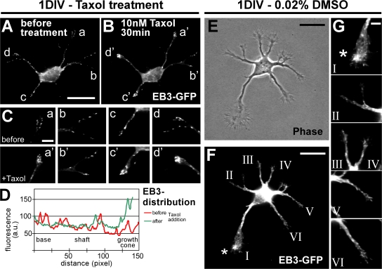
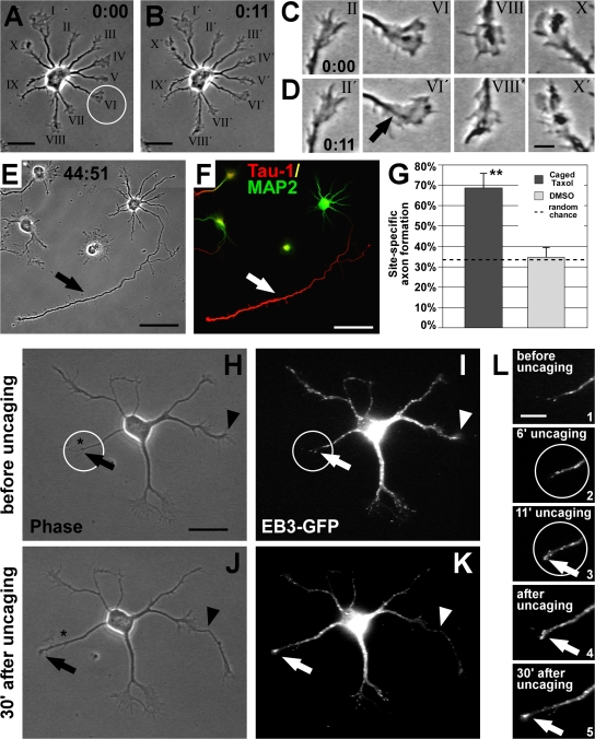
Axon formation is the initial step in establishing neuronal polarity. We examine here the role of microtubule dynamics in neuronal polarization using hippocampal neurons in culture. We see increased microtubule stability along the shaft in a single neurite before axon formation and in the axon of morphologically polarized cells. Loss of polarity or formation of multiple axons after manipulation of neuronal polarity regulators, synapses of amphids defective (SAD) kinases, and glycogen synthase kinase-3β correlates with characteristic changes in microtubule turnover. Consistently, changing the microtubule dynamics is sufficient to alter neuronal polarization. Application of low doses of the microtubule-destabilizing drug nocodazole selectively reduces the formation of future dendrites. Conversely, low doses of the microtubule-stabilizing drug taxol shift polymerizing microtubules from neurite shafts to process tips and lead to the formation of multiple axons. Finally, local stabilization of microtubules using a photoactivatable analogue of taxol induces axon formation from the activated area. Thus, local microtubule stabilization in one neurite is a physiological signal specifying neuronal polarization.
Overview
The claims and statistics of this paper haven’t been checked yet.
Free: it reads the open-access text and checks the first 5 claims that cite a source we can check. Create a free account to check every claim.
Integrity signals
Registry
Cites
Statistics
Figures


Verification
Claims the paper makes
The claims and statistics of this paper haven’t been checked yet.
Free: it reads the open-access text and checks the first 5 claims that cite a source we can check. Create a free account to check every claim.
Claims other papers make about this one
Each is checked against this paper’s own text: does it say what the citing paper says it does?
The source backs part of the claim, not all of it — or backs it for a different population, condition or comparison.
In the citing paper
“Restricted expression of acetylated tubulin near the centrosome defines a stable location for axon growth [77].”
From Abnormal accumulation of extracellular vesicles in hippocampal dystrophic axons and regulation by the primary cilia in Alzheimer's disease (2023) · cites this paper as [77]
Experimental — accuracy not yet measuredIn the citing paper
“Selective microtubule stabilization in one minor process determines axon specification (19) and only after this event, the dendritic microtubule network polarity becomes progressively mixed (18).”
From Axonal transport: Driving synaptic function (2019) · cites this paper as [19]
Self-check
Percentages, sums and reported differences re-computed from the paper’s own numbers.
The self-check needs the paper’s full text.
Citations
Cites
‘Green mice’ as a source of ubiquitous green cells
Okabe, M., Ikawa, M., Kominami, K., Nakanishi, T., & Nishimune, Y. (1997) · FEBS Letters · doi:10.1016/s0014-5793(97)00313-x · Cited by 2,581
GSK-3: tricks of the trade for a multi-tasking kinase
Doble, B. W., & Woodgett, J. R. (2003) · Journal of Cell Science · doi:10.1242/jcs.00384 · Cited by 2,084 · cited 5×
“The online version of this paper contains supplemental material.”
InfluentialThe establishment of polarity by hippocampal neurons in culture.
Dotti, C., Sullivan, C., & Banker, G. (1988) · The Journal of Neuroscience · doi:10.1523/jneurosci.08-04-01454.1988 · Cited by 1,862
The multifaceted roles of glycogen synthase kinase 3β in cellular signaling
Grimes, C. A., & Jope, R. S. (2001) · Progress in Neurobiology · doi:10.1016/s0301-0082(01)00011-9 · Cited by 1,423 · cited 2×
“Taxol-treated 2005 ; Yoshimura et al., 2005 ), which are known modulators of MT stability, yet they have various other cellular functions including the regulation of metabolism, signaling, gene transcription, and endocytosis ( Crump et al., 2001 ; Grimes and Jope, 2001 ).”
Microtubule Acetylation Promotes Kinesin-1 Binding and Transport
Reed, N. A., Cai, D., Blasius, T. L., Jih, G. T., Meyhofer, E., Gaertig, J., & Verhey, K. J. (2006) · Current Biology · doi:10.1016/j.cub.2006.09.014 · Cited by 919 · cited 1×
“…unpolymerized tubulin subunits, cells were simultaneously fi xed and permeabilized in PHEM buffer (60 mM Pipes, 25 mM Hepes, 5 mM EGTA, and 1 mM MgCl) containing 0.25% glutaraldehyde, 3.7% paraformaldehyde, 3.7% sucrose, and 0.1% Triton X-100 (adapted from Smith, 1994 ) and quenched as above.”
InfluentialGSK-3β Regulates Phosphorylation of CRMP-2 and Neuronal Polarity
Yoshimura, T., Kawano, Y., Arimura, N., Kawabata, S., Kikuchi, A., & Kaibuchi, K. (2005) · Cell · doi:10.1016/j.cell.2004.11.012 · Cited by 875 · cited 7×
“The online version of this paper contains supplemental material.”
InfluentialCdc42 regulates GSK-3β and adenomatous polyposis coli to control cell polarity
Etienne-Manneville, S., & Hall, A. (2003) · Nature · doi:10.1038/nature01423 · Cited by 835
CRMP-2 binds to tubulin heterodimers to promote microtubule assembly
Fukata, Y., Itoh, T. J., Kimura, T., Ménager, C., Nishimura, T., Shiromizu, T., Watanabe, H., Inagaki, N., Iwamatsu, A., Hotani, H., et al. (2002) · Nature Cell Biology · doi:10.1038/ncb825 · Cited by 761
Post-translational modifications regulate microtubule function
Westermann, S., & Weber, K. (2003) · Nature Reviews Molecular Cell Biology · doi:10.1038/nrm1260 · Cited by 717 · cited 2×
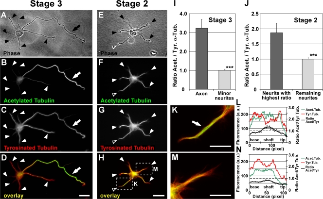
“On average from all stage 3 neurons, the axonal shaft showed a 3.2 ± 0.5-fold ratio increase of the fl uorescence intensities of acetylated versus tyrosinated MTs compared with the shafts of minor neurites (P < 0.001 by t test; Fig.”
InfluentialStepanova, T., Slemmer, J., Hoogenraad, C. C., Lansbergen, G., Dortland, B., De Zeeuw, C. I., Grosveld, F., van Cappellen, G., Akhmanova, A., & Galjart, N. (2003) · The Journal of Neuroscience · doi:10.1523/jneurosci.23-07-02655.2003 · Cited by 698 · cited 2×
“Taxol application rapidly stabilizes MTs and shifts dynamic MTs to the tips of processes To assess MT dynamics during axon formation, polymerizing MT plus ends were visualized by transfecting neurons with the GFP-tagged MT plus end – binding protein 3 (EB3; Stepanova et al., 2003 ).”
Research works printed but not linked: 3
- 1991, J. Cell Sci. Suppl., 15, 75
- 2005, Cell., 120, 123
- 2003, Nat. Rev. Mol. Cell Biol., 4, 938
Cited by
Items 101–200 of 593
ADF/Cofilin-Mediated Actin Turnover Promotes Axon Regeneration in the Adult CNS
Tedeschi, A., Dupraz, S., Curcio, M., Laskowski, C. J., Schaffran, B., Flynn, K. C., Santos, T. E., Stern, S., Hilton, B. J., Larson, M. J. E., et al. (2019) · Neuron · doi:10.1016/j.neuron.2019.07.007 · Cited by 101
“For example, moderate pharmacological stabilization of microtubules, which elicits axon growth in developing hippocampal neurons (Gomis-Ruth et al., 2008; van Beuningen et al., 2015; Witte et al., 2008), induces axon regeneration after spinal cord injury and leads to functional recovery (Hellal et al.”
BackgroundSelf-citationIntegrating Chemistry and Mechanics: The Forces Driving Axon Growth
Franze, K. (2020) · Annual Review of Cell and Developmental Biology · doi:10.1146/annurev-cellbio-100818-125157 · Cited by 117
Microtubule plus-end tracking proteins in neuronal development
van de Willige, D., Hoogenraad, C. C., & Akhmanova, A. (2016) · Cellular and Molecular Life Sciences · doi:10.1007/s00018-016-2168-3 · Cited by 98
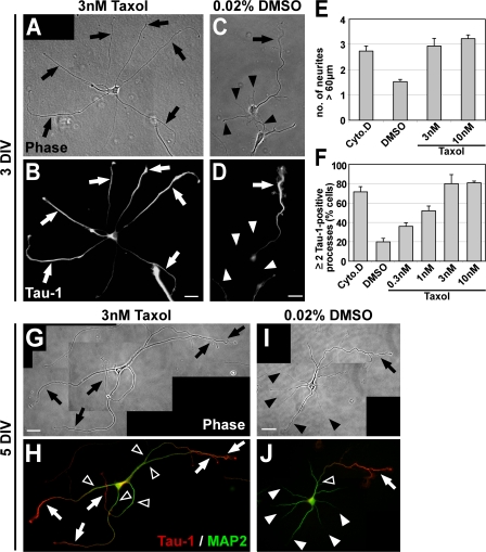
“The MT stabilizing drug taxol, on the other hand, is known to induce the formation of multiple axons in young dissociated neurons at concentrations as low as 3 nM [23], and nanomolar concentrations of both MT stabilizing and destabilizing drugs affect axon pathfinding [28].”
BackgroundFarías, G. G., Fréal, A., Tortosa, E., Stucchi, R., Pan, X., Portegies, S., Will, L., Altelaar, M., & Hoogenraad, C. C. (2019) · Neuron · doi:10.1016/j.neuron.2019.01.030 · Cited by 108
Schneider, B. P., Zhao, F., Wang, M., Stearns, V., Martino, S., Jones, V. E., Perez, E. A., Saphner, T., Wolff, A. C., Sledge, G. W., et al. (2012) · Journal of Clinical Oncology · doi:10.1200/jco.2011.39.8446 · Cited by 100
“The mechanism for this toxicity is not well understood.(9-17)In addition, no agent has been proven to definitively prevent or successfully reverse this adverse effect after it has occurred.”
BackgroundMüller‐Deku, A., Meiring, J. C. M., Loy, K., Kraus, Y., Heise, C., Bingham, R., Jansen, K. I., Qu, X., Bartolini, F., Kapitein, L. C., et al. (2020) · Nature Communications · doi:10.1038/s41467-020-18389-6 · Cited by 99
Microtubule Dynamics in Neuronal Development, Plasticity, and Neurodegeneration
Penazzi, L., Bakota, L., & Brandt, R. (2015) · International review of cytology · doi:10.1016/bs.ircmb.2015.09.004 · Cited by 110
Axon Growth of CNS Neurons in Three Dimensions Is Amoeboid and Independent of Adhesions
Santos, T. E., Schaffran, B., Broguière, N., Meyn, L., Zenobi‐Wong, M., & Bradke, F. (2020) · Cell Reports · doi:10.1016/j.celrep.2020.107907 · Cited by 103
Self-citationNeuronal polarization and the cytoskeleton
Neukirchen, D., & Bradke, F. (2011) · Seminars in Cell and Developmental Biology · doi:10.1016/j.semcdb.2011.08.007 · Cited by 107
Self-citationMAP1B Regulates Axonal Development by Modulating Rho-GTPase Rac1 Activity
Montenegro‐Venegas, C., Tortosa, E., Rosso, S. B., Peretti, D., Bollati, F. A., Bisbal, M., Jausoro, I., Ávila, J., Cáceres, A., & González-Billault, C. E. (2010) · Molecular Biology of the Cell · doi:10.1091/mbc.e09-08-0709 · Cited by 104
“Posttranslational modifications of -tubulin are needed for axon specification as indicated by studies showing increased acetylation of -tubulin during axon initiation (Witte et al., 2008).”
BackgroundHatanaka, Y., & Yamauchi, K. (2012) · Cerebral Cortex · doi:10.1093/cercor/bhr383 · Cited by 100
“cytoskeleton and microtubules (Tahirovic and Bradke 2009), and critical role of local microtubule stabilization is directly demonstrated (Witte et al. 2008).”
Establishing Neuronal Polarity with Environmental and Intrinsic Mechanisms
Yogev, S., & Shen, K. (2017) · Neuron · doi:10.1016/j.neuron.2017.10.021 · Cited by 108
Chen, J., Kholina, E. G., Szyk, A., Fedorov, V. A., Kovalenko, I. B., Гудимчук, Н. Б., & Roll‐Mecak, A. (2021) · Developmental Cell · doi:10.1016/j.devcel.2021.05.005 · Cited by 100
Uchida, S., Martel, G., Pavlowsky, A., Takizawa, S., Hevi, C., Watanabe, Y., Kandel, E. R., Alarcón, J. M., & Shumyatsky, G. P. (2014) · Nature Communications · doi:10.1038/ncomms5389 · Cited by 104
Tapia, M., Wandosell, F. G., & Garrido, J. J. (2010) · PLoS ONE · doi:10.1371/journal.pone.0012908 · Cited by 98
“A recent study has pointed that the ratio between acetylated tubulin versus tyrosinated tubulin is higher in the neurite specified as axon [6].”
BackgroundAguilar, A., Becker, L., Tedeschi, T. R., Heller, S., Iomini, C., & Nachury, M. V. (2014) · Molecular Biology of the Cell · doi:10.1091/mbc.e13-10-0609 · Cited by 105
“In mature neurons, acetylated microtubules are highly enriched in axons compared with dendrites, and there is conflicting evidence for acetylated microtubules playing a role in axonal specification and neuronal polarization (Witte et al., 2008; Li et al., 2012).”
BackgroundMEC-17 Deficiency Leads to Reduced α-Tubulin Acetylation and Impaired Migration of Cortical Neurons
Li, L., Wei, D., Wang, Q., Pan, J., Liu, R., Zhang, X., & Bao, L. (2012) · Journal of Neuroscience · doi:10.1523/jneurosci.0016-12.2012 · Cited by 96
“In the CNS, proper regulation of microtubule dynamics is essential for neurite outgrowth and neuronal migration during development (Ayala et al., 2007; Métin et al., 2008; Witte et al., 2008).”
BackgroundGlobal site-specific neddylation profiling reveals that NEDDylated cofilin regulates actin dynamics
Vogl, A. M., Phu, L., Becerra, R., Giusti, S. A., Verschueren, E., Hinkle, T. B., Bordenave, M. D., Adrian, M., Heidersbach, A. J., Yankilevich, P., et al. (2020) · Nature Structural & Molecular Biology · doi:10.1038/s41594-019-0370-3 · Cited by 92
Wang, H., Zheng, Z., Han, W., Yuan, Y., Li, Y., Zhou, K., Wang, Q., Xie, L., Xu, K., Zhang, H., et al. (2020) · Oxidative Medicine and Cellular Longevity · doi:10.1155/2020/9741369 · Cited by 98
“The ratio of Ace-tubulin to Tyrtubulin was performed to evaluate the relative ratio of stable to dynamic microtubules [44].”
BackgroundHirai, S.-I., Banba, Y., Satake, T., & Ohno, S. (2011) · Journal of Neuroscience · doi:10.1523/jneurosci.5038-10.2011 · Cited by 104
“To test whether the DLK–JNK pathway facilitates axon formation by regulating microtubule stability, we evaluated the ability of low-dose Taxol treatment (Witte et al., 2008) to offset axon formation defects caused by suppression of the DLK–JNK pathway.”
BackgroundInfluentialNeuronal polarity: remodeling microtubule organization
van Beuningen, S. F. B., & Hoogenraad, C. C. (2016) · Current Opinion in Neurobiology · doi:10.1016/j.conb.2016.02.003 · Cited by 98
Andrés‐Delgado, L., Antón, O. M., Bartolini, F., Ruiz-Sáenz, A., Correas, I., Gundersen, G. G., & Alonso, M. A. (2012) · The Journal of Cell Biology · doi:10.1083/jcb.201202137 · Cited by 90
“Despite the importance of MT detyrosination in the regulation of various cellular functions, such as axon outgrowth (Erck et al., 2005; Witte et al., 2008) and polarized recycling (Lin et al.”
Lei, W., Xing, S., Deng, C.-Y., Ju, X.-C., Jiang, X., & Luo, Z. (2012) · Cell Research · doi:10.1038/cr.2012.40 · Cited by 96
Spinal cord regeneration: where fish, frogs and salamanders lead the way, can we follow?
Quiroz, J. F. D., & Echeverri, K. (2013) · Biochemical Journal · doi:10.1042/bj20121807 · Cited by 96
Davare, M. A., Fortin, D. A., Saneyoshi, T., Nygaard, S., Kaech, S., Banker, G., Soderling, T. R., & Wayman, G. A. (2009) · The Journal of Neuroscience · doi:10.1523/jneurosci.1544-09.2009 · Cited by 86
“Microtubules and local microtubule stabilization in one neurite are suggested to be a physiological signal that specifies initial neuronal polarity (Witte et al., 2008).”
BackgroundNeukirchen, D., & Bradke, F. (2011) · Journal of Neuroscience · doi:10.1523/jneurosci.3983-10.2011 · Cited by 90
“As only modest but not strong stabilization of microtubules leads to axon formation (Witte et al., 2008), we sought a microtubule-binding factor that moderately changes the stability of microtubules by affecting their dynamics.”
BackgroundMethodSelf-citationDuan, A. R., Jonasson, E. M., Alberico, E. O., Li, C., Scripture, J. P., Miller, R. A., Alber, M., & Goodson, H. V. (2017) · Journal of Molecular Biology · doi:10.1016/j.jmb.2017.03.018 · Cited by 89
Kollins, K. M., Bell, R. L., Butts, M., & Withers, G. S. (2009) · Neural Development · doi:10.1186/1749-8104-4-26 · Cited by 89
“From these data, it appears that differential control over stability not only contributes to axon formation during the initiation of neuron polarity [ 6 ], but it also continues to distinguish dendrites from axons into maturity as well.”
BackgroundChen, S., Chen, J., Shi, H., Wei, M., Castaneda-Castellanos, D. R., Bultje, R. S., Pei, X., Kriegstein, A. R., Zhang, M., & Shi, S.-H. (2013) · Developmental Cell · doi:10.1016/j.devcel.2012.11.014 · Cited by 82
“…dynamics are differentially regulated at the neurite growth cones of a polarizing neuron and this differential regulation is critical for axon specification and the establishment of neuronal polarity (Bradke and Dotti, 1999; Stiess and Bradke, 2011; Witte and Bradke, 2008; Witte et al., 2008).”
InfluentialNeuronal maturation and axon regeneration: unfixing circuitry to enable repair
Hilton, B. J., Griffin, J. M., Fawcett, J. W., & Bradke, F. (2024) · Nature reviews. Neuroscience · doi:10.1038/s41583-024-00849-3 · Cited by 83
“Mitochondrial transport to the axon becomes stalled, partly owing to increased axonal expression of syntaphilin, a mitochondrial docking protein that decelerates mitochondrial transport along the axon 63 .”
Self-citationGumy, L. F., Chew, D. J., Tortosa, E., Katrukha, E. A., Kapitein, L. C., Tolkovsky, A. M., Hoogenraad, C. C., & Fawcett, J. W. (2013) · Journal of Neuroscience · doi:10.1523/jneurosci.5221-12.2013 · Cited by 85
“Kinesins bind to the microtubule lattice via their MD (Vale and Fletterick, 1997), and microtubule posttranslational modifications can regulate the interaction and localization of kinesin-1 on neuronal microtubules (Witte et al., 2008; Konishi and Setou, 2009; Hammond et al., 2010).”
BackgroundTranslocation of CaMKII to dendritic microtubules supports the plasticity of local synapses
Lemieux, M., Labrecque, S., Tardif, C., Labrie-Dion, É., LeBel, É., & Koninck, P. D. (2012) · The Journal of Cell Biology · doi:10.1083/jcb.201202058 · Cited by 99
The α-Tubulin gene TUBA1A in Brain Development: A Key Ingredient in the Neuronal Isotype Blend
Aiken, J., Buscaglia, G., Bates, E. A., & Moore, J. K. (2017) · Journal of Developmental Biology · doi:10.3390/jdb5030008 · Cited by 89
“Locally stabilizing microtubules in a particular neurite with stabilizing drugs results in that neurite becoming the axon [114].”
BackgroundBouchet, B. P., Noordstra, I., van Amersfoort, M., Katrukha, E. A., Ammon, Y.-C., ter Hoeve, N. D., Hodgson, L., Dogterom, M., Derksen, P. W. B., & Akhmanova, A. (2016) · Developmental Cell · doi:10.1016/j.devcel.2016.11.009 · Cited by 74
“SLAIN2 inactivation caused only limited defects in axon elongation (van der Vaart et al., 2011), and in fact low doses of paclitaxel can even promote axon outgrowth (Witte et al., 2008).”
CAMSAP3 maintains neuronal polarity through regulation of microtubule stability
Pongrakhananon, V., Saito, H., Hiver, S., Abe, T., Shioi, G., Meng, W., & Takeichi, M. (2018) · Proceedings of the National Academy of Sciences · doi:10.1073/pnas.1803875115 · Cited by 86
“It has been shown that local stabilization of the cytoskeleton is sufficient to initiate the extension of axons (6).”
BackgroundResultInfluentialDynamic Microtubules Catalyze Formation of Navigator-TRIO Complexes to Regulate Neurite Extension
van Haren, J., Boudeau, J., Schmidt, S., Basu, S., Liu, Z., Lammers, D., Demmers, J., Benhari, J., Grosveld, F. G., Debant, A., et al. (2014) · Current Biology · doi:10.1016/j.cub.2014.06.037 · Cited by 80
Wei, D., Gao, N., Li, L., Zhu, J.-X., Diao, L., Huang, J., Han, Q.-J., Wang, S., Xue, H., Wang, Q., et al. (2018) · Cerebral Cortex · doi:10.1093/cercor/bhx225 · Cited by 73
The role of altered protein acetylation in neurodegenerative disease
Kabir, F., Atkinson, R., Cook, A. L., Phipps, A. J., & King, A. E. (2023) · Frontiers in Aging Neuroscience · doi:10.3389/fnagi.2022.1025473 · Cited by 75
Wang, C., Gong, Z., Huang, X., Wang, J., Xia, K., Ying, L., Shu, J., Yu, C., Zhou, X., Li, F., et al. (2019) · Theranostics · doi:10.7150/thno.37601 · Cited by 75
“Primarily, polarization of neurons is regulated by the microtubule stability status [56].”
BackgroundLocal axonal function of STAT3 rescues axon degeneration in the pmn model of motoneuron disease
Selvaraj, B. T., Frank, N., Bender, F. L. P., Asan, E., & Sendtner, M. (2012) · Journal of Cell Biology · doi:10.1083/jcb.201203109 · Cited by 74
“Tyrosination of MTs has been shown as a characteristic feature of highly dynamic MTs, i.e., the MTs close to a moving growth cone (Witte et al., 2008).”
InfluentialThe Yin–Yang of Dendrite Morphology: Unity of Actin and Microtubules
Georges, P. C., Hadzimichalis, N. M., Sweet, E. S., & Firestein, B. L. (2008) · Molecular Neurobiology · doi:10.1007/s12035-008-8046-8 · Cited by 88
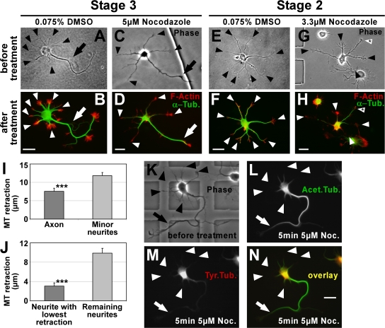
“In contrast, antagonizing MT polymerization with nocodazole specifically curtails dendritic outgrowth [29].”
BackgroundYin, D., Huang, Y.-H., Zhu, Y. B., & Wang, Y. (2008) · Journal of Neuroscience · doi:10.1523/jneurosci.1291-08.2008 · Cited by 84
“The mechanisms underlying the reorganization of membrane trafficking are complex and involve the destabilization of F-actin and the stabilization of microtubules in the axon (Yu and Baas, 1994; Bradke and Dotti, 1999; Witte et al., 2008).”
BackgroundA Role for Myosin VI in the Localization of Axonal Proteins
Lewis, T. L., Mao, T., & Arnold, D. B. (2011) · PLoS Biology · doi:10.1371/journal.pbio.1001021 · Cited by 76
“Additionally, tyrosination, a phenomenon that occurs in dendrites, but not axons, causes Kif5 to bind at lower affinity to dendritic microtubules in comparison to those present in the axon [34,35].”
BackgroundGhiretti, A. E., Thies, E., Tokito, M. K., Lin, T., Ostap, E. M., Kneussel, M., & Holzbaur, E. L. F. (2016) · Neuron · doi:10.1016/j.neuron.2016.10.003 · Cited by 74
“Given that axonal microtubules are generally less dynamic than microtubules in dendrites (Conde and Cáceres, 2009; Witte et al., 2008), the regulatory needs of dendritic microtubules are likely to be higher.”
HDAC6 and RhoA are novel players in Abeta-driven disruption of neuronal polarity
Tsushima, H., Emanuele, M., Polenghi, A., Esposito, A., Vassalli, M., Barberis, A., Difato, F., & Chieregatti, E. (2015) · Nature Communications · doi:10.1038/ncomms8781 · Cited by 71
Roles of intracellular fibroblast growth factors in neural development and functions
Zhang, X., Bao, L., Liu, Y., Wu, Q., & Li, S. (2012) · Science China Life Sciences · doi:10.1007/s11427-012-4412-x · Cited by 76
Podkowa, M., Zhao, X., Chow, C.-W., Coffey, E. T., Davis, R. J., & Attisano, L. (2010) · Molecular and Cellular Biology · doi:10.1128/mcb.01166-09 · Cited by 77
“Since microtubule stabilization is particularly important for axon formation (25, 52, 53), it would be interesting to determine whether BMPs, via regulation of JNK activity, also contribute to microtubule stability in axonal processes.”
BackgroundVelasco, S., Díez‐Revuelta, N., Hernández‐Iglesias, T., Kaltner, H., André, S., Gabius, H., & Abad‐Rodríguez, J. (2013) · Journal of Neurochemistry · doi:10.1111/jnc.12148 · Cited by 73
“(2013) 125, 49--62 It is well established that the transport of a plethora of essential molecules for axonal development and function depends on microtubule organization (Witte and Bradke 2008; Witte et al. 2008).”
BackgroundCdk5-Mediated Phosphorylation of Axin Directs Axon Formation during Cerebral Cortex Development
Fang, W., Ip, J. P. K., Li, R., Ng, Y. P., Lin, S., Chen, Y., Fu, A. K. Y., & Ip, N. Y. (2011) · Journal of Neuroscience · doi:10.1523/jneurosci.3120-11.2011 · Cited by 75
“Axon formation is regulated by the local stabilization of microtubules in one of the neuronal processes (Witte et al., 2008).”
BackgroundTonazzini, I., Meucci, S., Faraci, P., Beltram, F., & Cecchini, M. G. (2013) · Biomaterials · doi:10.1016/j.biomaterials.2013.04.039 · Cited by 74
Linking Cell Polarity to Cortical Development and Malformations
Hakanen, J., Ruiz-Reig, N., & Tissir, F. (2019) · Frontiers in Cellular Neuroscience · doi:10.3389/fncel.2019.00244 · Cited by 73
“The functional knowledge of Par3-Par6-aPKC, Crb-Pals1-Patj1, and Scribble -Lgl-Dlg polarity complexes in apical–basal polarity originates predominantly from studies in Caenorhabditis elegans and Drosophila melanogaster (Kemphues et al., 1988; Wodarz et al., 1995; Bilder and Perrimon, 2000).”
BackgroundA cryo-ET survey of microtubules and intracellular compartments in mammalian axons
Foster, H. E., Ventura Santos, C., & Carter, A. P. (2022) · Journal of Cell Biology · doi:10.1083/jcb.202103154 · Cited by 69
Garcez, P. P., Díaz-Alonso, J., Crespo‐Enríquez, I., Castro, D. S., Bell, D. M., & Guillemot, F. (2015) · Nature Communications · doi:10.1038/ncomms7474 · Cited by 67
“To further examine microtubule stability in Cenpj -silenced neurons, we tested microtubule resistance to nocodazole-induced depolymerization 31,32 in Tbr1-expressing cortical neurons electroporated ex vivo and maintained in culture for 3 days (Supplementary Fig.”
Differential neurite outgrowth is required for axon specification by cultured hippocampal neurons
Yamamoto, H., Demura, T., Morita, M., Banker, G. A., Tanii, T., & Nakamura, S. (2012) · Journal of Neurochemistry · doi:10.1111/jnc.12001 · Cited by 73
“Actin destabilization leads to enhanced polymerization and stabilization of microtubules, which play a critical role in neurite extension (Bradke and Dotti 1999; Andersen and Bi 2000; Witte et al. 2008).”
BackgroundMAP7 Regulates Axon Collateral Branch Development in Dorsal Root Ganglion Neurons
Tymanskyj, S. R., Yang, B. H., Falnikar, A., Lepore, A. C., & Ma, L. (2017) · Journal of Neuroscience · doi:10.1523/jneurosci.3260-16.2017 · Cited by 71
“…Bulinski et al., 2001; Gruber et al., 2001; Sung et al., 2008; Magnan et al., 2009; Metzger et al., 2012; Barlan et al., 2013; Gallaud et al., 2014; Yadav et al., 2014), but very little is known about its function in the nervous system, despite its wide expression in neurons (Fabre-Jonca et al.,…”
BackgroundINF1 Is a Novel Microtubule-associated Formin
Young, K. G., Thurston, S., Copeland, S. J., Smallwood, C. A., & Copeland, J. W. R. (2008) · Molecular Biology of the Cell · doi:10.1091/mbc.e08-05-0469 · Cited by 65
“One notable example is the necessary role that stable, acetylated microtubules play in the development of the neuronal axon (Witte et al., 2008).”
Unsain, N., Bordenave, M. D., Martínez, G. F., Jalil, S., von Bilderling, C., Barabas, F. M., Masullo, L. A., Johnstone, A. D., Barker, P. A., Bisbal, M., et al. (2018) · Scientific Reports · doi:10.1038/s41598-018-21232-0 · Cited by 66
Demchouk, A. O., Gardner, M. K., & Odde, D. J. (2011) · Cellular and Molecular Bioengineering · doi:10.1007/s12195-010-0155-6 · Cited by 64
Architecture and Dynamics of the Neuronal Secretory Network
Kennedy, M. J., & Hanus, C. (2019) · Annual Review of Cell and Developmental Biology · doi:10.1146/annurev-cellbio-100818-125418 · Cited by 63
“Indeed, in young neurons, local stabilization of microtubules is both necessary and sufficient for the specification of the axon (Witte et al. 2008).”
BackgroundRuschel, J., & Bradke, F. (2017) · Experimental Neurology · doi:10.1016/j.expneurol.2017.12.001 · Cited by 66
“As other CNS neurons also show enhanced axon growth using MSAs (Ertürk et al., 2007; Witte et al., 2008; Ruschel et al., 2015) we cannot rule out that different axonal tracts are involved in the functional improvement.”
Self-citationTsaneva-Atanasova, K. T., Burgo, A., Galli, T., & Holcman, D. (2009) · Biophysical Journal · doi:10.1016/j.bpj.2008.10.036 · Cited by 63
“(64), who show that microtubules in the axon are more stable than those in other neurites.”
BackgroundMechanisms of Axon Growth and Regeneration: Moving between Development and Disease
Bradke, F. (2022) · Journal of Neuroscience · doi:10.1523/jneurosci.1131-22.2022 · Cited by 61
“He also established causality between microtubule stability and axon growth in CNS neurons (Witte et al., 2008).”
BackgroundSelf-citationKappagantula, S., Andrews, M. R., Cheah, M., Abad-Rodriguez, J., Dotti, C. G., & Fawcett, J. W. (2014) · The Journal of Neuroscience · doi:10.1523/jneurosci.4432-13.2014 · Cited by 62
“Successful axon regeneration begins with growth cone formation involving coordinated proteolytic, cytoskeletal, and cell surface regulatory events (Bradke and Dotti, 1999; Long and Lemmon, 2000; Erez et al., 2007; Witte et al., 2008; Afshari et al., 2009; Bradke et al., 2012) as well as activation of local protein tra…”
BackgroundFan, C., Li, X., Zhao, Y., Xiao, Z., Xue, W., Sun, J., Li, X., Zhuang, Y., Chen, Y., & Dai, J. (2018) · Biomaterials Science · doi:10.1039/c8bm00363g · Cited by 61
JIP1 Mediates Anterograde Transport of Rab10 Cargos during Neuronal Polarization
Deng, C.-Y., Lei, W.-L., Xu, X.-H., Ju, X.-C., Liu, Y., & Luo, Z.-G. (2014) · The Journal of Neuroscience · doi:10.1523/jneurosci.4496-13.2014 · Cited by 60
“Actually, polarized microtubule assembly happens before morphological polarization (Witte et al., 2008; Lei et al., 2012).”
BackgroundRotenone-induced toxicity is mediated by Rho-GTPases in hippocampal neurons
Sanchez, M., Gastaldi, L., Remedi, M., Cáceres, A., & Landa, C. (2008) · Toxicological Sciences · doi:10.1093/toxsci/kfn092 · Cited by 60
Ibiricu, I., Huiskonen, J. T., Döhner, K., Bradke, F., Sodeik, B., & Grünewald, K. (2011) · PLoS Pathogens · doi:10.1371/journal.ppat.1002406 · Cited by 59
“Dissociated rat hippocampal neurons were prepared as described [83].”
MethodSelf-citationUsher, L., Johnstone, A. L., Ertürk, A., Hu, Y. J., Strikis, D., Wanner, I. B., Moorman, S., Lee, J.-W., Min, J., Ha, H., et al. (2010) · Journal of Neuroscience · doi:10.1523/jneurosci.0302-10.2010 · Cited by 60
“1% Triton X-100, essentially as described previously (Witte et al., 2008).”
BackgroundSelf-citationHiguero, A. M., Sánchez‐Ruiloba, L., Doglio, L. E., Portillo, F., Abad‐Rodríguez, J., Dotti, C. G., & Iglesias, T. (2009) · Journal of Biological Chemistry · doi:10.1074/jbc.m109.024703 · Cited by 64
Pan, X., Cao, Y., Stucchi, R., Hooikaas, P. J., Portegies, S., Will, L., Martin, M., Akhmanova, A., Harterink, M., & Hoogenraad, C. C. (2019) · Cell Reports · doi:10.1016/j.celrep.2019.01.084 · Cited by 54
“Low concentration of the microtubule stabilizing drug taxol has been shown to induce the formation of multiple axons, somewhat mimicking the process of axon initiation (Witte et al., 2008).”
BackgroundCytoskeletal control of axon domain assembly and function
Zhang, C., & Rasband, M. N. (2016) · Current Opinion in Neurobiology · doi:10.1016/j.conb.2016.05.001 · Cited by 57
Local Release of Paclitaxel from Aligned, Electrospun Microfibers Promotes Axonal Extension
Roman, J. A., Reucroft, I., Martin, R. A., Hurtado, A., & Mao, H. (2016) · Advanced Healthcare Materials · doi:10.1002/adhm.201600415 · Cited by 60
Li, J., Wang, Q., Wang, H., Wu, Y., Yin, J., Chen, J., Zheng, Z., Jiang, T., Xie, L., Wu, F., et al. (2017) · Journal of Neurotrauma · doi:10.1089/neu.2017.5205 · Cited by 65
“31 Second, microtubule dynamics are instructive for neurite formation and function, and are adenosine triphosphate (ATP)- dependent forces on the microtubules produced by molecular motor proteins.”
Role of VAMP7-Dependent Secretion of Reticulon 3 in Neurite Growth
Wojnacki, J., Nola, S., Bun, P., Cholley, B., Filippini, F., Pressé, M. T., Lipecka, J., Lam, S. M., Nguyen, J., Simon, A., et al. (2020) · Cell Reports · doi:10.1016/j.celrep.2020.108536 · Cited by 57
Molecular basis of axonal dysfunction and traffic impairments in CMT
Gentil, B. J., & Cooper, L. (2012) · Brain Research Bulletin · doi:10.1016/j.brainresbull.2012.05.003 · Cited by 61
Glioblastoma motility occurs in the absence of actin polymer
Panopoulos, A., Howell, M., Fotedar, R., & Margolis, R. L. (2011) · Molecular Biology of the Cell · doi:10.1091/mbc.e10-10-0849 · Cited by 57
“Microtubules also are involved in creating cell surface deformations in neural cells and thus are implicated in creating neural extensions (Kaech et al., 1996; Witte et al., 2008).”
BackgroundCell-Length-Dependent Microtubule Accumulation during Polarization
Davidow, D. S., & Odde, D. J. (2010) · Current Biology · doi:10.1016/j.cub.2010.04.040 · Cited by 56
Liebert, A. D., Chow, R. T., Bicknell, B. T., & Varigos, E. (2016) · Journal of Experimental Neuroscience · doi:10.4137/jen.s33444 · Cited by 56
Deletion of a kinesin I motor unmasks a mechanism of homeostatic branching control by neurotrophin-3
Auer, T. O., Xiao, T., Bercier, V., Gebhardt, C., Duroure, K., Concordet, J., Wyart, C., Suster, M. L., Kawakami, K., Wittbrodt, J., et al. (2015) · eLife · doi:10.7554/elife.05061 · Cited by 55
Microtubule Plus-End Tracking Protein CLASP2 Regulates Neuronal Polarity and Synaptic Function
Beffert, U., Dillon, G. M., Sullivan, J. M., Stuart, C. E., Gilbert, J. P., Kambouris, J. A., & Ho, A. (2012) · The Journal of Neuroscience · doi:10.1523/jneurosci.2108-12.2012 · Cited by 50
“Microtubules nucleated at the Golgi are preferentially oriented toward the leading edge and direct polarized trafficking in motile cells similar to the microtubule-dependent trafficking pathway during axon specification (Hoogenraad et al., 2001; Witte et al., 2008; Miller et al., 2009).”
BackgroundDownregulation of NAD-Dependent Deacetylase SIRT2 Protects Mouse Brain Against Ischemic Stroke
Xie, X. Q., Zhang, P., Tian, B., & Chen, X. (2016) · Molecular Neurobiology · doi:10.1007/s12035-016-0173-z · Cited by 54
“In neurons, acetylated microtubules are enriched in the axon compared to dendrites [24] and largely excluded from the dynamic growth cone periphery.”
BackgroundQin, T., Li, C., Xu, Y., Qin, Y., Jin, Y., He, R., Luo, Z., Zhao, J., Duan, C., Lü, H., et al. (2023) · Bioactive Materials · doi:10.1016/j.bioactmat.2023.11.013 · Cited by 59
“Therefore, the ratio of acetylated to tyro-sinated α -tubulin (A/T ratio) was used as a reliable method for quantitatively comparing microtubule stability properties [59,60].”
MethodInhibition of CRMP2 phosphorylation repairs CNS by regulating neurotrophic and inhibitory responses
Nagai, J., Owada, K., Kitamura, Y., Goshima, Y., & Ohshima, T. (2016) · Experimental Neurology · doi:10.1016/j.expneurol.2016.01.015 · Cited by 55
“2001), and microtubule stabilization is required for axon formation and neuronal polarization (Witte et al. 2008).”
Neuronal polarization in vivo: Growing in a complex environment
Funahashi, Y., Namba, T., Nakamuta, S., & Kaibuchi, K. (2014) · Current Opinion in Neurobiology · doi:10.1016/j.conb.2014.04.009 · Cited by 55
“A previous study demonstrated that the future axon has more stable microtubules in its shaft and that the stabilization of microtubules induces axon formation in cultured neurons, which suggests that the stabilization of microtubules is sufficient for axon specification [25].”
BackgroundAutoNeuriteJ: An ImageJ plugin for measurement and classification of neuritic extensions
Boulan, B., Béghin, A., Ravanello, C., Deloulme, J., Gory‐Fauré, S., Andrieux, A., Brocard, J., & Denarier, É. (2020) · PLoS ONE · doi:10.1371/journal.pone.0234529 · Cited by 53
Rojas, P. S., Neira, D., Muñoz, M., Lavandero, S., & Fiedler, J. L. (2014) · Journal of Neuroscience Research · doi:10.1002/jnr.23390 · Cited by 51
“Cells grown on coverslips were fixed and permeabilized at 3 DIV in accordance with methods published by Witte et al. (2008).”
MethodMicrotubule Plus-End Tracking Proteins SLAIN1/2 and ch-TOG Promote Axonal Development
van der Vaart, B., Franker, M. A. M., Kuijpers, M., Hua, S., Bouchet, B. P., Jiang, K., Grigoriev, I., Hoogenraad, C. C., & Akhmanova, A. S. (2012) · Journal of Neuroscience · doi:10.1523/jneurosci.1240-12.2012 · Cited by 54
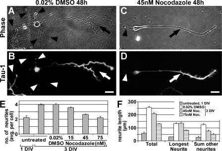
“Interestingly, the extent of reduction of axon length in our experiments was similar to that observed in another study where hippocampal neurons were treated with low doses of nocodazole from DIV1 to DIV3 (Witte et al., 2008).”
BackgroundResultKirszenblat, L., Neumann, B., Coakley, S., & Hilliard, M. A. (2012) · Molecular Biology of the Cell · doi:10.1091/mbc.e12-06-0441 · Cited by 55
“Ectopic neurite outgrowth is triggered by hyperstable microtubules Local stabilization of microtubules in immature neurites of dissociated neurons is sufficient to trigger axon formation (Witte et al., 2008).”
BackgroundThe cytoskeletal arrangements necessary to neurogenesis
Compagnucci, C., Piemonte, F., Sferra, A., Piermarini, E., & Bertini, E. (2016) · Oncotarget · doi:10.18632/oncotarget.6838 · Cited by 57
“Recently, MT stability has also been demonstrated to be a signal specifying neuronal polarization [32].”
BackgroundChia, J. X., Efimova, N., & Svitkina, T. (2016) · Molecular Biology of the Cell · doi:10.1091/mbc.e16-04-0253 · Cited by 51
“…are required in some cases (Tanaka et al., 1995; Dent and Kalil, 2001; Kaverina et al., 2002; Rajnicek et al., 2006), whereas stable microtubules are sufficient or even beneficial in other situations (Gundersen and Bulinski, 1988; Buck and Zheng, 2002; Witte et al., 2008; Lu et al., 2013).”
BackgroundSystemic epothilone D improves hindlimb function after spinal cord contusion injury in rats
Sandner, B., Puttagunta, R., Motsch, M., Bradke, F., Ruschel, J., Blesch, A., & Weidner, N. (2018) · Experimental Neurology · doi:10.1016/j.expneurol.2018.01.018 · Cited by 56
“of paclitaxel lead to moderate microtubule stabilization in axonal growth cones, which still allows microtubule polymerization at the plus end (Derry et al., 1995; Derry et al., 1997; Witte et al., 2008), enhances axonal growth in cultured neurons (Hellal et al.”
BackgroundSelf-citationThe functional architecture of axonal actin
Papandréou, M., & Leterrier, C. (2018) · Molecular and Cellular Neuroscience · doi:10.1016/j.mcn.2018.05.003 · Cited by 53
Rehbach, K., Kesavan, J., Hauser, S., Ritzenhofen, S., Jungverdorben, J., Schüle, R., Schöls, L. J., Peitz, M., & Brüstle, O. (2019) · Scientific Reports · doi:10.1038/s41598-019-45246-4 · Cited by 52
Casein kinase 2 and microtubules control axon initial segment formation
Sánchez-Ponce, D., Muñoz, A., & Garrido, J. J. (2010) · Molecular and Cellular Neuroscience · doi:10.1016/j.mcn.2010.09.005 · Cited by 57
“Indeed, CK2α and CK2α′ are polarized at the outgrowing axon, together with ankyrinG and pIκBα, where there is a higher ratio of acetylated versus tyrosinated tubulin when compared to other neurites (Witte et al., 2008).”
BackgroundNeuronal polarization in the developing cerebral cortex
Sakakibara, A., & Hatanaka, Y. (2015) · Frontiers in Neuroscience · doi:10.3389/fnins.2015.00116 · Cited by 51
“Stable microtubules within axons confer distinct characteristics based on their organization (Witte et al., 2008; Conde and Cáceres, 2009), and signaling molecules like LKB1 and SAD-A/B kinases trigger axonogenesis by changing the phosphorylation state of microtubule-associated proteins (MAPs),…”
BackgroundMicrotubules Modulate F-actin Dynamics during Neuronal Polarization
Zhao, B. Q., Meka, D. P., Scharrenberg, R., König, T., Schwanke, B., Kobler, O., Windhorst, S., Kreutz, M. R., Mikhaylova, M., & de Anda, F. C. (2017) · Scientific Reports · doi:10.1038/s41598-017-09832-8 · Cited by 52
Das, V., & Miller, J. H. (2012) · European Journal of Neuroscience · doi:10.1111/j.1460-9568.2012.08084.x · Cited by 55
“With paclitaxel, lower concentrations can cause a directional shift of polymerizing dynamic MTs towards the axonal tip, thereby favouring neurite outgrowth (Witte et al., 2008).”
BackgroundInfluentialLPA1, LPA2, LPA4, and LPA6 receptor expression during mouse brain development
Suckau, O., Groß, I., Schrötter, S., Yang, F., Luo, J., Wree, A., Chun, J. J. M., Baska, D., Baumgart, J., Kano, K., et al. (2019) · Developmental Dynamics · doi:10.1002/dvdy.23 · Cited by 50
“Drug concentration and duration of treatment were adjusted after Witte et al.99 After removal of the medium at the indicated time points (5, 15, 30 or 60 minutes) and washing with 1× PBS, cells were fixed in ice cold 4% PFA in 1× PBS containing 15% sucrose for five minutes at RT.”
MethodSymptoms: Chemotherapy-Induced Peripheral Neuropathy
Schneider, B. P., Hershman, D. L., & Loprinzi, C. L. (2015) · Advances in experimental medicine and biology · doi:10.1007/978-3-319-16366-6_6 · Cited by 50
Vinopal, S., Dupraz, S., Alfadil, E., Pietralla, T., Bendre, S., Stieß, M., Falk, S., Ortega, G. C., Maghelli, N., Tolić, I. M., et al. (2023) · Neuron · doi:10.1016/j.neuron.2023.01.020 · Cited by 49
“8, 11, 12 , 14, 110– 112 Decrease and increase of centrosomal microtubule nucleation Therefore, we investigated its role in the centrosome-related migratory block.”
BackgroundSelf-citation
Metadata
- Authors
- Witte, H., Neukirchen, D., & Bradke, F.
- Affiliations
- Max Planck Institute of Neurobiology (DE)
- Max Planck Society (DE)
- Venue
- The Journal of Cell Biology
- Publisher
- Rockefeller University Press
- Published
- 2008-02-11
- Volume
- 180
- Issue
- 3
- Pages
- 619-632
- ISSN
- 0021-9525 (ISSN-L)
- 1540-8140
- PMC
- PMC2234250
- OpenAlex
- W2084158780
- Cited by
- 564
- References
- 54
- Citations by year
- 2026: 22 · 2025: 21 · 2024: 19 · 2023: 25 · 2022: 35 · 2021: 36 · 2020: 49 · 2019: 27 · 2018: 27 · 2017: 29 · 2016: 38 · 2015: 38 · 2014: 24 · 2013: 24 · 2012: 39
- FWCI
- 15.86
- Type
- journal article
- Language
- en
- Open access
- green
- www.ncbi.nlm.nih.gov
- Licence
- cc-by-nc-sa
- Locations
- The Journal of Cell Biology (publishedVersion)
- PubMed Central (publishedVersion, cc-by-nc-sa)
- PubMed (publishedVersion)
- Max Planck Institute for Plasma Physics (submittedVersion)
- Max Planck Digital Library (submittedVersion)
- MPG.PuRe (Max Planck Society) (submittedVersion)
- Topics
- Life Sciences › Neuroscience › Cellular and Molecular Neuroscience › Axon Guidance and Neuronal Signaling
- Life Sciences › Neuroscience › Developmental Neuroscience › Neurogenesis and neuroplasticity mechanisms
- Life Sciences › Biochemistry, Genetics and Molecular Biology › Cell Biology › Microtubule and mitosis dynamics
- Keywords
- neuronal polarity, axon formation, microtubule stabilization, microtubule dynamics, hippocampal neurons, glycogen synthase kinase-3β, nocodazole, paclitaxel, dendrite formation
- MeSH terms
- Glycogen Synthase Kinase 3 beta, Animals, Cell Differentiation, Cells, Cultured, Central Nervous System, Hippocampus, Microtubules, Neurons, Nocodazole, Neurites, Cell Polarity, Paclitaxel, Protein Serine-Threonine Kinases, Mice, Knockout, Growth Cones, Intercellular Signaling Peptides and Proteins, Glycogen Synthase Kinase 3, Tubulin Modulators, Mice, Rats
About these checks
Registry, retraction, Cites and statistics results are computed, not judged, with one exception: a printed reference that no index lists is read once by a model before it counts as a citation error. Claims are extracted by a model and kept only when their quote appears verbatim in the text; each cited source is then checked, with a model’s help, for whether it says what the claim relies on. Those verdicts can be wrong — read the claim’s quote and the cited source before relying on one.
Statistics and arithmetic are recomputed, not judged.
Confidence bands are shown only for fields calibrated on our annotated test set. None is calibrated yet, so no band is shown.
What counts as a claim ›How we measure confidence ›How we check this ›
Are you an author, or do you see a problem with this page? Request a review
Data sources
Bibliographic data: Semantic Scholar, OpenAlex, Crossref.
Contains information from the Semantic Scholar Academic Graph Datasets, made available under the ODC Attribution License (ODC-BY 1.0).
Semantic Scholar’s data is described in: Kinney, R., et al. (2023). The Semantic Scholar Open Data Platform. arXiv:2301.10140.
