

Microtubule stabilization specifies initial neuronal polarization
Open accessThe DOI matches its Crossref record; 56 of the 56 references it cites were found in a registry or an index.
Abstract
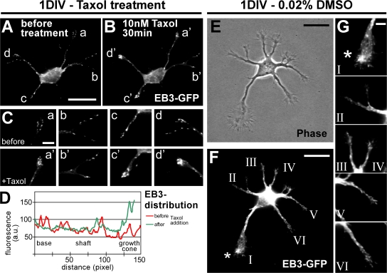
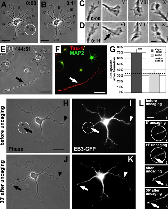
Axon formation is the initial step in establishing neuronal polarity. We examine here the role of microtubule dynamics in neuronal polarization using hippocampal neurons in culture. We see increased microtubule stability along the shaft in a single neurite before axon formation and in the axon of morphologically polarized cells. Loss of polarity or formation of multiple axons after manipulation of neuronal polarity regulators, synapses of amphids defective (SAD) kinases, and glycogen synthase kinase-3β correlates with characteristic changes in microtubule turnover. Consistently, changing the microtubule dynamics is sufficient to alter neuronal polarization. Application of low doses of the microtubule-destabilizing drug nocodazole selectively reduces the formation of future dendrites. Conversely, low doses of the microtubule-stabilizing drug taxol shift polymerizing microtubules from neurite shafts to process tips and lead to the formation of multiple axons. Finally, local stabilization of microtubules using a photoactivatable analogue of taxol induces axon formation from the activated area. Thus, local microtubule stabilization in one neurite is a physiological signal specifying neuronal polarization.
Overview
The claims and statistics of this paper haven’t been checked yet.
Free: it reads the open-access text and checks the first 5 claims that cite a source we can check. Create a free account to check every claim.
Integrity signals
Registry
Cites
Statistics
Figures


Verification
Claims the paper makes
The claims and statistics of this paper haven’t been checked yet.
Free: it reads the open-access text and checks the first 5 claims that cite a source we can check. Create a free account to check every claim.
Claims other papers make about this one
Each is checked against this paper’s own text: does it say what the citing paper says it does?
The source backs part of the claim, not all of it — or backs it for a different population, condition or comparison.
In the citing paper
“Restricted expression of acetylated tubulin near the centrosome defines a stable location for axon growth [77].”
From Abnormal accumulation of extracellular vesicles in hippocampal dystrophic axons and regulation by the primary cilia in Alzheimer's disease (2023) · cites this paper as [77]
Experimental — accuracy not yet measuredIn the citing paper
“Selective microtubule stabilization in one minor process determines axon specification (19) and only after this event, the dendritic microtubule network polarity becomes progressively mixed (18).”
From Axonal transport: Driving synaptic function (2019) · cites this paper as [19]
Self-check
Percentages, sums and reported differences re-computed from the paper’s own numbers.
The self-check needs the paper’s full text.
Citations
Cites
‘Green mice’ as a source of ubiquitous green cells
Okabe, M., Ikawa, M., Kominami, K., Nakanishi, T., & Nishimune, Y. (1997) · FEBS Letters · doi:10.1016/s0014-5793(97)00313-x · Cited by 2,581
GSK-3: tricks of the trade for a multi-tasking kinase
Doble, B. W., & Woodgett, J. R. (2003) · Journal of Cell Science · doi:10.1242/jcs.00384 · Cited by 2,084 · cited 5×
“The online version of this paper contains supplemental material.”
InfluentialThe establishment of polarity by hippocampal neurons in culture.
Dotti, C., Sullivan, C., & Banker, G. (1988) · The Journal of Neuroscience · doi:10.1523/jneurosci.08-04-01454.1988 · Cited by 1,862
The multifaceted roles of glycogen synthase kinase 3β in cellular signaling
Grimes, C. A., & Jope, R. S. (2001) · Progress in Neurobiology · doi:10.1016/s0301-0082(01)00011-9 · Cited by 1,423 · cited 2×
“Taxol-treated 2005 ; Yoshimura et al., 2005 ), which are known modulators of MT stability, yet they have various other cellular functions including the regulation of metabolism, signaling, gene transcription, and endocytosis ( Crump et al., 2001 ; Grimes and Jope, 2001 ).”
Microtubule Acetylation Promotes Kinesin-1 Binding and Transport
Reed, N. A., Cai, D., Blasius, T. L., Jih, G. T., Meyhofer, E., Gaertig, J., & Verhey, K. J. (2006) · Current Biology · doi:10.1016/j.cub.2006.09.014 · Cited by 919 · cited 1×
“…unpolymerized tubulin subunits, cells were simultaneously fi xed and permeabilized in PHEM buffer (60 mM Pipes, 25 mM Hepes, 5 mM EGTA, and 1 mM MgCl) containing 0.25% glutaraldehyde, 3.7% paraformaldehyde, 3.7% sucrose, and 0.1% Triton X-100 (adapted from Smith, 1994 ) and quenched as above.”
InfluentialGSK-3β Regulates Phosphorylation of CRMP-2 and Neuronal Polarity
Yoshimura, T., Kawano, Y., Arimura, N., Kawabata, S., Kikuchi, A., & Kaibuchi, K. (2005) · Cell · doi:10.1016/j.cell.2004.11.012 · Cited by 875 · cited 7×
“The online version of this paper contains supplemental material.”
InfluentialCdc42 regulates GSK-3β and adenomatous polyposis coli to control cell polarity
Etienne-Manneville, S., & Hall, A. (2003) · Nature · doi:10.1038/nature01423 · Cited by 835
CRMP-2 binds to tubulin heterodimers to promote microtubule assembly
Fukata, Y., Itoh, T. J., Kimura, T., Ménager, C., Nishimura, T., Shiromizu, T., Watanabe, H., Inagaki, N., Iwamatsu, A., Hotani, H., et al. (2002) · Nature Cell Biology · doi:10.1038/ncb825 · Cited by 761
Post-translational modifications regulate microtubule function
Westermann, S., & Weber, K. (2003) · Nature Reviews Molecular Cell Biology · doi:10.1038/nrm1260 · Cited by 717 · cited 2×
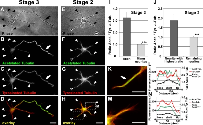
“On average from all stage 3 neurons, the axonal shaft showed a 3.2 ± 0.5-fold ratio increase of the fl uorescence intensities of acetylated versus tyrosinated MTs compared with the shafts of minor neurites (P < 0.001 by t test; Fig.”
InfluentialStepanova, T., Slemmer, J., Hoogenraad, C. C., Lansbergen, G., Dortland, B., De Zeeuw, C. I., Grosveld, F., van Cappellen, G., Akhmanova, A., & Galjart, N. (2003) · The Journal of Neuroscience · doi:10.1523/jneurosci.23-07-02655.2003 · Cited by 698 · cited 2×
“Taxol application rapidly stabilizes MTs and shifts dynamic MTs to the tips of processes To assess MT dynamics during axon formation, polymerizing MT plus ends were visualized by transfecting neurons with the GFP-tagged MT plus end – binding protein 3 (EB3; Stepanova et al., 2003 ).”
Research works printed but not linked: 3
- 1991, J. Cell Sci. Suppl., 15, 75
- 2005, Cell., 120, 123
- 2003, Nat. Rev. Mol. Cell Biol., 4, 938
Cited by
Items 201–300 of 593
Sgrò, F., Bianchi, F. T., Falcone, M., Pallavicini, G., Gai, M., Chiotto, A. M. A., Berto, G. E., Turco, E., Chang, Y., Huttner, W. Β., et al. (2015) · Cell Death and Differentiation · doi:10.1038/cdd.2015.142 · Cited by 49
“The results of previous studies21,26 and the present report indicate that neurogenic divisions are strongly dependent on CIT-K.”
BackgroundInfluentialBarsh, G. R., Isabella, A. J., & Moens, C. B. (2017) · Current Biology · doi:10.1016/j.cub.2017.11.022 · Cited by 47
Rnd1 Regulates Axon Extension by Enhancing the Microtubule Destabilizing Activity of SCG10
Li, Y., Ghavampur, S., Bondallaz, P., Will, L., Grenningloh, G., & ̈schel, A. W. P. (2008) · Journal of Biological Chemistry · doi:10.1074/jbc.m808126200 · Cited by 52
Nie, J., Sun, C., Faruque, O. M., Ye, G., Li, J., Liang, Q., Chang, Z., Yang, W., Han, X., & Shi, Y. (2012) · Journal of Biological Chemistry · doi:10.1074/jbc.m112.378372 · Cited by 46
Actin and Microtubule-Based Cytoskeletal Cues Direct Polarized Targeting of Proteins in Neurons
Arnold, D. B. (2009) · Science Signaling · doi:10.1126/scisignal.283pe49 · Cited by 46
“nesin Kif5 causes this motor protein to bind with a relatively low affinity to microtubules modified by tyrosination, a process that occurs more commonly in dendrites (22).”
The synaptic life of microtubules
Waites, C. L., Qu, X., & Bartolini, F. (2021) · Current Opinion in Neurobiology · doi:10.1016/j.conb.2021.03.004 · Cited by 48
Chen, H., Lin, G.-T., Huang, C.-K., & Fann, M. (2014) · Experimental Neurology · doi:10.1016/j.expneurol.2014.06.024 · Cited by 47
Marx, A., Godinez, W. J., Tsimashchuk, V., Bankhead, P., Rohr, K., & Engel, U. (2013) · Molecular Biology of the Cell · doi:10.1091/mbc.e12-08-0573 · Cited by 47
Mutation of Kinesin-6 Kif20b causes defects in cortical neuron polarization and morphogenesis
McNeely, K. C., Cupp, T. D., Little, J. N., Janisch, K. M., Shrestha, A., & Dwyer, N. D. (2017) · Neural Development · doi:10.1186/s13064-017-0082-5 · Cited by 46
“A key process driving neuron polarization is likely to be local stabilization and bundling of parallel microtubules [4, 28].”
BackgroundEffects of mutating α-tubulin lysine 40 on sensory dendrite development
Jenkins, B., Saunders, H. A. J., Record, H. L., Johnson-Schlitz, D. M., & Wildonger, J. (2017) · Journal of Cell Science · doi:10.1242/jcs.210203 · Cited by 46
TUBB3 and KIF21A in neurodevelopment and disease
Puri, D., Barry, B. J., & Engle, E. C. (2023) · Frontiers in Neuroscience · doi:10.3389/fnins.2023.1226181 · Cited by 46
“Neuronal development and maintenance is regulated by intracellular and extracellular factors (Yogev and Shen, 2017), and a pivotal role is played by the intracellular microtubule cytoskeleton (Witte et al., 2008; Conde and Cáceres, 2009).”
BackgroundThe novel formin FMNL3 is a cytoskeletal regulator of angiogenesis.
Hetheridge, C., Scott, A. N., Swain, R. K., Copeland, J. W. R., Higgs, H. N., Bicknell, R., & Mellor, H. (2012) · Journal of Cell Science · doi:10.1242/jcs.091066 · Cited by 47
“Similar parallel alignments of stable microtubules are found in elongating neurons (Witte et al., 2008).”
BackgroundInfluentialNagai, J., Baba, R., & Ohshima, T. (2016) · Molecular Neurobiology · doi:10.1007/s12035-016-0005-1 · Cited by 47
“Neuronal polarity has been shown to be regulated by CRMP2 [44], where microtubule stabilization is necessary for axon formation and neuronal polarization [83].”
Acute exposure to ustiloxin A affects growth and development of early life zebrafish, Danio rerio
Hu, Z., Dang, Y., Liu, C., Zhou, L., & Liu, H. (2019) · Chemosphere · doi:10.1016/j.chemosphere.2019.04.002 · Cited by 43
Rac-GTPases Regulate Microtubule Stability and Axon Growth of Cortical GABAergic Interneurons
Tivodar, S., Kalemaki, K., Kounoupa, Z., Vidaki, M., Theodorakis, Κ., Denaxa, M., Kessaris, N., de Curtis, I., Pachnis, V., & Karagogeos, D. (2014) · Cerebral Cortex · doi:10.1093/cercor/bhu037 · Cited by 42
“Moreover axons and minor neurites respond differently after treatment with taxol and have a different distribution of Ac-tubulin (Witte et al. 2008).”
BackgroundInfluentialDeceivingly dynamic: Learning-dependent changes in stathmin and microtubules
Uchida, S., & Shumyatsky, G. P. (2015) · Neurobiology of Learning and Memory · doi:10.1016/j.nlm.2015.07.011 · Cited by 45
Wnt Signaling Directs Neuronal Polarity and Axonal Growth
Stanganello, E., Zahavi, E. E., Burute, M., Smits, J., Jordens, I., Maurice, M. M., Kapitein, L. C., & Hoogenraad, C. C. (2019) · iScience · doi:10.1016/j.isci.2019.02.029 · Cited by 45
Liaci, C., Camera, M., Caslini, G., Rando, S., Contino, S., Romano, V., & Merlo, G. R. (2021) · International Journal of Molecular Sciences · doi:10.3390/ijms22116167 · Cited by 42
“The stable to dynamic microtubules ratio is significantly higher in the neurite that is meant to form the axon as compared to the other neurites, indicating that stable microtubules are essential for axon specification [289,290].”
BackgroundBerto, G. E., Iobbi, C., Camera, P., Scarpa, E., Iampietro, C., Bianchi, F. T., Gai, M., Sgrò, F., Cristofani, F., Gärtner, A., et al. (2014) · PLoS ONE · doi:10.1371/journal.pone.0093721 · Cited by 42
“This condition allows the penetration of microtubules, which can direct membrane flow from the Golgi to the growing tip [8,14].”
BackgroundCell Polarity in Cerebral Cortex Development—Cellular Architecture Shaped by Biochemical Networks
Hansen, A. H., Duellberg, C., Mieck, C., Loose, M., & Hippenmeyer, S. (2017) · Frontiers in Cellular Neuroscience · doi:10.3389/fncel.2017.00176 · Cited by 41
“Studies using drugs that either stabilized microtubules (Witte et al., 2008) or destabilized actin filaments (Bradke and Dotti, 1999) are sufficient to induce the formation of multiple axons, consistent with the view that the effects of the above mentioned circuits are transmitted via selective…”
ResultIvashko‐Pachima, Y., Maor-Nof, M., & Gozes, I. (2019) · PLoS ONE · doi:10.1371/journal.pone.0213666 · Cited by 44
Microtubule dynamics in healthy and injured neurons
Rolls, M. M., Thyagarajan, P., & Feng, C. (2020) · Developmental Neurobiology · doi:10.1002/dneu.22746 · Cited by 42
Strategies to neutralize RhoA/ROCK pathway after spinal cord injury
Roy, A., Pathak, Z., & Kumar, H. (2021) · Experimental Neurology · doi:10.1016/j.expneurol.2021.113794 · Cited by 42
Ngo, L., Haas, M., Qu, Z., Li, S., Zenker, J., Teng, K. S.-L., Gunnersen, J. M., Breuss, M. W., Habgood, M. D., Keays, D. A., et al. (2014) · Human Molecular Genetics · doi:10.1093/hmg/ddu238 · Cited by 41
Dynamic regulation of GEF-H1 localization at microtubules by Par1b/MARK2
Yoshimura, Y., & Miki, H. (2011) · Biochemical and Biophysical Research Communications · doi:10.1016/j.bbrc.2011.04.032 · Cited by 39
“Because the stabilization and post-translational modification of microtubules are crucial for the development of cell polarity [11,12], it is assumed that Par1b may contribute to cell polarization through the regulation of microtubules.”
BackgroundFibroblast growth factor signaling in axons: from development to disease
Tomé, D., Dias, M. S., Correia, J., & Almeida, R. D. (2023) · Cell Communication and Signaling · doi:10.1186/s12964-023-01284-0 · Cited by 40
“Increased MT stabilization induces the formation of a single axon from the multitude of primordial neuronal branches [50].”
BackgroundFormation and functions of asymmetric microtubule organization in polarized cells
Sugioka, K., & Sawa, H. (2012) · Current Opinion in Cell Biology · doi:10.1016/j.ceb.2012.05.007 · Cited by 45
Stimulating neuroregeneration as a therapeutic drug approach for traumatic brain injury
Mueller, B. K., Mueller, R., & Schoemaker, H. (2009) · British Journal of Pharmacology · doi:10.1111/j.1476-5381.2009.00220.x · Cited by 39
“…(homologues of the partitioning defective-1, PAR-1, serine/threonine kinases), c-Jun N-terminal kinases or collapsin-response mediator protein 2 because their inhibition or deletion has been shown to affect the process of microtubule stabilization and thereby of axon formation (Witte et al., 2008).”
BackgroundRole of calpain-mediated p53 truncation in semaphorin 3A-induced axonal growth regulation
Qin, Q., Liao, G., Baudry, M., & Bi, X. (2010) · Proceedings of the National Academy of Sciences · doi:10.1073/pnas.1008652107 · Cited by 39
“Besides actin filaments, microtubule dynamics are also critical for neurite outgrowth (3, 4).”
BackgroundNagai, J., Kitamura, Y., Owada, K., Yamashita, N., Takei, K., Goshima, Y., & Ohshima, T. (2015) · Scientific Reports · doi:10.1038/srep08269 · Cited by 39
Parcerisas, A., Pujadas, L., Ortega-Gascó, A., Perelló‐Amorós, B., Viais, R., Hino, K., Figueiro‐Silva, J., Torre, A. L., Trullás, R., Simó, S., et al. (2020) · Cerebral Cortex · doi:10.1093/cercor/bhz342 · Cited by 40
“To determine whether microtubule destabilization mediates NCAM2 silencing-induced dendritic alterations, we performed experiments using taxol to stabilize microtubules and reduce catastrophe rates (Witte et al. 2008).”
BackgroundMethodCAMSAP1 breaks the homeostatic microtubule network to instruct neuronal polarity
Zhou, Z., Xu, H., Li, Y., Yang, M., Zhang, R., Shiraishi, A., Kiyonari, H., Liang, X., Huang, X., Wang, Y., et al. (2020) · Proceedings of the National Academy of Sciences · doi:10.1073/pnas.1913177117 · Cited by 42
“The MT network goes through tremendous changes during neuronal polarization (6, 8).”
BackgroundInfluentialNovel α-tubulin mutation disrupts neural development and tubulin proteostasis
Hanson, M. G., Aiken, J., Sietsema, D. V., Sept, D., Bates, E. A., Niswander, L. A., & Moore, J. K. (2015) · Developmental Biology · doi:10.1016/j.ydbio.2015.11.022 · Cited by 39
“During neuronal morphogenesis, microtubules help to form and stabilize the axon by packing into highly stable, polarized arrays (Conde and Caceres, 2009; Witte et al., 2008).”
BackgroundCellular cartography: Towards an atlas of the neuronal microtubule cytoskeleton
Iwanski, M. K., & Kapitein, L. C. (2023) · Frontiers in Cell and Developmental Biology · doi:10.3389/fcell.2023.1052245 · Cited by 38
“This was demonstrated through the use of photoactivatable Taxol, which allowed microtubules to be selectively stabilized in one neurite, promoting that neurite to become the axon (Witte et al., 2008).”
MethodKim, J. S., Lilley, B. N., Zhang, C., Shokat, K. M., Sanes, J. R., & Zhen, M. (2008) · Neural Development · doi:10.1186/1749-8104-3-23 · Cited by 43
“The mammalian orthologs of SAD-1, SAD-A and SAD-B, have been shown to regulate neuronal polarity through phosphorylation of tau, a microtubule associated protein [22,23], and subsequent promotion of microtubule dynamics during axon extension [32].”
Wenzel, E. D., Speidell, A. P., Flowers, S. A., Wu, C., Avdoshina, V., & Mocchetti, I. (2019) · Cell Death and Disease · doi:10.1038/s41419-019-1920-7 · Cited by 39
Taran, A., Belikova, L. D., Lagarkova, M. A., & Alieva, I. B. (2020) · Cells · doi:10.3390/cells9061514 · Cited by 40
“Axon forms after neurite expansion due to the local stabilization of MTs in pre-axonal neurites [22].”
Backgroundα-TubK40me3 is required for neuronal polarization and migration by promoting microtubule formation
Xie, X., Wang, S., Li, M., Diao, L., Pan, X., Chen, J., Zou, W., Zhang, X., Feng, W., & Bao, L. (2021) · Nature Communications · doi:10.1038/s41467-021-24376-2 · Cited by 38
Quantitative Analysis of Microtubule Self-assembly Kinetics and Tip Structure
Prahl, L. S., Castle, B. T., Gardner, M. K., & Odde, D. J. (2014) · Methods in enzymology on CD-ROM/Methods in enzymology · doi:10.1016/b978-0-12-397924-7.00003-0 · Cited by 42
Wang, T., Wang, Q., Song, R., Zhang, Y., Yang, J., Wang, Y., Yuan, Y., Bian, J., Liu, X., Gu, J., et al. (2015) · Neurotoxicology and Teratology · doi:10.1016/j.ntt.2015.11.007 · Cited by 39
PTEN suppresses axon outgrowth by down-regulating the level of detyrosinated microtubules
Kath, C., Goñi‐Oliver, P., Müller, R., Schultz, C., Haucke, V., Eickholt, B. J., & Schmoranzer, J. (2018) · PLoS ONE · doi:10.1371/journal.pone.0193257 · Cited by 40
APC2 controls dendrite development by promoting microtubule dynamics
Kahn, O. I., Schätzle, P., van de Willige, D., Tas, R. P., Lindhout, F. W., Portegies, S., Kapitein, L. C., & Hoogenraad, C. C. (2018) · Nature Communications · doi:10.1038/s41467-018-05124-5 · Cited by 36
Eom, T.-Y., Stanco, A., Guo, J., Wilkins, G., Deslauriers, D., Yan, J. E., Monckton, C., Blair, J., Oon, E., Perez, A., et al. (2014) · Developmental Cell · doi:10.1016/j.devcel.2014.11.022 · Cited by 37
“Immunohistochemistry, Immunoprecipitation and In Situ Hybridization Cerebral cortical sections and cortical cells were immunolabeled as previously described (Witte et al., 2008; Yokota et al., 2009).”
Localized discrete breather modes in neuronal microtubules
Kavitha, L., Parasuraman, E., Muniyappan, A., Gopi, D., & Zdravković, S. (2017) · Nonlinear Dynamics · doi:10.1007/s11071-017-3359-7 · Cited by 35
“First, the stabilization of MTs is sufficient to induce axon formation during neuronal development, and they act as signal molecules for initial neuronal polarization [77].”
BackgroundAxonal plasticity in response to active forces generated through magnetic nano-pulling
Falconieri, A., Vincentiis, S. D., Cappello, V., Convertino, D., Das, R., Ghignoli, S., Figoli, S., Luin, S., Català-Castro, F., Marchetti, L., et al. (2022) · Cell Reports · doi:10.1016/j.celrep.2022.111912 · Cited by 39
Müller, M., Lutter, D., & Püschel, A. W. (2009) · Journal of Cell Science · doi:10.1242/jcs.058230 · Cited by 37
“The observation that minor neurites show differences in microtubule stability already at stage 2 supports this conclusion (Witte et al., 2008).”
ResultPar proteins and neuronal polarity
Insolera, R., Chen, S., & Shi, S. (2010) · Developmental Neurobiology · doi:10.1002/dneu.20867 · Cited by 38
“Proper polarization of neurons entails differential regulation of the actin and microtubule cytoskeletons at the tip of the growth cone in developing neurites so as to effect their growth rate (Baas, 1999; Bradke and Dotti, 1999; Witte et al., 2008).”
Spedden, E., Kaplan, D. L., & Staii, C. (2013) · Physical Biology · doi:10.1088/1478-3975/10/5/056002 · Cited by 34
“First, we measure the effects of Taxol, a drug that stabilizes microtubules against changes in aggregation and against temperature-induced changes in microtubule flexural rigidity [21, 22].”
BackgroundMethodLiu, Y., Lv, K., Li, Z., Yu, A. C. H., Chen, J., & Teng, J. (2012) · Journal of Biological Chemistry · doi:10.1074/jbc.m112.403451 · Cited by 40
Growth, collapse, and stalling in a mechanical model for neurite motility
Recho, P., Jérusalem, A., & Goriely, A. (2016) · Physical review. E · doi:10.1103/physreve.93.032410 · Cited by 33
Neurofilament depletion improves microtubule dynamics via modulation of Stat3/stathmin signaling
Yadav, P., Selvaraj, B. T., Bender, F. L. P., Behringer, M., Moradi, M., Sivadasan, R., Dombert, B., Blum, R., Asan, E., Sauer, M., et al. (2016) · Acta Neuropathologica · doi:10.1007/s00401-016-1564-y · Cited by 33
“In neurons, acetylated microtubules are found in more proximal parts of axons where microtubules are thought to be stable, and they appear to be excluded from axon terminals [66, 80].”
BackgroundThe +TIP Navigator-1 is an actin–microtubule crosslinker that regulates axonal growth cone motility
Sánchez-Huertas, C., Bonhomme, M., Falco, A., Fagotto‐Kaufmann, C., van Haren, J., Jeanneteau, F. D., Galjart, N., Debant, A., & Boudeau, J. (2020) · The Journal of Cell Biology · doi:10.1083/jcb.201905199 · Cited by 35
“For MT immunostaining (Witte et al., 2008), neurons were simultaneously permeabilized and fixed using 4%”
MethodMolecular Motors in Cargo Trafficking and Synapse Assembly
van den Berg, R., & Hoogenraad, C. C. (2012) · Advances in experimental medicine and biology · doi:10.1007/978-3-7091-0932-8_8 · Cited by 35
The ER ladder is a unique morphological feature of developing mammalian axons
Zamponi, E., Meehl, J. B., & Voeltz, G. K. (2022) · Developmental Cell · doi:10.1016/j.devcel.2022.05.002 · Cited by 32
The receptor tyrosine kinase Ror is required for dendrite regeneration in Drosophila neurons
Nye, D. M. R., Albertson, R. M., Weiner, A. T., Hertzler, J. I., Shorey, M., Goberdhan, D. C. I., WILSON, C. G., Janes, K. A., & Rolls, M. M. (2020) · PLoS Biology · doi:10.1371/journal.pbio.3000657 · Cited by 32
“Alternatively, the relatively high stability of axonal microtubules relative to dendritic ones [63, 64] could account for the difference in sensitivity to reduced nucleation.”
Background5'-UTR SNP of FGF13 causes translational defect and intellectual disability
Pan, X., Zhao, J., Zhou, Z., Chen, J., Yang, Z., Wu, Y., Bai, M., Jiao, Y., Yang, Y., Hu, X., et al. (2021) · eLife · doi:10.7554/elife.63021 · Cited by 35
“Regulation of microtubule dynamics is crucial for a neuronal transition from the multipolar to bipolar morphology, which is a prerequisite for initiating the neuron migration (Witte et al., 2008; Jaglin and Chelly, 2009; Li et al., 2012; Zhang et al., 2012).”
BackgroundMythili, Y., LaPointe, N. E., Lopus, M., Cox, C., Jordan, M. A., Feinstein, S. C., & Wilson, L. (2010) · Journal of Alzheimer s Disease · doi:10.3233/jad-2010-1335 · Cited by 31
“Relatively stable microtubules are normally associated with axons [19] and, when present in a neurite, can favor axon formation and neuronal polarization [20].”
HECT‐type E3 ubiquitin ligases in nerve cell development and synapse physiology
Ambrozkiewicz, M. C., & Kawabe, H. (2015) · FEBS Letters · doi:10.1016/j.febslet.2015.05.009 · Cited by 33
The role of selective transport in neuronal polarization
Namba, T., Nakamuta, S., Funahashi, Y., & Kaibuchi, K. (2011) · Developmental Neurobiology · doi:10.1002/dneu.20876 · Cited by 31
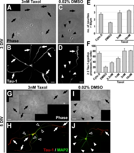
“…the treatment of Taxol which enhances tubulin acetylation, detyrosination and polyglutamylation leads to the accumulation of KIF5 motor domain not only at the axonal tip but also at the tips of other immature neurites and induces multiple axon formation (Witte et al., 2008; Hammond et al., 2010).”
BackgroundMicrotubules: A Key to Understand and Correct Neuronal Defects in CDKL5 Deficiency Disorder?
Barbiero, I., Rosa, R. D., & Kilstrup‐Nielsen, C. (2019) · International Journal of Molecular Sciences · doi:10.3390/ijms20174075 · Cited by 33
“Indeed, axon formation can be artificially induced treating unpolarised neurons with the MT-stabilising drug, taxol [28].”
BackgroundValencia, R. G., Mihailovska, E., Winter, L., Bauer, K., Fischer, I., Walko, G., Jorgačevski, J., Potokar, M., Zorec, R., & Wiche, G. (2020) · Neuropathology and Applied Neurobiology · doi:10.1111/nan.12635 · Cited by 33
Post-translational control of Myc function during differentiation
Conacci‐Sorrell, M., & Eisenman, R. N. (2011) · Cell Cycle · doi:10.4161/cc.10.4.14794 · Cited by 34
Li, L., Liu, B., Zhang, X., & Ye, L. (2015) · Biochemical and Biophysical Research Communications · doi:10.1016/j.bbrc.2015.02.036 · Cited by 29
Duan, A. R., & Goodson, H. V. (2012) · Molecular Biology of the Cell · doi:10.1091/mbc.e12-05-0374 · Cited by 29
“In neurons, MTs are linearly and densely packed along the length of the axon, inducing a global cellular polarity critical for neuronal signaling (Tanaka and Kirschner, 1991; Slaughter et al., 1997; Witte et al., 2008).”
Adaptive-Control Model for Neutrophil Orientation in the Direction of Chemical Gradients
Irimia, D., Balázsi, G., Agrawal, N., & Toner, M. (2009) · Biophysical Journal · doi:10.1016/j.bpj.2008.12.3967 · Cited by 33
“In neurons, the local stability of the microtubules is a signal for axonal and dendrite formation and neuronal polarization (73).”
BackgroundJeong, S., & Cho, G. (2016) · Biochemical and Biophysical Research Communications · doi:10.1016/j.bbrc.2016.11.031 · Cited by 29
Regulation of plasma membrane expansion during axon formation
Quiroga, S., Bisbal, M., & Cáceres, A. (2018) · Developmental Neurobiology · doi:10.1002/dneu.22553 · Cited by 31
A microtubule polymerase is required for microtubule orientation and dendrite pruning in Drosophila
Tang, Q., Rui, M., Bu, S., Wang, Y., Chew, L. Y., & Yu, F. (2020) · The EMBO Journal · doi:10.15252/embj.2019103549 · Cited by 30
“The samples were then quenched with 50 mM ammonium chloride for 5 min (Witte et al , 2008).”
Epothilones Improve Axonal Growth and Motor Outcomes after Stroke in the Adult Mammalian CNS
Kugler, C., Thielscher, C. S., Tambe, B. A., Schwarz, M. K., Halle, A., Bradke, F., & Petzold, G. C. (2020) · Cell Reports Medicine · doi:10.1016/j.xcrm.2020.100159 · Cited by 30
Self-citationPuri, D., Ponniah, K., Biswas, K., Basu, A., Dey, S., Lundquist, E. A., & Ghosh‐Roy, A. (2019) · The Journal of Cell Biology · doi:10.1083/jcb.202005080 · Cited by 28
A human IgM signals axon outgrowth: coupling lipid raft to microtubules
Xu, X., Warrington, A. E., Wright, B. R., Bieber, A. J., Keulen, V. P. V., Pease, L. R., & Rodriguez, M. (2011) · Journal of Neurochemistry · doi:10.1111/j.1471-4159.2011.07416.x · Cited by 29
“Cells were then lysed in ice-cold lysis buffer (50 mM Tris HCl, pH 7.4, 150 mM NaCl, 1 mM EDTA, 1% Triton X-100 and protease inhibitor cocktail) for 30 min.”
InfluentialDantas, T. J., Carabalona, A., Hu, D. J. K., & Vallee, R. B. (2016) · Cytoskeleton · doi:10.1002/cm.21293 · Cited by 31
“Centrosome and microtubule dynamics have been implicated in axon specification [Witte et al., 2008; de Anda et al., 2010].”
BackgroundRotenone inhibits axonogenesis via an Lfc/RhoA/ ROCK pathway in cultured hippocampal neurons
Bisbal, M., Remedi, M. V., Quassollo, G., Cáceres, A., & Sanchez, M. S. (2018) · Journal of Neurochemistry · doi:10.1111/jnc.14547 · Cited by 31
“For some experiments Taxol (3 nM; Sigma-Aldrich; Cat Number: T7191, PubChem CID: 36314) was also added to the culture medium, as described by Witte et al. (2008).”
BackgroundMethodInfluentialChen, Y., Zheng, J., Li, X., Zhu, L., Shao, Z., Yan, X., & Zhu, X. (2020) · Cell Reports · doi:10.1016/j.celrep.2020.107526 · Cited by 29
Axon Regeneration: A Subcellular Extension in Multiple Dimensions
Winter, C. C., He, Z., & Jacobi, A. (2021) · Cold Spring Harbor Perspectives in Biology · doi:10.1101/cshperspect.a040923 · Cited by 31
“These neurons can be cultured on different substrates (e.g., MAIs and CSPGs), thereby offering uniquemodels to probe into cellular mechanisms of axon regeneration (Witte et al. 2008; Al-Ali et al. 2017; Rodemer et al. 2020).”
BackgroundDong, X., Lü, Y., Hu, Q., Zeng, C., Zheng, J., Huang, J., Dong, H., Zou, P., Wang, T., Wu, Y., et al. (2025) · Small · doi:10.1002/smll.202412170 · Cited by 27
Wang, Q., Cai, H., Hu, Z., Wu, Y., Guo, X., Li, J., Wang, H., Liu, Y., Liu, Y., Xie, L., et al. (2018) · Journal of Neurotrauma · doi:10.1089/neu.2018.5966 · Cited by 28
Lee, T. W., Tumanov, S., Villas-Bôas, S. G., Montgomery, J. M., & Birch, N. P. (2014) · Journal of Neurochemistry · doi:10.1111/jnc.13009 · Cited by 29
“…EGTA, MgCl2) buffer made of 60 mM piperazine-N,N’-bis(2-ethanesulfonic acid) (PIPES), 25 mM N-2-hydroxyethylpiperazine-N-2-ethanesulfonic acid (HEPES), 5 mM EGTA,and 1 mM MgCl2, and containing 0.25% glutaraldehyde, 3.7% paraformaldehyde, 3.7% sucrose, and 0.1% Triton X-100 (Witte et al. 2008).”
InfluentialGärtner, A., Fornasiero, E. F., Valtorta, F., & Dotti, C. G. (2014) · Journal of Cell Science · doi:10.1242/jcs.149815 · Cited by 27
“Taking into account that MT dynamics is polarized in neurons in vitro during (and just shortly before) axon outgrowth, and that actin dynamics is polarized in neurons with several neurites and during neuronal migration (Bradke and Dotti, 1999; Sakakibara et al., 2014; Solecki et al., 2009; Witte et al., 2008) our new…”
BackgroundNucleating microtubules in neurons: Challenges and solutions
Lüders, J. (2020) · Developmental Neurobiology · doi:10.1002/dneu.22751 · Cited by 29
Wilkes, O. R., & Moore, A. W. (2020) · Frontiers in Cellular Neuroscience · doi:10.3389/fncel.2020.594199 · Cited by 28
Moving through the crowd. Where are we at understanding physiological axon growth?
Alfadil, E., & Bradke, F. (2022) · Seminars in Cell and Developmental Biology · doi:10.1016/j.semcdb.2022.07.001 · Cited by 27
Self-citationChemical and mechanical control of axon fasciculation and defasciculation
Breau, M. A., & Trembleau, A. (2022) · Seminars in Cell and Developmental Biology · doi:10.1016/j.semcdb.2022.06.014 · Cited by 31
Microtubule retrograde flow retains neuronal polarization in a fluctuating state
Schelski, M., & Bradke, F. (2022) · Science Advances · doi:10.1126/sciadv.abo2336 · Cited by 28
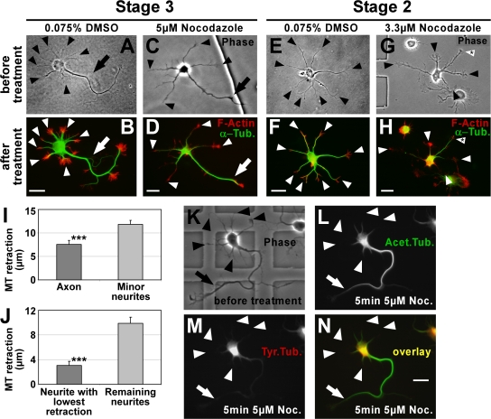
“Consistent with this view, two pharmacological manipulations that transform nongrowing neurites into multiple growing axons per cell (23, 28) slowed down MT-RF in neurites.”
BackgroundSelf-citationYang, R., Kong, E., Jin, J., Hergovich, A., & Püschel, A. W. (2014) · Journal of Cell Science · doi:10.1242/jcs.146696 · Cited by 25
“dynamics and thereby preferentially activates these kinases in the axon (Witte et al., 2008).”
BackgroundMicrotubule function and dysfunction in the nervous system
Jang, E., Choi, H., & Hur, E. (2024) · Molecules and Cells · doi:10.1016/j.mocell.2024.100111 · Cited by 31
“As newly formed neurites mature, one of the neurites accumulates tubulin acetylation and becomes the axon (Witte et al., 2008).”
Cytoskeletal Symmetry Breaking and Chirality: From Reconstituted Systems to Animal Development
Pohl, C. (2015) · Symmetry · doi:10.3390/sym7042062 · Cited by 26
“become clear that axon specification requires both a complex interplay between microtubule ends and cortical actin [70,71] strongly dependent on microtubule ends undergoing simultaneous reorganization via dynamic instability [72].”
BackgroundIvashko‐Pachima, Y., & Gozes, I. (2020) · Progress in molecular biology and translational science · doi:10.1016/bs.pmbts.2020.07.008 · Cited by 26
SUMOylation of α-tubulin is a novel modification regulating microtubule dynamics
Feng, W., Liu, R., Xie, X., Diao, L., Gao, N., Cheng, J., Zhang, X., Li, Y., & Bao, L. (2020) · Journal of Molecular Cell Biology · doi:10.1093/jmcb/mjaa076 · Cited by 26
“high doses of taxol is sufficient to block MT dynamics and neurite extension in cultured neurons (Letourneau and Ressler, 1984; Dehmelt et al., 2003; Witte et al., 2008), further supporting the importance of coordinated MT dynamics in neurite growth.”
BackgroundKIF2A deficiency causes early-onset neurodegeneration
Ruiz-Reig, N., Chehade, G., Hakanen, J., Aittaleb, M., Wierda, K. D. B., de Wit, J., Nguyen, L., Gailly, P. L., & Tissir, F. (2022) · Proceedings of the National Academy of Sciences · doi:10.1073/pnas.2209714119 · Cited by 27
“KIF2A is enriched in postmitotic neurons where it forms homodimers that bind to MTs and move toward the plus end, wherein they remove tubulin subunits in an adenosine 50-triphosphate–dependent manner (4, 12, 13).”
InfluentialMoreira‐Filho, C. A., Bando, S. Y., Bertonha, F. B., Iamashita, P., Silva, F. N., Costa, L. D. F., da Silva, A. V., Castro, L. H. M., & Tzu, W. H. (2015) · PLoS ONE · doi:10.1371/journal.pone.0128174 · Cited by 27
“FOHD1 coordinates actin filament and microtubule alignment to mediate cell elongation [67], being possibly involved in hippocampal neuronal polarization [68, 69].”
BackgroundXing, S., Liu, W., Huang, Z., Chen, L., Sun, K., Han, D. C., Zhang, W., & Jiang, X. (2010) · Electrophoresis · doi:10.1002/elps.201000133 · Cited by 25
Varberg, J. M., Padgett, L. R., Arrizabalaga, G., & Sullivan, W. J. (2016) · mSphere · doi:10.1128/msphere.00088-15 · Cited by 33
Rojas, P. S., Aguayo, F. I., Neira, D., Tejos‐Bravo, M., Aliaga, E., Muñoz, J. P., Parra, C. S., & Fiedler, J. L. (2017) · Molecular and Cellular Neuroscience · doi:10.1016/j.mcn.2017.09.009 · Cited by 26
“At early stage in culture (DIV 1-DIV 3), neurites do not express microtubule-associated protein 2 (MAP-2) immunoreactivity; nonetheless they can be immunostained with an antibody that recognize stable microtubules (acetylated-tubulin) (Witte et al., 2008).”
BackgroundSystemically treating spinal cord injury
Tran, A. P., & Silver, J. (2015) · Science · doi:10.1126/science.aab1615 · Cited by 25
Jakobs, M. A., Franze, K., & Zemel, A. (2015) · Frontiers in Cellular Neuroscience · doi:10.3389/fncel.2015.00441 · Cited by 27
“Furthermore, fast MT polymerization correlates with faster outgrowth (Baas and Ahmad, 1993; Witte et al., 2008; Lu et al., 2013).”
BackgroundMethodTAOK2 rescues autism-linked developmental deficits in a 16p11.2 microdeletion mouse model
Scharrenberg, R., Richter, M., Johanns, O., Meka, D. P., Rücker, T., Murtaza, N., Lindenmaier, Z., Ellegood, J., Naumann, A., Zhao, B. Q., et al. (2022) · Molecular Psychiatry · doi:10.1038/s41380-022-01785-3 · Cited by 24
Martinez-Carrera, L. A., Gabriel, E., Donohoe, C. D., Hölker, I., Mariappan, A., Storbeck, M., Uhlířová, M., Gopalakrishnan, J., & Wirth, B. (2018) · Human Molecular Genetics · doi:10.1093/hmg/ddy086 · Cited by 26
“larization, morphology and guidance of the axon (41,42).”
BackgroundResultCentrosome‐mediated microtubule remodeling during axon formation in human iPSC‐derived neurons
Lindhout, F. W., Portegies, S., Kooistra, R., Herstel, L. J., Stucchi, R., Hummel, J. J. A., Scheefhals, N., Katrukha, E. A., Altelaar, M. A. F., MacGillavry, H. D., et al. (2021) · The EMBO Journal · doi:10.15252/embj.2020106798 · Cited by 26
“Axonal outgrowth critically relies on local cytoskeleton reorganization and growth cone dynamics (Dotti et al, 1988; Witte et al, 2008).”
Clark, J. A., Chuckowree, J. A., Dyer, M. S., Dickson, T. C., & Blizzard, C. A. (2020) · Scientific Reports · doi:10.1038/s41598-020-57718-z · Cited by 25
Metadata
- Authors
- Witte, H., Neukirchen, D., & Bradke, F.
- Affiliations
- Max Planck Institute of Neurobiology (DE)
- Max Planck Society (DE)
- Venue
- The Journal of Cell Biology
- Publisher
- Rockefeller University Press
- Published
- 2008-02-11
- Volume
- 180
- Issue
- 3
- Pages
- 619-632
- ISSN
- 0021-9525 (ISSN-L)
- 1540-8140
- PMC
- PMC2234250
- OpenAlex
- W2084158780
- Cited by
- 564
- References
- 54
- Citations by year
- 2026: 22 · 2025: 21 · 2024: 19 · 2023: 25 · 2022: 35 · 2021: 36 · 2020: 49 · 2019: 27 · 2018: 27 · 2017: 29 · 2016: 38 · 2015: 38 · 2014: 24 · 2013: 24 · 2012: 39
- FWCI
- 15.86
- Type
- journal article
- Language
- en
- Open access
- green
- www.ncbi.nlm.nih.gov
- Licence
- cc-by-nc-sa
- Locations
- The Journal of Cell Biology (publishedVersion)
- PubMed Central (publishedVersion, cc-by-nc-sa)
- PubMed (publishedVersion)
- Max Planck Institute for Plasma Physics (submittedVersion)
- Max Planck Digital Library (submittedVersion)
- MPG.PuRe (Max Planck Society) (submittedVersion)
- Topics
- Life Sciences › Neuroscience › Cellular and Molecular Neuroscience › Axon Guidance and Neuronal Signaling
- Life Sciences › Neuroscience › Developmental Neuroscience › Neurogenesis and neuroplasticity mechanisms
- Life Sciences › Biochemistry, Genetics and Molecular Biology › Cell Biology › Microtubule and mitosis dynamics
- Keywords
- neuronal polarity, axon formation, microtubule stabilization, microtubule dynamics, hippocampal neurons, glycogen synthase kinase-3β, nocodazole, paclitaxel, dendrite formation
- MeSH terms
- Glycogen Synthase Kinase 3 beta, Animals, Cell Differentiation, Cells, Cultured, Central Nervous System, Hippocampus, Microtubules, Neurons, Nocodazole, Neurites, Cell Polarity, Paclitaxel, Protein Serine-Threonine Kinases, Mice, Knockout, Growth Cones, Intercellular Signaling Peptides and Proteins, Glycogen Synthase Kinase 3, Tubulin Modulators, Mice, Rats
About these checks
Registry, retraction, Cites and statistics results are computed, not judged, with one exception: a printed reference that no index lists is read once by a model before it counts as a citation error. Claims are extracted by a model and kept only when their quote appears verbatim in the text; each cited source is then checked, with a model’s help, for whether it says what the claim relies on. Those verdicts can be wrong — read the claim’s quote and the cited source before relying on one.
Statistics and arithmetic are recomputed, not judged.
Confidence bands are shown only for fields calibrated on our annotated test set. None is calibrated yet, so no band is shown.
What counts as a claim ›How we measure confidence ›How we check this ›
Are you an author, or do you see a problem with this page? Request a review
Data sources
Bibliographic data: Semantic Scholar, OpenAlex, Crossref.
Contains information from the Semantic Scholar Academic Graph Datasets, made available under the ODC Attribution License (ODC-BY 1.0).
Semantic Scholar’s data is described in: Kinney, R., et al. (2023). The Semantic Scholar Open Data Platform. arXiv:2301.10140.
