

The DOI matches its Crossref record; 51 of the 51 references it cites were found in a registry or an index.
PU.1 regulates the commitment of adult hematopoietic progenitors and restricts granulopoiesis
Open accessAbstract
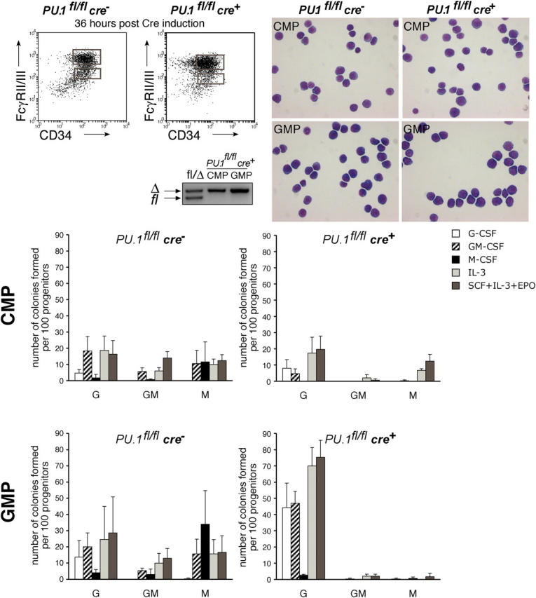
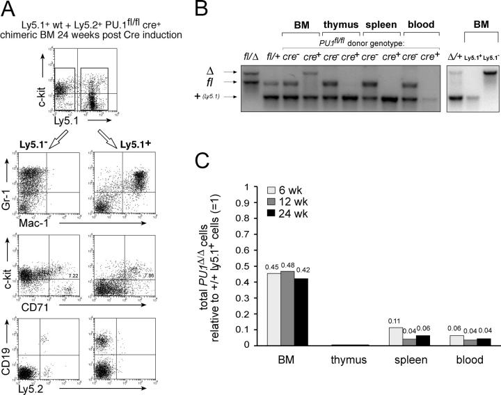
Although the transcription factor PU.1 is essential for fetal lymphomyelopoiesis, we unexpectedly found that elimination of the gene in adult mice allowed disturbed hematopoiesis, dominated by granulocyte production. Impaired production of lymphocytes was evident in PU.1-deficient bone marrow (BM), but myelocytes and clonogenic granulocytic progenitors that are responsive to granulocyte colony-stimulating factor or interleukin-3 increased dramatically. No identifiable common lymphoid or myeloid progenitor populations were discernable by flow cytometry; however, clonogenic assays suggested an overall increased frequency of blast colony-forming cells and BM chimeras revealed existence of long-term self-renewing PU.1-deficient cells that required PU.1 for lymphoid, but not granulocyte, generation. PU.1 deletion in granulocyte-macrophage progenitors, but not in common myeloid progenitors, resulted in excess granulocyte production; this suggested specific roles of PU.1 at different stages of myeloid development. These findings emphasize the distinct nature of adult hematopoiesis and reveal that PU.1 regulates the specification of the multipotent lymphoid and myeloid compartments and restrains, rather than promotes, granulopoiesis.
Overview
The claims and statistics of this paper haven’t been checked yet.
Run the check — free, no account needed. It reads the open-access text and checks the sources it cites.
Integrity signals
Registry
Cites
Statistics
Figures
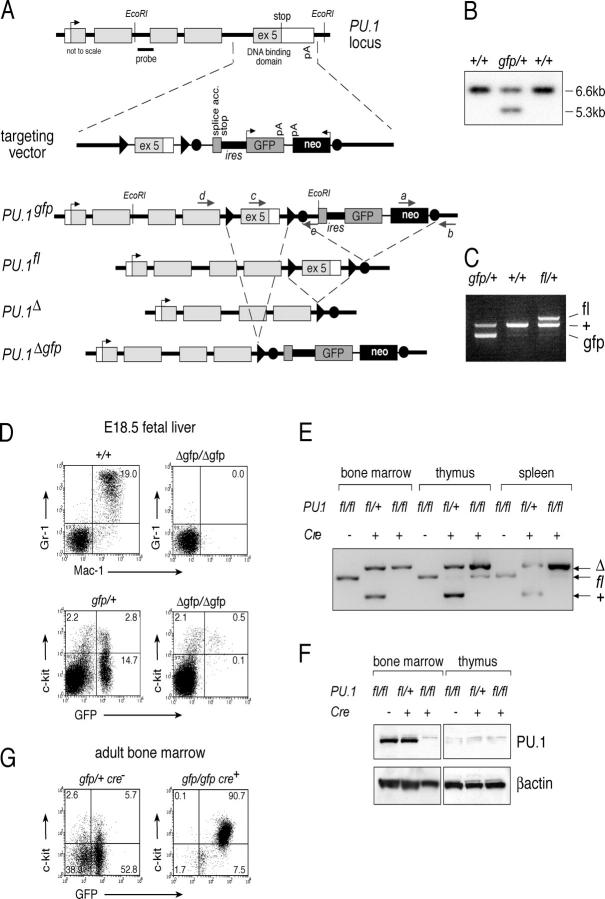
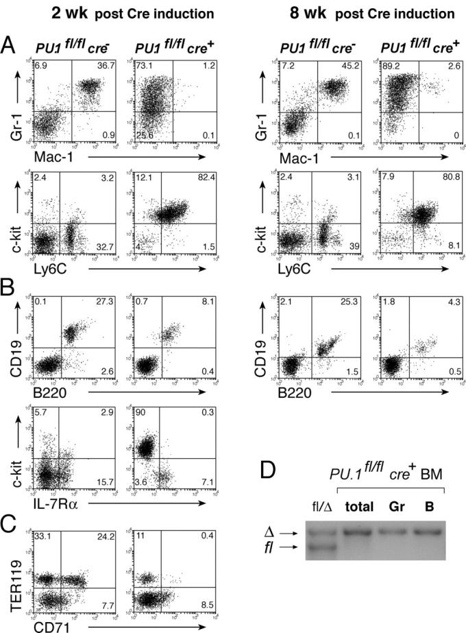
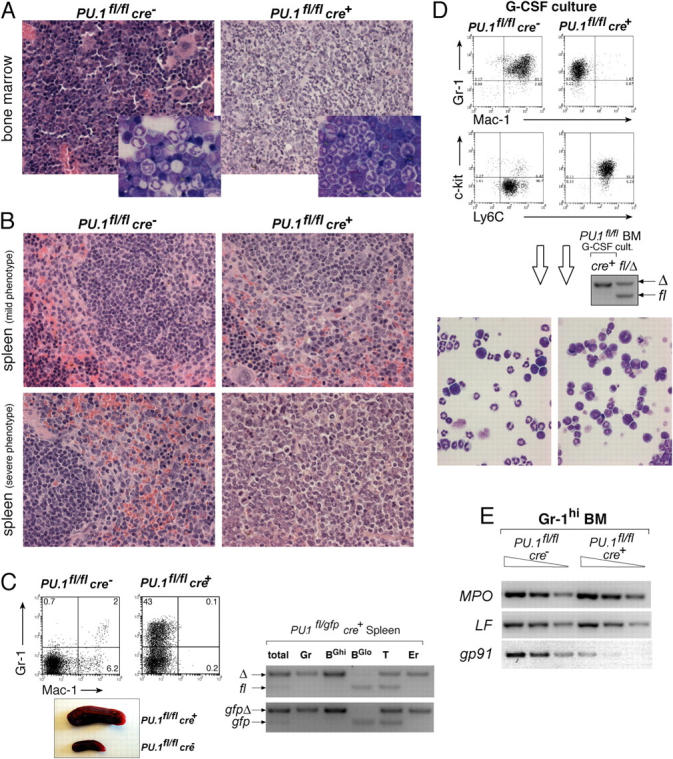
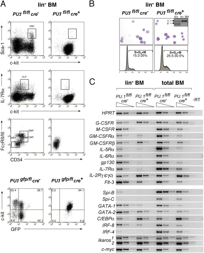
Verification
Claims the paper makes
The claims and statistics of this paper haven’t been checked yet.
Run the check — free, no account needed. It reads the open-access text and checks the sources it cites.
Claims other papers make about this one
Each is checked against this paper’s own text: does it say what the citing paper says it does?
The source backs part of the claim, not all of it — or backs it for a different population, condition or comparison.
In the citing paper
“Ets family transcription factor PU.1 is pivotal for normal hematopoiesis as it directs differentiation of hematopoietic progenitors to different cell lineages such as macrophage, granulocyte or B cell (1).”
From FBW7 Inhibits Myeloid Differentiation in Acute Myeloid Leukemia via GSK3-Dependent Ubiquitination of PU.1 (2021) · cites this paper as [1]
Experimental — accuracy not yet measuredThe source backs part of the claim, not all of it — or backs it for a different population, condition or comparison.
In the citing paper
“In preleukemic states (e.g., MDS), where PU.1 downregulation permits clonal expansion 33-35”
From USP7 sustains hematopoietic stem cell homeostasis partially via PU.1 stabilization (2026) · cites this paper as [35]
Experimental — accuracy not yet measuredThe source backs part of the claim, not all of it — or backs it for a different population, condition or comparison.
In the citing paper
“One such master regulator is the Ets-family transcription factor PU.1, which is indispensable for HSC function and the differentiation of cells within the myeloid as well as lymphoid lineages3–5.”
From Minimal PU.1 reduction induces a preleukemic state and promotes development of acute myeloid leukemia (2015) · cites this paper as [4]
Experimental — accuracy not yet measuredThe source backs part of the claim, not all of it — or backs it for a different population, condition or comparison.
In the citing paper
“its action is also important or indispensable for sustained generation of all known hematopoietic precursors that have lymphoid developmental potentials (1–9).”
From Mechanisms of Action of Hematopoietic Transcription Factor PU.1 in Initiation of T-Cell Development (2019) · cites this paper as [2]
Experimental — accuracy not yet measuredThe source backs part of the claim, not all of it — or backs it for a different population, condition or comparison.
In the citing paper
“All the hematopoietic progenitors that generate either B or T cells appear to originate from PU.1-expressing, PU.1-dependent prethymic cells (2, 27)”
From Mechanisms of Action of Hematopoietic Transcription Factor PU.1 in Initiation of T-Cell Development (2019) · cites this paper as [2]
Experimental — accuracy not yet measured
The source backs part of the claim, not all of it — or backs it for a different population, condition or comparison.
In the citing paper
“It probably extends for over 10 days for the thymocytes in young adult mice (153), where it is crucial for successful T-cell generation (2, 147).”
From Mechanisms of Action of Hematopoietic Transcription Factor PU.1 in Initiation of T-Cell Development (2019) · cites this paper as [2]
Experimental — accuracy not yet measuredThe source backs part of the claim, not all of it — or backs it for a different population, condition or comparison.
In the citing paper
“Dysregulation of the PU.1 transcriptional network is a common phenotype associated with hematological malignancy, particularly diseases of the myeloid lineage (Cook et al., 2004; Dakic et al., 2005; Koschmieder et al., 2005; Will et al., 2015; Sive et al., 2016; Aivalioti et al., 2022).”
From PU.1 is required to restrain myelopoiesis during chronic inflammatory stress (2023) · cites this paper as Dakic et al., 2005
Experimental — accuracy not yet measuredThe cited source says what the claim attributes to it.
In the citing paper
“PU.1 is a hematopoietic-specific and ETS family transcription factor that is essential for lymphoid and myeloid development25–28.”
From HIV-1 Vpr combats the PU.1-driven antiviral response in primary human macrophages (2024) · cites this paper as [27]
The cited source says what the claim attributes to it.
In the citing paper
“in postnatal mice, although T cell development is much more severely affected by PU.1 deletion than neutrophil development (2)”
From Mechanisms of Action of Hematopoietic Transcription Factor PU.1 in Initiation of T-Cell Development (2019) · cites this paper as [2]
The cited source says what the claim attributes to it.
In the citing paper
“These findings are consistent with prior work indicating PU.1 can constrain myelopoietic activity (DeKoter et al., 1998; Dakic et al., 2005)”
From PU.1 is required to restrain myelopoiesis during chronic inflammatory stress (2023) · cites this paper as Dakic et al., 2005
In the citing paper
“PU.1 binds to purine-rich sequence (PU-box) and activates gene expression during immune cell development [24, 25].”
From Transcriptional and Epigenetic Regulation of Microglia in Health and Disease (2019) · cites this paper as [25]
Self-check
Percentages, sums and reported differences re-computed from the paper’s own numbers.
The self-check needs the paper’s full text.
Citations
Cites
A clonogenic common myeloid progenitor that gives rise to all myeloid lineages
Akashi, K., Traver, D., Miyamoto, T., & Weissman, I. L. (2000) · Nature · doi:10.1038/35004599 · Cited by 2,617
Identification of Clonogenic Common Lymphoid Progenitors in Mouse Bone Marrow
Kondo, M., Weissman, I. L., & Akashi, K. (1997) · Cell · doi:10.1016/s0092-8674(00)80453-5 · Cited by 2,253
Inducible Gene Targeting in Mice
Kühn, R., Schwenk, F., Aguet, M., & Rajewsky, K. (1995) · Science · doi:10.1126/science.7660125 · Cited by 1,641
Requirement of transcription factor PU.1 in the development of multiple hematopoietic lineages
Scott, E. W., Simon, M. C., Anastasi, J., & Singh, H. (1994) · Science · doi:10.1126/science.8079170 · Cited by 1,532
Schwenk, F., Baron, U., & Rajewsky, K. (1995) · Nucleic Acids Research · doi:10.1093/nar/23.24.5080 · Cited by 1,289
Targeted disruption of the PU.1 gene results in multiple hematopoietic abnormalities
McKercher, S. R., Torbett, B. E., Anderson, K. L., Henkel, G. W., Vestal, D. J., Baribault, H., Klemsz, M., Feeney, A. J., Wu, G. E., Paige, C. J., et al. (1996) · The EMBO Journal · doi:10.1002/j.1460-2075.1996.tb00949.x · Cited by 1,130
Biology of Hematopoietic Stem Cells and Progenitors: Implications for Clinical Application
Kondo, M., Wagers, A. J., Manz, M. G., Prohaska, S. S., Scherer, D. C., Beilhack, G. F., Shizuru, J. A., & Weissman, I. L. (2003) · Annual Review of Immunology · doi:10.1146/annurev.immunol.21.120601.141007 · Cited by 1,025
Regulation of B lymphocyte and macrophage development by graded expression of PU.1
DeKoter, R. P., & Singh, H. (2000) · Science · doi:10.1126/science.288.5470.1439 · Cited by 749
Holtschke, T., Löhler, J., Kanno, Y., Fehr, T., Giese, N., Rosenbauer, F., Lou, J., Knobeloch, K.-P., Gabriele, L., Waring, J. F., et al. (1996) · Cell · doi:10.1016/s0092-8674(00)81348-3 · Cited by 644
Widespread recombinase expression using FLPeR (Flipper) mice
Farley, F. W., Soriano, P. M., Steffen, L. S., & Dymecki, S. M. (2000) · genesis · doi:10.1002/1526-968x(200011/12)28:3/4 · Cited by 602
Ichikawa, M., Asai, T., Saito, T., Yamamoto, G., Seo, S., Yamazaki, I., Yamagata, T., Mitani, K., Chiba, S., Hirai, H., et al. (2004) · Nature Medicine · doi:10.1038/nm997 · Cited by 596
Zhang, P., Iwasaki-Arai, J., Iwasaki, H., Fenyus, M. L., Dayaram, T., Owens, B. M., Shigematsu, H., Levantini, E., Huettner, C. S., Lekstrom-Himes, J. A., et al. (2004) · Immunity · doi:10.1016/j.immuni.2004.11.006 · Cited by 526
Acute myeloid leukemia induced by graded reduction of a lineage-specific transcription factor, PU.1.
Rosenbauer, F., Wagner, K., Kutok, J. L., Iwasaki, H., Le Beau, M. M., Okuno, Y., Akashi, K., Fiering, S., & Tenen, D. G. (2004) · Nature Genetics · doi:10.1038/ng1361 · Cited by 506
D'Amico, A., & Wu, L. (2003) · The Journal of Experimental Medicine · doi:10.1084/jem.20030107 · Cited by 386
Self-citationDahl, R., Walsh, J. C., Lancki, D., Laslo, P., Iyer, S. R., Singh, H., & Simon, M. C. (2003) · Nature Immunology · doi:10.1038/ni973 · Cited by 380
Mikkola, H. K. A., Klintman, J., Yang, H., Hock, H., Schlaeger, T. M., Fujiwara, Y., & Orkin, S. H. (2003) · Nature · doi:10.1038/nature01345 · Cited by 371
Dynamic regulation of PU.1 expression in multipotent hematopoietic progenitors
Nutt, S. L., Metcalf, D., D'Amico, A., Polli, M., & Wu, L. (2005) · The Journal of Experimental Medicine · doi:10.1084/jem.20041535 · Cited by 325
Self-citationPU.1 Regulates Expression of the Interleukin-7 Receptor in Lymphoid Progenitors
DeKoter, R. P., Lee, H.-J., & Singh, H. (2002) · Immunity · doi:10.1016/s1074-7613(02)00269-8 · Cited by 319
The proto-oncogene PU.1 regulates expression of the myeloid-specific CD11b promoter
Pahl, H. L., Scheibe, R. J., Zhang, D. E., Chen, H. M., Galson, D. L., Maki, R. A., & Tenen, D. G. (1993) · Journal of Biological Chemistry · doi:10.1016/s0021-9258(18)53496-7 · Cited by 301
DeKoter, R. P., Walsh, J. C., & Singh, H. (1998) · The EMBO Journal · doi:10.1093/emboj/17.15.4456 · Cited by 296 · cited 3×
“1 / from fetal liver (28, 29, 31), and provided a potential explanation of the increased granulopoiesis.”
BackgroundSOCS3 Is a Critical Physiological Negative Regulator of G-CSF Signaling and Emergency Granulopoiesis
Croker, B. A., Metcalf, D., Robb, L., Wei, W., Mifsud, S., DiRago, L., Cluse, L. A., Sutherland, K. D., Hartley, L., Williams, E., et al. (2004) · Immunity · doi:10.1016/s1074-7613(04)00022-6 · Cited by 285
Self-citationScott, E. W., Fisher, R. C., Olson, M. C., Kehrli, E. W., Simon, M. C., & Singh, H. (1997) · Immunity · doi:10.1016/s1074-7613(00)80287-3 · Cited by 270
Assembling a Gene Regulatory Network for Specification of the B Cell Fate
Medina, K. L., Pongubala, J. M. R., Reddy, K. L., Lancki, D. W., DeKoter, R. P., Kieslinger, M., Grosschedl, R., & Singh, H. (2004) · Developmental Cell · doi:10.1016/j.devcel.2004.08.006 · Cited by 244
Lee, C.-K., Raz, R., Gimeno, R., Gertner, R., Wistinghausen, B., Takeshita, K., DePinho, R. A., & Levy, D. E. (2002) · Immunity · doi:10.1016/s1074-7613(02)00336-9 · Cited by 242
Mebius, R. E., Miyamoto, T., Christensen, J. L., Domen, J., Cupedo, T., Weissman, I. L., & Akashi, K. (2001) · The Journal of Immunology · doi:10.4049/jimmunol.166.11.6593 · Cited by 241
(1995) · PubMed · Cited by 206
Myeloid Development Is Selectively Disrupted in PU.1 Null Mice
Anderson, K. L., Smith, K. A., Conners, K., McKercher, S. R., Maki, R. A., & Torbett, B. E. (1998) · Blood · doi:10.1182/blood.v91.10.3702 · Cited by 193
Cytokine Receptor Expression on Hematopoietic Stem and Progenitor Cells
McKinstry, W. J., Li, C., Rasko, J. E. J., Nicola, N. A., Johnson, G. R., & Metcalf, D. (1997) · Blood · doi:10.1182/blood.v89.1.65 · Cited by 184
Self-citationImpairment of T and B Cell Development by Treatment with a Type I Interferon
Lin, Q., Dong, C., & Cooper, M. D. (1998) · The Journal of Experimental Medicine · doi:10.1084/jem.187.1.79 · Cited by 163
Fisher, R. C., & Scott, E. W. (1998) · STEM CELLS · doi:10.1002/stem.160025 · Cited by 158
T Cell Progenitors Emerge Earlier Than B Cell Progenitors in the Murine Fetal Liver
Kawamoto, H., Ikawa, T., Ohmura, K., Fujimoto, S., & Katsura, Y. (2000) · Immunity · doi:10.1016/s1074-7613(00)80196-x · Cited by 157
The key role of PU.1/SPI-1 in B cells, myeloid cells and macrophages
Lloberas, J., Soler, C., & Celada, A. (1999) · Immunology Today · doi:10.1016/s0167-5699(99)01442-5 · Cited by 142
Neutrophils Deficient in PU.1 Do Not Terminally Differentiate or Become Functionally Competent
Anderson, K. L., Smith, K. A., Pio, F., Torbett, B. E., & Maki, R. A. (1998) · Blood · doi:10.1182/blood.v92.5.1576 · Cited by 142
Cook, W. D., McCaw, B. J., Herring, C., John, D. L., Foote, S. J., Nutt, S. L., & Adams, J. M. (2004) · Blood · doi:10.1182/blood-2004-06-2234 · Cited by 140
Self-citationIndependent regulation of the two Pax5 alleles during B-cell development
Nutt, S. L., Vambrie, S., Steinlein, P., Kozmík, Z., Rolink, A. G., Weith, A., & Busslinger, M. (1999) · Nature Genetics · doi:10.1038/7720 · Cited by 135
Self-citationZhang, D., Hohaus, S., Voso, M. T., Chen, H.-M., Smith, L. T., Hetherington, C. J., & Tenen, D. G. (1996) · Current topics in microbiology and immunology · doi:10.1007/978-3-642-85232-9_14 · Cited by 131
Fetal liver myelopoiesis occurs through distinct, prospectively isolatable progenitor subsets
Traver, D. J., Miyamoto, T., Christensen, J. L., Iwasaki-Arai, J., Akashi, K., & Weissman, I. L. (2001) · Blood · doi:10.1182/blood.v98.3.627 · Cited by 126
PU.1 determines the self-renewal capacity of erythroid progenitor cells
Back, J., Dierich, A., Bronn, C., Kastner, P., & Chan, S. (2004) · Blood · doi:10.1182/blood-2003-11-4089 · Cited by 123
Scheller, M., Foerster, J., Heyworth, C. M., Waring, J. F., Löhler, J., Gilmore, G. L., Shadduck, R. K., Dexter, T. M., & Horak, I. (1999) · Blood · doi:10.1182/blood.v94.11.3764 · Cited by 111
Suzuki, S., Kumatori, A., Haagen, I.-A., Fujii, Y., Sadat, M., Jun, H. L., Tsuji, Y., Roos, D., & Nakamura, M. (1998) · Proceedings of the National Academy of Sciences · doi:10.1073/pnas.95.11.6085 · Cited by 99
Anderson, K. L., Smith, K. A., Perkin, H. B., Hermanson, G., Anderson, C.-G., Jolly, D. J., Maki, R. A., & Torbett, B. E. (1999) · Blood · doi:10.1182/blood.v94.7.2310.419k34_2310_2318 · Cited by 90
Feinman, R., Qiu, W. Q., Pearse, R. N., Nikolajczyk, B. S., Sen, R., Sheffery, M., & Ravetch, J. V. (1994) · The EMBO Journal · doi:10.1002/j.1460-2075.1994.tb06696.x · Cited by 88
PU.1/Interferon Regulatory Factor Interactions; Mechanisms of Transcriptional Regulation
Marecki, S., & Fenton, M. J. (2000) · Cell Biochemistry and Biophysics · doi:10.1385/cbb:33:2:127 · Cited by 74
Disabled-2 is transcriptionally regulated by ICSBP and augments macrophage spreading and adhesion
Rosenbauer, F., Kallies, A., Scheller, M., Knobeloch, K., Rock, C. O., Schwieger, M., Stocking, C., & Horak, I. (2002) · The EMBO Journal · doi:10.1093/emboj/21.3.211 · Cited by 59
Commitment to the Monocytic Lineage Occurs in the Absence of the Transcription Factor PU.1
Henkel, G. W., McKercher, S. R., Leenen, P. J. M., & Maki, R. A. (1999) · Blood · doi:10.1182/blood.v93.9.2849 · Cited by 53
Metcalf, D., Willson, T. A., Hilton, D. J., Rago, L. D., & Mifsud, S. (1995) · PubMed · Cited by 46
Self-citationMetcalf, D., Rago, L. D., & Mifsud, S. (2002) · Stem Cells · doi:10.1002/stem.200552 · Cited by 36
Self-citationde Haan, G., Loeffler, M. W., & NIJHOF, W. (1992) · PubMed · Cited by 34
PU.1 is a lineage-specific regulator of tyrosine phosphatase CD45
Anderson, K. L., Nelson, S. L., Perkin, H. B., Smith, K. A., Klemsz, M. J., & Torbett, B. E. (2001) · Journal of Biological Chemistry · doi:10.1074/jbc.m009133200 · Cited by 32
PU.1 supports proliferation of immature erythroid progenitors
Fisher, R. C., Slayton, W. B., Chien, C. D., Guthrie, S. M., Bray, C. L., & Scott, E. W. (2003) · Leukemia Research · doi:10.1016/s0145-2126(03)00178-4 · Cited by 29
The transcription factor PU.1 does not regulate lineage commitment but has lineage-specific effects
McKercher, S. R., Henkel, G. W., & Maki, R. A. (1999) · Journal of Leukocyte Biology · doi:10.1002/jlb.66.5.727 · Cited by 28
Research works printed but not linked: 5
- 2004, Blood., 104, 3437
- 1996, Curr. Top. Microbiol. Immunol., 211, 137
- 1995, Oncogene., 11, 1549
- 1995, Leukemia., 9, 1556
- 1992, Exp. Hematol., 20, 600
Cited by
Sorted by citations
A Lineage of Myeloid Cells Independent of Myb and Hematopoietic Stem Cells
Schulz, C., Perdiguero, E. G., Chorro, L., Szabo-Rogers, H., Cagnard, N., Kierdorf, K., Prinz, M., Wu, B., Jacobsen, S. E. W., Pollard, J. W., et al. (2012) · Science · doi:10.1126/science.1219179 · Cited by 2,578
Blood Monocytes: Development, Heterogeneity, and Relationship with Dendritic Cells
Auffray, C., Sieweke, M. H., & Geissmann, F. (2009) · Annual Review of Immunology · doi:10.1146/annurev.immunol.021908.132557 · Cited by 1,744
Weng, H., Huang, H., Wu, H., Qin, X., Zhao, B. S., Dong, L., Shi, H., Skibbe, J. R., Shen, C., Hu, C., et al. (2017) · Cell stem cell · doi:10.1016/j.stem.2017.11.016 · Cited by 979
RNA N6-methyladenosine modification in cancers: current status and perspectives
Deng, X., Su, R. J., Weng, H., Huang, H., Li, Z., & Chen, J. (2018) · Cell Research · doi:10.1038/s41422-018-0034-6 · Cited by 855
Transcription factors in myeloid development: balancing differentiation with transformation
Rosenbauer, F., & Tenen, D. G. (2007) · Nature Reviews Immunology · doi:10.1038/nri2024 · Cited by 606
Metadata
- Authors
- Dakic, A., Metcalf, D., Di Rago, L., Mifsud, S., Wu, L., & Nutt, S. L.
- Affiliations
- Walter and Eliza Hall Institute of Medical Research (AU)
- The Walter and Eliza Hall Institute of Medical Research, Parkville, Victoria 3050, Australia
- Venue
- The Journal of Experimental Medicine
- Publisher
- Rockefeller University Press
- Published
- 2005-05-02
- Volume
- 201
- Issue
- 9
- Pages
- 1487-1502
- ISSN
- 0022-1007 (ISSN-L)
- 1540-9538
- PMC
- PMC2213186
- OpenAlex
- W2136610354
- Cited by
- 297
- References
- 51
- Citations by year
- 2026: 4 · 2025: 4 · 2024: 15 · 2023: 13 · 2022: 6 · 2021: 9 · 2020: 7 · 2019: 12 · 2018: 13 · 2017: 17 · 2016: 9 · 2015: 18 · 2014: 15 · 2013: 26 · 2012: 21
- FWCI
- 10.47
- Type
- journal article
- Language
- en
- Open access
- green
- europepmc.org
- Licence
- cc-by-nc-sa
- Locations
- The Journal of Experimental Medicine (publishedVersion)
- Europe PMC (PubMed Central) (publishedVersion, cc-by-nc-sa)
- PubMed Central (publishedVersion, cc-by-nc-sa)
- PubMed (publishedVersion)
- Minerva Access (University of Melbourne) (submittedVersion, cc-by-nc-sa)
- Funders
- National Institutes of Health (CA22556)
- Medical Research Council
- National Health and Medical Research Council
- Cancer Council Victoria
- National Cancer Institute (R37CA022556, CA22556, R01CA022556)
- Topics
- Health Sciences › Medicine › Hematology › Hematopoietic Stem Cell Transplantation
- Health Sciences › Medicine › Hematology › Acute Myeloid Leukemia Research
- Health Sciences › Medicine › Hematology › Blood disorders and treatments
- Keywords
- PU.1, granulopoiesis, hematopoietic progenitor cells, CFU-GM, common myeloid progenitors, lymphocyte production, granulocyte colony-stimulating factor, interleukin-3, bone marrow chimeras, transcription factors, adult hematopoiesis
- MeSH terms
- Proto-Oncogene Protein Spi-1, Animals, Antibodies, Monoclonal, Blood Cell Count, Cell Differentiation, Cells, Cultured, Colony-Forming Units Assay, Flow Cytometry, Genetic Vectors, Genotype, Granulocytes, Hematopoietic Stem Cells, Mice, Inbred C57BL, Mice, Transgenic, Proto-Oncogene Proteins, Blotting, Western, Trans-Activators, DNA Primers, Reverse Transcriptase Polymerase Chain Reaction, Myeloid Cells, Green Fluorescent Proteins, Mice
About these checks
Registry, retraction, Cites and statistics results are computed, not judged, with one exception: a printed reference that no index lists is read once by a model before it counts as a citation error. Claims are extracted by a model and kept only when their quote appears verbatim in the text; each cited source is then checked, with a model’s help, for whether it says what the claim relies on. Those verdicts can be wrong — read the claim’s quote and the cited source before relying on one.
Statistics and arithmetic are recomputed, not judged.
Confidence bands are shown only for fields calibrated on our annotated test set. None is calibrated yet, so no band is shown.
What counts as a claim ›How we measure confidence ›How we check this ›
Are you an author, or do you see a problem with this page? Request a review
