Cited by 195

The DOI matches its Crossref record; 3 of the 3 references it cites were found in a registry or an index.

microRNAs associated with the different human Argonaute proteins

Dueck, A., Ziegler, C., Eichner, A., Berezikov, E., Meister, G.·2012·Nucleic Acids Research

Overview

The claims and statistics of this paper haven’t been checked yet.

Run the check — free, no account needed. It reads the open-access text and checks the sources it cites.

A few free checks a day · papers already verified open instantly · results are public

Integrity signals

Registry

Crossref · 10.1093/nar/gks705

Cites

3 of 3 references verified0 citation errors · 0 not checkable

Statistics

Figures

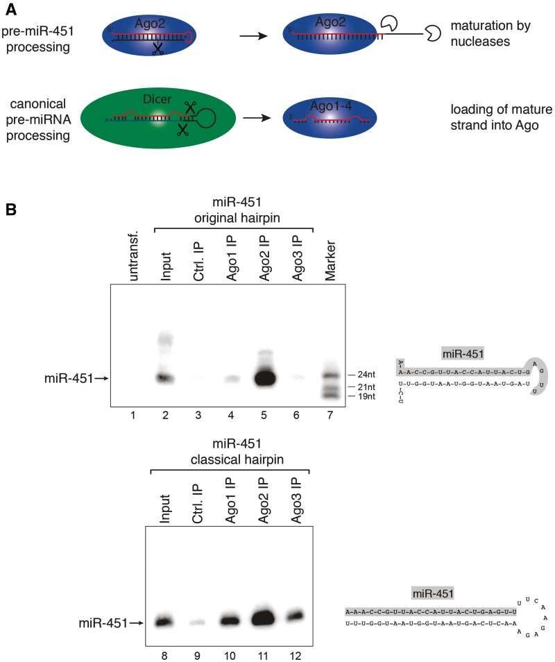
Figure 1. miR-451 associates with Ago2 only. (A) Schematic representation of Ago2-mediated miR-451 processing (upper part) and canonical pre-miRNA processing by Dicer (lower part). (B) Constructs expressing wildtype (wt) pre-miR-451 (upper panel) or miR-451 embedded into a classical Dicer-dependent hairpin (lower panel) were transfected into HEK 293 cells. Ago1 (Lanes 4 and 10), Ago2 (Lanes 5 and 11) and Ago3 (Lanes 6 and 12) were immunoprecipitated using specific monoclonal antibodies. Co-immunoprecipitated miRNAs were analyzed by northern blotting against miR-451. Lanes 3 and 9 show control IPs, Lanes 2 and 8 show input samples and Lane 7 a size marker. (C) RNA was extracted from the indicated mouse tissues and miR-451 was detected by northern blotting. (D) Endogenous mAgo1 (Lanes 4 and 8) or mAgo2 (Lanes 5 and 9) were immunoprecipitated from spleen or liver and co-immunoprecipitated miR-451 (upper panel) or miR-19b (lower panel) was analyzed by northern blotting. Lanes 3 and 7 show control IPs, Lanes 2 and 6 input samples and Lanes 1 and 10 size markers.
Figure 1. miR-451 associates with Ago2 only. (A) Schematic representation of Ago2-mediated miR-451 processing (upper part) and canonical pre-miRNA processing by Dicer (lower part). (B) Constructs expressing wildtype (wt) pre-miR-451 (upper panel) or miR-451 embedded into a classical Dicer-dependent hairpin (lower panel) were transfected into HEK 293 cells. Ago1 (Lanes 4 and 10), Ago2 (Lanes 5 and 11) and Ago3 (Lanes 6 and 12) were immunoprecipitated using specific monoclonal antibodies. Co-immunoprecipitated miRNAs were analyzed by northern blotting against miR-451. Lanes 3 and 9 show control IPs, Lanes 2 and 8 show input samples and Lane 7 a size marker. (C) RNA was extracted from the indicated mouse tissues and miR-451 was detected by northern blotting. (D) Endogenous mAgo1 (Lanes 4 and 8) or mAgo2 (Lanes 5 and 9) were immunoprecipitated from spleen or liver and co-immunoprecipitated miR-451 (upper panel) or miR-19b (lower panel) was analyzed by northern blotting. Lanes 3 and 7 show control IPs, Lanes 2 and 6 input samples and Lanes 1 and 10 size markers.CC BY-NC · PubMed Central
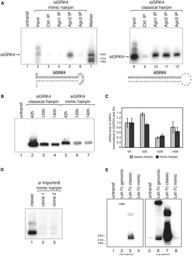
Figure 2. Pre-miR-451-based shRNA expression. (A) An siRNA against GRK4 was either embedded into a pre-miR-451-like hairpin (left part) or a classical Dicer-dependent hairpin (right part). Both constructs were transfected into HEK 293 cells and Ago1 (Lanes 4 and 10), Ago2 (Lanes 5 and 11) or Ago3 (Lanes 6 and 12) was immunoprecipitated from the lysates. GRK4 siRNA incorporation was analyzed by northern blotting. Lanes 3 and 9 show control IPs, Lanes 2 and 8 input samples and Lane 7 a size marker. (B) Both GRK4 shRNAs described in (A) were transfected into HEK 293 cells. Total RNA was extracted at the indicated time points and GRK4 siRNA production was analyzed by northern blotting using a probe complementary to the guide strand of the GRK4 siRNA. (C) Samples described in (B) were analyzed by qPCR for knockdown of GRK4 at the indicated time points. (D) siRNAs against Importin 8 were embedded into a pre-miR-451-like hairpin (two different clones were tested, Lanes 2 and 3) or a classical Dicer-like shRNA (Lane 1). The constructs were transfected into HEK 293 cells and siRNA production was analyzed by northern blotting. (E) Relative expression levels of let-7c were tested by transfecting the genomically encoded (‘genomic’) (Lanes 2 and 6), the classical Dicer-like shRNA (‘classic’) (Lanes 3 and 7) or a pre-451-like hairpin (‘mimic’) (Lanes 4 and 8). As a background control, RNA from untransfected HEK 293 was used (Lanes 1 and 5).
Figure 2. Pre-miR-451-based shRNA expression. (A) An siRNA against GRK4 was either embedded into a pre-miR-451-like hairpin (left part) or a classical Dicer-dependent hairpin (right part). Both constructs were transfected into HEK 293 cells and Ago1 (Lanes 4 and 10), Ago2 (Lanes 5 and 11) or Ago3 (Lanes 6 and 12) was immunoprecipitated from the lysates. GRK4 siRNA incorporation was analyzed by northern blotting. Lanes 3 and 9 show control IPs, Lanes 2 and 8 input samples and Lane 7 a size marker. (B) Both GRK4 shRNAs described in (A) were transfected into HEK 293 cells. Total RNA was extracted at the indicated time points and GRK4 siRNA production was analyzed by northern blotting using a probe complementary to the guide strand of the GRK4 siRNA. (C) Samples described in (B) were analyzed by qPCR for knockdown of GRK4 at the indicated time points. (D) siRNAs against Importin 8 were embedded into a pre-miR-451-like hairpin (two different clones were tested, Lanes 2 and 3) or a classical Dicer-like shRNA (Lane 1). The constructs were transfected into HEK 293 cells and siRNA production was analyzed by northern blotting. (E) Relative expression levels of let-7c were tested by transfecting the genomically encoded (‘genomic’) (Lanes 2 and 6), the classical Dicer-like shRNA (‘classic’) (Lanes 3 and 7) or a pre-451-like hairpin (‘mimic’) (Lanes 4 and 8). As a background control, RNA from untransfected HEK 293 was used (Lanes 1 and 5).CC BY-NC · PubMed Central
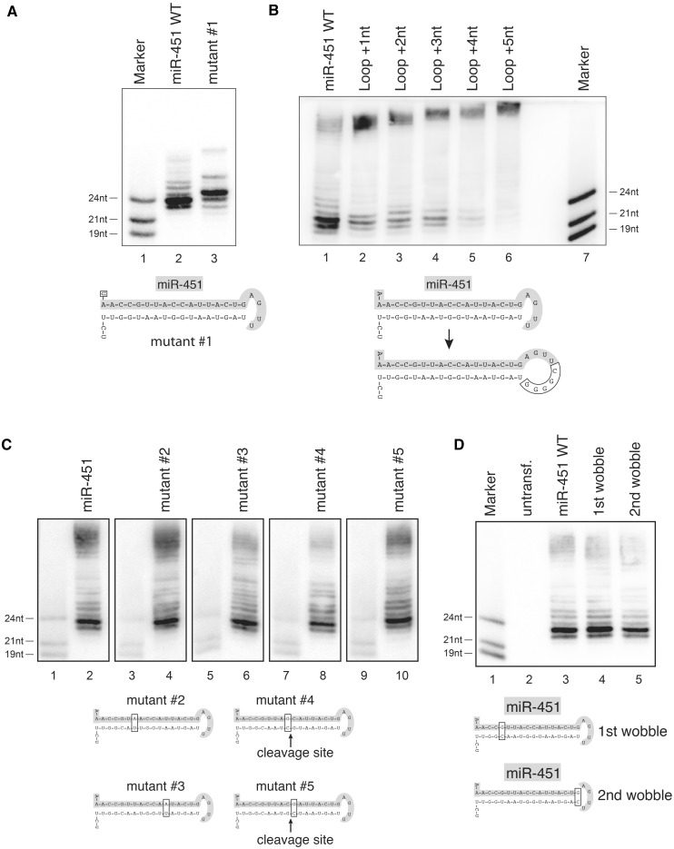
Figure 3. Structure and sequence requirements of miR-451 for Ago2-mediated cleavage. (A) A wt miR-451 construct (Lane 2) or a mutant with a changed 5′ end (Lane 3, A to U mutation, mutant #1) were transfected into HEK 293 cells and processing was analyzed by northern blotting. Lane 1 shows a size marker. (B) Nucleotides were inserted into the loop of pre-miR-451 (Lanes 2 to 6) and the constructs were analyzed as described in (A). (C) Mutants of the wt pre-miR-451 hairpin were constructed to test the sequence specificity of Ago2-mediated processing. Constructs were transfected into HEK 293 cells and analyzed on individual northern blots, since each mutation alters the resulting mature sequence. Equal amounts of size marker were loaded to provide comparability. (D) G/U wobble base pairs at the 5′ end (Lane 4) and near the loop (Lane 5) were mutated to G/C Watson–Crick base pairs and the constructs analyzed as in (A).
Figure 3. Structure and sequence requirements of miR-451 for Ago2-mediated cleavage. (A) A wt miR-451 construct (Lane 2) or a mutant with a changed 5′ end (Lane 3, A to U mutation, mutant #1) were transfected into HEK 293 cells and processing was analyzed by northern blotting. Lane 1 shows a size marker. (B) Nucleotides were inserted into the loop of pre-miR-451 (Lanes 2 to 6) and the constructs were analyzed as described in (A). (C) Mutants of the wt pre-miR-451 hairpin were constructed to test the sequence specificity of Ago2-mediated processing. Constructs were transfected into HEK 293 cells and analyzed on individual northern blots, since each mutation alters the resulting mature sequence. Equal amounts of size marker were loaded to provide comparability. (D) G/U wobble base pairs at the 5′ end (Lane 4) and near the loop (Lane 5) were mutated to G/C Watson–Crick base pairs and the constructs analyzed as in (A).CC BY-NC · PubMed Central
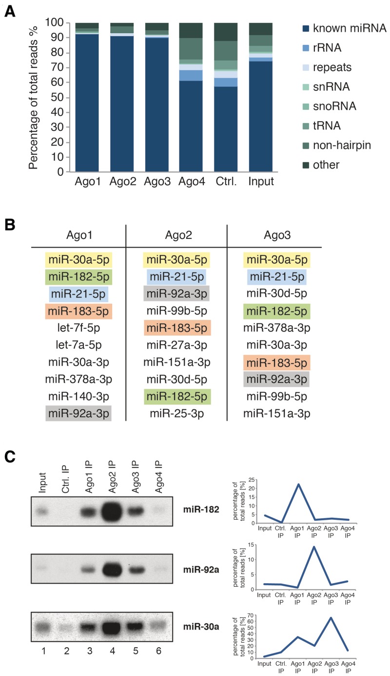
Figure 4. Deep-sequencing analysis of small RNAs associated with endogenous Ago proteins. (A) Composition of the small RNA libraries from the input sample and immunoprecipitated Ago1, Ago2, Ago3, Ago4 and control (an unrelated antibody against the complement system protein RmC from rat). (B) Table of the 10 most abundant miRNAs associated with Ago1, Ago2 or Ago3. The colors indicate the same miRNA. (C) Comparison of the sorting profile from the deep-sequencing data (right side) for miR-182 (top panel), miR-92a (middle panel, right) and miR-30a (lower panel) with profiles resulting from northern blotting (left side).
Figure 4. Deep-sequencing analysis of small RNAs associated with endogenous Ago proteins. (A) Composition of the small RNA libraries from the input sample and immunoprecipitated Ago1, Ago2, Ago3, Ago4 and control (an unrelated antibody against the complement system protein RmC from rat). (B) Table of the 10 most abundant miRNAs associated with Ago1, Ago2 or Ago3. The colors indicate the same miRNA. (C) Comparison of the sorting profile from the deep-sequencing data (right side) for miR-182 (top panel), miR-92a (middle panel, right) and miR-30a (lower panel) with profiles resulting from northern blotting (left side).CC BY-NC · PubMed Central
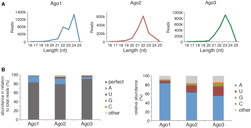
Figure 5. Ago proteins show different global small RNA binding patterns and nontemplated 3′ extension profiles. (A) Length distribution patterns of Ago1 (left), Ago2 (middle) and Ago3 (right). (B) Left side: Ago1, Ago2 and Ago3 associated miRNAs were analyzed for nontemplated 3′ extensions, values were calculated in relation to all miRNA reads of the respective library. Perfect: no nontemplated 3′ extensions; A, U, G, C: amount of single nucleotide 3′ extensions; other: sum of all other 3′ extensions (≥2 nt). Right side: each single nontemplated 3′ extension was set in relation to the total number of nontemplated 3′ extensions in the respective library (Ago1, Ago2 or Ago3).
Figure 5. Ago proteins show different global small RNA binding patterns and nontemplated 3′ extension profiles. (A) Length distribution patterns of Ago1 (left), Ago2 (middle) and Ago3 (right). (B) Left side: Ago1, Ago2 and Ago3 associated miRNAs were analyzed for nontemplated 3′ extensions, values were calculated in relation to all miRNA reads of the respective library. Perfect: no nontemplated 3′ extensions; A, U, G, C: amount of single nucleotide 3′ extensions; other: sum of all other 3′ extensions (≥2 nt). Right side: each single nontemplated 3′ extension was set in relation to the total number of nontemplated 3′ extensions in the respective library (Ago1, Ago2 or Ago3).CC BY-NC · PubMed Central
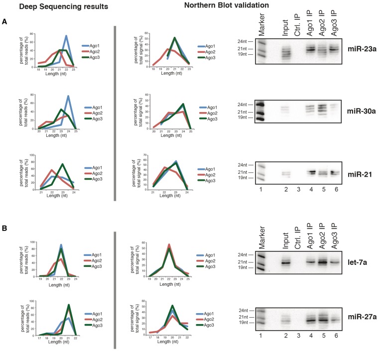
Figure 6. Ago proteins show distinct length profiles for individual miRNAs. The length pattern of individual miRNAs was extracted from the deep-sequencing data (left side) and compared against the length profiles seen by northern blotting (right side). For this, northern blotting signals for each miRNA band were quantified; the total signal from each lane/each Ago was set to 100%. The length of a miRNA in northern blotting was assessed by comparison with the size marker. miRNA length profiles were separated into: (A) miRNA length profiles showing divergent Ago association and (B) miRNA length profiles showing matching Ago1–3 association.
Figure 6. Ago proteins show distinct length profiles for individual miRNAs. The length pattern of individual miRNAs was extracted from the deep-sequencing data (left side) and compared against the length profiles seen by northern blotting (right side). For this, northern blotting signals for each miRNA band were quantified; the total signal from each lane/each Ago was set to 100%. The length of a miRNA in northern blotting was assessed by comparison with the size marker. miRNA length profiles were separated into: (A) miRNA length profiles showing divergent Ago association and (B) miRNA length profiles showing matching Ago1–3 association.CC BY-NC · PubMed Central

Verification

Claims the paper makes

The claims and statistics of this paper haven’t been checked yet.

Run the check — free, no account needed. It reads the open-access text and checks the sources it cites.

A few free checks a day · papers already verified open instantly · results are public

Claims other papers make about this one

Each is checked against this paper’s own text: does it say what the citing paper says it does?

Self-check

Percentages, sums and reported differences re-computed from the paper’s own numbers.

The self-check needs the paper’s full text.

Citations

Cites

Research works printed but not linked: 50

  1. Bartel, 2009, Cell, 136, 215, 10.1016/j.cell.2009.01.002 · DOI · Paper page
  2. Carthew, 2009, Cell, 136, 642, 10.1016/j.cell.2009.01.035 · DOI
  3. Kim, 2009, Nature reviews. Molecular cell biology, 10, 126, 10.1038/nrm2632 · DOI · Paper page
  4. Siomi, 2009, Nature; Physical Science (London), 457, 396, 10.1038/nature07754 · DOI
  5. Peters, 2007, Molecular cell, 26, 611, 10.1016/j.molcel.2007.05.001 · DOI · Paper page
  6. Ender, 2010, Journal of Cell Science, 123, 1819, 10.1242/jcs.055210 · DOI
  7. Hutvagner, 2008, Nature reviews. Molecular cell biology, 9, 22, 10.1038/nrm2321 · DOI
  8. Jinek, 2009, Nature; Physical Science (London), 457, 405, 10.1038/nature07755 · DOI
  9. Meister, 2004, Molecular cell, 15, 185, 10.1016/j.molcel.2004.07.007 · DOI · Paper page
  10. 2004, Science, 305, 1437, 10.1126/science.1102513 · DOI · Paper page
  11. Matranga, 2005, Cell, 123, 607, 10.1016/j.cell.2005.08.044 · DOI · Paper page
  12. Rand, 2005, Cell, 123, 621, 10.1016/j.cell.2005.10.020 · DOI
  13. Leuschner, 2006, EMBO Reports, 7, 314, 10.1038/sj.embor.7400637 · DOI
  14. Ruby, 2007, Nature; Physical Science (London), 448, 83, 10.1038/nature05983 · DOI · Paper page
  15. Okamura, 2007, Cell, 130, 89, 10.1016/j.cell.2007.06.028 · DOI
  16. Berezikov, 2007, Molecular cell, 28, 328, 10.1016/j.molcel.2007.09.028 · DOI
  17. Ender, 2008, Molecular cell, 32, 519, 10.1016/j.molcel.2008.10.017 · DOI
  18. 2009, RNA, 15, 1233, 10.1261/rna.1528909 · DOI
  19. Scott, 2009, 5, e1000507, 10.1371/journal.pcbi.1000507 · DOI
  20. Saraiya, 2008, 4, e1000224, 10.1371/journal.ppat.1000224 · DOI
  21. 2010, Science, 328, 1694, 10.1126/science.1190809 · DOI
  22. Cheloufi, 2010, Nature; Physical Science (London), 465, 584, 10.1038/nature09092 · DOI · Paper page
  23. 2008, REV GENET, 9, 102, 10.1038/nrg2290 · DOI
  24. Meister, 2004, Nature; Physical Science (London), 431, 343, 10.1038/nature02873 · DOI
  25. Dorsett, 2004, Nature reviews. Drug discovery, 3, 318, 10.1038/nrd1345 · DOI
  26. Forstemann, 2007, Cell, 130, 287, 10.1016/j.cell.2007.05.056 · DOI
  27. Tomari, 2007, Cell, 130, 299, 10.1016/j.cell.2007.05.057 · DOI
  28. 2011, RNA, 17, 737, 10.1261/rna.2348111 · DOI
  29. Wu, 2008, Current biology : CB, 18, 1327, 10.1016/j.cub.2008.07.072 · DOI
  30. Svoboda, 2007, Current opinion in molecular therapeutics, 9, 248
  31. Beitzinger, 2007, 4, 76, 10.4161/rna.4.2.4640 · DOI · Paper page
  32. 2008, RNA, 14, 1244, 10.1261/rna.973808 · DOI
  33. Weinmann, 2009, Cell, 136, 496, 10.1016/j.cell.2008.12.023 · DOI
  34. 2010, Journal of Virology, 84, 10266, 10.1128/JVI.01119-10 · DOI
  35. Pall, 2008, 3, 1077, 10.1038/nprot.2008.67 · DOI
  36. Lin, 2005, Nucleic Acids Research, 33, 4527, 10.1093/nar/gki762 · DOI
  37. 2010, PNAS, 107, 15163, 10.1073/pnas.1006432107 · DOI
  38. Frank, 2010, Nature; Physical Science (London), 465, 818, 10.1038/nature09039 · DOI · Paper page
  39. Burroughs, 2011, 8, 158, 10.4161/rna.8.1.14300 · DOI
  40. 2008, Genome Research, 18, 1787, 10.1101/gr.077578.108 · DOI
  41. Jackson, 2010, Nature reviews. Drug discovery, 9, 57, 10.1038/nrd3010 · DOI
  42. Eiring, 2010, Cell, 140, 652, 10.1016/j.cell.2010.01.007 · DOI
  43. 2011, RNA, 17, 1697, 10.1261/rna.2799511 · DOI · Paper page
  44. 2012, Nucleic Acids Research, 40, 6808, 10.1093/nar/gks293 · DOI
  45. Heo, 2008, Molecular cell, 32, 276, 10.1016/j.molcel.2008.09.014 · DOI
  46. Heo, 2009, Cell, 138, 696, 10.1016/j.cell.2009.08.002 · DOI · Paper page
  47. Hagan, 2009, Nature structural & molecular biology, 16, 1021, 10.1038/nsmb.1676 · DOI · Paper page
  48. 2011, Genome Research, 21, 1450, 10.1101/gr.118059.110 · DOI
  49. 2010, Nucleic Acids Research, 38, e34, 10.1093/nar/gkp1127 · DOI
  50. 2010, Genome Research, 20, 1398, 10.1101/gr.106054.110 · DOI

Cited by

Sorted by citations

See all 195

Metadata

Authors
Dueck, A., Ziegler, C., Eichner, A., Berezikov, E., & Meister, G.
Venue
Nucleic Acids Research
Published
2012-07-25
Volume
40
Issue
19
Pages
9850-9862
ISSN
  • 0305-1048
  • 1362-4962
Cited by
195
References
3
Language
en

About these checks

Registry, retraction, Cites and statistics results are computed, not judged, with one exception: a printed reference that no index lists is read once by a model before it counts as a citation error. Claims are extracted by a model and kept only when their quote appears verbatim in the text; each cited source is then checked, with a model’s help, for whether it says what the claim relies on. Those verdicts can be wrong — read the claim’s quote and the cited source before relying on one.

Statistics and arithmetic are recomputed, not judged.

Confidence bands are shown only for fields calibrated on our annotated test set. None is calibrated yet, so no band is shown.

What counts as a claim ›How we measure confidence ›How we check this ›

Are you an author, or do you see a problem with this page? Request a review