

The DOI matches its Crossref record; 65 of the 65 references it cites were found in a registry or an index.
SARS-CoV-2 escape in vitro from a highly neutralizing COVID-19 convalescent plasma
Preprint — published as 10.1073/pnas.2103154118
Abstract
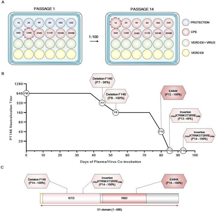
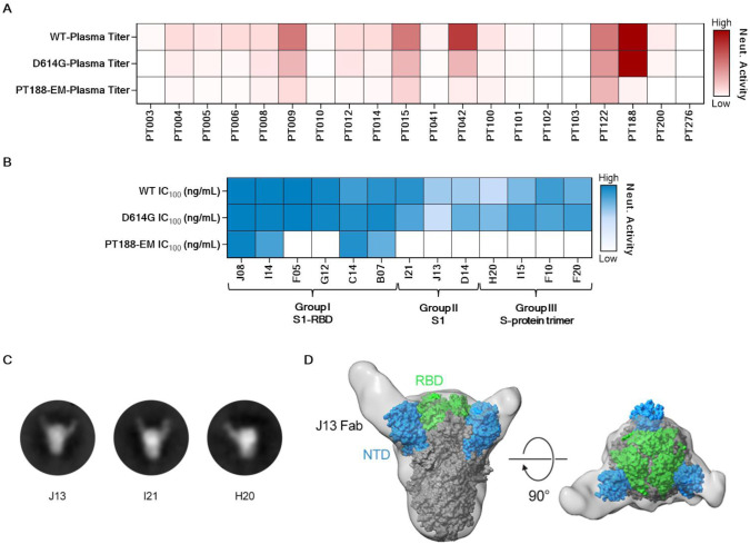
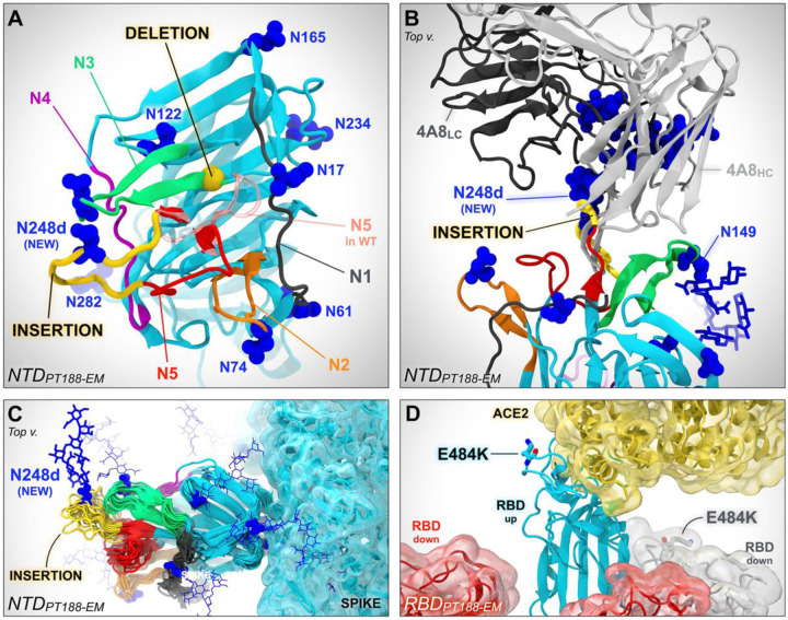
To investigate the evolution of SARS-CoV-2 in the immune population, we co-incubated authentic virus with a highly neutralizing plasma from a COVID-19 convalescent patient. The plasma fully neutralized the virus for 7 passages, but after 45 days, the deletion of F140 in the spike N-terminal domain (NTD) N3 loop led to partial breakthrough. At day 73, an E484K substitution in the receptor-binding domain (RBD) occurred, followed at day 80 by an insertion in the NTD N5 loop containing a new glycan sequon, which generated a variant completely resistant to plasma neutralization. Computational modeling predicts that the deletion and insertion in loops N3 and N5 prevent binding of neutralizing antibodies. The recent emergence in the United Kingdom and South Africa of natural variants with similar changes suggests that SARS-CoV-2 has the potential to escape an effective immune response and that vaccines and antibodies able to control emerging variants should be developed. One Sentence Summary: Three mutations allowed SARS-CoV-2 to evade the polyclonal antibody response of a highly neutralizing COVID-19 convalescent plasma.
Overview
The claims and statistics of this paper haven’t been checked yet.
Run the check — free, no account needed. It reads the open-access text and checks the sources it cites.
Integrity signals
Registry
Cites
Statistics
Figures
Verification
Claims the paper makes
The claims and statistics of this paper haven’t been checked yet.
Run the check — free, no account needed. It reads the open-access text and checks the sources it cites.
Claims other papers make about this one
Each is checked against this paper’s own text: does it say what the citing paper says it does?
In the citing paper
“As seen in natural infection, a majority of the antibodies tested (9/17) were at least ten-fold less effective against pseudotyped viruses carrying the E484K mutation7,12,29.”
From mRNA vaccine-elicited antibodies to SARS-CoV-2 and circulating variants (2021) · cites this paper as [29]
In the citing paper
“Human neutralizing monoclonal antibodies to the SARS-CoV-2 RBD can be categorized as belonging to 4 different classes based on their target regions on the RBD29.”
From mRNA vaccine-elicited antibodies to SARS-CoV-2 and circulating variants (2021) · cites this paper as [29]
In the citing paper
“Fab-S complexes exhibited multiple RBD-binding orientations recognizing either ‘up’/‘down’ (Fig 4a–j) or solely ‘up’ (Fig 4k–n) RBD conformations, consistent with structurally defined antibody classes from natural infection (Fig 4o)29.”
From mRNA vaccine-elicited antibodies to SARS-CoV-2 and circulating variants (2021) · cites this paper as [29]
In the citing paper
“A475V is resistant to class 1 antibodies (structurally defined as described29); E484K and Q493R are resistant to class 2 antibodies7,8,12,13,29,30,32,33; while R346S, N439K, and N440K are resistant to class 3 antibodies7,8,12,13,29,33.”
From mRNA vaccine-elicited antibodies to SARS-CoV-2 and circulating variants (2021) · cites this paper as [29]
In the citing paper
“These antibodies target epitopes that overlap or are closely associated with RBD residues K417, E484 and N501.”
From mRNA vaccine-elicited antibodies to SARS-CoV-2 and circulating variants (2021) · cites this paper as [29]
In the citing paper
“SARS-CoV-2 vaccines have recently been licensed that target the spike (S) protein, either using mRNA or adenovirus vector technology with protection rates ranging from 62 to 95%9–11.”
From Sensitivity of SARS-CoV-2 B.1.1.7 to mRNA vaccine-elicited antibodies (2021) · cites this paper as [9]
Self-check
Percentages, sums and reported differences re-computed from the paper’s own numbers.
The self-check needs the paper’s full text.
Citations
Cites
VMD: visual molecular dynamics
Humphrey, W., Dalke, A., & Schulten, K. (1996) · Journal of Molecular Graphics · doi:10.1016/0263-7855(96)00018-5 · Cited by 66,952
Particle mesh Ewald: An N⋅log(N) method for Ewald sums in large systems
Darden, T., York, D., & Pedersen, L. (1993) · The Journal of Chemical Physics · doi:10.1063/1.464397 · Cited by 29,638
Ryckaert, J.-P., Ciccotti, G., & Berendsen, H. J. C. (1977) · Journal of Computational Physics · doi:10.1016/0021-9991(77)90098-5 · Cited by 21,430
Comparative Protein Modelling by Satisfaction of Spatial Restraints
Šali, A., & Blundell, T. L. (1993) · Journal of Molecular Biology · doi:10.1006/jmbi.1993.1626 · Cited by 12,977 · cited 1×
“The S1 subunit presents two highly immunogenic domains, the N-terminal domain (NTD) and the RBD, which are the major targets of polyclonal and monoclonal neutralizing antibodies (4, 9, 10).”
cryoSPARC: algorithms for rapid unsupervised cryo-EM structure determination
Punjani, A., Rubinstein, J. L., Fleet, D. J., & Brubaker, M. A. (2017) · Nature Methods · doi:10.1038/nmeth.4169 · Cited by 11,626
Structure, function and antigenicity of the SARS-CoV-2 spike glycoprotein
Walls, A. C., Park, Y.-J., Tortorici, M. A., Wall, A., McGuire, A. T., & Veesler, D. (2020) · doi:10.1101/2020.02.19.956581 · Cited by 8,586 · cited 1×
“The main target of vaccines and mAbs is the SARS-CoV-2 spike protein (S-protein), a large class I trimeric fusion protein which plays a key role in viral pathogenesis (3, 5, 6).”
CHARMM36m: an improved force field for folded and intrinsically disordered proteins
Huang, J., Rauscher, S., Nawrocki, G., Ran, T., Feig, M., de Groot, B. L., Grubmüller, H., & MacKerell, A. D. (2016) · Nature Methods · doi:10.1038/nmeth.4067 · Cited by 7,334
Structure of the SARS-CoV-2 spike receptor-binding domain bound to the ACE2 receptor
Lan, J., Ge, J., Yu, J., Shan, S., Zhou, H., Fan, S., Zhang, Q., Shi, X., Wang, Q., Zhang, L., et al. (2020) · Nature · doi:10.1038/s41586-020-2180-5 · Cited by 5,983
Constant pressure molecular dynamics algorithms
Martyna, G. J., Tobias, D. J., & Klein, M. L. (1994) · The Journal of Chemical Physics · doi:10.1063/1.467468 · Cited by 5,845 · cited 1×
“clinical settings where an immunocompromised COVID-19 patient, after 154 days of infection, presented different variants of the virus including the E484K substitution (25).”
Structural basis for the recognition of SARS-CoV-2 by full-length human ACE2
Yan, R., Zhang, Y., Li, Y., Xia, L., Guo, Y., & Zhou, Q. (2020) · Science · doi:10.1126/science.abb2762 · Cited by 5,036 · cited 1×
“The S1 subunit presents two highly immunogenic domains, the N-terminal domain (NTD) and the RBD, which are the major targets of polyclonal and monoclonal neutralizing antibodies (4, 9, 10).”
CHARMM36 all-atom additive protein force field: Validation based on comparison to NMR data
Huang, J., & MacKerell, A. D. (2013) · Journal of Computational Chemistry · doi:10.1002/jcc.23354 · Cited by 4,537
PROPKA3: Consistent Treatment of Internal and Surface Residues in Empirical p K a Predictions
Olsson, M. H. M., Søndergaard, C. R., Rostkowski, M., & Jensen, J. H. (2011) · Journal of Chemical Theory and Computation · doi:10.1021/ct100578z · Cited by 4,425
Constant pressure molecular dynamics simulation: The Langevin piston method
Feller, S., Zhang, Y., Pastor, R., & Brooks, B. (1995) · The Journal of Chemical Physics · doi:10.1063/1.470648 · Cited by 4,271
An mRNA Vaccine against SARS-CoV-2 — Preliminary Report
Jackson, L. A., Anderson, E. J., Rouphael, N. G., Roberts, P. C., Makhene, M., Coler, R. N., McCullough, M. P., Chappell, J. D., Denison, M. R., Stevens, L. J., et al. (2020) · New England Journal of Medicine · doi:10.1056/nejmoa2022483 · Cited by 3,237
Scalable molecular dynamics on CPU and GPU architectures with NAMD
Phillips, J. C., Hardy, D. J., Maia, J. D. C., Stone, J. E., Ribeiro, J. V., Bernardi, R. C., Buch, R., Fiorin, G., Hénin, J., Jiang, W., et al. (2020) · The Journal of Chemical Physics · doi:10.1063/5.0014475 · Cited by 3,236
GISAID: Global initiative on sharing all influenza data – from vision to reality
Shu, Y., & McCauley, J. (2017) · Eurosurveillance · doi:10.2807/1560-7917.es.2017.22.13.30494 · Cited by 3,039 · cited 1×
“Indeed, S-protein variants have been recently described in the United Kingdom and South Africa (11, 12), and the Global Initiative on Sharing All Influenza Data (GISAID) database, reports more than 1,100 amino acid changes in the S-protein (13, 14).”
Structural and Functional Basis of SARS-CoV-2 Entry by Using Human ACE2
Wang, Q., Zhang, Y., Wu, L., Niu, S., Song, C., Zhang, Z., Lu, G., Qiao, C., Hu, Y., Yuen, K.-Y., et al. (2020) · Cell · doi:10.1016/j.cell.2020.03.045 · Cited by 2,892 · cited 1×
“The S1 subunit presents two highly immunogenic domains, the N-terminal domain (NTD) and the RBD, which are the major targets of polyclonal and monoclonal neutralizing antibodies (4, 9, 10).”
Tegally, H., Wilkinson, E., Giovanetti, M., Iranzadeh, A., Fonseca, V., Giandhari, J., Doolabh, D., Pillay, S., San, E. J., Msomi, N., et al. (2020) · doi:10.1101/2020.12.21.20248640 · Cited by 2,535 · cited 3×
“The recent isolation of SARS-CoV-2 variants in the United Kingdom and South Africa with deletions in or near the NTD loops shows that what we describe here can occur in the human population.”
Folegatti, P. M., Ewer, K. J., Aley, P. K., Angus, B., Becker, S., Belij-Rammerstorfer, S., Bellamy, D., Bibi, S., Bittaye, M., Clutterbuck, E. A., et al. (2020) · The Lancet · doi:10.1016/s0140-6736(20)31604-4 · Cited by 2,419
Brownian dynamics with hydrodynamic interactions
Ermak, D., & McCammon, J. (1978) · The Journal of Chemical Physics · doi:10.1063/1.436761 · Cited by 2,238 · cited 1×
“These results are remarkable because while escape mutants can be easily isolated when viruses are incubated with single monoclonal antibodies, usually a combination of two mAbs is sufficient to eliminate the evolution of escape variants and because SARS-CoV-2 shows a very low estimated evolutionary rate of mutation as…”
SARS-CoV-2 vaccines in development
Krammer, F. (2020) · Nature · doi:10.1038/s41586-020-2798-3 · Cited by 2,098 · cited 1×
“Thanks to an incredible scientific and financial effort, several prophylactic and therapeutic tools, such as vaccines and monoclonal antibodies (mAbs), have been developed in less than one year to combat this pandemic (1-4).”
Data, disease and diplomacy: GISAID's innovative contribution to global health
Elbe, S., & Buckland‐Merrett, G. (2017) · Global Challenges · doi:10.1002/gch2.1018 · Cited by 1,928 · cited 2×
“limited and the mutation E484K that we found is one of the most frequent mutations to escape monoclonal antibodies and among the most common RBD mutations described in experimental settings as well as in natural isolates posted in the GISAD database (13, 26, 27).”
Site-specific glycan analysis of the SARS-CoV-2 spike
Watanabe, Y., Allen, J. D., Wrapp, D., McLellan, J. S., & Crispin, M. (2020) · Science · doi:10.1126/science.abb9983 · Cited by 1,538
Self-citationA neutralizing human antibody binds to the N-terminal domain of the Spike protein of SARS-CoV-2
Chi, X., Yan, R., Zhang, J., Zhang, G., Zhang, Y., Hao, M., Zhang, Z., Fan, P., Dong, Y., Yang, Y., et al. (2020) · Science · doi:10.1126/science.abc6952 · Cited by 1,385 · cited 1×
“The SARS-CoV-2 S-protein is composed of two subunits: S1, which contains the receptor-binding domain (RBD) responsible for the interaction with receptors on the host cells, and S2, which mediates membrane fusion and viral entry (7, 8).”
Structure-based design of prefusion-stabilized SARS-CoV-2 spikes
Hsieh, C.-L., Goldsmith, J. A., Schaub, J. M., DiVenere, A. M., Kuo, H.-C., Javanmardi, K., Le, K. C., Wrapp, D., Lee, A. G., Liu, Y., et al. (2020) · Science · doi:10.1126/science.abd0826 · Cited by 1,303
Self-citationBaum, A., Fulton, B. O., Wloga, E., Copin, R., Pascal, K. E., Russo, V., Giordano, S., Lanza, K., Negron, N., Ni, M., et al. (2020) · Science · doi:10.1126/science.abd0831 · Cited by 1,253 · cited 1×
“…incubated with single monoclonal antibodies, usually a combination of two mAbs is sufficient to eliminate the evolution of escape variants and because SARS-CoV-2 shows a very low estimated evolutionary rate of mutation as this virus encodes a proofreading exoribonuclease machinery (21, 22)(23, 24).”
Persistence and Evolution of SARS-CoV-2 in an Immunocompromised Host
Choi, B., Choudhary, M. C., Regan, J., Sparks, J. A., Padera, R. F., Qiu, X., Solomon, I. H., Kuo, H.-H., Boucau, J., Bowman, K., et al. (2020) · New England Journal of Medicine · doi:10.1056/nejmc2031364 · Cited by 1,246 · cited 6×
“…RBD, the possibility to escape is limited and the mutation E484K that we found is one of the most frequent mutations to escape monoclonal antibodies and among the most common RBD mutations described in experimental settings as well as in natural isolates posted in the GISAD database (13, 26, 27).”
InfluentialRogers, T. F., Zhao, F., Huang, D., Beutler, N., Burns, A., He, W.-T., Limbo, O., Smith, C., Song, G., Woehl, J., et al. (2020) · Science · doi:10.1126/science.abc7520 · Cited by 1,230 · cited 1×
“The SARS-CoV-2 S-protein is composed of two subunits: S1, which contains the receptor-binding domain (RBD) responsible for the interaction with receptors on the host cells, and S2, which mediates membrane fusion and viral entry (7, 8).”
SARS-CoV-2 Infection Depends on Cellular Heparan Sulfate and ACE2
Clausen, T. M., Sandoval, D. R., Spliid, C. B., Pihl, J., Perrett, H. R., Painter, C. D., Narayanan, A., Majowicz, S. A., Kwong, E. M., McVicar, R. N., et al. (2020) · Cell · doi:10.1016/j.cell.2020.09.033 · Cited by 1,197
Structures and distributions of SARS-CoV-2 spike proteins on intact virions
Ke, Z., Otón, J., Qu, K., Cortese, M., Žíla, V., McKeane, L., Nakane, T., Zivanov, J., Neufeldt, C. J., Cerikan, B., et al. (2020) · Nature · doi:10.1038/s41586-020-2665-2 · Cited by 1,140 · cited 1×
“The main target of vaccines and mAbs is the SARS-CoV-2 spike protein (S-protein), a large class I trimeric fusion protein which plays a key role in viral pathogenesis (3, 5, 6).”
Potently neutralizing and protective human antibodies against SARS-CoV-2
Zost, S. J., Gilchuk, P., Case, J. B., Binshtein, E., Chen, R. E., Nkolola, J. P., Schäfer, A., Reidy, J. X., Trivette, A., Nargi, R. S., et al. (2020) · Nature · doi:10.1038/s41586-020-2548-6 · Cited by 1,041
Greaney, A. J., Starr, T. N., Gilchuk, P., Zost, S. J., Binshtein, E., Loes, A. N., Hilton, S. K., Huddleston, J., Eguia, R., Crawford, K. H. D., et al. (2021) · Cell Host & Microbe · doi:10.1016/j.chom.2020.11.007 · Cited by 1,016
Greaney, A. J., Starr, T. N., Gilchuk, P., Zost, S. J., Binshtein, E., Loes, A. N., Hilton, S. K., Huddleston, J., Eguia, R. T., Crawford, K. H. D., et al. (2020) · bioRxiv · doi:10.1101/2020.09.10.292078 · Cited by 948 · cited 1×
“…RBD, the possibility to escape is limited and the mutation E484K that we found is one of the most frequent mutations to escape monoclonal antibodies and among the most common RBD mutations described in experimental settings as well as in natural isolates posted in the GISAD database (13, 26, 27).”
Beyond Shielding: The Roles of Glycans in the SARS-CoV-2 Spike Protein
Casalino, L., Gaieb, Z., Goldsmith, J. A., Hjorth, C. K., Dommer, A. C., Harbison, A. M., Fogarty, C. A., de Barros, E. P., Taylor, B. C., McLellan, J. S., et al. (2020) · ACS Central Science · doi:10.1021/acscentsci.0c01056 · Cited by 925
Self-citationcisTEM, user-friendly software for single-particle image processing
Grant, T. S., Rohou, A. L., & Grigorieff, N. (2018) · eLife · doi:10.7554/elife.35383 · Cited by 879
Neutralizing Antibodies against SARS-CoV-2 and Other Human Coronaviruses
Jiang, S., Hillyer, C., & Du, L. (2020) · Trends in Immunology · doi:10.1016/j.it.2020.03.007 · Cited by 847 · cited 1×
“Thanks to an incredible scientific and financial effort, several prophylactic and therapeutic tools, such as vaccines and monoclonal antibodies (mAbs), have been developed in less than one year to combat this pandemic (1-4).”
Structural Basis for RNA Replication by the SARS-CoV-2 Polymerase
Wang, Q., Wu, J., Wang, H., Gao, Y., Liu, Q., Mu, A., Ji, W., Yan, L., Zhu, Y., Zhu, C., et al. (2020) · Cell · doi:10.1016/j.cell.2020.05.034 · Cited by 773
cisTEM: User-friendly software for single-particle image processing
Grant, T., Rohou, A., & Grigorieff, N. (2017) · bioRxiv · doi:10.1101/257618 · Cited by 735
CHARMM Additive All-Atom Force Field for Glycosidic Linkages between Hexopyranoses
Guvench, O., Hatcher, E., Venable, R. M., Pastor, R. W., & MacKerell, A. D. (2009) · Journal of Chemical Theory and Computation · doi:10.1021/ct900242e · Cited by 667
Structure-based Design of Prefusion-stabilized SARS-CoV-2 SpikesPreprint
Hsieh, C., Goldsmith, J., Schaub, J. M., et al. (2020) · bioRxiv · doi:10.1101/2020.05.30.125484 · Cited by 588
Self-citationA systematic review of SARS-CoV-2 vaccine candidates
Dong, Y., Dai, T., Wei, Y., Zhang, L., Zheng, M., & Zhou, F. (2020) · Signal Transduction and Targeted Therapy · doi:10.1038/s41392-020-00352-y · Cited by 555
REGN-COV2 antibodies prevent and treat SARS-CoV-2 infection in rhesus macaques and hamsters
Baum, A., Ajithdoss, D., Copin, R., Zhou, A., Lanza, K., Negron, N., Ni, M., Wei, Y., Mohammadi, K., Musser, B., et al. (2020) · Science · doi:10.1126/science.abe2402 · Cited by 517 · cited 1×
“…RBD, the possibility to escape is limited and the mutation E484K that we found is one of the most frequent mutations to escape monoclonal antibodies and among the most common RBD mutations described in experimental settings as well as in natural isolates posted in the GISAD database (13, 26, 27).”
Zhang, M. (2004) · Glycobiology · doi:10.1093/glycob/cwh106 · Cited by 482 · cited 1×
“The introduction of a glycan is a well-known immunogenic escape strategy described in influenza (29), HIV-1 and other viruses (30-32), although to our knowledge this finding presents the first patient-derived escape mutant utilizing this mechanism for SARS-CoV-2.”
Deducing the N- and O-glycosylation profile of the spike protein of novel coronavirus SARS-CoV-2
Shajahan, A., Supekar, N. T., Gleinich, A. S., & Azadi, P. (2020) · Glycobiology · doi:10.1093/glycob/cwaa042 · Cited by 478
Coronavirus RNA Proofreading: Molecular Basis and Therapeutic Targeting
Robson, F., Khan, K. S., Le, T. K., Paris, C., Demirbag, S., Barfuss, P., Rocchi, P., & Ng, W. (2020) · Molecular Cell · doi:10.1016/j.molcel.2020.07.027 · Cited by 404 · cited 1×
“…incubated with single monoclonal antibodies, usually a combination of two mAbs is sufficient to eliminate the evolution of escape variants and because SARS-CoV-2 shows a very low estimated evolutionary rate of mutation as this virus encodes a proofreading exoribonuclease machinery (21, 22)(23, 24).”
Structural analysis of full-length SARS-CoV-2 spike protein from an advanced vaccine candidate
Bangaru, S., Ozorowski, G., Turner, H. L., Antanasijevic, A., Huang, D., Wang, X., Torres, J. L., Diedrich, J. K., Tian, J.-H., Portnoff, A. D., et al. (2020) · Science · doi:10.1126/science.abe1502 · Cited by 338 · cited 1×
“The SARS-CoV-2 S-protein is composed of two subunits: S1, which contains the receptor-binding domain (RBD) responsible for the interaction with receptors on the host cells, and S2, which mediates membrane fusion and viral entry (7, 8).”
Bar‐On, Y., Gruell, H., Schoofs, T., Pai, J. A., Nogueira, L., Butler, A. L., Millard, K. G., Lehmann, C., Suárez, I., Oliveira, T. Y., et al. (2018) · Nature Medicine · doi:10.1038/s41591-018-0186-4 · Cited by 267 · cited 1×
“…incubated with single monoclonal antibodies, usually a combination of two mAbs is sufficient to eliminate the evolution of escape variants and because SARS-CoV-2 shows a very low estimated evolutionary rate of mutation as this virus encodes a proofreading exoribonuclease machinery (21, 22)(23, 24).”
SARS-CoV-2 Infection Depends on Cellular Heparan Sulfate and ACE2Preprint
Clausen, T. M., Sandoval, D. R., Spliid, C. B., Pihl, J., Painter, C. D., Thacker, B. E., Glass, C. A., Narayanan, A., Majowicz, S. A., Yang, Z., et al. (2020) · bioRxiv (Cold Spring Harbor Laboratory) · doi:10.1101/2020.07.14.201616 · Cited by 229 · cited 1×
“The SARS-CoV-2 S-protein is composed of two subunits: S1, which contains the receptor-binding domain (RBD) responsible for the interaction with receptors on the host cells, and S2, which mediates membrane fusion and viral entry (7, 8).”
Neutralizing Antibodies against SARS-CoV-2 and Other Human Coronaviruses
Jiang, S., Hillyer, C. D., & Du, L. (2020) · Trends in Immunology · doi:10.1016/j.it.2020.04.008 · Cited by 203
Extremely potent human monoclonal antibodies from COVID-19 convalescent patients
Andreano, E., Nicastri, E., Paciello, I., Pileri, P., Manganaro, N., Piccini, G., Manenti, A., Pantano, E., Kabanova, A., Troisi, M., et al. (2021) · Cell · doi:10.1016/j.cell.2021.02.035 · Cited by 199
Self-citationAI-Driven Multiscale Simulations Illuminate Mechanisms of SARS-CoV-2 Spike DynamicsPreprint
Casalino, L., Dommer, A. C., Gaieb, Z., et al. (2020) · bioRxiv · doi:10.1101/2020.11.19.390187 · Cited by 178
Self-citationManenti, A., Maggetti, M., Casa, E., Martinuzzi, D., Torelli, A., Trombetta, C. M., Marchi, S., & Montomoli, E. (2020) · Journal of Medical Virology · doi:10.1002/jmv.25986 · Cited by 176 · cited 1×
“effort, several prophylactic and therapeutic tools, such as vaccines and monoclonal antibodies (mAbs), have been developed in less than one year to combat this pandemic (1-4).”
Self-citationStructural analysis of full-length SARS-CoV-2 spike protein from an advanced vaccine candidate
Bangaru, S., Ozorowski, G., Turner, H., et al. (2020) · bioRxiv · doi:10.1101/2020.08.06.234674 · Cited by 161
The flexibility of ACE2 in the context of SARS-CoV-2 infection
de Barros, E. P., Casalino, L., Gaieb, Z., Dommer, A. C., Wang, Y., Fallon, L., Raguette, L. E., Belfon, K. A. A., Simmerling, C. L., & Amaro, R. E. (2020) · Biophysical Journal · doi:10.1016/j.bpj.2020.10.036 · Cited by 120
Self-citationWang, W., Nie, J., Prochnow, C., Truong, C., Jia, Z., Wang, S., Chen, X. S., & Wang, Y. (2013) · Retrovirology · doi:10.1186/1742-4690-10-14 · Cited by 114 · cited 1×
“The introduction of a glycan is a well-known immunogenic escape strategy described in influenza (29), HIV-1 and other viruses (30-32), although to our knowledge this finding presents the first patient-derived escape mutant utilizing this mechanism for SARS-CoV-2.”
Human Influenza A Virus Hemagglutinin Glycan Evolution Follows a Temporal Pattern to a Glycan Limit
Altman, M. O., Angel, M., Košík, I., Trovão, N. S., Zost, S. J., Gibbs, J. S., Casalino, L., Amaro, R. E., Hensley, S. E., Nelson, M. I., et al. (2019) · mBio · doi:10.1128/mbio.00204-19 · Cited by 100 · cited 1×
“The introduction of a glycan is a well-known immunogenic escape strategy described in influenza (29), HIV-1 and other viruses (30-32), although to our knowledge this finding presents the first patient-derived escape mutant utilizing this mechanism for SARS-CoV-2.”
Self-citationStructural Basis of SARS-CoV-2 and SARS-CoV Antibody Interactions
Gavor, E., Choong, Y. K., Er, S. Y., Sivaraman, H., & Sivaraman, J. (2020) · Trends in Immunology · doi:10.1016/j.it.2020.09.004 · Cited by 83
Structures, conformations and distributions of SARS-CoV-2 spike protein trimers on intact virionsPreprint
Ke, Z., Otón, J., Qu, K., et al. (2020) · bioRxiv (Cold Spring Harbor Laboratory) · doi:10.1101/2020.06.27.174979 · Cited by 63
The roles of glycans in the SARS-CoV-2 spike protein
Casalino, L., & Amaro, R. E. (2022) · Biophysical Journal · doi:10.1016/j.bpj.2021.11.504 · Cited by 59 · cited 2×
“limited and the mutation E484K that we found is one of the most frequent mutations to escape monoclonal antibodies and among the most common RBD mutations described in experimental settings as well as in natural isolates posted in the GISAD database (13, 26, 27).”
Self-citationGenome evolution of SARS-CoV-2 and its virological characteristics
Nakagawa, S., & Miyazawa, T. (2020) · Inflammation and Regeneration · doi:10.1186/s41232-020-00126-7 · Cited by 52
The flexibility of ACE2 in the context of SARS-CoV-2 infectionPreprint
Barros, E. P., Casalino, L., Gaieb, Z., et al. (2020) · bioRxiv · doi:10.1101/2020.09.16.300459 · Cited by 35 · cited 1×
“This viral concentration is approximately three logs more than what is conventionally used in microneutralization assays (15-19).”
Self-citationOne-hit models for virus inactivation studies.
Kundi, M. (1999) · Antiviral Research · doi:10.1016/s0166-3542(99)00008-x · Cited by 32 · cited 2×
“The main target of vaccines and mAbs is the SARS-CoV-2 spike protein (S-protein), a large class I trimeric fusion protein which plays a key role in viral pathogenesis (3, 5, 6).”
Molecular features of IGHV3-53-encoded antibodies elicited by SARS-CoV-2
Fagiani, F., Catanzaro, M., & Lanni, C. (2020) · Signal Transduction and Targeted Therapy · doi:10.1038/s41392-020-00287-4 · Cited by 18
Wang, W., Zirkle, B., Nie, J., Ma, J., Gao, K., Chen, X. S., Huang, W., Kong, W., & Wang, Y. (2015) · JAIDS Journal of Acquired Immune Deficiency Syndromes · doi:10.1097/qai.0000000000000595 · Cited by 16
Extremely potent human monoclonal antibodies from convalescent Covid-19 patientsPreprint
Andreano, E., Nicastri, E., Paciello, I., et al. (2020) · bioRxiv · doi:10.1101/2020.10.07.328302 · Cited by 12 · cited 2×
“This viral concentration is approximately three logs more than what is conventionally used in microneutralization assays (15-19).”
Self-citation
Research works printed but not linked: 2
- A. Manenti et al., Evaluation of SARS-CoV-2 neutralizing antibodies using a CPE-based colorimetric live virus micro-neutralization assay in human serum samples. Journal of medical virology, (2020). · DOI
- X. Chi et al., A neutralizing human antibody binds to the N-terminal domain of the Spike protein of SARS-CoV-2. 369, 650–655 (2020). · DOI · Paper page
Cited by
Sorted by citations
SARS-CoV-2 variants, spike mutations and immune escape
Harvey, W. T., Carabelli, A. M., Jackson, B., Gupta, R. K., Thomson, E. C., Harrison, E. M., Ludden, C., Reeve, R., Rambaut, A., COVID-19 Genomics UK (COG-UK) Consortium, et al. (2021) · Nature Reviews Microbiology · doi:10.1038/s41579-021-00573-0 · Cited by 3,528
Antibody resistance of SARS-CoV-2 variants B.1.351 and B.1.1.7
Wang, P., Nair, M. S., Liu, L., Iketani, S., Luo, Y., Guo, Y., Wang, M., Yu, J., Zhang, B., Kwong, P. D., et al. (2021) · Nature · doi:10.1038/s41586-021-03398-2 · Cited by 2,312
mRNA vaccine-elicited antibodies to SARS-CoV-2 and circulating variants
Wang, Z., Schmidt, F., Weisblum, Y., Muecksch, F., Barnes, C. O., Finkin, S., Schaefer-Babajew, D., Cipolla, M., Gaebler, C., Lieberman, J. A., et al. (2021) · Nature · doi:10.1038/s41586-021-03324-6 · Cited by 1,343
Greaney, A. J., Loes, A. N., Crawford, K. H. D., Starr, T. N., Malone, K. D., Chu, H. Y., & Bloom, J. D. (2021) · Cell Host & Microbe · doi:10.1016/j.chom.2021.02.003 · Cited by 1,178
“For example, mutations and deletions in the NTD can affect plasma antibody neutralization (Andreano et al., 2020; Kemp et al., 2020b; Liu et al., 2020a;McCarthy et al., 2020; Voss et al., 2020).”
InfluentialEvidence of escape of SARS-CoV-2 variant B.1.351 from natural and vaccine-induced sera
Zhou, D., Dejnirattisai, W., Supasa, P., Liu, C., Mentzer, A. J., Ginn, H. M., Zhao, Y., Duyvesteyn, H. M. E., Tuekprakhon, A., Nutalai, R., et al. (2021) · Cell · doi:10.1016/j.cell.2021.02.037 · Cited by 992
“Furthermore, serial passage of virus in sub-neutralizing concentrations of immune plasma led to the emergence of the deletion of F140 and the creation of a new N-linked glycosylation sequon in the NTD together with the E484K RBD mutation (Andreano et al., 2020).”
Metadata
- Authors
- Andreano, E., Piccini, G., Licastro, D., Casalino, L., Johnson, N. V., Paciello, I., Monego, S. D., Pantano, E., Manganaro, N., Manenti, A., Manna, R., Casa, E., Hyseni, I., Benincasa, L., Montomoli, E., Amaro, R. E., McLellan, J. S., & Rappuoli, R.
- Publisher
- openRxiv
- Published
- 2020-12-28
- PMC
- PMC7781313
- References
- 56
- Type
- Preprint
About these checks
Registry, retraction, Cites and statistics results are computed, not judged, with one exception: a printed reference that no index lists is read once by a model before it counts as a citation error. Claims are extracted by a model and kept only when their quote appears verbatim in the text; each cited source is then checked, with a model’s help, for whether it says what the claim relies on. Those verdicts can be wrong — read the claim’s quote and the cited source before relying on one.
Statistics and arithmetic are recomputed, not judged.
Confidence bands are shown only for fields calibrated on our annotated test set. None is calibrated yet, so no band is shown.
What counts as a claim ›How we measure confidence ›How we check this ›
Are you an author, or do you see a problem with this page? Request a review
