

Monocyte-derived macrophages orchestrate multiple cell-type interactions to repair necrotic liver lesions in disease models
Open accessThe DOI matches its Crossref record; 68 of the 68 references it cites were found in a registry or an index.
Abstract
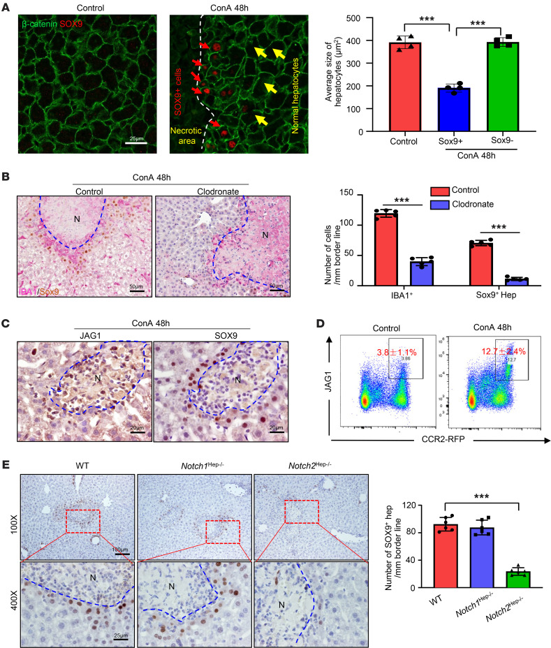
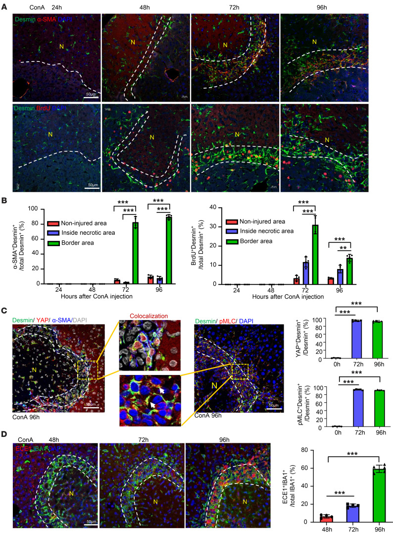
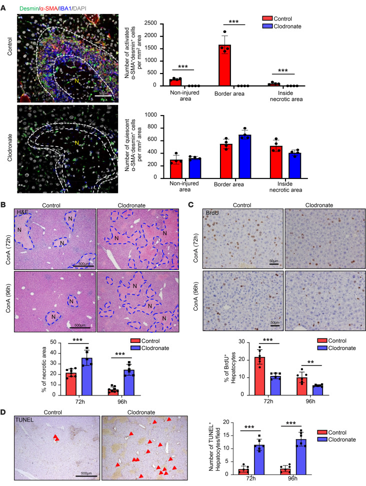
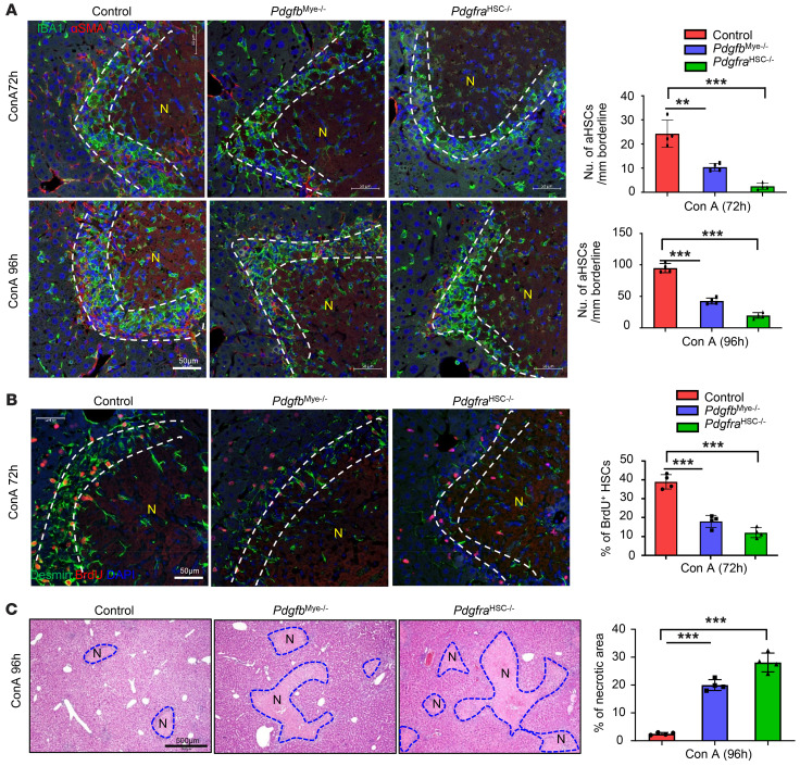
The liver can fully regenerate after partial resection, and its underlying mechanisms have been extensively studied. The liver can also rapidly regenerate after injury, with most studies focusing on hepatocyte proliferation; however, how hepatic necrotic lesions during acute or chronic liver diseases are eliminated and repaired remains obscure. Here, we demonstrate that monocyte-derived macrophages (MoMFs) were rapidly recruited to and encapsulated necrotic areas during immune-mediated liver injury and that this feature was essential in repairing necrotic lesions. At the early stage of injury, infiltrating MoMFs activated the Jagged1/notch homolog protein 2 (JAG1/NOTCH2) axis to induce cell death-resistant SRY-box transcription factor 9+ (SOX9+) hepatocytes near the necrotic lesions, which acted as a barrier from further injury. Subsequently, necrotic environment (hypoxia and dead cells) induced a cluster of complement 1q-positive (C1q+) MoMFs that promoted necrotic removal and liver repair, while Pdgfb+ MoMFs activated hepatic stellate cells (HSCs) to express α-smooth muscle actin and induce a strong contraction signal (YAP, pMLC) to squeeze and finally eliminate the necrotic lesions. In conclusion, MoMFs play a key role in repairing the necrotic lesions, not only by removing necrotic tissues, but also by inducing cell death-resistant hepatocytes to form a perinecrotic capsule and by activating α-smooth muscle actin-expressing HSCs to facilitate necrotic lesion resolution.
Overview
The claims and statistics of this paper haven’t been checked yet.
Free: it reads the open-access text and checks the first 5 claims that cite a source we can check. Create a free account to check every claim.
Integrity signals
Registry
Cites
Statistics
Figures





Verification
Claims the paper makes
The claims and statistics of this paper haven’t been checked yet.
Free: it reads the open-access text and checks the first 5 claims that cite a source we can check. Create a free account to check every claim.
Claims other papers make about this one
Each is checked against this paper’s own text: does it say what the citing paper says it does?
The source backs part of the claim, not all of it — or backs it for a different population, condition or comparison.
In the citing paper
“For instance, signals like IFN-γ and GM-CSF drive polarization towards a classically activated (M1) phenotype, producing pro-inflammatory cytokines (e.g., IL-1β, TNF-α) that exacerbate tissue damage.”
From Macrophage duality in liver diseases: heterogeneity, plasticity, and the quest for precision therapeutics (2026) · cites this paper as [21]
Experimental — accuracy not yet measuredThe source backs part of the claim, not all of it — or backs it for a different population, condition or comparison.
In the citing paper
“In contrast, IL-4 and IL-13 promote an alternatively activated (M2) phenotype, characterized by the secretion of IL-10 and TGF-β, which facilitate inflammation resolution and tissue repair (21, 22).”
From Macrophage duality in liver diseases: heterogeneity, plasticity, and the quest for precision therapeutics (2026) · cites this paper as [21]
Experimental — accuracy not yet measuredThe source backs part of the claim, not all of it — or backs it for a different population, condition or comparison.
In the citing paper
“Feng et al. revealed the synergistic action of two MoMFs subsets during ConA injury resolution. C1q+ MoMFs clear necrotic tissue via complement-mediated phagocytosis, while Pdgfb+ MoMFs secrete platelet-derived growth factor (PDGF) to activate hepatic stellate cells (HSCs), generating contraction signal that help squeeze and eliminate necrotic areas (21).”
From Macrophage duality in liver diseases: heterogeneity, plasticity, and the quest for precision therapeutics (2026) · cites this paper as [21]
Experimental — accuracy not yet measuredThe check wasn’t confident either way, so it gives no verdict — never disputed.
In the citing paper
“Consequently, KCs depletion impairs these MoMFs response (21, 30).”
From Macrophage duality in liver diseases: heterogeneity, plasticity, and the quest for precision therapeutics (2026) · cites this paper as [21]
Experimental — accuracy not yet measuredIn the citing paper
“The same study also demonstrated the mobilization of activated HSCs to form a scar around the necrotic area, representing another example of how multiplexed technologies now support the exploration of distinct cellular responses occurring concomitantly.”
From Inflammation and immunity in liver homeostasis and disease: a nexus of hepatocytes, nonparenchymal cells and immune cells (2025) · doi:10.1038/s41423-025-01313-7 · cites this paper as [344]
1 more claim about this paper
Create a free account to see every claim other papers make about this one.
Self-check
Percentages, sums and reported differences re-computed from the paper’s own numbers.
The self-check needs the paper’s full text.
Citations
Cites
Fiji: an open-source platform for biological-image analysis
Schindelin, J., Arganda-Carreras, I., Frise, E., Kaynig, V., Longair, M., Pietzsch, T., Preibisch, S., Rueden, C., Saalfeld, S., Schmid, B., et al. (2012) · Nature Methods · doi:10.1038/nmeth.2019 · Cited by 66,890 · cited 8×
“Acquired images were processed and analyzed using Image J FIJI (60) and the FIJI plugin HyperStackReg V5.”
BackgroundMethodInfluentialIntegrated analysis of multimodal single-cell data
Hao, Y., Hao, S., Andersen-Nissen, E., Mauck, W. M., Zheng, S., Butler, A., Lee, M. J., Wilk, A. J., Darby, C., Zagar, M., et al. (2020) · doi:10.1101/2020.10.12.335331 · Cited by 15,508 · cited 1×
“0) was used to further analyze single cell RNAseq data (61).”
MethodHafemeister, C., & Satija, R. (2019) · Genome Biology · doi:10.1186/s13059-019-1874-1 · Cited by 4,479 · cited 1×
“UMI counts for each gene were normalized and highly variable genes were 657 identified via the SCTransform function using default parameters (62).”
Methodilastik: interactive machine learning for (bio)image analysis
Berg, S., Kutra, D., Kroeger, T., Straehle, C. N., Kausler, B. X., Haubold, C., Schiegg, M., Ales, J., Beier, T., Rudy, M., et al. (2019) · Nature Methods · doi:10.1038/s41592-019-0582-9 · Cited by 3,368 · cited 1×
“3post3) (64), and CellProfiler (version 4.”
Mechanisms of hepatic stellate cell activation
Tsuchida, T., & Friedman, S. L. (2017) · Nature Reviews Gastroenterology & Hepatology · doi:10.1038/nrgastro.2017.38 · Cited by 2,909
Michalopoulos, G. K., & DeFrances, M. C. (1997) · Science · doi:10.1126/science.276.5309.60 · Cited by 2,828
Resolving the fibrotic niche of human liver cirrhosis at single-cell level
Ramachandran, P., Dobie, R., Wilson-Kanamori, J. R., Dora, E. F., Henderson, B. E. P., Luu, N. T., Portman, J. R., Matchett, K. P., Brice, M., Marwick, J. A., et al. (2019) · Nature · doi:10.1038/s41586-019-1631-3 · Cited by 1,784 · cited 6×
“For example, NAMs/LAMs had more overlapped signature genes with C1q+ MoMFs than SAMs; however, C1q+ MoMFs lack several NAM/LAM signature genes (Cd9, Cd36, Fabp4, Mmp12, H2Ab1) and lack several SAM signature genes (Il1b, Cxcr4, Pdgfb, Vegfa, Hes1) (Supplemental Figure 16F).”
BackgroundInfluentialSelf-citationMechanobiology of YAP and TAZ in physiology and disease
Panciera, T., Azzolin, L., Cordenonsi, M., & Piccolo, S. (2017) · Nature Reviews Molecular Cell Biology · doi:10.1038/nrm.2017.87 · Cited by 1,461 · cited 2×
“Interestingly, aHSCs also expressed high levels of nuclear YAP and phosphorylated myosin light chain (pMLC) (Figure 4C), 2 key signals for smooth muscle cell contraction (31, 32).”
BackgroundMederacke, I., Hsu, C. C., Troeger, J. S., Huebener, P., Mu, X., Dapito, D. H., Pradere, J.-P., & Schwabe, R. F. (2013) · Nature Communications · doi:10.1038/ncomms3823 · Cited by 1,443 · cited 1×
“Robert F 579 Schwabe from Columbia University, New York, NY (58).”
BackgroundLipid-associated macrophages control metabolic homeostasis in a Trem2-dependent manner
Jaitin, D. A., Adlung, L., Thaiss, C. A., Weiner, A., Li, B., Descamps, H., Lundgren, P., Bleriot, C., Liu, Z., Deczkowska, A., et al. (2019) · Cell · doi:10.1016/j.cell.2019.05.054 · Cited by 1,377 · cited 6×
“For example, NAMs/LAMs had more overlapped signature genes with C1q+ MoMFs than SAMs; however, C1q+ MoMFs lack several NAM/LAM signature genes (Cd9, Cd36, Fabp4, Mmp12, H2Ab1) and lack several SAM signature genes (Il1b, Cxcr4, Pdgfb, Vegfa, Hes1) (Supplemental Figure 16F).”
BackgroundInfluential
Cited by
Metabolic reprogramming in liver fibrosis
Horn, P., & Tacke, F. (2024) · Cell Metabolism · doi:10.1016/j.cmet.2024.05.003 · Cited by 432
Self-citationLiver diseases: epidemiology, causes, trends and predictions
Gan, C., Yuan, Y., Shen, H., Gao, J., Kong, X., Che, Z., Guo, Y., Wang, H., Dong, E., & Xiao, J. (2025) · Signal Transduction and Targeted Therapy · doi:10.1038/s41392-024-02072-z · Cited by 310
“346,347 Besides, mediators secreted by hepatocytes, such as IL-17, IL-1 β , and extracellular vesicles (EVs), mediate in fl ammatory macrophage in fi ltration and the development of ALD and MASLD.”
Self-citationAlcohol-associated liver disease
Mackowiak, B., Fu, Y., Maccioni, L., & Gao, B. (2024) · Journal of Clinical Investigation · doi:10.1172/jci176345 · Cited by 359
“However, recent studies have identified many subsets of macrophages, with some of them playing an important role in promoting liver repair and fibrosis resolution (38, 39), thus, selective inhibition of inflammatory macrophage infiltration may achieve better clinical outcomes for ALD treatment.”
BackgroundSelf-citationImmune microenvironment in hepatocellular carcinoma: from pathogenesis to immunotherapy
Seyhan, D., Allaire, M., Fu, Y., Conti, F., Wang, X. W., Gao, B., & Lafdil, F. (2025) · Cellular and Molecular Immunology · doi:10.1038/s41423-025-01308-4 · Cited by 164
“The use of mIF has been successful in characterizing the TME in the context of various liver-associated diseases, including alcohol-associated hepatitis [311], necrotic liver formation [312], and liver aging [313].”
Self-citationMolecular mechanisms in liver repair and regeneration: from physiology to therapeutics
Ma, X., Huang, T., Chen, X., Li, Q., Liao, M., Fu, L., Huang, J., Yuan, K., Wang, Z., & Zeng, Y. (2025) · Signal Transduction and Targeted Therapy · doi:10.1038/s41392-024-02104-8 · Cited by 133
(2025) · doi:10.1038/s41423-025-01313-7 · Cited by 97
“As such, multiplex immuno fl uorescence revealed the crucial role of Jagged-1-expressing monocyte-derived macrophages in inducing Sox9 expression in hepatocytes, allowing liver repair mechanisms to engage in a murine model of acute concanavalin-A-induced liver injury [344].”
Self-citationThe spatial and temporal activation of macrophages during fibrosis
Behmoaras, J. V., Mulder, K., Ginhoux, F., & Petretto, E. G. (2025) · Nature reviews. Immunology · doi:10.1038/s41577-025-01186-x · Cited by 86
“On the other hand, Pdgfb + monocyte-derived macrophages in the mouse liver induce contraction of hepatic stellate cells 35 , while CD150 + monocyte-derived cells in the gut induce a disease-specific fibroblast population in human Crohn’s disease 147 .”
Coordinated action of a gut–liver pathway drives alcohol detoxification and consumption
Fu, Y., Mackowiak, B., Lin, Y., Maccioni, L., Lehner, T., Pan, H., Guan, Y., Godlewski, G., Lu, H., Chen, C., et al. (2024) · Nature Metabolism · doi:10.1038/s42255-024-01063-2 · Cited by 71
Self-citationAntifibrotic therapies for metabolic dysfunction-associated steatotic liver disease
Schwabe, R. F., Tacke, F., Sugimoto, A., & Friedman, S. L. (2025) · JHEP Reports · doi:10.1016/j.jhepr.2025.101421 · Cited by 70
“190 The therapeutic potential of fi brolytic macrophages has been demonstrated in mice and is currently being investigated in patients.”
Self-citationLiver macrophages: development, dynamics, and functions
Nusse, Y., & Kubes, P. (2025) · Cellular & Molecular Immunology · doi:10.1038/s41423-025-01298-3 · Cited by 50
“LAM-like macrophages are also observed in T-Cell mediated liver damage (where they appear related to the C1q + debris clearing monocyte-derived macrophages) [164], but whether Kupffer cells can adopt a LAM phenotype in this model has not been de fi nitively explored.”
Yang, Y., Xu, L., Atkins, C. L., Kuhlman, L., Zhao, J., Jeong, J., Wen, Y., Moreno, N. F., Kim, K. H., An, Y., et al. (2024) · Gut · doi:10.1136/gutjnl-2024-332033 · Cited by 51
“57 It is known that macrophages adopt an inflammatory phenotype when they first enter an injured or infected tissue.”
Self-citationZhang, L., Deng, W., Huang, Q., Zhang, J., Wang, Y., Zhou, T., Xing, L., & Jiang, H. (2024) · Advanced Materials · doi:10.1002/adma.202311474 · Cited by 59
Guillot, A., & Tacke, F. (2024) · Hepatology Communications · doi:10.1097/hc9.0000000000000491 · Cited by 45
“[54] Accordingly, it was evidenced that two distinct populations of MoMFs were involved, one controlling tissue scarring by phagocy-tosing cellular debris and activating fibroblasts through platelet-derived growth factor subunit B, the other one by inducing SRY-box transcription factor 9 expression…”
BackgroundSelf-citationInflammation in Alcohol-Associated Hepatitis: Pathogenesis and Therapeutic Targets
Feng, D., Hwang, S., Guillot, A., Wang, Y., Guan, Y., Chen, C., Maccioni, L., & Gao, B. (2024) · Cellular and Molecular Gastroenterology and Hepatology · doi:10.1016/j.jcmgh.2024.04.009 · Cited by 44
“90 The uptake of dead cells by liver macrophages not only resolves the necrotic lesion but also converts macrophages from a proin-fl ammatory phenotype to an anti-in fl ammatory phenotype with enhanced IL-10 production.”
BackgroundSelf-citationThe evidence-based multifaceted roles of hepatic stellate cells in liver diseases: A concise review
Kim, J., & Kim, Y. J. (2024) · Life Sciences · doi:10.1016/j.lfs.2024.122547 · Cited by 43
Immunologic role of macrophages in sepsis-induced acute liver injury
Xu, W., Hou, H., Yang, W., Tang, W., & Sun, L. (2024) · International Immunopharmacology · doi:10.1016/j.intimp.2024.113492 · Cited by 40
Metabolic reprogramming of immune cells in MASH
Ma, C., Wang, S., Dong, B., & Tian, Y. (2025) · Hepatology · doi:10.1097/hep.0000000000001371 · Cited by 35
Feng, D., Guan, Y., Wang, Y., Maccioni, L., Mackowiak, B., & Gao, B. (2025) · eGastroenterology · doi:10.1136/egastro-2025-100189 · Cited by 37
“Furthermore, by integrating scRNA-seq analysis with immunohistochemical staining verification, we identified several populations of necrotic lesion-associated macrophages that have different signature genes and surround necrotic liver lesions, playing an important role in promoting necrotic lesion…”
Self-citationKim, S., Kim, Y. K., Kim, S. A., Choi, Y.-S., Lee, I., Joo, H., Kim, J., Kwon, M., Park, S., Jo, M., et al. (2025) · bioRxiv · doi:10.1038/s41467-025-57133-w · Cited by 25
“Acute and severe liver injury drives substantial migration of myeloid cells into the liver, where they differentiate into macrophages that play essential roles in both exacerbating liver injury and promoting tissue repair alongside resident macrophages, such as Kupffer cells 19,26 .”
Wang, Y., Guan, Y., Feng, D., Maccioni, L., Parra, M. A., Peiffer, B., Mackowiak, B., Kuwabara, T., Mori, K., Mukoyama, M., et al. (2025) · Cellular & Molecular Immunology · doi:10.1038/s41423-025-01343-1 · Cited by 23
“In support of this, we previously demonstrated that C1q ⁺ macrophages promote necrotic liver lesion resolution by removing dead cells in a model of ConA-induced acute liver injury [16, 42].”
Self-citationZhao, J., Hou, L., Dery, K. J., Yuan, X., Kim, K. H., Kupiec‐Weglinski, J. W., Hall, D. R., Thornley, C. J., Hobeika, M. J., Eltzschig, H. K., et al. (2025) · Hepatology · doi:10.1097/hep.0000000000001609 · Cited by 35
Self-citationHe, J., Du, C., Li, C., Li, W., Qiu, J., Ma, M., Chen, Y., & Zhang, Q. (2025) · Redox Biology · doi:10.1016/j.redox.2025.103657 · Cited by 24
Liu, Y., Liu, T., Zhang, F., & Gao, Y. (2023) · International Journal of Biological Sciences · doi:10.7150/ijbs.87975 · Cited by 23
“Recent research has shown that mononuclear cells have the ability to infiltrate liver tissue and convert into macrophages derived from mononuclear cells, thereby playing an important role in repair following acute liver necrosis [32].”
Guan, D., Huang, P., Liu, X., Li, Q., Zhang, X., Liu, N., Wang, Y., Wan, Y., Chai, J., Cai, S., et al. (2024) · Journal of Advanced Research · doi:10.1016/j.jare.2024.11.020 · Cited by 24
Detecting Cellular Microstructural Changes of Liver Fibrosis with Time-Dependent Diffusion MRI
Zhang, L., Long, X., Chen, L., Han, X., Zhang, X., Sun, P., Yang, Y., Fan, J., Wang, J., Lei, Z., et al. (2024) · Radiology · doi:10.1148/radiol.240343 · Cited by 18
Reprogramming macrophages to treat liver diseases
Simón-Codina, B., Cacho-Pujol, J., Moles, A., & Melgar‐Lesmes, P. (2024) · Hepatology · doi:10.1097/hep.0000000000001160 · Cited by 22
“By contrast, LY6C - resolutive MDMs can replace SAMs to facilitate fibrosis resolution, [129] since they counteract fibrosis by deacti-vating HSCs (senescence, apoptosis, or reversion to a quiescent state).”
Natural antibodies are required for clearance of necrotic cells and recovery from acute liver injury
Mattos, M. S., Vandendriessche, S., Schuermans, S., Feyaerts, L., Hövelmeyer, N., Waisman, A., & Marques, P. E. (2024) · JHEP Reports · doi:10.1016/j.jhepr.2024.101013 · Cited by 20
“6 However, the mechanisms underlying the clearance of necrotic debris from the liver are poorly understood, preventing its therapeutic use in acute liver disorders such as drug-induced liver injury, ischemia-reperfusion injury and liver graft dysfunction.”
BackgroundZhang, J., Chen, W., Song, K., Song, K., Kolls, J. K., & Wu, T. (2024) · The FASEB Journal · doi:10.1096/fj.202400813rr · Cited by 17
“…in the liver, 31 and the released C1q proteins can induce he-patic fibrosis through mediating the activation of hepatic stellate cells, 32,33 our data revealed that YAP activation via Mst1/2 deletion enhances both the pro-inflammatory and tissue repairing function of Kupffer-1 and - 2…”
An, R., Zhu, Z., Chen, Y., Guan, W., Wang, J., & Ren, H. (2025) · Advanced Science · doi:10.1002/advs.202408974 · Cited by 20
The role of matrix metalloproteinases in liver function and disease
Mitropoulou, G., Kompoura, V., Saffioti, F., & Mavroeidis, V. K. (2025) · Frontiers in Bioscience-Landmark · doi:10.31083/fbl27127 · Cited by 14
Macrophage heterogeneity and plasticity in liver injury and repair mechanisms
Zhang, T., Horn, P., Peiseler, M., & Tacke, F. (2025) · Stem Cells · doi:10.1093/stmcls/sxaf072 · Cited by 16
Self-citationLiver Macrophage Diversity in Health and Disease
Horn, P., & Tacke, F. (2024) · Results and problems in cell differentiation · doi:10.1007/978-3-031-65944-7_7 · Cited by 17
Self-citationMicrobiome-derived bile acids as endogenous regenerative mediators in liver repair
Pandey, S. N., Goyal, K., Rana, M., Menon, S. V., Ray, S., Ali, H., Kumbhar, P., Disouza, J., Singh, S. K., Gupta, G., et al. (2025) · Regenerative Therapy · doi:10.1016/j.reth.2025.08.011 · Cited by 12
“…McConnell et al. (2023) used VE-cadherin – Cre to delete FGFR1 in sinusoidal endothelial cells, which reduced HGF secretion by 40 % and delayed DNA synthesis by over 30 %, demonstrating that endothelial angiocrine support is indispensable for synchronizing vascular and parenchymal regrowth [31].”
Bai, B., Bao, W., Wang, Y., Shaha, A., Kisseleva, T., He, L., Sun, L., Jerez, S., Shah, V. H., Wang, X., et al. (2025) · Cellular and Molecular Gastroenterology and Hepatology · doi:10.1016/j.jcmgh.2025.101587 · Cited by 15
“75,76 Kupffer cell activation is regarded as an initial event in response to liver injury, and Kupffer cell-derived cytokines, such as TGF b 1, act on HSCs to induce their activation.”
Self-citationGuan, Y., Kim, Y. S., Wang, Y., Cho, Y. E., Xiang, X., Kim, S., Yao, T., Feng, D., Hwang, S., & Gao, B. (2025) · Journal of Clinical Investigation · doi:10.1172/jci190635 · Cited by 14
“Additionally, we have previously reported a subset of necrotic lesion–associated macrophages involved in concanavalin A-induced liver necrosis lesion resolution (53).”
Self-citationGonzalez‐Sanchez, E., Vaquero, J., Caballero‐Díaz, D., Grzelak, J., Fusté, N. P., Bertrán, E., Amengual, J., García‐Sáez, J., Martín‐Mur, B., Gut, M. O., et al. (2024) · The Journal of Pathology · doi:10.1002/path.6299 · Cited by 13
“Experimental animal models indicate that macrophages are critical for fi brosis regression because they play key roles in repairing the necrotic lesions, not only by removing necrotic tissues but also by inducing cell death-resistant hepatocytes and exerting anti-in fl ammatory actions [26,27].”
BackgroundYang, T., Zhang, Y., Duan, C., Liu, H., Wang, D., Liang, Q., Chen, X., Ma, J., Cheng, K., Chen, Y., et al. (2025) · Experimental & Molecular Medicine · doi:10.1038/s12276-024-01371-3 · Cited by 11
Xu, F., Li, Z., Zhang, W., Li, Q., Li, D., Meng, H., Shen, F., & Fu, H. (2024) · Anesthesiology · doi:10.1097/aln.0000000000005206 · Cited by 13
Avalos, B., Kulbe, J. R., Ford, M. K., Laird, A. E., Walter, K., Mante, M., Florio, J. B., Boustani, A., Chaillon, A., Schlachetzki, J. C. M., et al. (2024) · Viruses · doi:10.3390/v16101509 · Cited by 11
“The inhibition of PPAR γ by GW 9662 was observed to impact macrophage differentiation and inflammatory cytokine production, indicating its regulatory function in immune responses [122].”
BackgroundMyeloid cell path to malignancy: insights into liver cancer
Ramirez, C. F. A., & Akkari, L. (2025) · Trends in cancer · doi:10.1016/j.trecan.2025.02.006 · Cited by 12
Targeting cathepsin C ameliorates murine acetaminophen-induced liver injury
Raith, J., Bachmann, M., Gonther, S., Stülb, H., Aghdassi, A. A., Pham, C. T. N., & Mühl, H. (2024) · Theranostics · doi:10.7150/thno.96092 · Cited by 9
“Data presented suggest that cathepsin B (CtsB) supports resolution in the late phase (96 h) of concanavalin A-induced liver injury [ 69 ] .”
BackgroundDuan, H., Zhang, R., Asikaer, A., Pan, L., Wang, S., Huang, K., Lou, D., Wang, Y., Lin, Z., & Shen, Y. (2025) · International Immunopharmacology · doi:10.1016/j.intimp.2025.114620 · Cited by 9
Lee, H. W., Yang, X., Jin, P.-R., Won, K., Kim, C. H., & Jeong, H. (2024) · Drug Metabolism and Disposition · doi:10.1124/dmd.123.001541 · Cited by 9
“However, at the later stage (48-72 h), these macrophages are critical in tissue repair and regeneration by promoting the resolution and elimination of necrotic lesions (Jaeschke and Ramachandran, 2020a; Feng et al., 2023).”
BackgroundMacrophages in necrotic liver lesion repair: opportunities for therapeutical applications
Wang, Y., Rodrigues, R. M., Chen, C., Feng, D., Maccioni, L., & Gao, B. (2024) · American Journal of Physiology-Cell Physiology · doi:10.1152/ajpcell.00053.2024 · Cited by 11
Self-citationLiu, R., Cao, L., Zhou, Y., Li, Y., Wang, T., Meng, Z., Yang, T., Feng, C., Yang, Q., Liu, X., et al. (2025) · Phytomedicine · doi:10.1016/j.phymed.2025.157349 · Cited by 10
The Role of Immune Cells in Liver Regeneration
Wen, Y. (2023) · Livers · doi:10.3390/livers3030029 · Cited by 9
Targeting Macrophage Phenotype for Treating Heart Failure: A New Approach
Shi, M., Hui, Y., Li, Y., Guo, Z., & Wei, J. (2024) · Drug Design Development and Therapy · doi:10.2147/dddt.s486816 · Cited by 9
“28 Human monocytes are divided into three subpopulations based on the expression of CD14 and CD16.”
BackgroundKoelsch, N. J., Mirshahi, F., Aqbi, H. F., Seneshaw, M., Idowu, M. O., Olex, A. L., Sanyal, A. J., & Manjili, M. H. (2025) · Liver International · doi:10.1111/liv.70110 · Cited by 7
Liver as a key organ for systemic antimicrobial defense
Yao, T., Maccioni, L., Guan, Y., Rodrigues, R. M., & Gao, B. (2025) · Hepatology Communications · doi:10.1097/hc9.0000000000000814 · Cited by 7
“[157] To date, however, the exact function of MoMFs in combating infection after KC impairment remains inconclusive.”
Self-citationInfluencing factors and mechanism of hepatocyte regeneration
Zhang, X., Li, S., Hao, L., Jia, F., Yu, F., & Hu, X. (2025) · Journal of Translational Medicine · doi:10.1186/s12967-025-06278-9 · Cited by 7
“[138] 2023.08 Jagged1/notch Monocyte-derived macrophages (MoMFs) can activate the Jagged1/notch 2 axis in the early stage of injury, prevent further damage of hepatocytes, and play a key role in repairing necrotic lesions.”
InfluentialMacrophages clear out necrotic liver lesions: a new magic trick revealed
Rodrigues, R. M., Boeckmans, J., & Vanhaecke, T. (2023) · eGastroenterology · doi:10.1136/egastro-2023-100024 · Cited by 6
Liu, M., Zhu, Y., Li, Z., Yu, Y., Wang, D., Wang, Y., Jiang, X., & Li, B. (2025) · Cell Death and Disease · doi:10.1038/s41419-025-07925-1 · Cited by 6
Zeng, T., Huang, Y., He, H., Chang, J., Tao, S., Liu, Z., Thorne, R. F., Chen, W., Liu, K., Sun, X., et al. (2026) · Journal of Hepatology · doi:10.1016/j.jhep.2026.03.012 · Cited by 6
Cellular Senescence in Acute Liver Injury: What Happens to the Young Liver?
He, K., Zhou, D., Pu, Z., Chen, S., Shen, Y., Zhao, S., Qian, X., Hu, Q., Wu, X., Xie, Z., et al. (2024) · Aging and Disease · doi:10.14336/ad.2024.0586 · Cited by 8
“In ALI, immune cells accumulate in the liver [92, 93].”
Tanabe, M., Hosono, K., Yamashita, A., Ito, Y., Majima, M., Narumiya, S., Kusano, C., & Amano, H. (2024) · Inflammation and Regeneration · doi:10.1186/s41232-024-00356-z · Cited by 6
“Furthermore, control mice had more infiltrated macrophages than TP △ mac mice, suggesting that accumulation of macrophages in the necrotic area would be required for adequate removal of necrotic tissues and consequently for liver recovery and regeneration [22, 32].”
BackgroundChen, J., Yang, S., Shou, D., Liu, B., Li, S., Luo, T., Chen, H., Huang, C., & Zhou, Y. (2025) · Biomedicines · doi:10.3390/biomedicines13071636 · Cited by 6
“…early stages of liver injury, however, monocyte-derived macrophages rapidly accumulate along the periphery of necrotic areas under the chemotaxis of CCL2 , inducing a SOX9 + hepatocyte-resistant wall that encloses necrotic lesions, thereby protecting undamaged hepatocytes from further injury [47].”
Nguyen, K., Ding, W., & Ni, H. (2025) · eGastroenterology · doi:10.1136/egastro-2025-100205 · Cited by 6
“13 It would be particularly interesting to investigate whether these SOX9 + hepatocytes are the same population as ANXA2 + hepatocytes in ALF.”
The versatile interplay between steatotic liver disease and liver cancer
Rahbari, M., Gris‐Oliver, A., Ramadori, P., Aicher, D., Sia, D., MALEK, N. P., Loomba, R. S., Bataller, R. A., Llovet, J. M., & Heikenwälder, M. F. (2026) · Nature reviews. Cancer · doi:10.1038/s41568-026-00918-y · Cited by 4
Iwasaka, T., Morita, K., Azuma, I., Nakagawa, T., Nakashima, E., Kamei, T., Kato, Y., Kusuhara, H., & Mizuno, T. (2026) · Toxicological Sciences · doi:10.1101/2024.01.25.577198 · Cited by 4
Necrotic Liver Lesion Resolution: Another Mode of Liver Regeneration
Chen, C., Feng, D., Wang, Y., Yao, T., Mackowiak, B., & Gao, B. (2024) · Seminars in Liver Disease · doi:10.1055/a-2358-9505 · Cited by 3
Self-citationIncreased serum GM-CSF at diagnosis of biliary atresia is associated with improved biliary drainage
Taylor, S. A., Harpavat, S., Gromer, K. D., Andreev, V. P., Loomes, K. M., Bezerra, J. A., Jarasvaraparn, C., Wang, K. S., Horslen, S. P., Rosenthal, P. J., et al. (2025) · Pediatric Research · doi:10.1038/s41390-025-03804-9 · Cited by 3
SOX9: a novel janus-faced regulator in immunity and its promise as a therapeutic target
Shi, Q., & He, S. (2025) · Frontiers in Molecular Biosciences · doi:10.3389/fmolb.2025.1700812 · Cited by 3
“…G. et al., 2024). in the Intestinal tissues of patients with inflammatory bowel disease (IBD), including Crohn’s disease (Kobayashi et al., 2024; Zhuang et al., 2022), and as well as in conditions such as Sjögren’s syndrome abnormalities in SOX9 expression are frequently noted (Xiang et al., 2023).”
Yu, F., Qin, Z., Yu, W., Zhou, T., Cao, C., Xie, Y., Zhang, P., Liu, H., He, W., Cheng, A., et al. (2026) · Molecular Cancer · doi:10.1186/s12943-025-02546-w · Cited by 7
“Macrophages in the C57BL/6 mice were depleted using clodronate-containing liposomes according to a published protocol [61].”
Lynch, R. W., Louwe, P. A., Martens, L., Candela, M. E., Bain, C. C., Hawley, C. A., Henderson, N. C., Ramachandran, P., Bénézech, C., Scott, C. L., et al. (2026) · Cell Reports · doi:10.1016/j.celrep.2026.117136 · Cited by 3
Unified platform for multiplex immunofluorescence across liver tissues and engineered models
Leonardi, B. F., Guo, Y., Amiridze, N., Lan, T., Ahmed, Y. A., Berger, H., Tacke, F., Kohlhepp, M. S., & Guillot, A. (2026) · eGastroenterology · doi:10.1136/egastro-2026-100379 · Cited by 3
“27 We further used mIF for in-depth characterisation of histological changes in mouse models of liver injury to guide the design of a primary cell-based liver- on- a-chip model.”
Self-citationSchuermans, S., Quanico, J. P., Kestens, C., Vandendriessche, S., Slowikowski, E., Crijns, M. L., Pörtner, N., Berghmans, N., Baggerman, G., Mattos, M. S., et al. (2025) · Cellular and Molecular Life Sciences · doi:10.1007/s00018-025-05720-x · Cited by 3
“A subset of these macrophages, particularly those expressing C1q, exhibit strong phagocytic activity and is essential for clearing necrotic debris [44].”
Fan, X., Lin, J., Liu, H., Deng, Q., Zheng, Y., Wang, X., & Yang, L. (2024) · International Immunopharmacology · doi:10.1016/j.intimp.2024.113437 · Cited by 3
Self-citationSchulze, S., Keshvari, S., Miller, G. C., Bridle, K. R., Hume, D. A., & Irvine, K. M. (2025) · World Journal of Gastroenterology · doi:10.3748/wjg.v31.i24.108234 · Cited by 3
Chen, H., Wang, H., Huang, J., Qiu, G., Zhang, Z., Xu, L., Ma, X., Wang, Z., Chen, X., & Zeng, Y. (2025) · Acta Pharmaceutica Sinica B · doi:10.1016/j.apsb.2025.07.025 · Cited by 2
“Previous studies have shown that the accumulation of monocyte-derived macrophages is crucial for the repair of necrotic liver regions 50 .”
Self-citationSun, W., Zhou, F., Shi, C., Xu, C., Wang, Z., & Guo, X. (2025) · Journal of Hepatocellular Carcinoma · doi:10.2147/jhc.s538192 · Cited by 2
“SII was calculated as platelets×neutrophils/lymphocytes, and monocytes are reported to play crucial role in repairing necrotic liver lesions, 12 thus should be added into the inflammatory response index.”
Notch signaling in liver diseases: mechanistic insights and therapeutic implications
Shi, Q., Lou, N., Huang, D., & Fu, L. (2026) · Frontiers in Cell and Developmental Biology · doi:10.3389/fcell.2026.1770031 · Cited by 2
Tang, F., Tang, Y., Li, L., Zhou, F., Sun, C., Ouyang, B., Huang, D., Li, M., He, W., Pei, G., et al. (2026) · Journal of Ethnopharmacology · doi:10.1016/j.jep.2026.121652 · Cited by 2
Hu, Y., Hou, J., Cai, Y., Ni, C., Xing, Z., Li, J., Zhou, Z., Xu, G., Zeng, X., Jia, J., et al. (2026) · Archives of Pharmacal Research · doi:10.1007/s12272-026-01623-y · Cited by 3
“The polarization state of MoMFs and the network of cytokines they secrete constitute a core pathway regulating the hepatic immune microenvironment (Guilliams and Scott 2022; Feng et al. 2023).”
Chen, L., Chen, Y., Zhao, M., Yang, X., Sutte, K., Lu, M., Chen, J., Zheng, X., Yang, D., & Liu, J. (2026) · Frontiers in Immunology · doi:10.3389/fimmu.2026.1814879 · Cited by 1
Li, R., Shu, Y., Yan, Y., Zhu, J., Cheng, Z., Zhang, J., Zhu, L., Qiao, Y., & Sun, Q. X. (2025) · International Journal of Molecular Sciences · doi:10.3390/ijms26104903 · Cited by 1
“Further investigation is required to elucidate the spatiotemporal regulation of macrophage phenotypes and their interplay during acute liver injury resolution [12,13].”
Hu, Q., Wang, X., You, Y., Liu, J., Lan, B., Chen, F., Wen, H., Cheng, H., Zhuo, W., Xu, T., et al. (2025) · Neoplasia · doi:10.1016/j.neo.2025.101210 · Cited by 3
“Sequential multiplex immunofluorescence staining with more than 3 markers on formalin-fixed, paraffin-embedded tissue sections was performed as described [34].”
Self-citationKuroda, Y., Ito, Y., Nishizawa, N., Tanabe, M., Yamashita, A., Hosono, K., Kamata, M., Satoh, M., Kumamoto, Y., Hiki, N., et al. (2025) · Experimental Cell Research · doi:10.1016/j.yexcr.2025.114728 · Cited by 1
Shi, X., Mekis, G., Simms, K. J., Gao, B., & Zhang, P. (2025) · Pharmacology & Therapeutics · doi:10.1016/j.pharmthera.2025.108959 · Cited by 1
Self-citationWang, J., Lin, F., Xu, Y., Gong, L., & Peng, Y. (2026) · European Journal of Radiology · doi:10.1016/j.ejrad.2026.112899 · Cited by 1
Targeting NOTCH2-JAG1 juxtacrine signaling reverses macrophage-mediated tumor resistance to taxolPreprint
Yu, F., Zhou, Q., Zhou, T., Xie, Y., Zhang, P., He, W., Yu, W., Cheng, A., Liu, H., Wu, Q., et al. (2024) · bioRxiv (Cold Spring Harbor Laboratory) · doi:10.1101/2024.07.08.602467 · Cited by 1
Lee, Y., Goo, J., Cho, S., Kim, S. A., Nam, G., Kim, I., Park, J.-W., Jeong, C., & Kim, I.-S. (2024) · bioRxiv (Cold Spring Harbor Laboratory) · doi:10.1101/2024.08.29.608900 · Cited by 4
“16,18 HIF1α in monocytes/macrophages has been shown to upregulate genes related to antimicrobial activity and tissue remodeling.”
BackgroundImmunohistochemical characterization of cells of the mononuclear phagocyte system in the mouse
Bertola, L., Valenti, A. G., Cappelleri, A., Canesi, S., Scanziani, E., Recordati, C., & Minoli, L. (2025) · Veterinary Pathology · doi:10.1177/03009858251352581
“44 Interestingly, particularly in the perinecrotic area, cells appeared to express both M1 (MHC-II, iNOS) and M2 (argi-nase 1, HO-1, and Ym1) markers.”
Liu, J., Deng, M., He, X., Ma, J., Zhang, L., Yang, X., Dai, J., Dong, S., Zhang, Y., Zhang, Z., et al. (2026) · Journal of Pharmaceutical Analysis · doi:10.1016/j.jpha.2025.101540
“Emerging evidence implicates macrophage-mediated inflammation as a central factor in metabolic dysfunction associated with liver diseases [14 – 16].”
Roles and mechanistic insights into bile acids in sepsis pathogenesis
Bao, Y., Zhao, X., An, Z., & Tang, C. (2026) · Genes & Diseases · doi:10.1016/j.gendis.2026.102051
Elimination of intramuscular immunoglobin accumulation alleviates Duchenne Muscular DystrophyPreprint
Zhang, L., Guo, W., Huang, M., Liang, Z. Y., Zhu, E., Li, G., Chang, C., Guo, X. S., Sun, W., Li, J., et al. (2026) · bioRxiv (Cold Spring Harbor Laboratory) · doi:10.64898/2026.02.11.703666
Self-citationTransient steatosis reprograms MDMs for liver repair
Woodhoo, A., & Varela‐Rey, M. (2026) · Nature Metabolism · doi:10.1038/s42255-026-01479-y
Inflammatory monocytes constrain YAP-induced cell proliferation
Singh, S., Tahmasian, N., Liu, J., Wang, W., Haas, L., Zhu, Y., Griswold, M. M., Brodsky, N. N., Liu, S., Kalish, B. T., et al. (2026) · Science Advances · doi:10.1126/sciadv.aee8580
“These clusters express high levels of complement ( C1qa , C1qb , and C1qc ), suggesting that they may be phagocytosing target cells ( 55 , 56 ).”
Spatial immune niche remodeling of the neutrophil-macrophage axis inchronic liver disease
Ding, J., Xiong, X., Zhang, C., Fang, H., & Li, S. (2026) · Frontiers in Cell and Developmental Biology · doi:10.3389/fcell.2026.1902627
“Most of the fine inflammatory regulatory pathways reviewed in this review are derived from animal experiments such as mice; however, there is a huge difference in evolution between human and mouse myeloid cell systems (Feng et al., 2023).”
Sun, W., Wang, L., Zhu, Y., Zhou, Y., Ying, L., Yang, W., Liu, C., Jiang, X., Li, X., & Cheng, X. (2026) · Cell Reports · doi:10.1016/j.celrep.2026.117927
Jiang, X., Ma, J., Liu, Z., & Chen, J. (2026) · Frontiers in Pharmacology · doi:10.3389/fphar.2026.1955615
“Although the early inflammatory response can support host defense and tissue repair (Duffield et al., 2005; Feng et al., 2023), persistent injury or failure to resolve inflammation can shift this response toward chronic inflammation.”
Macrophage-derived IL-27 sustains autoreactive CD8 + cytotoxic T cells in autoimmune hepatitisPreprint
Hao, K., Baptista, S. G., Beaumond, A., Schafer, D. M., Gao, K. M., Kaja, P. L., Chiang, K., Nündel, K., Kumar, S. V., Sherman, M. S., et al. (2026) · bioRxiv (Cold Spring Harbor Laboratory) · doi:10.64898/2026.09.25.754422
Metadata
- Authors
- Feng, D., Xiang, X., Guan, Y., Guillot, A., Lu, H., Chang, C., He, Y., Wang, H., Pan, H., Ju, C., Colgan, S. P., Tacke, F., Wang, X. W., Kunos, G., & Gao, B.
- Affiliations
- National Institutes of Health (US)
- The University of Texas Health Science Center at Houston (US)
- University of Colorado Anschutz (US)
- Charité – Universitätsmedizin Berlin (DE)
- Corresponding author
- Gao, Bin
- ORCID
- Venue
- Journal of Clinical Investigation
- Published
- 2023-08-01
- Volume
- 133
- Issue
- 15
- ISSN
- 0021-9738 (ISSN-L)
- 1558-8238
- PMC
- PMC10378165
- OpenAlex
- W4381429881
- Cited by
- 93
- References
- 67
- Citations by year
- 2026: 19 · 2025: 41 · 2024: 30 · 2023: 3
- FWCI
- 15.72
- Type
- journal article
- Language
- en
- Open access
- gold
- doi.org
- Licence
- Locations
- Journal of Clinical Investigation (publishedVersion, cc-by)
- PubMed Central (publishedVersion, cc-by)
- PubMed (publishedVersion)
- Digital Commons-TMC (Texas Medical Center) (submittedVersion)
- DOAJ (DOAJ: Directory of Open Access Journals) (submittedVersion, cc-by-sa)
- Funders
- National Institute on Alcohol Abuse and Alcoholism (The intramural program)
- Topics
- Health Sciences › Medicine › Hepatology › Liver physiology and pathology
- Health Sciences › Medicine › Hepatology › Liver Diseases and Immunity
- Life Sciences › Pharmacology, Toxicology and Pharmaceutics › Pharmacology › Drug-Induced Hepatotoxicity and Protection
- Keywords
- monocyte-derived macrophages, liver regeneration, liver necrosis, hepatic stellate cells, YAP signaling, alpha-smooth muscle actin, hypoxia, immune-mediated liver injury, macrophage heterogeneity
- MeSH terms
- Actins, Humans, Liver, Liver Neoplasms, Macrophages, Necrosis, Hepatocytes, Hepatic Stellate Cells
About these checks
Registry, retraction, Cites and statistics results are computed, not judged, with one exception: a printed reference that no index lists is read once by a model before it counts as a citation error. Claims are extracted by a model and kept only when their quote appears verbatim in the text; each cited source is then checked, with a model’s help, for whether it says what the claim relies on. Those verdicts can be wrong — read the claim’s quote and the cited source before relying on one.
Statistics and arithmetic are recomputed, not judged.
Confidence bands are shown only for fields calibrated on our annotated test set. None is calibrated yet, so no band is shown.
What counts as a claim ›How we measure confidence ›How we check this ›
Are you an author, or do you see a problem with this page? Request a review
Data sources
Bibliographic data: Semantic Scholar, OpenAlex, Crossref.
Contains information from the Semantic Scholar Academic Graph Datasets, made available under the ODC Attribution License (ODC-BY 1.0).
Semantic Scholar’s data is described in: Kinney, R., et al. (2023). The Semantic Scholar Open Data Platform. arXiv:2301.10140.
