

Monocyte-derived macrophages orchestrate multiple cell-type interactions to repair necrotic liver lesions in disease models
Open accessThe DOI matches its Crossref record; 68 of the 68 references it cites were found in a registry or an index.
Abstract
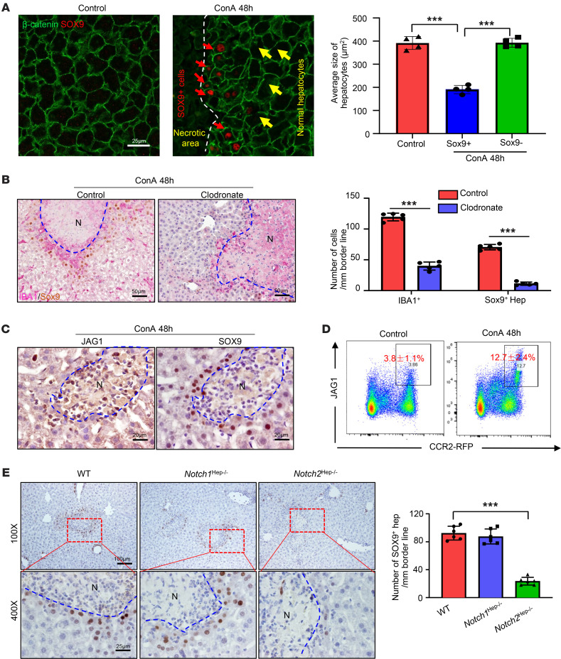
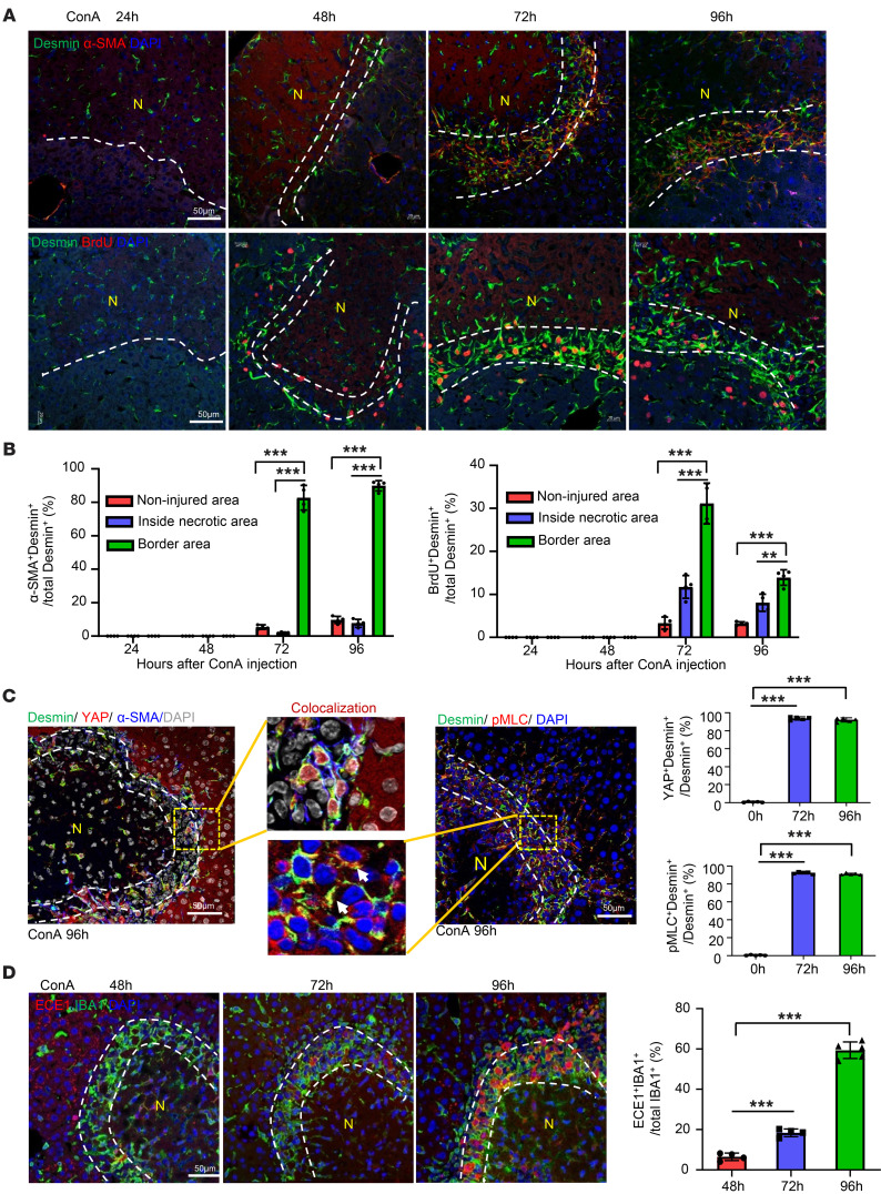
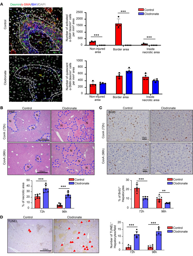
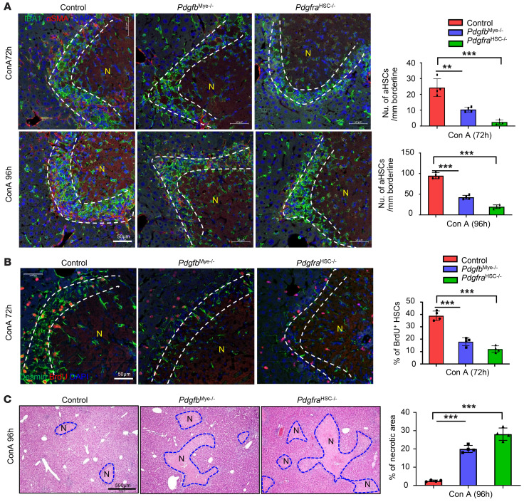
The liver can fully regenerate after partial resection, and its underlying mechanisms have been extensively studied. The liver can also rapidly regenerate after injury, with most studies focusing on hepatocyte proliferation; however, how hepatic necrotic lesions during acute or chronic liver diseases are eliminated and repaired remains obscure. Here, we demonstrate that monocyte-derived macrophages (MoMFs) were rapidly recruited to and encapsulated necrotic areas during immune-mediated liver injury and that this feature was essential in repairing necrotic lesions. At the early stage of injury, infiltrating MoMFs activated the Jagged1/notch homolog protein 2 (JAG1/NOTCH2) axis to induce cell death-resistant SRY-box transcription factor 9+ (SOX9+) hepatocytes near the necrotic lesions, which acted as a barrier from further injury. Subsequently, necrotic environment (hypoxia and dead cells) induced a cluster of complement 1q-positive (C1q+) MoMFs that promoted necrotic removal and liver repair, while Pdgfb+ MoMFs activated hepatic stellate cells (HSCs) to express α-smooth muscle actin and induce a strong contraction signal (YAP, pMLC) to squeeze and finally eliminate the necrotic lesions. In conclusion, MoMFs play a key role in repairing the necrotic lesions, not only by removing necrotic tissues, but also by inducing cell death-resistant hepatocytes to form a perinecrotic capsule and by activating α-smooth muscle actin-expressing HSCs to facilitate necrotic lesion resolution.
Overview
The claims and statistics of this paper haven’t been checked yet.
Free: it reads the open-access text and checks the first 5 claims that cite a source we can check. Create a free account to check every claim.
Integrity signals
Registry
Cites
Statistics
Figures





Verification
Claims the paper makes
The claims and statistics of this paper haven’t been checked yet.
Free: it reads the open-access text and checks the first 5 claims that cite a source we can check. Create a free account to check every claim.
Claims other papers make about this one
Each is checked against this paper’s own text: does it say what the citing paper says it does?
The source backs part of the claim, not all of it — or backs it for a different population, condition or comparison.
In the citing paper
“For instance, signals like IFN-γ and GM-CSF drive polarization towards a classically activated (M1) phenotype, producing pro-inflammatory cytokines (e.g., IL-1β, TNF-α) that exacerbate tissue damage.”
From Macrophage duality in liver diseases: heterogeneity, plasticity, and the quest for precision therapeutics (2026) · cites this paper as [21]
Experimental — accuracy not yet measuredThe source backs part of the claim, not all of it — or backs it for a different population, condition or comparison.
In the citing paper
“In contrast, IL-4 and IL-13 promote an alternatively activated (M2) phenotype, characterized by the secretion of IL-10 and TGF-β, which facilitate inflammation resolution and tissue repair (21, 22).”
From Macrophage duality in liver diseases: heterogeneity, plasticity, and the quest for precision therapeutics (2026) · cites this paper as [21]
Experimental — accuracy not yet measuredThe source backs part of the claim, not all of it — or backs it for a different population, condition or comparison.
In the citing paper
“Feng et al. revealed the synergistic action of two MoMFs subsets during ConA injury resolution. C1q+ MoMFs clear necrotic tissue via complement-mediated phagocytosis, while Pdgfb+ MoMFs secrete platelet-derived growth factor (PDGF) to activate hepatic stellate cells (HSCs), generating contraction signal that help squeeze and eliminate necrotic areas (21).”
From Macrophage duality in liver diseases: heterogeneity, plasticity, and the quest for precision therapeutics (2026) · cites this paper as [21]
Experimental — accuracy not yet measuredThe check wasn’t confident either way, so it gives no verdict — never disputed.
In the citing paper
“Consequently, KCs depletion impairs these MoMFs response (21, 30).”
From Macrophage duality in liver diseases: heterogeneity, plasticity, and the quest for precision therapeutics (2026) · cites this paper as [21]
Experimental — accuracy not yet measuredIn the citing paper
“The same study also demonstrated the mobilization of activated HSCs to form a scar around the necrotic area, representing another example of how multiplexed technologies now support the exploration of distinct cellular responses occurring concomitantly.”
From Inflammation and immunity in liver homeostasis and disease: a nexus of hepatocytes, nonparenchymal cells and immune cells (2025) · doi:10.1038/s41423-025-01313-7 · cites this paper as [344]
1 more claim about this paper
Create a free account to see every claim other papers make about this one.
Self-check
Percentages, sums and reported differences re-computed from the paper’s own numbers.
The self-check needs the paper’s full text.
Citations
Cites
Fiji: an open-source platform for biological-image analysis
Schindelin, J., Arganda-Carreras, I., Frise, E., Kaynig, V., Longair, M., Pietzsch, T., Preibisch, S., Rueden, C., Saalfeld, S., Schmid, B., et al. (2012) · Nature Methods · doi:10.1038/nmeth.2019 · Cited by 66,890 · cited 8×
“Acquired images were processed and analyzed using Image J FIJI (60) and the FIJI plugin HyperStackReg V5.”
BackgroundMethodInfluentialIntegrated analysis of multimodal single-cell data
Hao, Y., Hao, S., Andersen-Nissen, E., Mauck, W. M., Zheng, S., Butler, A., Lee, M. J., Wilk, A. J., Darby, C., Zagar, M., et al. (2020) · doi:10.1101/2020.10.12.335331 · Cited by 15,508 · cited 1×
“0) was used to further analyze single cell RNAseq data (61).”
MethodHafemeister, C., & Satija, R. (2019) · Genome Biology · doi:10.1186/s13059-019-1874-1 · Cited by 4,479 · cited 1×
“UMI counts for each gene were normalized and highly variable genes were 657 identified via the SCTransform function using default parameters (62).”
Methodilastik: interactive machine learning for (bio)image analysis
Berg, S., Kutra, D., Kroeger, T., Straehle, C. N., Kausler, B. X., Haubold, C., Schiegg, M., Ales, J., Beier, T., Rudy, M., et al. (2019) · Nature Methods · doi:10.1038/s41592-019-0582-9 · Cited by 3,368 · cited 1×
“3post3) (64), and CellProfiler (version 4.”
Mechanisms of hepatic stellate cell activation
Tsuchida, T., & Friedman, S. L. (2017) · Nature Reviews Gastroenterology & Hepatology · doi:10.1038/nrgastro.2017.38 · Cited by 2,909
Michalopoulos, G. K., & DeFrances, M. C. (1997) · Science · doi:10.1126/science.276.5309.60 · Cited by 2,828
Resolving the fibrotic niche of human liver cirrhosis at single-cell level
Ramachandran, P., Dobie, R., Wilson-Kanamori, J. R., Dora, E. F., Henderson, B. E. P., Luu, N. T., Portman, J. R., Matchett, K. P., Brice, M., Marwick, J. A., et al. (2019) · Nature · doi:10.1038/s41586-019-1631-3 · Cited by 1,784 · cited 6×
“For example, NAMs/LAMs had more overlapped signature genes with C1q+ MoMFs than SAMs; however, C1q+ MoMFs lack several NAM/LAM signature genes (Cd9, Cd36, Fabp4, Mmp12, H2Ab1) and lack several SAM signature genes (Il1b, Cxcr4, Pdgfb, Vegfa, Hes1) (Supplemental Figure 16F).”
BackgroundInfluentialSelf-citationMechanobiology of YAP and TAZ in physiology and disease
Panciera, T., Azzolin, L., Cordenonsi, M., & Piccolo, S. (2017) · Nature Reviews Molecular Cell Biology · doi:10.1038/nrm.2017.87 · Cited by 1,461 · cited 2×
“Interestingly, aHSCs also expressed high levels of nuclear YAP and phosphorylated myosin light chain (pMLC) (Figure 4C), 2 key signals for smooth muscle cell contraction (31, 32).”
BackgroundMederacke, I., Hsu, C. C., Troeger, J. S., Huebener, P., Mu, X., Dapito, D. H., Pradere, J.-P., & Schwabe, R. F. (2013) · Nature Communications · doi:10.1038/ncomms3823 · Cited by 1,443 · cited 1×
“Robert F 579 Schwabe from Columbia University, New York, NY (58).”
BackgroundLipid-associated macrophages control metabolic homeostasis in a Trem2-dependent manner
Jaitin, D. A., Adlung, L., Thaiss, C. A., Weiner, A., Li, B., Descamps, H., Lundgren, P., Bleriot, C., Liu, Z., Deczkowska, A., et al. (2019) · Cell · doi:10.1016/j.cell.2019.05.054 · Cited by 1,377 · cited 6×
“For example, NAMs/LAMs had more overlapped signature genes with C1q+ MoMFs than SAMs; however, C1q+ MoMFs lack several NAM/LAM signature genes (Cd9, Cd36, Fabp4, Mmp12, H2Ab1) and lack several SAM signature genes (Il1b, Cxcr4, Pdgfb, Vegfa, Hes1) (Supplemental Figure 16F).”
BackgroundInfluentialGodoy, P., Hewitt, N. J., Albrecht, U., Andersen, M. E., Ansari, N., Bhattacharya, S., Bode, J. G., Bolleyn, J., Borner, C., Böttger, J., et al. (2013) · Archives of Toxicology · doi:10.1007/s00204-013-1078-5 · Cited by 1,280 · cited 1×
“In contrast, hepatocytes near the portal area, which express very low levels of CYP2E1, are resistant to APAP-induced liver injury (45).”
BackgroundA T cell-dependent experimental liver injury in mice inducible by concanavalin A.
Tiegs, G., Hentschel, J., & Wendel, A. (1992) · Journal of Clinical Investigation · doi:10.1172/jci115836 · Cited by 1,074 · cited 1×
“Thus, in the current study, we used a well-established immune-mediated acute liver-injury model induced by concanavalin A (ConA) (7) to study the resolution of necrotic lesions and their repair by inflammatory cells.”
MethodLiver regeneration: biological and pathological mechanisms and implications
Michalopoulos, G. K., & Bhushan, B. (2021) · Nature Reviews Gastroenterology & Hepatology · doi:10.1038/s41575-020-0342-4 · Cited by 998 · cited 2×
“Liver regeneration has also been studied in a wide variety 48 of acute liver injury models that are associated with liver necrotic lesions (2-5), which are 49 not observed in the PHx model.”
BackgroundMethodXu, D., Emoto, N., Giaid, A., Slaughter, C., Kaw, S., deWit, D., & Yanagisawa, M. (1994) · Cell · doi:10.1016/0092-8674(94)90425-1 · Cited by 921 · cited 2×
“ECE1 is a 530 key membrane-bound metalloendopeptiase that cleaves ET-1 to biologically active 531 peptides that play a key role in inducing HSC contraction (32, 33).”
BackgroundCellProfiler™: Free, Versatile Software for Automated Biological Image Analysis
Lamprecht, M. R., Sabatini, D. M., & Carpenter, A. E. (2007) · BioTechniques · doi:10.2144/000112257 · Cited by 895
Furuyama, K., Kawaguchi, Y., Akiyama, H., Horiguchi, M., Kodama, S., Kuhara, T., Hosokawa, S., Elbahrawy, A., Soeda, T., Koizumi, M., et al. (2011) · Nature Genetics · doi:10.1038/ng.722 · Cited by 834 · cited 2×
“SOX9 is a marker for LPCs and biliary epithelial cells, but is not expressed in mature hepatocytes (23), playing a critical role in controlling specification of hepatoblasts to bile duct cells (BDCs) via the activation of notch homolog protein (NOTCH) (24, 25).”
BackgroundXiong, X., Kuang, H., Ansari, S., Liu, T., Gong, J., Wang, S., Zhao, X.-Y., Ji, Y., Li, C., Guo, L., et al. (2019) · Molecular Cell · doi:10.1016/j.molcel.2019.07.028 · Cited by 784 · cited 6×
“For example, NAMs/LAMs had more overlapped signature genes with C1q+ MoMFs than SAMs; however, C1q+ MoMFs lack several NAM/LAM signature genes (Cd9, Cd36, Fabp4, Mmp12, H2Ab1) and lack several SAM signature genes (Il1b, Cxcr4, Pdgfb, Vegfa, Hes1) (Supplemental Figure 16F).”
BackgroundInfluentialWang, G., & Semenza, G. (1993) · Blood · doi:10.1182/blood.v82.12.3610.3610 · Cited by 766
Macrophage heterogeneity in tissues: phenotypic diversity and functions
Gordon, S., Plüddemann, A., & Martinez Estrada, F. (2014) · Immunological Reviews · doi:10.1111/imr.12223 · Cited by 715 · cited 2×
“Both Kupffer cells and infiltrating 80 MoMFs have been characterized as not only proinflammatory cells but also having a key 81 role in tissue repair, depending on the stage of injury and recovery process (11-13).”
BackgroundWen, Y., Lambrecht, J., Ju, C., & Tacke, F. (2021) · Cellular & Molecular Immunology · doi:10.1038/s41423-020-00558-8 · Cited by 695
Self-citationApoptosis and necroptosis in the liver: a matter of life and death
Schwabe, R. F., & Luedde, T. (2018) · Nature Reviews Gastroenterology & Hepatology · doi:10.1038/s41575-018-0065-y · Cited by 582 · cited 2×
“), causing liver necrosis that manifests in massive necrotic lesions (8, 9).”
BackgroundWang, J., & Kubes, P. (2016) · Cell · doi:10.1016/j.cell.2016.03.009 · Cited by 537 · cited 2×
“Research in the last three decades has revealed that macrophages are highly 426 adaptable in their ability to respond to a variety of threats to the organism by differentiating 427 into subsets with unique gene expression profiles, exerting multiple functions including 428 promoting liver injury repair (11-13, 20).”
BackgroundNF- kappa B and Sp1 regulate transcription of the human monocyte chemoattractant protein-1 gene.
Ueda, A., Okuda, K., Ohno, S., Shirai, A., Igarashi, T., Matsunaga, K., Fukushima, J., Kawamoto, S., Ishigatsubo, Y., & Okubo, T. (1994) · The Journal of Immunology · doi:10.4049/jimmunol.153.5.2052 · Cited by 523 · cited 9×
“The important roles of CCL2/CCR2 in protecting against ConAinduced liver injury have been previously well documented (18, 19).”
BackgroundInfluentialHybrid Periportal Hepatocytes Regenerate the Injured Liver without Giving Rise to Cancer
Font-Burgada, J., Shalapour, S., Ramaswamy, S., Hsueh, B., Rossell, D., Umemura, A., Taniguchi, K., Nakagawa, H., Valasek, M. A., Ye, L., et al. (2015) · Cell · doi:10.1016/j.cell.2015.07.026 · Cited by 486 · cited 1×
“229 Previous studies reported that SOX9+ hepatocytes located in the periportal area had 230 greater regenerative potential compared with hepatocytes elsewhere in the liver (29, 30).”
BackgroundRobust cellular reprogramming occurs spontaneously during liver regeneration
Yanger, K., Zong, Y., Maggs, L. R., Shapira, S. N., Maddipati, R., Aiello, N. M., Thung, S. N., Wells, R. G., Greenbaum, L. E., & Stanger, B. Z. (2013) · Genes & Development · doi:10.1101/gad.207803.112 · Cited by 484 · cited 1×
“9 cells in the liver were labeled with EYFP (Supplemental Figure 9A, B) (27, 28).”
BackgroundBlériot, C., Dupuis, T., Jouvion, G., Eberl, G., Disson, O., & Lecuit, M. (2015) · Immunity · doi:10.1016/j.immuni.2014.12.020 · Cited by 471 · cited 1×
“In addition, both Kupffer cell proliferation 548 and MoMF differentiation have been shown to contribute Kupffer cell restoration in several 549 liver injury models (53-55).”
BackgroundHes binding to STAT3 mediates crosstalk between Notch and JAK–STAT signalling
Kamakura, S., Oishi, K., Yoshimatsu, T., Nakafuku, M., Masuyama, N., & Gotoh, Y. (2004) · Nature Cell Biology · doi:10.1038/ncb1138 · Cited by 423 · cited 14×
“Second, we found that SOX9+ hepatocytes expressed high levels of activated STAT3 and its downstream antiapoptotic protein BCL-xL.”
BackgroundInfluentialTran, S., Baba, I., Poupel, L., Dussaud, S., Moreau, M., Gélineau, A., Marcelin, G., Magréau-Davy, E., Ouhachi, M., Lesnik, P., et al. (2020) · Immunity · doi:10.1016/j.immuni.2020.06.003 · Cited by 400 · cited 1×
“In addition, both Kupffer cell proliferation 548 and MoMF differentiation have been shown to contribute Kupffer cell restoration in several 549 liver injury models (53-55).”
BackgroundProtective Role of Kupffer Cells in Acetaminophen-Induced Hepatic Injury in Mice
Ju, C., Reilly, T. P., Bourdi, M., Radonovich, M. F., Brady, J. N., George, J. W., & Pohl, L. R. (2002) · Chemical Research in Toxicology · doi:10.1021/tx0255976 · Cited by 360 · cited 1×
“For 82 example, depletion of liver macrophages before injection of acetaminophen (APAP) or 83 inhibition of MoMF recruitment early after APAP reduced APAP-induced liver injury, 84 whereas depletion of macrophages post APAP injection accelerated liver injury (14-16).”
BackgroundSelf-citationMossanen, J. C., Krenkel, O., Ergen, C., Govaere, O., Liepelt, A., Puengel, T., Heymann, F., Kalthoff, S., Lefebvre, E., Eulberg, D., et al. (2016) · Hepatology · doi:10.1002/hep.28682 · Cited by 325 · cited 1×
“For 82 example, depletion of liver macrophages before injection of acetaminophen (APAP) or 83 inhibition of MoMF recruitment early after APAP reduced APAP-induced liver injury, 84 whereas depletion of macrophages post APAP injection accelerated liver injury (14-16).”
BackgroundSelf-citationLiver regeneration and inflammation: from fundamental science to clinical applications
Campana, L., Esser, H., Huch, M., & Forbes, S. (2021) · Nature Reviews Molecular Cell Biology · doi:10.1038/s41580-021-00373-7 · Cited by 307 · cited 3×
“For example, how 54 are the hepatocytes adjacent to the necrotic lesions protected from further injury? How are 55 the dead cells and debris removed from the necrotic lesions? How are the necrotic lesions 56 eliminated? Do hepatocytes or liver progenitor cells migrate inside the necrotic lesions to 57 repair the lesio…”
BackgroundMethodTanimizu, N., & Miyajima, A. (2004) · Journal of Cell Science · doi:10.1242/jcs.01169 · Cited by 291 · cited 2×
“SOX9 is a marker for liver progenitor cells and 161 biliary epithelial cells but is not expressed in mature hepatocytes (23), playing a critical 162 role in controlling specification of hepatoblasts to bile duct cells via the activation of 163 NOTCH (24, 25).”
BackgroundChronic Liver Injury Induces Conversion of Biliary Epithelial Cells into Hepatocytes
Deng, X., Zhang, X., Li, W., Feng, R.-X., Li, L., Yi, G.-R., Zhang, X.-N., Yin, C., Yu, H.-Y., Zhang, J.-P., et al. (2018) · Cell Stem Cell · doi:10.1016/j.stem.2018.05.022 · Cited by 289 · cited 1×
“229 Previous studies reported that SOX9+ hepatocytes located in the periportal area had 230 greater regenerative potential compared with hepatocytes elsewhere in the liver (29, 30).”
BackgroundImportance of Kupffer Cells for T-Cell-Dependent Liver Injury in Mice
Schümann, J., Wolf, D., Pahl, A., Brune, K., Papadopoulos, T., van Rooijen, N., & Tiegs, G. (2000) · The American Journal of Pathology · doi:10.1016/s0002-9440(10)64804-3 · Cited by 284
REVIEW ARTICLE. CELL SURFACE CARBOHYDRATES AS PROGNOSTIC MARKERS IN HUMAN CARCINOMAS
Dabelsteen, E. (1996) · The Journal of Pathology · doi:10.1002/(sici)1096-9896(199608)179:4<358::aid-path564>3.0.co;2-t · Cited by 252
Hepatocellular carcinoma originates from hepatocytes and not from the progenitor/biliary compartment
Mu, X., Español-Suñer, R., Mederacke, I., Affò, S., Manco, R., Sempoux, C., Lemaigre, F. P., Adili, A., Yuan, D., Weber, A., et al. (2015) · Journal of Clinical Investigation · doi:10.1172/jci77995 · Cited by 215 · cited 1×
“9 cells in the liver were labeled with EYFP (Supplemental Figure 9A, B) (27, 28).”
BackgroundAlternatively activated macrophages promote resolution of necrosis following acute liver injury
Starkey Lewis, P., Campana, L., Aleksieva, N., Cartwright, J. A., Mackinnon, A., O'Duibhir, E., Kendall, T., Vermeren, M., Thomson, A., Gadd, V., et al. (2020) · Journal of Hepatology · doi:10.1016/j.jhep.2020.02.031 · Cited by 209 · cited 3×
“Both Kupffer cells and infiltrating 80 MoMFs have been characterized as not only proinflammatory cells but also having a key 81 role in tissue repair, depending on the stage of injury and recovery process (11-13).”
BackgroundSafety profile of autologous macrophage therapy for liver cirrhosis
Moroni, F., Dwyer, B. J., Graham, C., Pass, C., Bailey, L., Ritchie, L., Mitchell, D., Glover, A., Laurie, A., Doig, S., et al. (2019) · Nature Medicine · doi:10.1038/s41591-019-0599-8 · Cited by 202 · cited 1×
“Indeed, such therapies have been tested for the treatment of liver diseases both in preclinical models and in patients (13, 56, 57).”
BackgroundCysteine cathepsins in extracellular matrix remodeling: Extracellular matrix degradation and beyond
Vizovišek, M., Fonović, M., & Turk, B. (2019) · Matrix Biology · doi:10.1016/j.matbio.2018.01.024 · Cited by 200 · cited 1×
“18 proteins and the extracellular matrix (50).”
Danger-Associated Molecular Patterns (DAMPs): the Derivatives and Triggers of Inflammation
Patel, S. (2018) · Current Allergy and Asthma Reports · doi:10.1007/s11882-018-0817-3 · Cited by 197 · cited 1×
“Interestingly, only the hepatocytes around the perimeter of the necrotic area express 443 high levels of CCL2, which may be due to the activation of NF-κB stimulated by danger444 associated molecule patterns (DAMPs) (46) released by the necrotic tissue.”
BackgroundThe Fulminant Form of Epidemic Hepatitis
(1947) · New England Journal of Medicine · doi:10.1056/nejm194705082361908 · Cited by 183 · cited 1×
“Although necrotic lesions of the liver had been characterized almost 8 decades ago (6), how they are eliminated and repaired remains largely unknown.”
BackgroundSparks, E. E., Huppert, K. A., Brown, M. A., Washington, M. K., & Huppert, S. S. (2010) · Hepatology · doi:10.1002/hep.23431 · Cited by 140 · cited 2×
“SOX9 is a marker for liver progenitor cells and 161 biliary epithelial cells but is not expressed in mature hepatocytes (23), playing a critical 162 role in controlling specification of hepatoblasts to bile duct cells via the activation of 163 NOTCH (24, 25).”
BackgroundComplement in removal of the dead – balancing inflammation
Martin, M., & Blom, A. M. (2016) · Immunological Reviews · doi:10.1111/imr.12462 · Cited by 130 · cited 1×
“C1q is well known as playing a key role in removing dead cells (34).”
BackgroundGuillot, A., Winkler, M., Silva Afonso, M., Aggarwal, A., Lopez, D., Berger, H., Kohlhepp, M. S., Liu, H., Özdirik, B., Eschrich, J., et al. (2023) · Hepatology · doi:10.1097/hep.0000000000000270 · Cited by 116 · cited 1×
“counting and quantification as previously described (66, 67) (Supple-”
MethodSelf-citationGuo, J., Hu, Z., Yan, F., Lei, S., Li, T., Li, X., Xu, C., Sun, B., Pan, C., & Chen, L. (2020) · Free Radical Biology and Medicine · doi:10.1016/j.freeradbiomed.2020.08.015 · Cited by 108 · cited 1×
“As illustrated in Supplemental Figure 21, reduced PDGFb expression was found in MoMFs from Hif1amye–/– mice, which is consistent with several reports that PDGFβ is upregulated by hypoxia (43, 44).”
BackgroundC1q and phagocytosis: the perfect complement to a good meal
Galvan, M. D., Greenlee-Wacker, M. C., & Bohlson, S. S. (2012) · Journal of Leukocyte Biology · doi:10.1189/jlb.0212099 · Cited by 106
Tiegs, G. (2007) · Zeitschrift für Gastroenterologie · doi:10.1055/s-2006-927397 · Cited by 106 · cited 1×
“62 Injection of ConA, a T cell mitogen, rapidly activates T cells and subsequently activates 63 other types of immune cells (NKT, neutrophils, Kupffer cells) that release a variety of pro64 inflammatory cytokines (TNF-α, IFN-γ, IL-4, IL-5 etc), causing liver necrosis that 65 manifests in massive necrotic lesions (8, 9…”
BackgroundPatterns of necrosis in liver disease
Krishna, M. (2017) · Clinical Liver Disease · doi:10.1002/cld.653 · Cited by 105 · cited 1×
“In addition, liver necrotic lesions are frequently observed in human liver diseases, such as viral hepatitis, alcohol-associated liver disease, nonalcoholic fatty liver disease, autoimmune liver disease, and drug-induced liver injury (4, 5).”
BackgroundROLE OF MACROPHAGES IN ACETAMINOPHEN (PARACETAMOL)-INDUCED HEPATOTOXICITY
GOLDIN, R. D., RATNAYAKA, I. D., BREACH, C. S., BROWN, I. N., & WICKRAMASINGHE, S. N. (1996) · The Journal of Pathology · doi:10.1002/(sici)1096-9896(199608)179:4<432::aid-path609>3.0.co;2-s · Cited by 104 · cited 1×
“For 82 example, depletion of liver macrophages before injection of acetaminophen (APAP) or 83 inhibition of MoMF recruitment early after APAP reduced APAP-induced liver injury, 84 whereas depletion of macrophages post APAP injection accelerated liver injury (14-16).”
BackgroundZhang, S. X. L., Gozal, D., Sachleben, L. R., Rane, M. J., Klein, J. B., & Gozal, E. (2003) · The FASEB Journal · doi:10.1096/fj.02-1111fje · Cited by 102 · cited 1×
“As illustrated in Supplemental Figure 21, reduced PDGFb expression was 396 found in MoMFs from Hif1amye-/- mice, which is consistent with several reports that 397 PDGFβ is upregulated by hypoxia (43, 44).”
ResultMafB is a critical regulator of complement component C1q
Tran, M. T., Hamada, M., Jeon, H., Shiraishi, R., Asano, K., Hattori, M., Nakamura, M., Imamura, Y., Tsunakawa, Y., Fujii, R., et al. (2017) · Nature Communications · doi:10.1038/s41467-017-01711-0 · Cited by 102 · cited 2×
“Furthermore, our scRNA 497 data revealed that C1q+ macrophages express MAFB, a key transcription factor that 498 controls C1q expression (41), and expression of MAFB was markedly diminished in 499 MoMFs in Hif1amye-/- mice, suggesting that HIF1α controls C1q expression by inducing 500 MAFB in MoMFs.”
BackgroundNotch signaling regulates tubular morphogenesis during repair from biliary damage in mice
Fiorotto, R., Raizner, A., Morell, C. M., Torsello, B., Scirpo, R., Fabris, L., Spirli, C., & Strazzabosco, M. (2013) · Journal of Hepatology · doi:10.1016/j.jhep.2013.02.025 · Cited by 95 · cited 1×
“JAG1, a 191 cell surface protein that interacts with neurogenic locus notch homolog protein (NOTCH) 192 receptors, has been implicated in the lineage specification of LPCs/BDCs by promoting 193 SOX9 expression via the activation of NOTCH in a cell-cell contact manner (26).”
BackgroundMa, J., Guillot, A., Yang, Z., Mackowiak, B., Hwang, S., Park, O., Peiffer, B. J., Ahmadi, A. R., Melo, L., Kusumanchi, P., et al. (2022) · Journal of Clinical Investigation · doi:10.1172/jci157780 · Cited by 90 · cited 1×
“Multiplex immunofluorescence staining with 680 more than three markers was performed as previously described (63).”
MethodSelf-citationConcanavalin A-induced liver injury triggers hepatocyte proliferation.
Trautwein, C., Rakemann, T., Malek, N. P., Plümpe, J., Tiegs, G., & Manns, M. P. (1998) · Journal of Clinical Investigation · doi:10.1172/jci504 · Cited by 87
Gao, R. Y., Wang, M., Liu, Q., Feng, D., Wen, Y., Xia, Y., Colgan, S. P., Eltzschig, H. K., & Ju, C. (2020) · Hepatology · doi:10.1002/hep.30954 · Cited by 79
Self-citationAjuebor, M. N., Hogaboam, C. M., Le, T., & Swain, M. G. (2003) · The Journal of Immunology · doi:10.4049/jimmunol.170.10.5252 · Cited by 68 · cited 1×
“Blockage of MoMF recruitment also markedly 88 exacerbated ConA-induced liver injury (18, 19).”
BackgroundEffects of Kupffer cell-depletion on Concanavalin A-induced hepatitis
Hatano, M., Sasaki, S., Ohata, S., Shiratsuchi, Y., Yamazaki, T., Nagata, K., & Kobayashi, Y. (2008) · Cellular Immunology · doi:10.1016/j.cellimm.2008.02.003 · Cited by 56 · cited 3×
“Similarly, depletion of liver macrophages before ConA injection diminished ConA-induced liver injury (17), while their depletion after ConA injection exacerbated liver injury, as demonstrated in the current study.”
BackgroundInfluentialWeng, H.-L., Cai, X., Yuan, X., Liebe, R., Dooley, S., Li, H., & Wang, T.-L. (2015) · Frontiers in Physiology · doi:10.3389/fphys.2015.00178 · Cited by 50 · cited 2×
“In addition, liver necrotic lesions are frequently observed 50 in human liver diseases, such as viral hepatitis, alcohol-associated liver disease, 51 nonalcoholic fatty liver disease, autoimmune liver disease, and drug-induced liver injury 52 (4, 5).”
BackgroundAhmed, Y. A., Fu, Y., Rodrigues, R. M., He, Y., Guan, Y., Guillot, A., Ren, R., Feng, D., Hidalgo, J., Ju, C., et al. (2021) · Cellular and Molecular Immunology · doi:10.1038/s41423-021-00731-7 · Cited by 48 · cited 1×
“In addition, both Kupffer cell proliferation 548 and MoMF differentiation have been shown to contribute Kupffer cell restoration in several 549 liver injury models (53-55).”
BackgroundSelf-citationGuillot, A., Kohlhepp, M., Bruneau, A., Heymann, F., & Tacke, F. (2020) · Cancers · doi:10.3390/cancers12092449 · Cited by 46 · cited 1×
“4) (65) for cell counting and 683 quantification as described before (66, 67) and Supplemental Figure 23.”
BackgroundSelf-citationMacrophages as a Cell-Based Therapy for Liver Disease
Starkey Lewis, P. J., Moroni, F., & Forbes, S. J. (2019) · Seminars in Liver Disease · doi:10.1055/s-0039-1688502 · Cited by 36
Cre-inducible human CD59 mediates rapid cell ablation after intermedilysin administration
Feng, D., Dai, S., Liu, F., Ohtake, Y., Zhou, Z., Wang, H., Zhang, Y., Kearns, A., Peng, X., Zhu, F., et al. (2016) · Journal of Clinical Investigation · doi:10.1172/jci84921 · Cited by 36 · cited 1×
“ihCD59 mice were described 580 previously (59).”
Self-citationMammalian legumain - A lysosomal cysteine protease with extracellular functions?
Lunde, N. N., Bosnjak, T., Solberg, R., & Johansen, H. T. (2019) · Biochimie · doi:10.1016/j.biochi.2019.06.002 · Cited by 29
Myosin Mediates Contractile Force Generation by Hepatic Stellate Cells in Response to Endothelin-1
Saab, S., Tam, S. P., Tran, B. N., Melton, A. C., Tangkijvanich, P., Wong, H. C., & Yee, H. F. (2002) · Journal of Biomedical Science · doi:10.1007/bf02254988 · Cited by 28 · cited 4×
“ECE1 is a 530 key membrane-bound metalloendopeptiase that cleaves ET-1 to biologically active 531 peptides that play a key role in inducing HSC contraction (32, 33).”
BackgroundInfluentialEndothelin-converting enzyme is a plausible target gene for hypoxia-inducible factor
Khamaisi, M., Toukan, H., Axelrod, J. H., Rosenberger, C., Skarzinski, G., Shina, A., Meidan, R., Koesters, R., Rosen, S., Walkinshaw, G., et al. (2014) · Kidney International · doi:10.1038/ki.2014.362 · Cited by 26 · cited 11×
“Collectively, necrosis-associated hypoxia activates the HIF1α/MAFB/C1q axis in MoMFs, resulting in accumulation of C1q+ MoMFs with strong phagocytic activity to remove necrotic tissues.”
BackgroundInfluentialSánchez, A., Factor, V. M., Espinoza, L. A., Schroeder, I. S., & Thorgeirsson, S. S. (2004) · Hepatology · doi:10.1002/hep.20363 · Cited by 26 · cited 1×
“The protective effect of SOX9 is likely 467 mediated via the induction of pSTAT3 and BCL2 as SOX9+ hepatocytes express higher 468 levels of pSTAT3 and BCL-2 than SOX9- hepatocytes, which is in agreement with a 469 previous report that SOX9+ liver progenitor cells are resistant to cytokine induced apoptosis 470 (48).”
ResultIdentification of cathepsin B as a novel target of hypoxia-inducible factor-1-alpha in HepG2 cells
Xiaofei, C., Li, Y., Zhou, D., Dong, C., Zhao, F., & Weilin, W. (2018) · Biochemical and Biophysical Research Communications · doi:10.1016/j.bbrc.2018.06.116 · Cited by 22 · cited 11×
“Collectively, necrosis-associated hypoxia activates the HIF1α/MAFB/C1q axis in MoMFs, resulting in accumulation of C1q+ MoMFs with strong phagocytic activity to remove necrotic tissues.”
BackgroundInfluentialLee, Y., Yi, H., Suh, Y., Byun, J.-S., Eun, H. S., Kim, S. Y., Seo, W., Jeong, J., Choi, W., Kim, M., et al. (2015) · Molecules and Cells · doi:10.14348/molcells.2015.0218 · Cited by 14 · cited 1×
“Blockage of MoMF recruitment also markedly 88 exacerbated ConA-induced liver injury (18, 19).”
Background
Cited by
Metabolic reprogramming in liver fibrosis
Horn, P., & Tacke, F. (2024) · Cell Metabolism · doi:10.1016/j.cmet.2024.05.003 · Cited by 432
Self-citationLiver diseases: epidemiology, causes, trends and predictions
Gan, C., Yuan, Y., Shen, H., Gao, J., Kong, X., Che, Z., Guo, Y., Wang, H., Dong, E., & Xiao, J. (2025) · Signal Transduction and Targeted Therapy · doi:10.1038/s41392-024-02072-z · Cited by 310
“346,347 Besides, mediators secreted by hepatocytes, such as IL-17, IL-1 β , and extracellular vesicles (EVs), mediate in fl ammatory macrophage in fi ltration and the development of ALD and MASLD.”
Self-citationAlcohol-associated liver disease
Mackowiak, B., Fu, Y., Maccioni, L., & Gao, B. (2024) · Journal of Clinical Investigation · doi:10.1172/jci176345 · Cited by 359
“However, recent studies have identified many subsets of macrophages, with some of them playing an important role in promoting liver repair and fibrosis resolution (38, 39), thus, selective inhibition of inflammatory macrophage infiltration may achieve better clinical outcomes for ALD treatment.”
BackgroundSelf-citationImmune microenvironment in hepatocellular carcinoma: from pathogenesis to immunotherapy
Seyhan, D., Allaire, M., Fu, Y., Conti, F., Wang, X. W., Gao, B., & Lafdil, F. (2025) · Cellular and Molecular Immunology · doi:10.1038/s41423-025-01308-4 · Cited by 164
“The use of mIF has been successful in characterizing the TME in the context of various liver-associated diseases, including alcohol-associated hepatitis [311], necrotic liver formation [312], and liver aging [313].”
Self-citationMolecular mechanisms in liver repair and regeneration: from physiology to therapeutics
Ma, X., Huang, T., Chen, X., Li, Q., Liao, M., Fu, L., Huang, J., Yuan, K., Wang, Z., & Zeng, Y. (2025) · Signal Transduction and Targeted Therapy · doi:10.1038/s41392-024-02104-8 · Cited by 133
Metadata
- Authors
- Feng, D., Xiang, X., Guan, Y., Guillot, A., Lu, H., Chang, C., He, Y., Wang, H., Pan, H., Ju, C., Colgan, S. P., Tacke, F., Wang, X. W., Kunos, G., & Gao, B.
- Affiliations
- National Institutes of Health (US)
- The University of Texas Health Science Center at Houston (US)
- University of Colorado Anschutz (US)
- Charité – Universitätsmedizin Berlin (DE)
- Corresponding author
- Gao, Bin
- ORCID
- Venue
- Journal of Clinical Investigation
- Published
- 2023-08-01
- Volume
- 133
- Issue
- 15
- ISSN
- 0021-9738 (ISSN-L)
- 1558-8238
- PMC
- PMC10378165
- OpenAlex
- W4381429881
- Cited by
- 93
- References
- 67
- Citations by year
- 2026: 19 · 2025: 41 · 2024: 30 · 2023: 3
- FWCI
- 15.72
- Type
- journal article
- Language
- en
- Open access
- gold
- doi.org
- Licence
- Locations
- Journal of Clinical Investigation (publishedVersion, cc-by)
- PubMed Central (publishedVersion, cc-by)
- PubMed (publishedVersion)
- Digital Commons-TMC (Texas Medical Center) (submittedVersion)
- DOAJ (DOAJ: Directory of Open Access Journals) (submittedVersion, cc-by-sa)
- Funders
- National Institute on Alcohol Abuse and Alcoholism (The intramural program)
- Topics
- Health Sciences › Medicine › Hepatology › Liver physiology and pathology
- Health Sciences › Medicine › Hepatology › Liver Diseases and Immunity
- Life Sciences › Pharmacology, Toxicology and Pharmaceutics › Pharmacology › Drug-Induced Hepatotoxicity and Protection
- Keywords
- monocyte-derived macrophages, liver regeneration, liver necrosis, hepatic stellate cells, YAP signaling, alpha-smooth muscle actin, hypoxia, immune-mediated liver injury, macrophage heterogeneity
- MeSH terms
- Actins, Humans, Liver, Liver Neoplasms, Macrophages, Necrosis, Hepatocytes, Hepatic Stellate Cells
About these checks
Registry, retraction, Cites and statistics results are computed, not judged, with one exception: a printed reference that no index lists is read once by a model before it counts as a citation error. Claims are extracted by a model and kept only when their quote appears verbatim in the text; each cited source is then checked, with a model’s help, for whether it says what the claim relies on. Those verdicts can be wrong — read the claim’s quote and the cited source before relying on one.
Statistics and arithmetic are recomputed, not judged.
Confidence bands are shown only for fields calibrated on our annotated test set. None is calibrated yet, so no band is shown.
What counts as a claim ›How we measure confidence ›How we check this ›
Are you an author, or do you see a problem with this page? Request a review
Data sources
Bibliographic data: Semantic Scholar, OpenAlex, Crossref.
Contains information from the Semantic Scholar Academic Graph Datasets, made available under the ODC Attribution License (ODC-BY 1.0).
Semantic Scholar’s data is described in: Kinney, R., et al. (2023). The Semantic Scholar Open Data Platform. arXiv:2301.10140.
