

Decoding of grasping information from neural signals recorded using peripheral intrafascicular interfaces
Open accessThe DOI matches its Crossref record; 43 of the 43 references it cites were found in a registry or an index; 1 of the 16 sources its claims cite were checked for support.
Abstract
BACKGROUND: The restoration of complex hand functions by creating a novel bidirectional link between the nervous system and a dexterous hand prosthesis is currently pursued by several research groups. This connection must be fast, intuitive, with a high success rate and quite natural to allow an effective bidirectional flow of information between the user's nervous system and the smart artificial device. This goal can be achieved with several approaches and among them, the use of implantable interfaces connected with the peripheral nervous system, namely intrafascicular electrodes, is considered particularly interesting. METHODS: Thin-film longitudinal intra-fascicular electrodes were implanted in the median and ulnar nerves of an amputee's stump during a four-week trial. The possibility of decoding motor commands suitable to control a dexterous hand prosthesis was investigated for the first time in this research field by implementing a spike sorting and classification algorithm. RESULTS: The results showed that motor information (e.g., grip types and single finger movements) could be extracted with classification accuracy around 85% (for three classes plus rest) and that the user could improve his ability to govern motor commands over time as shown by the improved discrimination ability of our classification algorithm. CONCLUSIONS: These results open up new and promising possibilities for the development of a neuro-controlled hand prosthesis.
Overview
CoChat Summary
TL;DR
Thin-film longitudinal intrafascicular electrodes implanted in an amputee’s median and ulnar nerves allowed decoding of grasp-related motor commands, including grip types and single-finger movement. Classification accuracy reached about 85% for three movement classes plus rest, with performance improving over two days and with daily classifier retraining.
Key findings
- Motor information such as grip types and single-finger movements could be extracted from tfLIFE signals; the paper reports classification accuracy around 85% for three classes plus rest (Table 1: rest vs little vs pinch vs palmar = 85%; Table 2 with retraining: little, pinch, palmar plus rest = 0.85).
- Best recognition ratios varied by task: rest vs pinch = 100%, rest vs palmar = 95%, rest vs little = 93%, rest vs little vs palmar = 92%, rest vs little vs pinch = 90%, rest vs pinch vs palmar = 87%, and rest vs activity = 87%.
- Without daily retraining, performance was lower (0.72 for little, pinch, palmar plus rest) than with retraining (0.85); the user also improved classification performance over two training days.
- Increasing the number of recording channels improved classification performance up to a limit, after which performance degraded due to overfitting.
Methods
- Single case: P.P., a 26-year-old male with left arm amputation two years before implantation; two tfLIFE4s were implanted in the median and ulnar nerves during a four-week trial, with motor-signal trials conducted in the last week.
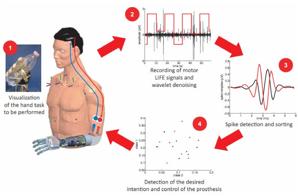
- The subject was visually cued to perform palmar grasp, pinch grasp, little-finger flexion, or rest; LIFE motor signals were recorded via 4-channel amplifiers, wavelet-denoised, spike-sorted into templates, and classified with a one-against-one SVM using leave-one-out validation and relative spike-rate features.
- Inter-day robustness was tested by training the classifier on the first day of recordings and applying it unchanged on subsequent days, compared with daily retraining.
Limitations
- Stated in the paper: time limitations prevented increasing the number of grip types; the learning effect was observed only for two consecutive days and needs confirmation in longer tests; future chronic experiments with an implanted hand prosthesis are needed.
- Stated in the paper: some unexpected channels contributed to best classification, possibly because the user could not deliver the natural (“correct”) neurophysiological commands.
- Evident from the text: this is a single-amputee case, so generalizability is not established; the no-retraining vs retraining comparison also indicates that signal time-variance or recalibration needs may remain despite the reported stability.
Model-written summary. Check it against the paper before relying on it.
Integrity signals
Registry
Cites
Statistics
Figures



Verification
Claims the paper makes
1 Valid · 0 Not valid · 7 Not checked · 17 Own results and interpretations
The cited sources support only part of this claim — read each source’s verdict below.
In the paper
“In fact, after appropriate control in experimental models to test their biocompatibility and efficacy [18], LIFEs have been recently used to control artificial devices [6,7,22-25] showing good results during short-term trials with human amputees.”
§ Decoding of grasping information from neural signals recorded using peripheral intrafascicular interfaces > I. Introduction
What the sources say
- Not checked yetcites [6] source
- Not checked yetcites [7] source
- Not checked yetcites [22] source
- Partly supportedcites [23] sourceExperimental — accuracy not yet measured
The INTRODUCTION of Kemere et al. (2008) does support the general part of the claim: prototype implementations 'have demonstrated that monkeys and humans can achieve some level of real-time control over motor ... or communication ... prostheses' recorded with 'chronically implanted microelectrode arrays' (citing Kennedy and Bakay 1998; Kennedy et al. 2000; Hochberg et al. 2006; Serruya et al. 2002; Taylor et al. 2002 — the intracortical/LIFE-type systems). But the population it describes is patients with spinal cord injury or neurodegenerative disease who are paralyzed (its Hochberg reference is titled '...by a human with tetraplegia'; Kennedy 1998 is '...from a paralyzed patient'), not amputees, and the paper nowhere reports short-term trials in amputees or grades their results as good — it explicitly hedges to 'some level' of control and frames these systems as requiring human intervention and suffering performance losses when neural-state transitions are undetected (INTRODUCTION; RESULTS). The 'human amputees' and 'good results during short-term trials' elements are therefore not attributable to this source. (LLM, conf=0.75)
- Not checked yetcites [24]
- Not checked yetcites [25]
plainstates a cause- Asserted: passes — The authors put it forward as true.
- Checkable: passes — It can be checked against evidence.
- Load-bearing: unclear — It is unclear whether the paper’s conclusions depend on it.
- Based: passes — The paper gives a reason why it holds.
A result the paper reports from its own data — it cites no other source for it, so there is none to check. The quote shows it is in the paper.
In the paper
“the user could improve his ability to govern motor commands over time as shown by the improved discrimination ability of our classification algorithm.”
- Also stated in: § IV Discussion · plain
“our amputee subject was able to improve his performance during the consecutive trials.”
§ Decoding of grasping information from neural signals recorded using peripheral intrafascicular interfaces > Abstract > Results
plainstates a causeSingle case- Asserted: passes — The authors put it forward as true.
- Checkable: passes — It can be checked against evidence.
- Load-bearing: passes — The paper’s conclusions depend on it.
- Based: passes — The paper gives a reason why it holds.
- Also stated in: § IV Discussion · plain
In the paper
“The quality of the recording after denoising can be considered good similarly to the considerations done in [35].”
§ Decoding of grasping information from neural signals recorded using peripheral intrafascicular interfaces > III. Results
What the sources say
- Not checked yetcites [35]
plainSingle case- Asserted: passes — The authors put it forward as true.
- Checkable: unclear — It is unclear whether it could be checked against evidence.
- Load-bearing: passes — The paper’s conclusions depend on it.
- Based: passes — The paper gives a reason why it holds.
A result the paper reports from its own data — it cites no other source for it, so there is none to check. The quote shows it is in the paper.
In the paper
“In most cases, a combination of features coming from all the three tfLIFE channels is required to obtain the best recognition ratio (rr).”
§ Decoding of grasping information from neural signals recorded using peripheral intrafascicular interfaces > III. Results
plainstates a causeSingle case- Asserted: passes — The authors put it forward as true.
- Checkable: passes — It can be checked against evidence.
- Load-bearing: passes — The paper’s conclusions depend on it.
- Based: passes — The paper gives a reason why it holds.
In the paper
“This spike-based approach was used because it already showed to allow high classification accuracy in animal experiments [28].”
§ Decoding of grasping information from neural signals recorded using peripheral intrafascicular interfaces > II. Materials and methods > C. Off-line Processing algorithms of LIFE signals
What the sources say
- Not checked yetcites [28]
plainstates a cause- Asserted: passes — The authors put it forward as true.
- Checkable: passes — It can be checked against evidence.
- Load-bearing: passes — The paper’s conclusions depend on it.
- Based: passes — The paper gives a reason why it holds.
5 more claims
Create a free account to see all 25 claims and to verify the 15 references still open.
- It may state two separate claims as one. 10
- The quote only partly states this claim. 2
- Probably not a substantial claim. 5
Claims other papers make about this one
Each is checked against this paper’s own text: does it say what the citing paper says it does?
The source backs part of the claim, not all of it — or backs it for a different population, condition or comparison.
In the citing paper
“Nevertheless, ENG signals have a low signal-to-noise ratio and limited stability.[363]”
From Flexible Electronics and Devices as Human–Machine Interfaces for Medical Robotics (2022) · cites this paper as [363]
Experimental — accuracy not yet measuredThe source backs part of the claim, not all of it — or backs it for a different population, condition or comparison.
In the citing paper
“For example, intrafascicular electrodes, such as the transverse intrafascicular multichannel electrode (TIME) and the longitudinal intrafascicular electrode (LIFE), transduce signals that have amplitudes less than 100 µVpp [62, 66, 71, 79, 82, 109].”
From Implantable neurotechnologies: a review of integrated circuit neural amplifiers (2016) · cites this paper as [82]
Experimental — accuracy not yet measuredThe source backs part of the claim, not all of it — or backs it for a different population, condition or comparison.
In the citing paper
“Due to the noise constraints placed on recording instrumentation, electrophysiological recordings from the PNS are largely performed using commercial bench-top low-noise amplifier setups [75, 82, 109] that require subjects to be tethered to the equipment.”
From Implantable neurotechnologies: a review of integrated circuit neural amplifiers (2016) · cites this paper as [82]
Experimental — accuracy not yet measuredThe source backs part of the claim, not all of it — or backs it for a different population, condition or comparison.
In the citing paper
“It has been implanted in clinical trials, and has demonstrated good performance with prosthetic arms. Multiple electrodes were implanted in different nerves and the amputees improved their control with training [91, 92].”
From Interfaces with the peripheral nervous system for the control of a neuroprosthetic limb: a review (2020) · cites this paper as [91]
Experimental — accuracy not yet measuredIn the citing paper
“Most recently, Micera et al. used peripheral nerve stimulation to evoke graded sensory perceptions in a human amputee subject while using the subject’s residual muscle activity to decode movement intention and actuate a robotic arm (Fig. 10) [82, 109].”
From Implantable neurotechnologies: a review of integrated circuit neural amplifiers (2016) · cites this paper as [82]
Self-check
Percentages, sums and reported differences re-computed from the paper’s own numbers.
Each contradiction between two passages listed here was confirmed by an independent check.
Rated minor: it would not touch a result — a value no result rests on, a p-value that stays on the same side of the significance level, or a conflict that is clerical, resolved elsewhere in the paper, or outside its own findings.
In the paper
§ Decoding of grasping information from neural signals recorded using peripheral intrafascicular interfaces > II. Materials and methods > B. Experimental protocol
“two tfLIFE4s (Figure 1) were inserted in the ulnar and the median nerves 45° obliquely”
§ Decoding of grasping information from neural signals recorded using peripheral intrafascicular interfaces > III. Results
“M1 an M2 indicate the two tfLIFE implanted in the median nerve whereas U indicates the tfLIFE implanted in the ulnar nerve.”
Citations
Cites
Cortes, C., & Vapnik, V. (1995) · Machine Learning · doi:10.1007/bf00994018 · Cited by 59,421
Cortes, C., & Vapnik, V. (1995) · Machine-mediated learning · doi:10.1023/a:1022627411411 · Cited by 48,445
LIBSVM: A library for support vector machines
Chang, C.-C., & Lin, C.-J. (2011) · ACM Transactions on Intelligent Systems and Technology · doi:10.1145/1961189.1961199 · Cited by 40,918
Translation-Invariant De-Noising
Coifman, R. R., & Donoho, D. L. (1995) · Lecture Notes in Statistics · doi:10.1007/978-1-4612-2544-7_9 · Cited by 1,914
A robust, real-time control scheme for multifunction myoelectric control
Englehart, K., & Hudgins, B. (2003) · IEEE Transactions on Biomedical Engineering · doi:10.1109/tbme.2003.813539 · Cited by 1,743
Cortical control of a prosthetic arm for self-feeding
Velliste, M., Perel, S., Spalding, M. C., Whitford, A. S., & Schwartz, A. B. (2008) · Nature · doi:10.1038/nature06996 · Cited by 1,591
Targeted Muscle Reinnervation for Real-time Myoelectric Control of Multifunction Artificial Arms
Kuiken, T. A. (2009) · JAMA · doi:10.1001/jama.2009.116 · Cited by 1,089
Navarro, X., Krueger, T. B., Lago, N., Micera, S., Stieglitz, T., & Dario, P. (2005) · Journal of the Peripheral Nervous System · doi:10.1111/j.1085-9489.2005.10303.x · Cited by 845 · cited 2×
“Several invasive PNS interfaces have been developed in the past [18].”
BackgroundSelf-citationHuang, Y., Englehart, K. B., Hudgins, B., & Chan, A. D. C. (2005) · IEEE Transactions on Biomedical Engineering · doi:10.1109/tbme.2005.856295 · Cited by 643
Control of Multifunctional Prosthetic Hands by Processing the Electromyographic Signal
Zecca, M., Micera, S., Carrozza, M. C., & Dario, P. (2002) · Critical Reviews? in Biomedical Engineering · doi:10.1615/critrevbiomedeng.v30.i456.80 · Cited by 606
Self-citation
Cited by
Flexible Electronics and Devices as Human–Machine Interfaces for Medical Robotics
Heng, W., Solomon, S., & Gao, W. (2022) · Advanced Materials · doi:10.1002/adma.202107902 · Cited by 609
Toward higher-performance bionic limbs for wider clinical use
Farina, D., Vujaklija, I., Brånemark, R., Bull, A. M. J., Dietl, H., Graimann, B., Hargrove, L. J., Hoffmann, K.-P., Huang, H., Ingvarsson, T., et al. (2021) · Nature Biomedical Engineering · doi:10.1038/s41551-021-00732-x · Cited by 263
Self-citationWendelken, S., Page, D. M., Davis, T., Wark, H. A. C., Kluger, D. T., Duncan, C., Warren, D. J., Hutchinson, D. T., & Clark, G. A. (2017) · Journal of NeuroEngineering and Rehabilitation · doi:10.1186/s12984-017-0320-4 · Cited by 216
Implantable neurotechnologies: a review of micro- and nanoelectrodes for neural recording
Patil, A. C., & Thakor, N. V. (2016) · Medical & Biological Engineering & Computing · doi:10.1007/s11517-015-1430-4 · Cited by 175
Multi-Modal Sensing Techniques for Interfacing Hand Prostheses: A Review
Fang, Y., Hettiarachchi, N., Zhou, D., & Liu, H. (2015) · IEEE Sensors Journal · doi:10.1109/jsen.2015.2450211 · Cited by 172
Metadata
- Authors
- Micera, S., Rossini, P. M., Rigosa, J., Citi, L., Carpaneto, J., Raspopovic, S., Tombini, M., Cipriani, C., Assenza, G., Carrozza, M. C., Hoffmann, K.-P., Yoshida, K., Navarro, X., & Dario, P.
- Affiliations
- Scuola Superiore Sant'Anna (IT)
- Agostino Gemelli University Polyclinic (IT)
- Università Campus Bio-Medico (IT)
- Fraunhofer Institute for Biomedical Engineering (DE)
- Indiana University – Purdue University Indianapolis (US)
- Universitat Autònoma de Barcelona (ES)
- Corresponding author
- Micera, Silvestro
- ORCID
- Venue
- Journal of NeuroEngineering and Rehabilitation
- Published
- 2011
- Volume
- 8
- Issue
- 1
- Pages
- 53
- ISSN
- 1743-0003 (ISSN-L)
- PMC
- PMC3177892
- OpenAlex
- W2164012737
- Cited by
- 100
- References
- 43
- Citations by year
- 2025: 2 · 2024: 3 · 2023: 7 · 2022: 5 · 2021: 13 · 2020: 8 · 2019: 12 · 2018: 11 · 2017: 6 · 2016: 10 · 2015: 5 · 2014: 5 · 2013: 8 · 2012: 5
- FWCI
- 2.38
- Type
- journal article
- Language
- en
- Open access
- gold
- doi.org
- Licence
- cc-by
- Locations
- Journal of NeuroEngineering and Rehabilitation (publishedVersion, cc-by)
- PubMed Central (publishedVersion, cc-by)
- PubMed (publishedVersion)
- Open Access at Essex (University of Essex) (acceptedVersion)
- SDL (submittedVersion, cc-by-nc-nd)
- BOA (University of Milano-Bicocca) (submittedVersion, other-oa)
- Dipòsit Digital de Documents de la UAB (Universitat Autònoma de Barcelona) (submittedVersion, cc-by)
- LA Referencia (Red Federada de Repositorios Institucionales de Publicaciones Científicas) (submittedVersion, cc-by)
- DOAJ (DOAJ: Directory of Open Access Journals) (submittedVersion)
- Infoscience (Ecole Polytechnique Fédérale de Lausanne) (submittedVersion)
- Infoscience (Ecole Polytechnique Fédérale de Lausanne) (submittedVersion, cc-by-nc-nd)
- Fraunhofer-Publica (Fraunhofer-Gesellschaft) (submittedVersion)
- PubliCatt (Università Cattolica del Sacro Cuore) (submittedVersion, other-oa)
- CINECA IRIS Institutional Research Information System (Sant'Anna School of Advanced Studies) (submittedVersion, other-oa)
- Repository for Publications and Research Data (ETH Zurich) (submittedVersion, cc-by)
- ETH Zürich Research Collection (cc-by)
- Topics
- Life Sciences › Neuroscience › Cellular and Molecular Neuroscience › Neuroscience and Neural Engineering
- Physical Sciences › Engineering › Biomedical Engineering › Muscle activation and electromyography studies
- Life Sciences › Neuroscience › Cognitive Neuroscience › EEG and Brain-Computer Interfaces
- Keywords
- intrafascicular electrodes, peripheral nerve interfaces, neural decoding, amputees, median nerve, ulnar nerve, spike sorting, spike classification, neuroplasticity
- MeSH terms
- Adult, Algorithms, Artificial Limbs, Electrodes, Implanted, Hand, Humans, Male, Prosthesis Design, Robotics, User-Computer Interface, Hand Strength
About these checks
Registry, retraction, Cites and statistics results are computed, not judged, with one exception: a printed reference that no index lists is read once by a model before it counts as a citation error. Claims are extracted by a model and kept only when their quote appears verbatim in the text; each cited source is then checked, with a model’s help, for whether it says what the claim relies on. Those verdicts can be wrong — read the claim’s quote and the cited source before relying on one.
The claim verdicts this report counts are experimental: their accuracy is not yet measured.
A language model drew the claims from the paper on 2026-10-04, under version 3 of the claim definition (model deepseek-v4.1-flash, prompt extract-v3).
A second model labelled each claim on 2026-10-04 (model jev-1.13-20260917, definition revision gold-guideline-v0.2).
Cited sources are checked with a language model’s help (qwen3.8-omni-flash).
Statistics and arithmetic are recomputed, not judged.
Confidence bands are shown only for fields calibrated on our annotated test set. None is calibrated yet, so no band is shown.
What counts as a claim ›How we measure confidence ›How we check this ›
Are you an author, or do you see a problem with this page? Request a review
How to cite
Cite CoChat for the checks on this page, and the paper for its own findings.
- This page
- CoChat. (2026). Citation check: Decoding of grasping information from neural signals recorded using peripheral intrafascicular interfaces. https://app.cochat.ai/papers/10.1186/1743-0003-8-53
- The paper
- Micera, S., Rossini, P. M., Rigosa, J., Citi, L., Carpaneto, J., Raspopovic, S., Tombini, M., Cipriani, C., Assenza, G., Carrozza, M. C., Hoffmann, K.-P., Yoshida, K., Navarro, X., & Dario, P. (2011). Decoding of grasping information from neural signals recorded using peripheral intrafascicular interfaces. Journal of NeuroEngineering and Rehabilitation. https://doi.org/10.1186/1743-0003-8-53
Data sources
Bibliographic data: Semantic Scholar, OpenAlex, Crossref.
Contains information from the Semantic Scholar Academic Graph Datasets, made available under the ODC Attribution License (ODC-BY 1.0).
Semantic Scholar’s data is described in: Kinney, R., et al. (2023). The Semantic Scholar Open Data Platform. arXiv:2301.10140.
