

The DOI matches its Crossref record; 85 of the 85 references it cites were found in a registry or an index.
Microglia in the aging brain: relevance to neurodegeneration
Open accessAbstract
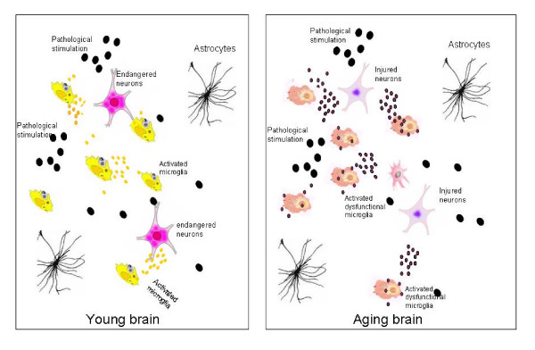
Microglia cells are the brain counterpart of macrophages and function as the first defense in the brain. Although they are neuroprotective in the young brain, microglia cells may be primed to react abnormally to stimuli in the aged brain and to become neurotoxic and destructive during neurodegeneration. Aging-induced immune senescence occurs in the brain as age-associated microglia senescence, which renders microglia to function abnormally and may eventually promote neurodegeneration. Microglia senescence is manifested by both morphological changes and alterations in immunophenotypic expression and inflammatory profile. These changes are likely caused by microinvironmental factors, but intrinsic factors cannot yet be completely excluded. Microglia senescence appears to underlie the switching of microglia from neuroprotective in the young brain to neurotoxic in the aged brain. The hypothesis of microglia senescence during aging offers a novel perspective on their roles in aging-related neurodegeneration. In Parkinson's disease and Alzheimer's disease, over-activation of microglia may play an active role in the pathogenesis because microglia senescence primes them to be neurotoxic during the development of the diseases.
Overview
The claims and statistics of this paper haven’t been checked yet.
Run the check — free, no account needed. It reads the open-access text and checks the sources it cites.
Integrity signals
Registry
Cites
Statistics
Figures
Verification
Claims the paper makes
The claims and statistics of this paper haven’t been checked yet.
Run the check — free, no account needed. It reads the open-access text and checks the sources it cites.
Claims other papers make about this one
Each is checked against this paper’s own text: does it say what the citing paper says it does?
In the citing paper
“The ageing brain is also characterised by structural and functional changes to microglial cells, which are the resident immune cell population of the central nervous system (CNS) and are the CNS equivalent of macrophages. They are activated by brain injury and local and systemic inflammation and become primed (hyper-responsive) to small stimuli with ageing, which can potentially cause damage and”
From Frailty in elderly people (2013) · cites this paper as [26]
Self-check
Percentages, sums and reported differences re-computed from the paper’s own numbers.
The self-check needs the paper’s full text.
Citations
Cites
Resting microglial cells are highly dynamic surveillants of brain parenchyma in vivo
Nimmerjahn, A., Kirchhoff, F., & Helmchen, F. (2005) · Science · doi:10.1126/science.1110647 · Cited by 5,827 · cited 2×
“By using in vivo two-photon imaging techniques, Nimmerjahn et al[37].”
BackgroundMethodInflamm‐aging: An Evolutionary Perspective on Immunosenescence
FRANCESCHI, C., BONAFÈ, M., VALENSIN, S., OLIVIERI, F., DE LUCA, M., OTTAVIANI, E., & DE BENEDICTIS, G. (2000) · Annals of the New York Academy of Sciences · doi:10.1111/j.1749-6632.2000.tb06651.x · Cited by 5,358 · cited 2×
“Extrinsic factors might include neurotransmitter, oxidative stress, neuroendocrine factors[10,68], hormones, and pathological factors such as abnormal protein aggregation [52] that exists in the microenvironment where microglia live.”
BackgroundMicroglia: a sensor for pathological events in the CNS
Kreutzberg, G. W. (1996) · Trends in Neurosciences · doi:10.1016/0166-2236(96)10049-7 · Cited by 4,396 · cited 1×
“When triggered by appropriate stimulation, microglia rapidly transformed from a resting state to an activated state with deramified shape and enhanced antigen presentation, which has been graded to a series of stages[18].”
BackgroundMicroglia: Active sensor and versatile effector cells in the normal and pathologic brain
Hanisch, U.-K., & Kettenmann, H. (2007) · Nature Neuroscience · doi:10.1038/nn1997 · Cited by 3,676 · cited 1×
“Microglia are distributed ubiquitously throughout the brain and function as resident macrophages and antigen-presenting cells in the CNS[35,36].”
BackgroundMicroglia and inflammation-mediated neurodegeneration: Multiple triggers with a common mechanism
Block, M. L., & Hong, J.-S. (2005) · Progress in Neurobiology · doi:10.1016/j.pneurobio.2005.06.004 · Cited by 1,557 · cited 1×
“These studies indicate that microglia are capable of being both neurotrophic and neurotoxic, depending on the specific stimulus, the severity of injury, and the environment [31-34].”
BackgroundMicroglial Dysfunction and Defective β-Amyloid Clearance Pathways in Aging Alzheimer's Disease Mice
Hickman, S. E., Allison, E. K., & El Khoury, J. (2008) · The Journal of Neuroscience · doi:10.1523/jneurosci.0616-08.2008 · Cited by 1,329 · cited 1×
“These genes include Abbinding scavenger receptors A (SRA), CD36, receptor for advanced glycosylation end products (RAGE), Abdegrading enzymes (insulysin, neprilysin) and MMP9 [74].”
BackgroundThe Endocrine Regulation of Aging by Insulin-like Signals
Tatar, M., Bartke, A., & Antebi, A. (2003) · Science · doi:10.1126/science.1081447 · Cited by 1,327 · cited 1×
“Insulin-like signaling pathway has been shown to regulate life span in worms, flies, and mice[ 72 ].”
BackgroundDown-Regulation of the Macrophage Lineage Through Interaction with OX2 (CD200)
Hoek, R. M., Ruuls, S. R., Murphy, C. A., Wright, G. J., Goddard, R., Zurawski, S. M., Blom, B., Homola, M. E., Streit, W. J., Brown, M. H., et al. (2000) · Science · doi:10.1126/science.290.5497.1768 · Cited by 1,022 · cited 1×
“Microglia activation is normally controlled under physiological conditions of the CNS through neuron-glia contact, CD200R-CD200 pathway[19], neuronal electrical activity[20], and/or some soluble neurotransmitters [21].”
BackgroundStreit, W. J., Braak, H., Xue, Q.-S., & Bechmann, I. (2009) · Acta Neuropathologica · doi:10.1007/s00401-009-0556-6 · Cited by 727
Dystrophic microglia in the aging human brain
Streit, W. J., Sammons, N. W., Kuhns, A. J., & Sparks, D. L. (2004) · Glia · doi:10.1002/glia.10319 · Cited by 705 · cited 2×
“”[50] Characteristics of “dystrophic” microglia that were observed in the aged brain include deramification (loss of finely branched cytoplasmic processes), cytoplasmic beading/spheroid formation, shortened and twisted cytoplasmic processes, and instances of partial or complete cytoplasmic fragmentation[50].”
Background
Cited by
Sorted by citations
Items 101–200 of 235
Age-Related Midbrain Inflammation and Senescence in Parkinson’s Disease
Russo, T., & Rießland, M. (2022) · Frontiers in Aging Neuroscience · doi:10.3389/fnagi.2022.917797 · Cited by 38
“There is evidence for an overactivation of microglia in PD patients, which could point to microglia being primed to become neurotoxic (Luo et al., 2010).”
BackgroundHong, B., Ohtake, Y., Itokazu, T., & Yamashita, T. (2024) · Cell Death Discovery · doi:10.1038/s41420-024-01816-8 · Cited by 38
Biomarkers for Microglial Activation in Alzheimer′s Disease
Lautner, R., Mattsson, N., Schöll, M., Augutis, K., Blennow, K., Olsson, B., & Zetterberg, H. (2011) · International Journal of Alzheimer s Disease · doi:10.4061/2011/939426 · Cited by 38
“It is still unknown why and when, in the course of AD, microglia switch from being beneficial to becoming neurotoxic, but age-related disturbance of the physiological function and regulation of microglia (immunosenescence) has been suggested to play an important role in the AD pathogenesis [6].”
BackgroundVillar, M. F. Z., Hanotte, J. L., Pardo, J., Morel, G. R., Mazzolini, G. D., García, M. G., & Reggiani, P. C. (2019) · Molecular Neurobiology · doi:10.1007/s12035-019-01729-z · Cited by 37
Accelerated aging in major depression: the role of nitro-oxidative stress
Luca, M., Luca, A., & Calandra, C. (2013) · Oxidative Medicine and Cellular Longevity · doi:10.1155/2013/230797 · Cited by 37
Lucke-Wold, B. P., Turner, R. C., Lucke-Wold, N., Rosen, C. L., & Huber, J. D. (2012) · Europe PMC (PubMed Central) · Cited by 36
Yang, H., Feng, G., Liang, Z., Vitale, A. V., Jiao, X., Ju, G., & You, S. (2012) · Neurochemistry International · doi:10.1016/j.neuint.2012.04.020 · Cited by 37
“…serum; DMEM, Dulbecco ethylenediamine tetraacetic species; ERK, extracellular ase; EGF, epidermal growth r necrosis factor. x: +86 29 83246270. ang), yousiwei@fmmu.edu.cn changes in their surrounding micro-environment and thus play a major role in maintaining the CNS homeostasis (Luo et al., 2010).”
Leptin and ghrelin: Sewing metabolism onto neurodegeneration
de Candia, P., & Matarese, G. (2017) · Neuropharmacology · doi:10.1016/j.neuropharm.2017.12.025 · Cited by 38
Kermanshahi, S., Ghanavati, G., Mesrabadi, M. A., Gholami, M., Ulloa, L., Motaghinejad, M., & Safari, S. (2020) · Neurochemical Research · doi:10.1007/s11064-020-03134-8 · Cited by 36
Yang, S., Liu, W., Lü, S., Tian, Y., Wang, W., Ling, T.-J., & Liu, R. (2016) · ACS Chemical Neuroscience · doi:10.1021/acschemneuro.6b00091 · Cited by 36
An, Y., Li, Y., Hou, Y., Huang, S., & Pei, G. (2022) · Oxidative Medicine and Cellular Longevity · doi:10.1155/2022/3086010 · Cited by 37
“Cell senescence also leads to microglia dysfunction, resulting in inaccurate response to external stimuli and neurodegeneration worsening [8, 10, 11].”
Microglia and modifiable life factors: Potential contributions to cognitive resilience in aging
Duggan, M. R., & Parikh, V. (2021) · Behavioural Brain Research · doi:10.1016/j.bbr.2021.113207 · Cited by 38
New Th2 adjuvants for preventive and active immunotherapy of neurodegenerative proteinopathies
Marciani, D. J. (2014) · Drug Discovery Today · doi:10.1016/j.drudis.2014.02.015 · Cited by 34
Jia, Z., Xue, R., Ma, S., Xu, J., Guo, S., Li, S., Zhang, E., Wang, J., & Yang, J. (2015) · Molecular Neurobiology · doi:10.1007/s12035-015-9438-1 · Cited by 35
Miller, A. P., Shah, A. S., Aperi, B. V., Budde, M. D., Pintar, F. A., Tarima, S. S., Kurpad, S. N., Stemper, B. D., & Glavaski‐Joksimovic, A. (2015) · Frontiers in Neurology · doi:10.3389/fneur.2015.00020 · Cited by 33
“Alongside astrogliosis, microglial activation has been strongly associated with various neurodegenerative disorders, including blast (32, 38, 120) and non-blast TBI (131), Parkinson’s disease (132, 133), and Alzheimer’s disease (134, 135), among many others.”
Harackiewicz, O., & Grembecka, B. (2024) · Journal of Integrative Neuroscience · doi:10.31083/j.jin2311203 · Cited by 39
“Microglial activity changes with aging as these cells become more predisposed to transition into inflammatory phenotypes [107,108].”
Pardo, J., Abba, M. C., Lacunza, E., Francelle, L., Morel, G. R., Outeiro, T. F., & Goya, R. G. (2017) · Hippocampus · doi:10.1002/hipo.22703 · Cited by 32
“, 2009), with microglia playing a central role in this immune dysregulation (Luo et al., 2010; Norden et al., 2013).”
BackgroundAutophagy and NLRP3 inflammasome crosstalk in neuroinflammation in aged bovine brains
Biase, D. D., Piegari, G., Prisco, F., Cimmino, I., Pirozzi, C., Raso, G. M., Oriente, F., Grieco, E., Papparella, S., & Paciello, O. (2020) · Journal of Cellular Physiology · doi:10.1002/jcp.29426 · Cited by 32
Stojiljkovic, M. R., Schmeer, C. W., & Witte, O. W. (2022) · Neuroscience · doi:10.1016/j.neuroscience.2022.02.018 · Cited by 31
“Moreover, microglial senescence seems to switch these cells from neuropro- /licenses/by/4.0/). tective in the young brain to neurotoxic in the aged brain (Luo et al., 2010).”
BackgroundInfluentialMiller, S., Campbell, C. E., Jimenez-Corea, H. A., Wu, G., & Logan, R. A. (2022) · Frontiers in Neuroscience · doi:10.3389/fnins.2022.824191 · Cited by 34
“microglia and produce an enhanced neuroinflammatory response to injury, compared to young brains (Sugama et al., 2003; Wasserman and Schlichter, 2008; Luo et al., 2010).”
BackgroundFujikawa, R., & Jinno, S. (2022) · European Journal of Neuroscience · doi:10.1111/ejn.15812 · Cited by 29
“For instance, de-ramified microglia were found in aged brains, and they were distinguished from hypertrophic microglia by retracted and thickened processes (Luo et al., 2010).”
BackgroundMaroon, J. C., LePere, D. B., Blaylock, R. L., & Bost, J. W. (2012) · The Physician and Sportsmedicine · doi:10.3810/psm.2012.11.1990 · Cited by 32
Amiraslani, B., Sabouni, F., Abbasi, S., et al. (1996) · PubMed · doi:10.6091/ibj.1012.2012 · Cited by 31
“mongst three types of glial cells, including microglia, oligodandrocytes and astrocytes, it seems that, microglial cells with a mesenchymal origin [1-3] are the first-line defense [4, 5] in the central nervous system.”
BackgroundLiu, J., Zhou, L., Wang, Y.-Z., Wang, X., Zhou, Y., Ho, W., & Li, J. (2016) · Journal of Immunology Research · doi:10.1155/2016/4962351 · Cited by 30
Translocator protein in late stage Alzheimer’s disease and Dementia with Lewy bodies brains
Xu, J., Sun, J., Perrin, R. J., Mach, R. H., Bales, K. R., Morris, J. C., Benzinger, T. L. S., & Holtzman, D. M. (2019) · Annals of Clinical and Translational Neurology · doi:10.1002/acn3.50837 · Cited by 31
Microglia in the normally aged hippocampus
Choi, J. H., & Won, M. (2011) · Laboratory Animal Research · doi:10.5625/lar.2011.27.3.181 · Cited by 29
“Numerous studies and reviews have reported that numerical, morphological and functional changes in microglia are apparently changed in the normal aging brain and in the aged brain with diseases [1,2,6,12,17-21].”
BackgroundMotawaj, M., Peoc’h, K., Callebert, J., & Arrang, J. (2010) · Journal of Alzheimer s Disease · doi:10.3233/jad-2010-100381 · Cited by 30
“Recent findings indicate that microglia becomes also more reactive during normal aging and it was suggested that this age-associated microglia senescence eventually promotes neurodegeneration [69–71].”
Pathways towards an effective immunotherapy for Parkinson’s disease
Hutter-Saunders, J. A. L., Mosley, R. L., & Gendelman, H. E. (2011) · Expert Review of Neurotherapeutics · doi:10.1586/ern.11.163 · Cited by 28
Model Senescent Microglia Induce Disease Related Changes in α-Synuclein Expression and Activity
Angelova, D. M., & Brown, D. R. (2018) · Biomolecules · doi:10.3390/biom8030067 · Cited by 30
Cognitive resilience in Alzheimer’s disease: Mechanism and potential clinical intervention
Jia, B., Xu, Y., & Zhu, X. (2025) · Ageing Research Reviews · doi:10.1016/j.arr.2025.102711 · Cited by 30
Cornejo, F., & Rommy von Bernhardi, M. (2013) · Mediators of Inflammation · doi:10.1155/2013/895651 · Cited by 25
Li, Y., & Ju, D. (2016) · Elsevier eBooks · doi:10.1016/b978-0-12-804598-5.00012-x · Cited by 25
Honig, M. G., Dorian, C. C., Worthen, J. D., Micetich, A. C., Mulder, I. A., Sanchez, K. B., Pierce, W. F., Mar, N. A. D., & Reiner, A. J. (2020) · European Journal of Neuroscience · doi:10.1111/ejn.14711 · Cited by 26
Ensheathing glia promote increased lifespan and healthy brain aging
Sheng, L., Shields, E. J., Gospočić, J., Sorida, M., Ju, L., Byrns, C. N., Carranza, F. R., Berger, S. L., Bonini, N. M., & Bonasio, R. (2023) · Aging Cell · doi:10.1111/acel.13803 · Cited by 24
Hadad, N., & Lévy, R. (2017) · NeuroMolecular Medicine · doi:10.1007/s12017-017-8459-5 · Cited by 24
“Although microglia activation is normally controlled under physiological conditions of the CNS through neuron–glia interaction, in the healthy aged brain microglia lose control and acquire a proinflammatory M1 profile resulting in deleterious and neurotoxic consequences provoked by their excessive production of pro-in…”
InfluentialThe Impact of Non-Neurotropic Influenza Strains on the Brain: A Role for Microglial Priming?
Barbosa‐Silva, M. C., Santos, L. E., & Rangel, B. (2018) · Journal of Neuroscience · doi:10.1523/jneurosci.1368-18.2018 · Cited by 23
“Priming of microglia has been shown to occur indirectly, through the release of peripheral cytokines that reach the CNS, and even as a result of atypical insults, such as the mild inflammation that accompanies normal aging (Luo et al., 2010).”
BackgroundNeuroinflammation and Alzheimer's disease: Unravelling the molecular mechanisms
Kakkar, A., Singh, H., Singh, B. K., Kumar, A., Mishra, A. K., & Chopra, H. (2025) · Journal of Alzheimer s Disease · doi:10.1177/13872877251374353 · Cited by 26
“33,34 In this context, microglia were revealed to cluster in regions immediately surrounding A β deposits by elegant immunohistochemical studies.”
Sinha, M. S., Bir, A., Banerjee, A., Bhowmick, P., & Chakrabarti, S. (2015) · Neurochemistry International · doi:10.1016/j.neuint.2015.10.003 · Cited by 22
Sorrentino, S., Ascari, R., Maderna, E., Catania, M., Ghetti, B. F., Tagliavini, F., Giaccone, G., & Fede, G. D. (2021) · International Journal of Molecular Sciences · doi:10.3390/ijms22052780 · Cited by 23
“Moreover, the age-associated microglia senescence can negatively impact the microglia functionality and eventually trigger and sustain neurodegeneration [79,80].”
BackgroundMonzón-Sandoval, J., Burlacu, E., Agarwal, D., Handel, A. E., Wei, L., Davis, J., Cowley, S. A., Cader, M. Z., & Webber, C. (2022) · Disease Models & Mechanisms · doi:10.1242/dmm.049349 · Cited by 18
“The normal functioning of microglia is key to brain homeostasis, while their functional disruption, prolonged activation or ageing may contribute to pathological conditions (Luo et al., 2010).”
Serum hs-CRP Levels are Increased in de Novo Parkinson's Disease Independently from Age of Onset
Song, I., Cho, H.-J., Kim, J.-S., Park, I.-S., & Lee, K. (2014) · European Neurology · doi:10.1159/000363570 · Cited by 19
Stevenson, A. J., McCartney, D. L., Shireby, G. L., Hillary, R. F., King, D., Tzioras, M., Wrobel, N., McCafferty, S., Murphy, L., McColl, B. W., et al. (2022) · European Journal of Neuroscience · doi:10.1111/ejn.15661 · Cited by 20
“This altered phenotype can lead to 389 exaggerated neuro-inflammation in response to peripheral or central immune challenges which can 390 precipitate neuro-toxicity, and thus, degeneration (40, 41).”
BackgroundReduction of the ATPase inhibitory factor 1 (IF1) leads to visual impairment in vertebrates
Martín-Jiménez, R., Faccenda, D., Allen, E., Reichel, H. B., Arcos, L., Ferraina, C., Strobbe, D., Russell, C., & Campanella, M. (2018) · Cell Death and Disease · doi:10.1038/s41419-018-0578-x · Cited by 17
Kwon, H. J., Kim, D., Seo, K., Kim, Y. G., Han, S. I., Kang, T., Soh, M., & Hyeon, T. (2018) · Angewandte Chemie · doi:10.1002/ange.201805052 · Cited by 18
Seiffert, P., Derejczyk, J., Kawa, J., Marcisz, C., Czernek, M., Szymszal, J., Kapko, W., Bugdol, M. N., Torbus, A., & Stȩpień-Wyrobiec, O. (2017) · Biogerontology · doi:10.1007/s10522-017-9716-6 · Cited by 16
Zhuang, H., Li, Q., Sun, C., Xu, D., Gan, G., Zhang, C., Chen, C., Yuan, Y., Liu, L., Xiao, Y. M., et al. (2023) · Brain Behavior and Immunity · doi:10.1016/j.bbi.2023.09.002 · Cited by 15
Stress regulates Alzheimer’s disease progression via selective enrichment of CD8+ T cells
Feng, Y., Fan, J., Cheng, Y., Dai, Q., & Ma, S. (2023) · Cell Reports · doi:10.1016/j.celrep.2023.113313 · Cited by 19
Cho, H. J., Verbridge, S. S., Davalos, R. V., & Lee, Y. W. (2018) · Annals of Biomedical Engineering · doi:10.1007/s10439-018-2004-z · Cited by 15
Chen, Y., Cai, M., Xu, S. Q., Meng, M., Ren, X., Yang, J. W., Dong, Y.-Q., Liu, X., Yang, J., & Xiong, X.-D. (2016) · Oncotarget · doi:10.18632/oncotarget.10170 · Cited by 15
“Besides, senescence has been implicated as a detrimental factor for agerelated diseases such as cardiovascular disease, diabetes, neurodegenerative disease, cancer and many other diseases [9-12].”
BackgroundSerine Racemase Expression Differentiates Aging from Alzheimer’s Brain
Wu, S., Zhou, J., Zhang, H., & Barger, S. W. (2022) · Current Alzheimer Research · doi:10.2174/1567205019666220805105106 · Cited by 14
Wang, P., Mi, Y., Yu, H. N., Teng, X., Jin, S., Xiao, L., Xue, H., Tian, D., Guo, Q., & Wu, Y. (2024) · Biomedicine & Pharmacotherapy · doi:10.1016/j.biopha.2024.116549 · Cited by 15
Daghestani, H. N., Zhu, G., Johnston, P. A., Shinde, S. N., Brodsky, J. L., & Day, B. W. (2011) · Assay and Drug Development Technologies · doi:10.1089/adt.2010.0367 · Cited by 14
Chamniansawat, S., & Chongthammakun, S. (2014) · NeuroToxicology · doi:10.1016/j.neuro.2014.11.005 · Cited by 14
Marx, M., Diestel, S., Bozon, M., Keglowich, L., Drouot, N., Bouché, E., Frébourg, T., Minz, M., Saugier-Véber, P., Castellani, V., et al. (2012) · Neurogenetics · doi:10.1007/s10048-011-0307-4 · Cited by 14
Neurodegeneration, demyelination, and astrogliosis in rat spinal cord by chronic lead treatment
Villa‐Cedillo, S. A., Nava-Hernández, M. P., Soto‐Domínguez, A., Hernández‐Ibarra, J. A., Perez‐Trujillo, J. J., & Cárdenas, O. S. (2019) · Cell Biology International · doi:10.1002/cbin.11147 · Cited by 13
“Microglia act as the first and main form of active immune defense in the brain and spinal cord (Luo et al., 2010).”
Advances in proteomic phenotyping of microglia in neurodegeneration
Sunna, S. N., Bowen, C. A., Ramelow, C. C., Santiago, J. V., Kumar, P., & Rangaraju, S. (2023) · PROTEOMICS · doi:10.1002/pmic.202200183 · Cited by 13
Endocannabinoid Signaling for GABAergic-Microglia (Mis)Communication in the Brain Aging
Carrera, J., Tomberlin, J., Kurtz, J., Karakaya, E., Bostancıklıoğlu, M., & Albayram, Ö. (2021) · Frontiers in Neuroscience · doi:10.3389/fnins.2020.606808 · Cited by 12
“The ECS is comprised of endogenous cannabinoid receptor ligands, biosynthetic and metabolic enzymes, and G-protein coupled cannabinoid type 1 receptor (CB1) and cannabinoid type 2 receptor (CB2) expressed in neuron and microglia, respectively (Luongo et al., 2010; Albayram et al., 2011; Figure 1).”
BackgroundMesenchymal stem cell therapy improves spatial memory and hippocampal structure in aging rats
Villar, M. F. Z., Lehmann, M., García, M. G., Mazzolini, G. D., Morel, G. R., Cónsole, G. M., Podhajcer, O. L., Reggiani, P. C., & Goya, R. G. (2019) · Behavioural Brain Research · doi:10.1016/j.bbr.2019.04.001 · Cited by 12
“Immune reactivity and inflammatory processes increase with aging in the brain [18], with microglia playing a central role in this immune dysregulation [19, 20].”
BackgroundAdministration of Glucosylceramide Ameliorated the Memory Impairment in Aged Mice
Lee, Y., Oliynyk, S., Jung, J., Han, J. J., & Oh, S. (2013) · Evidence-based Complementary and Alternative Medicine · doi:10.1155/2013/824120 · Cited by 12
Aging-induced alterations in microglial cells and their impact on neurodegenerative disorders
Singh, H., Gupta, R., Gupta, M., & Ahmad, A. (2025) · Molecular Biology Reports · doi:10.1007/s11033-025-10623-y · Cited by 11
“Decreased soma volume, less active tissue sampling, retraction of processes, decreased synaptic contact, abnormalities in the cytoplasm and non-uniform tissue distribution are an indication of microglial senescence [4, 13, 52].”
InfluentialPerdaens, O., & Pesch, V. V. (2024) · International Journal of Molecular Sciences · doi:10.3390/ijms252312637 · Cited by 12
Börner, J. H., Rawashdeh, O., & Rami, A. (2021) · Antioxidants · doi:10.3390/antiox10091330 · Cited by 12
“Possible consequences of these microglial alterations with aging might be a dysregulated response to injuries, changes in neuroprotective functions, and an increase in neurotoxic inflammatory responses [47].”
BackgroundHuang, C., Lu, X., Wang, J. L., Tong, L., Ling, Y., Jiang, B., Yang, R., & Zhang, W. (2016) · Neuroscience · doi:10.1016/j.neuroscience.2016.06.018 · Cited by 11
Involvement of CtBP2 in LPS-induced microglial activation
Zhang, G., Yan, Y., Kang, L., et al. (2012) · Journal of Molecular Histology · doi:10.1007/s10735-012-9399-x · Cited by 11
“Microglia are resident macrophages of the brain and spinal cord, and thus act as the first and main form of active immune defense in the central nervous system (CNS) (Luo et al. 2010).”
BackgroundFeng, J., Liu, W., Feng, D., Chitrakar, B., Chen, X., Sang, Y., & Wang, X. (2024) · Journal of Functional Foods · doi:10.1016/j.jff.2024.106441 · Cited by 10
Phorbol myristate acetate induces cellular senescence in rat microglia in vitro
Cao, D., Li, X.-H., Luo, X., Yu, H., Wan, L.-S., Wei, L., & Ren, Y. (2020) · International Journal of Molecular Medicine · doi:10.3892/ijmm.2020.4587 · Cited by 11
Self-citationJia, J., Liu, C., Han, Y., Han, H., Zhong, M., & Gao, Y. (2020) · Human & Experimental Toxicology · doi:10.1177/0960327120930069 · Cited by 10
“TNF-a is also functioned by these roles of mediating systemic inflammation and stimulating the acute phase reaction.(21) As shown in Figure 2, when compared to BC group, the expression of IL-1b, TNF-a, and IL-6 increased significantly (p < 0.”
ResultMarchese, E., Maria, V. D., Samengo, D., Pani, G., Michetti, F., & Geloso, M. C. (2017) · Neurochemical Research · doi:10.1007/s11064-017-2233-9 · Cited by 10
Cell ageing: a flourishing field for neurodegenerative diseases
Brites, D. M. T. O. (2015) · AIMS molecular science · doi:10.3934/molsci.2015.3.225 · Cited by 10
“Dopaminergic neurons seem to be particularly vulnerable to oxidative stress by the deregulated microglia [76] and the reduced clearance of Aβ peptide by the senescent microglia contributes to the pathogenesis of AD [77].”
BackgroundMacKenzie, J. L., Ivanova, N., Nell, H. J., Giordano, C. R., Terlecky, S. R., Agca, C., Ağca, Y., Walton, P. A., Whitehead, S. N., & Cechetto, D. F. (2022) · Neuroscience · doi:10.1016/j.neuroscience.2022.01.026 · Cited by 9
Tissue-specific features of microglial innate immune responses
Timmerman, R., Burm, S. M., & Bajramovic, J. J. (2020) · Neurochemistry International · doi:10.1016/j.neuint.2020.104924 · Cited by 10
Microglia: The Bodyguard and the Hunter of the Adult Neurogenic Niche
Valero, J., Eiriz, M. F., Santos, T., Neiva, I., Ferreira, R., & Malva, J. O. (2012) · Humana Press eBooks · doi:10.1007/978-1-61779-940-2_14 · Cited by 9
Assale, T. A., Afrang, N., Wißfeld, J., Cuevas-Ríos, G., Klaus, C., Linnartz‐Gerlach, B., & Neumann, H. (2024) · Frontiers in Neuroscience · doi:10.3389/fnins.2024.1504765 · Cited by 10
“Chronic reactive microgliosis and the resulting neuroinflammation have been implicated in age-related neurodegeneration, which can lead to dementia (Luo et al., 2010).”
Gut dysbiosis leads to cognitive decline through CNTF-mediated activation of microglia in mice
Krishnapriya, Rishikesh, R., Kishanchand, D. K., Haack, I., Hosseini, S., Deepti, A., Sudhadevi, T., Michaelsen‐Preusse, K., Sivan, U., Körte, M., et al. (2025) · BMC Biology · doi:10.1186/s12915-025-02454-x · Cited by 8
Wang, Y., Yang, L. C., & Yang, D. (2015) · Neurochemical Research · doi:10.1007/s11064-015-1624-z · Cited by 8
Npc1 deficiency impairs microglia function via TREM2-mTOR signaling in Niemann-Pick disease type C
Qiao, L., Han, X., Ru, D., Shang, X., Xiao, L., Gao, G., Zhang, C., Kang, J., Su, X., Liu, Y., et al. (2024) · Biochimica et Biophysica Acta (BBA) - Molecular Basis of Disease · doi:10.1016/j.bbadis.2024.167478 · Cited by 8
Melatonin Delays Brain Aging by Decreasing the Nitric Oxide Level
AKBULUT, K. G., Güney, Ş., Çeti̇n, F., Akgun, N., Aktaş, S. H., & Akbulut, H. (2013) · Neurophysiology · doi:10.1007/s11062-013-9368-3 · Cited by 7
“by lipopolysaccharides was found during aging [34].”
Establishing the relationship between brain cellular senescence and brain structure
Lund, A. N., Kopell, B. H., Golestani, N., Vornholt, E., Thompson, R. C., Liharska, L. E., Luo, Y., Cheng, E., Park, Y. J., Fennessy, B., et al. (2026) · Cell · doi:10.1016/j.cell.2025.10.014 · Cited by 6
Wu, C. J., Bendriem, R. M., Freed, W. J., & Lee, C. (2021) · Restorative Neurology and Neuroscience · doi:10.3233/rnn-211161 · Cited by 7
Anti-inflammatory role of GM1 and modulatory effects of gangliosides on microglia functionsPreprint
Galleguillos, D., Wang, Q., Steinberg, N., Shrivastava, G., Dhami, K., Rubinstein, K., Giuliani, F., Churchward, M. A., Power, C., Todd, K. G., et al. (2020) · bioRxiv (Cold Spring Harbor Laboratory) · doi:10.1101/2020.03.04.975862 · Cited by 6
“With ageing, homeostatic and neurotrophic activities of microglia decline or become progressively impaired [8], promoting the onset and/or progression of neurodegenerative conditions [9-11].”
BackgroundAgeing, Neurodegeneration and the Endocannabinoid System
Fernández‐Ruíz, J., Sagredo, O., Gómez‐Ruíz, M., & de Lago, E. (2025) · Current topics in behavioral neurosciences · doi:10.1007/7854_2025_597 · Cited by 5
Effect of novel quercetin pivaloyl ester on functions of adult rat microglia
Kuniaková, M., Mrvová, N., Knezl, V., & Račková, L. (2015) · Biologia · doi:10.1515/biolog-2015-0082 · Cited by 5
Natural products and the aging process
Ergin, V., Bali, E. B., Hariry, R. E., & Karasu, Ç. (2013) · Hormone Molecular Biology and Clinical Investigation · doi:10.1515/hmbci-2013-0031 · Cited by 5
Predictive Value of Inflammation Markers for Frailty in Older Patients with CVD
Hu, S., Lai, X., Shi, Y., Chai, H., Guo, Z., Liu, D., & Cui, C. (2025) · Clinical Interventions in Aging · doi:10.2147/cia.s502617 · Cited by 4
“57,58 59–61 8.”
Polyphenol Supplementation Reverses Age-Related Changes in Microglial Signaling Cascades
Jalloh, A., Flowers, A., Hudson, C., Chaput, D., Guergues, J., Stevens, S. M., & Bickford, P. C. (2021) · International Journal of Molecular Sciences · doi:10.3390/ijms22126373 · Cited by 5
“Primed microglia are hyper-responsive to pro-inflammatory stimuli and show a blunted response to anti-inflammatory signals [11].”
BackgroundLiu, L., Luo, X., YU, H.-M., Feng, Y., Ren, Y., Yin, Y.-F., Shang, H., & He, Z. (2015) · Molecular Medicine Reports · doi:10.3892/mmr.2015.4412 · Cited by 4
“The neurotoxic effects of the activated microglia in the aged neurodegenerative brain may result in age-associated microglia senescence (4).”
Self-citationAn introduction to innate immunity in the central nervous system
Harry, G. J., Childers, G. M., & McPherson, C. A. (2018) · Advances in neurotoxicology · doi:10.1016/bs.ant.2018.10.002 · Cited by 3
Ahn, J. H., Luo, W., Kim, J., et al. (2010) · Assay and Drug Development Technologies · doi:10.1089/adt.2010.0327 · Cited by 3
Does the MK2-dependent Production of TNFα Regulate mGluR-dependent Synaptic Plasticity?
Hogg, E. L., Müller, J., & Corrêa, S. A. L. (2015) · Current Neuropharmacology · doi:10.2174/1570159x13666150624165939 · Cited by 3
“associated with increased microglia activity, this is thought to cause an over production of TNFα in the normal aging brain [38,39].”
Allogeneic Mesenchymal Stem Cells as a Treatment for Aging Frailty
Tompkins, B. A., Landin, A. M., Florea, V., Natsumeda, M., Rieger, A. C., Balkan, W., Schulman, I. H., & Hare, J. M. (2017) · InTech eBooks · doi:10.5772/intechopen.69194 · Cited by 3
“Concomitant with changes in the immune system, microglial cells, which are the resident immune cells of the central nervous system, are also structurally and functionally altered with aging and undergo senescence, likely causing damage and neuronal death [8].”
BackgroundSingle cell analysis of the aging female hypothalamusPreprint
Hajdarovic, K. H., Yu, D., Hassell, L.-A., Evans, S. A., Packer, S., Neretti, N., & Webb, A. E. (2021) · bioRxiv (Cold Spring Harbor Laboratory) · doi:10.1101/2021.03.07.434282 · Cited by 3
Alrouji, M., Alshammari, M. S., Alhajlah, S., Syed, T., Ashraf, G. M., Dinislam, K., Shahwan, M. J., & Shamsi, A. (2026) · Engineering · doi:10.1016/j.eng.2025.12.030 · Cited by 3
Impact of sex differences on microglial function in Alzheimer’s disease
Figueiredo, E. V., Azevedo, C. V., Penitente, A. R., Pedrosa, V. R., da Silva, V. R. C., & Longo, B. M. (2026) · Alzheimer s Research & Therapy · doi:10.1186/s13195-026-02099-0 · Cited by 2
Prenatal-induced psychopathologies: All roads lead to microglia
Lebow, M., Kuperman, Y., & Chen, A. (2023) · Elsevier eBooks · doi:10.1016/b978-0-12-817558-3.00016-0 · Cited by 2
Frailty Syndrome in the Elderly and Omega-3 Polyunsaturated Fatty Acids
Cin, P., & Büyükuslu, N. (2020) · Ankara Medical Journal · doi:10.5505/amj.2020.92260 · Cited by 2
Melatonina sintetizada por microglias de cerebelo em cultura regula o processo de fagocitose
dos Santos, A. A. (2015) · Digital Library of Theses and Dissertations (Universidade de São Paulo) · doi:10.11606/d.41.2015.tde-17072015-153623 · Cited by 2
Reagin, K. L., Lee, R., & Funk, K. E. (2025) · Current Protocols · doi:10.1002/cpz1.70199 · Cited by 1
“Aged microglia also show increased phagocytic ability and prolonged proliferation and activation, even after resolution of the inflammatory event (Antignano et al., 2023; Godbout et al., 2005; Luo et al., 2010; Miller & Streit, 2007; Xu et al., 2022).”
Zhao, M., Zhang, J., Yang, Q., Chen, L., Shen, H., Pu, Y., Wei, W., Han, Y., & Zhang, P. (2025) · Frontiers in Aging Neuroscience · doi:10.3389/fnagi.2025.1570482 · Cited by 1
“Although microglia cells are generally favorable initially in performing immune functions, prolonged or excessive activation can lead to cytotoxic eects (Luo et al., 2010; McGeer and McGeer, 2001).”
Guo, H., Peng, X., Zhang, X. C., Ruganzu, J. B., Wu, X., Zhao, M., Pengbo, Y., Ji, S., & Yang, W. (2026) · Experimental Cell Research · doi:10.1016/j.yexcr.2026.114920 · Cited by 1
Waghmare, A., Rahangdale, S. R., Khare, K., Taksande, B. G., Umekar, M. J., & Mangrulkar, S. V. (2026) · Molecular and Cellular Neuroscience · doi:10.1016/j.mcn.2026.104075 · Cited by 1
Metadata
- Authors
- Luo, X.-G., Ding, J.-Q., & Chen, S.-D.
- Venue
- Molecular Neurodegeneration
- Published
- 2010-03-24
- Volume
- 5
- Issue
- 1
- Pages
- 12-12
- ISSN
- 1750-1326 (ISSN-L)
- PMC
- PMC2852379
- OpenAlex
- W2156427445
- Cited by
- 236
- References
- 85
- Citations by year
- 2026: 7 · 2025: 12 · 2024: 10 · 2023: 11 · 2022: 17 · 2021: 14 · 2020: 15 · 2019: 11 · 2018: 20 · 2017: 13 · 2016: 18 · 2015: 19 · 2014: 12 · 2013: 22 · 2012: 15
- FWCI
- 10.86
- Type
- journal article
- Language
- en
- Open access
- gold
- doi.org
- Licence
- cc-by
- Locations
- Molecular Neurodegeneration (publishedVersion, cc-by)
- PubMed Central (publishedVersion, cc-by)
- PubMed (publishedVersion)
- DOAJ (DOAJ: Directory of Open Access Journals) (submittedVersion, cc-by-sa)
- Topics
- Life Sciences › Neuroscience › Neurology › Neuroinflammation and Neurodegeneration Mechanisms
- Life Sciences › Biochemistry, Genetics and Molecular Biology › Molecular Biology › Inflammasome and immune disorders
- Life Sciences › Neuroscience › Neurology › Neurological Disease Mechanisms and Treatments
- Keywords
- microglia, brain aging, neurodegeneration, microglial senescence, immunosenescence, neuroinflammation, neuroprotection, Parkinson's disease, Alzheimer's disease, microglial activation, neurotoxicity
About these checks
Registry, retraction, Cites and statistics results are computed, not judged, with one exception: a printed reference that no index lists is read once by a model before it counts as a citation error. Claims are extracted by a model and kept only when their quote appears verbatim in the text; each cited source is then checked, with a model’s help, for whether it says what the claim relies on. Those verdicts can be wrong — read the claim’s quote and the cited source before relying on one.
Statistics and arithmetic are recomputed, not judged.
Confidence bands are shown only for fields calibrated on our annotated test set. None is calibrated yet, so no band is shown.
What counts as a claim ›How we measure confidence ›How we check this ›
Are you an author, or do you see a problem with this page? Request a review
