

The DOI matches its Crossref record; 5 of the 5 references it cites were found in a registry or an index.
Astrocytic Ca2+ signals are required for the functional integrity of tripartite synapses
Abstract
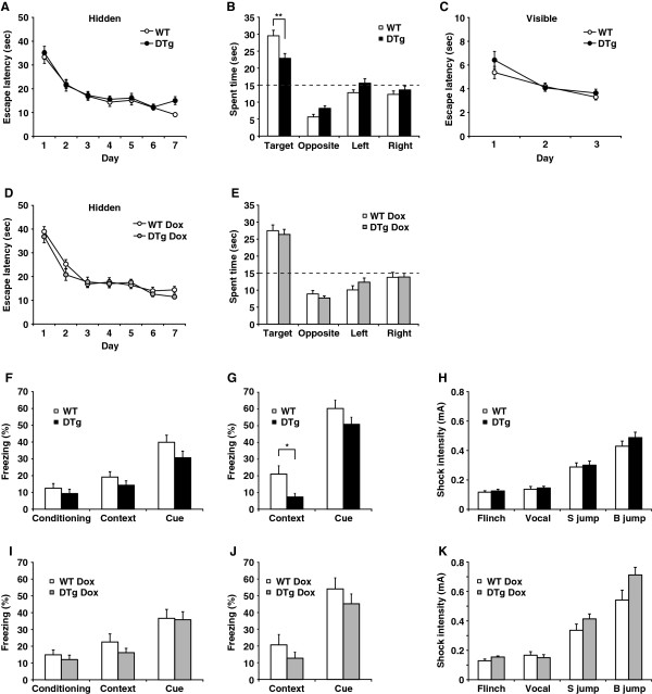
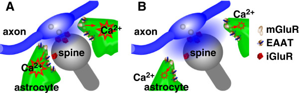
Background: Neuronal activity alters calcium ion (Ca 2+) dynamics in astrocytes, but the physiologic relevance of these changes is controversial. To examine this issue further, we generated an inducible transgenic mouse model in which the expression of an inositol 1,4,5-trisphosphate absorbent, “IP 3 sponge”, attenuates astrocytic Ca 2+signaling. Results: Attenuated Ca 2+activity correlated with reduced astrocytic coverage of asymmetric synapses in the hippocampal CA1 region in these animals. The decreased astrocytic ‘protection’ of the synapses facilitated glutamate ‘spillover’, which was reflected by prolonged glutamate transporter currents in stratum radiatum astrocytes and enhanced N-methyl-D-aspartate receptor currents in CA1 pyramidal neurons in response to burst stimulation. These mice also exhibited behavioral impairments in spatial reference memory and remote contextual fear memory, in which hippocampal circuits are involved. Conclusions: Our findings suggest that IP 3-mediated astrocytic Ca 2+signaling correlates with the formation of functional tripartite synapses in the hippocampus.
Overview
The claims and statistics of this paper haven’t been checked yet.
Run the check — free, no account needed. It reads the open-access text and checks the sources it cites.
Integrity signals
Registry
Cites
Statistics
Figures
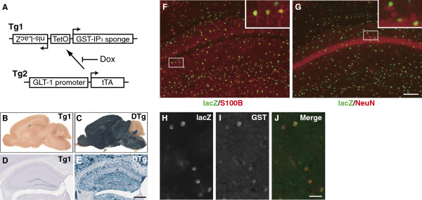
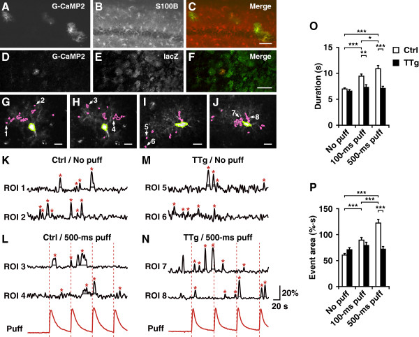
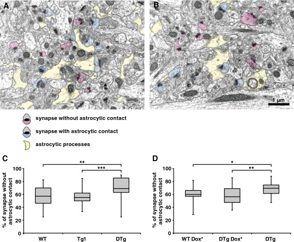
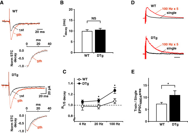
Verification
Claims the paper makes
The claims and statistics of this paper haven’t been checked yet.
Run the check — free, no account needed. It reads the open-access text and checks the sources it cites.
Claims other papers make about this one
Each is checked against this paper’s own text: does it say what the citing paper says it does?
The source backs part of the claim, not all of it — or backs it for a different population, condition or comparison.
In the citing paper
“The evidence indicates that astrocyte atrophy and reduced synaptic coverage are key factors in cognitive disorders76,77.”
From Cholesterol-driven pathological astrocytic responses in diabetes-associated cognitive impairment through astrocytic SCAP accumulation and NF-κB–C3 signaling modulation (2025) · cites this paper as [76]
Experimental — accuracy not yet measuredIn the citing paper
“Astrocyte atrophy results in decreased territory size and synaptic coverage, impairing synaptic plasticity and contributing to cognitive dysfunction.”
Self-check
Percentages, sums and reported differences re-computed from the paper’s own numbers.
The self-check needs the paper’s full text.
Citations
Cites
Glutamate Induces Calcium Waves in Cultured Astrocytes: Long-Range Glial Signaling
Cornell-Bell, A. H., Finkbeiner, S. M., Cooper, M. S., & Smith, S. J. (1990) · Science · doi:10.1126/science.1967852 · Cited by 1,846
Long-term potentiation depends on release of d-serine from astrocytes
Henneberger, C., Papouin, T., Oliet, S. H. R., & Rusakov, D. A. (2010) · Nature · doi:10.1038/nature08673 · Cited by 1,389
The tripartite synapse: roles for gliotransmission in health and disease
Halassa, M. M., Fellin, T., & Haydon, P. G. (2007) · Trends in Molecular Medicine · doi:10.1016/j.molmed.2006.12.005 · Cited by 852
Astrocytes Mediate In Vivo Cholinergic-Induced Synaptic Plasticity
Navarrete, M., Perea, G., de Sevilla, D. F., Gómez-Gonzalo, M., Núñez, A., Martín, E. D., & Araque, A. (2012) · PLoS Biology · doi:10.1371/journal.pbio.1001259 · Cited by 387
Astrocyte Calcium Signaling Transforms Cholinergic Modulation to Cortical Plasticity In Vivo
Takata, N., Mishima, T., Hisatsune, C., Nagai, T., Ebisui, E., Mikoshiba, K., & Hirase, H. (2011) · The Journal of Neuroscience · doi:10.1523/jneurosci.5289-11.2011 · Cited by 382
Self-citation
Research works printed but not linked: 59
- Venance L, Stella N, Glowinski J, Giaume C: Mechanism involved in initiation and propagation of receptor-induced intercellular calcium signaling in cultured rat astrocytes. J Neurosci. 1997, 17: 1981-1992. · DOI
- Verkhratsky A, Orkand RK, Kettenmann H: Glial calcium: homeostasis and signaling function. Physiol Rev. 1998, 78: 99-141. · DOI
- Agulhon C, Petravicz J, McMullen AB, Sweger EJ, Minton SK, Taves SR, Casper KB, Fiacco TA, McCarthy KD: What is the role of astrocyte calcium in neurophysiology?. Neuron. 2008, 59: 932-946. 10.1016/j.neuron.2008.09.004. · DOI
- Petravicz J, Fiacco TA, McCarthy KD: Loss of IP3 receptor-dependent Ca2+ increases in hippocampal astrocytes does not affect baseline CA1 pyramidal neuron synaptic activity. J Neurosci. 2008, 28: 4967-4973. 10.1523/JNEUROSCI.5572-07.2008. · DOI
- Agulhon C, Fiacco TA, McCarthy KD: Hippocampal short- and long-term plasticity are not modulated by astrocyte Ca2+ signaling. Science. 2010, 327: 1250-1254. 10.1126/science.1184821. · DOI
- Takata N, Hirase H: Cortical layer 1 and layer 2/3 astrocytes exhibit distinct calcium dynamics in vivo. PLoS One. 2008, 3: e2525-10.1371/journal.pone.0002525. · DOI
- Schummers J, Yu H, Sur M: Tuned responses of astrocytes and their influence on hemodynamic signals in the visual cortex. Science. 2008, 320: 1638-1643. 10.1126/science.1156120. · DOI
- Wang X, Lou N, Xu Q, Tian GF, Peng WG, Han X, Kang J, Takano T, Nedergaard M: Astrocytic Ca2+ signaling evoked by sensory stimulation in vivo. Nat Neurosci. 2006, 9: 816-823. 10.1038/nn1703. · DOI · Paper page
- Hirase H, Qian L, Bartho P, Buzsaki G: Calcium dynamics of cortical astrocytic networks in vivo. PLoS Biol. 2004, 2: E96-10.1371/journal.pbio.0020096. · DOI
- Uchiyama T, Yoshikawa F, Hishida A, Furuichi T, Mikoshiba K: A novel recombinant hyperaffinity inositol 1,4,5-trisphosphate (IP(3)) absorbent traps IP(3), resulting in specific inhibition of IP(3)-mediated calcium signaling. J Biol Chem. 2002, 277: 8106-8113. 10.1074/jbc.M108337200. · DOI
- He J, Ma L, Kim S, Nakai J, Yu CR: Encoding gender and individual information in the mouse vomeronasal organ. Science. 2008, 320: 535-538. 10.1126/science.1154476. · DOI
- Martin DI, Whitelaw E: The vagaries of variegating transgenes. Bioessays. 1996, 18: 919-923. 10.1002/bies.950181111. · DOI · Paper page
- Karpen GH: Position-effect variegation and the new biology of heterochromatin. Curr Opin Genet Dev. 1994, 4: 281-291. 10.1016/S0959-437X(05)80055-3. · DOI
- Sarrazin N, Di Blasi F, Roullot-Lacarriere V, Rouge-Pont F, Le Roux A, Costet P, Revest JM, Piazza PV: Transcriptional effects of glucocorticoid receptors in the dentate gyrus increase anxiety-related behaviors. PLoS One. 2009, 4: e7704-10.1371/journal.pone.0007704. · DOI
- Verkhratsky A, Kettenmann H: Calcium signalling in glial cells. Trends Neurosci. 1996, 19: 346-352. 10.1016/0166-2236(96)10048-5. · DOI
- Takei K, Shin RM, Inoue T, Kato K, Mikoshiba K: Regulation of nerve growth mediated by inositol 1,4,5-trisphosphate receptors in growth cones. Science. 1998, 282: 1705-1708. · DOI
- Araque A, Parpura V, Sanzgiri RP, Haydon PG: Tripartite synapses: glia, the unacknowledged partner. Trends Neurosci. 1999, 22: 208-215. 10.1016/S0166-2236(98)01349-6. · DOI · Paper page
- Diamond JS: Deriving the glutamate clearance time course from transporter currents in CA1 hippocampal astrocytes: transmitter uptake gets faster during development. J Neurosci. 2005, 25: 2906-2916. 10.1523/JNEUROSCI.5125-04.2005. · DOI
- Rusakov DA, Kullmann DM: Extrasynaptic glutamate diffusion in the hippocampus: ultrastructural constraints, uptake, and receptor activation. J Neurosci. 1998, 18: 3158-3170. · DOI · Paper page
- Wu YW, Grebenyuk S, McHugh TJ, Rusakov DA, Semyanov A: Backpropagating action potentials enable detection of extrasynaptic glutamate by NMDA receptors. Cell Rep. 2012, 1: 495-505. 10.1016/j.celrep.2012.03.007. · DOI
- Jarrard LE: On the role of the hippocampus in learning and memory in the rat. Behav Neural Biol. 1993, 60: 9-26. 10.1016/0163-1047(93)90664-4. · DOI · Paper page
- Frankland PW, Bontempi B: The organization of recent and remote memories. Nat Rev Neurosci. 2005, 6: 119-130. · DOI · Paper page
- Goshen I, Brodsky M, Prakash R, Wallace J, Gradinaru V, Ramakrishnan C, Deisseroth K: Dynamics of retrieval strategies for remote memories. Cell. 2011, 147: 678-689. 10.1016/j.cell.2011.09.033. · DOI
- Suzuki W, Naya Y: Two routes for remembering the past. Cell. 2011, 147: 493-495. 10.1016/j.cell.2011.10.005. · DOI
- Usui-Aoki K, Matsumoto K, Koganezawa M, Kohatsu S, Isono K, Matsubayashi H, Yamamoto MT, Ueda R, Takahashi K, Saigo K, et al: Targeted expression of Ip3 sponge and Ip3 dsRNA impaires sugar taste sensation in Drosophila. J Neurogenet. 2005, 19: 123-141. 10.1080/01677060600569713. · DOI
- Iwasaki H, Chiba K, Uchiyama T, Yoshikawa F, Suzuki F, Ikeda M, Furuichi T, Mikoshiba K: Molecular characterization of the starfish inositol 1,4,5-trisphosphate receptor and its role during oocyte maturation and fertilization. J Biol Chem. 2002, 277: 2763-2772. 10.1074/jbc.M108839200. · DOI
- Mayford M, Bach ME, Huang YY, Wang L, Hawkins RD, Kandel ER: Control of memory formation through regulated expression of a CaMKII transgene. Science. 1996, 274: 1678-1683. 10.1126/science.274.5293.1678. · DOI · Paper page
- Hirrlinger J, Hulsmann S, Kirchhoff F: Astroglial processes show spontaneous motility at active synaptic terminals in situ. Eur J Neurosci. 2004, 20: 2235-2239. 10.1111/j.1460-9568.2004.03689.x. · DOI
- Nishiyama H, Knopfel T, Endo S, Itohara S: Glial protein S100B modulates long-term neuronal synaptic plasticity. Proc Natl Acad Sci U S A. 2002, 99: 4037-4042. 10.1073/pnas.052020999. · DOI
- Theodosis DT, Poulain DA, Oliet SH: Activity-dependent structural and functional plasticity of astrocyte-neuron interactions. Physiol Rev. 2008, 88: 983-1008. 10.1152/physrev.00036.2007. · DOI · Paper page
- Haber M, Zhou L, Murai KK: Cooperative astrocyte and dendritic spine dynamics at hippocampal excitatory synapses. J Neurosci. 2006, 26: 8881-8891. 10.1523/JNEUROSCI.1302-06.2006. · DOI
- Wang YF, Hatton GI: Astrocytic plasticity and patterned oxytocin neuronal activity: dynamic interactions. J Neurosci. 2009, 29: 1743-1754. 10.1523/JNEUROSCI.4669-08.2009. · DOI
- Cornell-Bell AH, Thomas PG, Smith SJ: The excitatory neurotransmitter glutamate causes filopodia formation in cultured hippocampal astrocytes. Glia. 1990, 3: 322-334. 10.1002/glia.440030503. · DOI
- Genoud C, Quairiaux C, Steiner P, Hirling H, Welker E, Knott GW: Plasticity of astrocytic coverage and glutamate transporter expression in adult mouse cortex. PLoS Biol. 2006, 4: e343-10.1371/journal.pbio.0040343. · DOI
- Panatier A, Theodosis DT, Mothet JP, Touquet B, Pollegioni L, Poulain DA, Oliet SH: Glia-derived D-serine controls NMDA receptor activity and synaptic memory. Cell. 2006, 125: 775-784. 10.1016/j.cell.2006.02.051. · DOI · Paper page
- Lavialle M, Aumann G, Anlauf E, Prols F, Arpin M, Derouiche A: Structural plasticity of perisynaptic astrocyte processes involves ezrin and metabotropic glutamate receptors. Proc Natl Acad Sci U S A. 2011, 108: 12915-12919. 10.1073/pnas.1100957108. · DOI
- Bernstein M, Behnisch T, Balschun D, Reymann KG, Reiser G: Pharmacological characterisation of metabotropic glutamatergic and purinergic receptors linked to Ca2+ signalling in hippocampal astrocytes. Neuropharmacology. 1998, 37: 169-178. 10.1016/S0028-3908(98)00012-4. · DOI
- Ahmed Z, Lewis CA, Faber DS: Glutamate stimulates release of Ca2+ from internal stores in astroglia. Brain Res. 1990, 516: 165-169. 10.1016/0006-8993(90)90914-W. · DOI
- Schoepp DD, Jane DE, Monn JA: Pharmacological agents acting at subtypes of metabotropic glutamate receptors. Neuropharmacology. 1999, 38: 1431-1476. 10.1016/S0028-3908(99)00092-1. · DOI
- Zur Nieden R, Deitmer JW: The role of metabotropic glutamate receptors for the generation of calcium oscillations in rat hippocampal astrocytes in situ. Cereb Cortex. 2006, 16: 676-687. · DOI
- Valeyev NV, Downing AK, Skorinkin AI, Campbell ID, Kotov NV: A calcium dependent de-adhesion mechanism regulates the direction and rate of cell migration: a mathematical model. In Silico Biol. 2006, 6: 545-572. · DOI
- Woo DH, Han KS, Shim JW, Yoon BE, Kim E, Bae JY, Oh SJ, Hwang EM, Marmorstein AD, Bae YC, et al: TREK-1 and Best1 channels mediate fast and slow glutamate release in astrocytes upon GPCR activation. Cell. 2012, 151: 25-40. 10.1016/j.cell.2012.09.005. · DOI
- Navarrete M, Araque A: Endocannabinoids potentiate synaptic transmission through stimulation of astrocytes. Neuron. 2010, 68: 113-126. 10.1016/j.neuron.2010.08.043. · DOI
- Parpura V, Zorec R: Gliotransmission: Exocytotic release from astrocytes. Brain Res Rev. 2010, 63: 83-92. 10.1016/j.brainresrev.2009.11.008. · DOI
- Papouin T, Ladepeche L, Ruel J, Sacchi S, Labasque M, Hanini M, Groc L, Pollegioni L, Mothet JP, Oliet SH: Synaptic and Extrasynaptic NMDA Receptors Are Gated by Different Endogenous Coagonists. Cell. 2012, 150: 633-646. 10.1016/j.cell.2012.06.029. · DOI
- Barbour B: An evaluation of synapse independence. J Neurosci. 2001, 21: 7969-7984. · DOI
- Ayala JE, Niswender CM, Luo Q, Banko JL, Conn PJ: Group III mGluR regulation of synaptic transmission at the SC-CA1 synapse is developmentally regulated. Neuropharmacology. 2008, 54: 804-814. 10.1016/j.neuropharm.2007.12.009. · DOI
- Hawasli AH, Benavides DR, Nguyen C, Kansy JW, Hayashi K, Chambon P, Greengard P, Powell CM, Cooper DC, Bibb JA: Cyclin-dependent kinase 5 governs learning and synaptic plasticity via control of NMDAR degradation. Nat Neurosci. 2007, 10: 880-886. 10.1038/nn1914. · DOI
- Ohshima T, Ogura H, Tomizawa K, Hayashi K, Suzuki H, Saito T, Kamei H, Nishi A, Bibb JA, Hisanaga S, et al: Impairment of hippocampal long-term depression and defective spatial learning and memory in p35 mice. J Neurochem. 2005, 94: 917-925. 10.1111/j.1471-4159.2005.03233.x. · DOI
- Gladding CM, Fitzjohn SM, Molnar E: Metabotropic glutamate receptor-mediated long-term depression: molecular mechanisms. Pharmacol Rev. 2009, 61: 395-412. 10.1124/pr.109.001735. · DOI · Paper page
- Bach ME, Hawkins RD, Osman M, Kandel ER, Mayford M: Impairment of spatial but not contextual memory in CaMKII mutant mice with a selective loss of hippocampal LTP in the range of the theta frequency. Cell. 1995, 81: 905-915. 10.1016/0092-8674(95)90010-1. · DOI
- Hanna A, Iremonger K, Das P, Dickson D, Golde T, Janus C: Age-related increase in amyloid plaque burden is associated with impairment in conditioned fear memory in CRND8 mouse model of amyloidosis. Alzheimers Res Ther. 2012, 4: 21-10.1186/alzrt124. · DOI · Paper page
- Okun E, Barak B, Saada-Madar R, Rothman SM, Griffioen KJ, Roberts N, Castro K, Mughal MR, Pita MA, Stranahan AM, et al: Evidence for a developmental role for TLR4 in learning and memory. PLoS One. 2012, 7: e47522-10.1371/journal.pone.0047522. · DOI
- Mizuno K, Giese KP: Hippocampus-dependent memory formation: do memory type-specific mechanisms exist?. J Pharmacol Sci. 2005, 98: 191-197. 10.1254/jphs.CRJ05005X. · DOI
- Frankland PW, Bontempi B, Talton LE, Kaczmarek L, Silva AJ: The involvement of the anterior cingulate cortex in remote contextual fear memory. Science. 2004, 304: 881-883. 10.1126/science.1094804. · DOI
- Toyoda H, Li XY, Wu LJ, Zhao MG, Descalzi G, Chen T, Koga K, Zhuo M: Interplay of amygdala and cingulate plasticity in emotional fear. Neural Plast. 2011, 2011: 813749- · DOI
- Sassa T, Gomi H, Itohara S: Postnatal expression of Cdkl2 in mouse brain revealed by LacZ inserted into the Cdkl2 locus. Cell Tissue Res. 2004, 315: 147-156. 10.1007/s00441-003-0828-8. · DOI
- Sidman RL, Angevine JBJ, Pierce ET: Atlas of the Mouse Brain and Spinal Cord. 1971, Harvard University Press, Cambridge, MA
- Bergles DE, Jahr CE: Synaptic activation of glutamate transporters in hippocampal astrocytes. Neuron. 1997, 19: 1297-1308. 10.1016/S0896-6273(00)80420-1. · DOI
Cited by
Sorted by citations
Gliotransmitters Travel in Time and Space
Araque, A., Carmignoto, G., Haydon, P. G., Oliet, S. H. R., Robitaille, R., & Volterra, A. (2014) · Neuron · doi:10.1016/j.neuron.2014.02.007 · Cited by 1,308
Astrocyte function from information processing to cognition and cognitive impairment
Santello, M., Toni, N., & Volterra, A. (2019) · Nature Neuroscience · doi:10.1038/s41593-018-0325-8 · Cited by 737
Astrocyte Ca2+ signalling: an unexpected complexity
Volterra, A., Liaudet, N., & Savtchouk, I. (2014) · Nature Reviews Neuroscience · doi:10.1038/nrn3725 · Cited by 446
Improved tools to study astrocytes
Yu, X., Nagai, J., & Khakh, B. S. (2020) · Nature Reviews Neuroscience · doi:10.1038/s41583-020-0264-8 · Cited by 298
Do stars govern our actions? Astrocyte involvement in rodent behavior
Oliveira, J. F., Sardinha, V. M., Guerra-Gomes, S., Araque, A., & Sousa, N. (2015) · Trends in Neurosciences · doi:10.1016/j.tins.2015.07.006 · Cited by 189
Metadata
- Authors
- Tanaka, M., Shih, P.-Y., Gomi, H., Yoshida, T., Nakai, J., Ando, R., Furuichi, T., Mikoshiba, K., Semyanov, A., & Itohara, S.
- Venue
- Molecular Brain
- Published
- 2013-01-28
- Volume
- 6
- Issue
- 1
- ISSN
- 1756-6606
- PMC
- PMC3563617
- Cited by
- 127
- References
- 5
- Language
- en
About these checks
Registry, retraction, Cites and statistics results are computed, not judged, with one exception: a printed reference that no index lists is read once by a model before it counts as a citation error. Claims are extracted by a model and kept only when their quote appears verbatim in the text; each cited source is then checked, with a model’s help, for whether it says what the claim relies on. Those verdicts can be wrong — read the claim’s quote and the cited source before relying on one.
Statistics and arithmetic are recomputed, not judged.
Confidence bands are shown only for fields calibrated on our annotated test set. None is calibrated yet, so no band is shown.
What counts as a claim ›How we measure confidence ›How we check this ›
Are you an author, or do you see a problem with this page? Request a review
