

The DOI matches its Crossref record; 57 of the 58 references it cites were found in a registry or an index.
Time-dependent effects of CX3CR1 in a mouse model of mild traumatic brain injury
Open accessAbstract
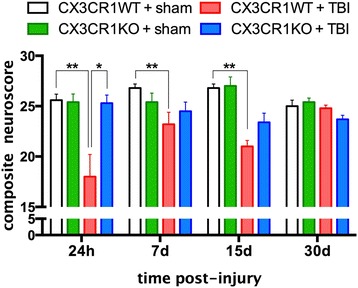
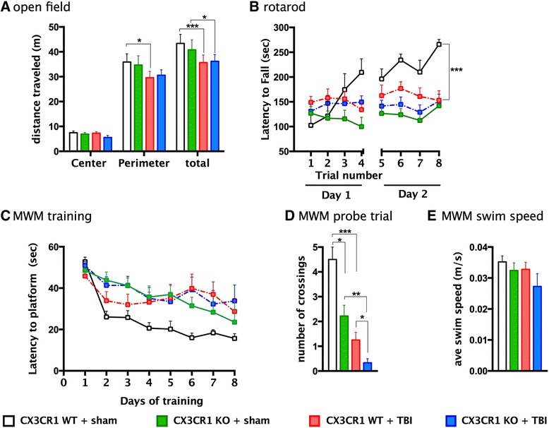
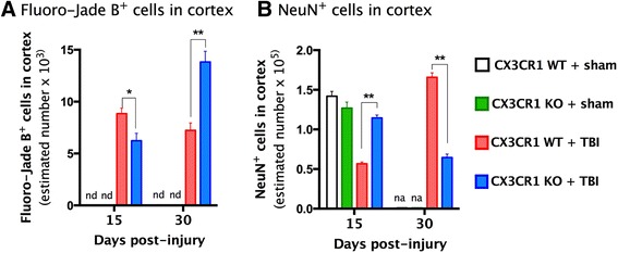
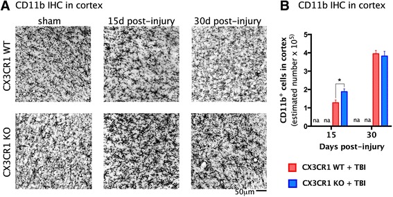
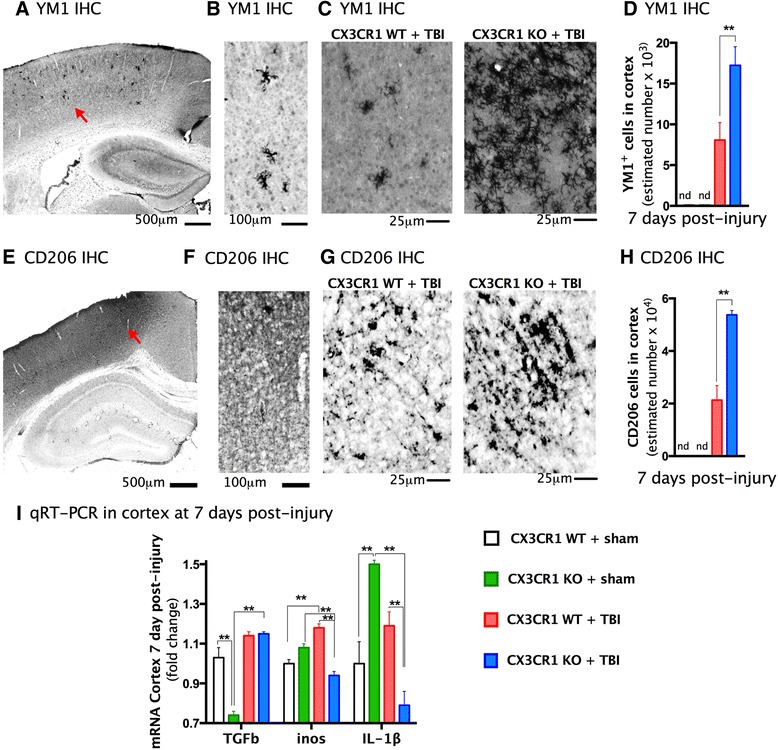
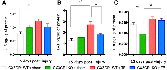
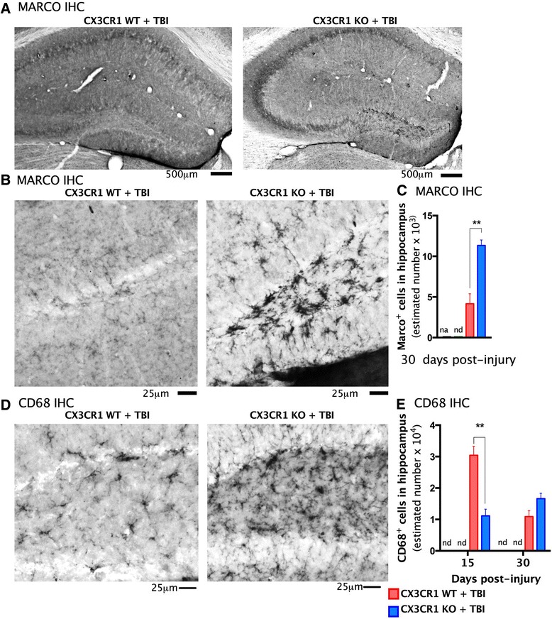
BACKGROUND: Neuroinflammation is an important secondary mechanism that is a key mediator of the long-term consequences of neuronal injury that occur in traumatic brain injury (TBI). Microglia are highly plastic cells with dual roles in neuronal injury and recovery. Recent studies suggest that the chemokine fractalkine (CX3CL1, FKN) mediates neural/microglial interactions via its sole receptor CX3CR1. CX3CL1/CX3CR1 signaling modulates microglia activation, and depending upon the type and time of injury, either protects or exacerbates neurological diseases. METHODS: In this study, mice deficient in CX3CR1 were subjected to mild controlled cortical impact injury (CCI), a model of TBI. We evaluated the effects of genetic deletion of CX3CR1 on histopathology, cell death/survival, microglia activation, and cognitive function for 30 days post-injury. RESULTS: During the acute post-injury period (24 h-15 days), motor deficits, cell death, and neuronal cell loss were more profound in injured wild-type than in CX3CR1(-/-) mice. In contrast, during the chronic period of 30 days post-TBI, injured CX3CR1(-/-) mice exhibited greater cognitive dysfunction and increased neuronal death than wild-type mice. The protective and deleterious effects of CX3CR1 were associated with changes in microglia phenotypes; during the acute phase CX3CR1(-/-) mice showed a predominant anti-inflammatory M2 microglial response, with increased expression of Ym1, CD206, and TGFβ. In contrast, increased M1 phenotypic microglia markers, Marco, and CD68 were predominant at 30 days post-TBI. CONCLUSION: Collectively, these novel data demonstrate a time-dependent role for CX3CL1/CX3CR1 signaling after TBI and suggest that the acute and chronic responses to mild TBI are modulated in part by distinct microglia phenotypes.
Overview
The claims and statistics of this paper haven’t been checked yet.
Run the check — free, no account needed. It reads the open-access text and checks the sources it cites.
Integrity signals
Registry
Cites
Statistics
Figures
Verification
Claims the paper makes
The claims and statistics of this paper haven’t been checked yet.
Run the check — free, no account needed. It reads the open-access text and checks the sources it cites.
Claims other papers make about this one
Each is checked against this paper’s own text: does it say what the citing paper says it does?
The source backs part of the claim, not all of it — or backs it for a different population, condition or comparison.
In the citing paper
“After a single CCI TBI, Cx3cr1−/− mice have improved motor recovery and decreased neuronal loss through 15 days post-injury (DPI).”
From The Inflammatory Continuum of Traumatic Brain Injury and Alzheimer’s Disease (2018) · cites this paper as [80]
Experimental — accuracy not yet measuredThe cited source says what the claim attributes to it.
In the citing paper
“By 30 DPI, however, these Cx3cr1−/− mice have worse cognitive dysfunction and neuronal loss compared to wild-type controls.”
From The Inflammatory Continuum of Traumatic Brain Injury and Alzheimer’s Disease (2018) · cites this paper as [80]
The cited source says what the claim attributes to it.
In the citing paper
“These changes are directly associated with an altered and time-dependent inflammatory profile in microglia (80).”
From The Inflammatory Continuum of Traumatic Brain Injury and Alzheimer’s Disease (2018) · cites this paper as [80]
Self-check
Percentages, sums and reported differences re-computed from the paper’s own numbers.
The self-check needs the paper’s full text.
Citations
Cites
The chemokine system in diverse forms of macrophage activation and polarization
MANTOVANI, A., SICA, A., SOZZANI, S., ALLAVENA, P., VECCHI, A., & LOCATI, M. (2004) · Trends in Immunology · doi:10.1016/j.it.2004.09.015 · Cited by 6,534 · cited 1×
“M1 microglia are generally considered pro-inflammatory whereas, M2 microglia are generally anti-inflammatory [29 – 34].”
Alternative activation of macrophages
Gordon, S. (2003) · Nature Reviews Immunology · doi:10.1038/nri978 · Cited by 6,189 · cited 1×
“M1 microglia are generally considered pro-inflammatory whereas, M2 microglia are generally anti-inflammatory [29–34].”
Blood Monocytes Consist of Two Principal Subsets with Distinct Migratory Properties
Geissmann, F., Jung, S., & Littman, D. R. (2003) · Immunity · doi:10.1016/s1074-7613(03)00174-2 · Cited by 3,504 · cited 1×
“Conversely, monocytes of the subset Ly6C low /CX3CR1 high are CCR2 deficient and are dependent on CX3CR1 for recruitment [51 – 53].”
West, M. J., Slomianka, L., & Gundersen, H. J. G. (1991) · The Anatomical Record · doi:10.1002/ar.1092310411 · Cited by 2,994 · cited 1×
“All cell counts were obtained using the Optical Fractionator method of unbiased stereological counting techniques [39].”
Jung, S., Aliberti, J., Graemmel, P., Sunshine, M. J., Kreutzberg, G. W., Sher, A., & Littman, D. R. (2000) · Molecular and Cellular Biology · doi:10.1128/mcb.20.11.4106-4114.2000 · Cited by 2,632 · cited 1×
“Indeed, deleting CX3CR1 [4] exacerbates microglial neurotoxicity induced by systemic inflammation in rodent models of Parkinson’s disease and amyotrophic lateral sclerosis [5].”
Nahrendorf, M., Swirski, F. K., Aikawa, E., Stangenberg, L., Wurdinger, T., Figueiredo, J.-L., Libby, P., Weissleder, R., & Pittet, M. J. (2007) · The Journal of Experimental Medicine · doi:10.1084/jem.20070885 · Cited by 2,300 · cited 2×
“For example, in a model of myocardial infarction, tissue repair requires recruitment of Ly6C and then Ly6C monocytes [53].”
Kigerl, K. A., Gensel, J. C., Ankeny, D. P., Alexander, J. K., Donnelly, D. J., & Popovich, P. G. (2009) · The Journal of Neuroscience · doi:10.1523/jneurosci.3257-09.2009 · Cited by 2,130 · cited 2×
“Ly6C monocytes are proteolitic, degrade injured tissue, and give rise to M1 macrophages in vivo [35, 56].”
Arnold, L., Henry, A., Poron, F., Baba-Amer, Y., van Rooijen, N., Plonquet, A., Gherardi, R. K., & Chazaud, B. (2007) · The Journal of Experimental Medicine · doi:10.1084/jem.20070075 · Cited by 2,002 · cited 1×
“CX3CR1 monocytes may differentiate from Ly6C monocytes in tissue and thus may not require CX3CR1 for recruitment [54, 55].”
Microglia Shape Adult Hippocampal Neurogenesis through Apoptosis-Coupled Phagocytosis
Sierra, A., Encinas, J. M., Deudero, J. J. P., Chancey, J. H., Enikolopov, G., Overstreet-Wadiche, L. S., Tsirka, S. E., & Maletic-Savatic, M. (2010) · Cell Stem Cell · doi:10.1016/j.stem.2010.08.014 · Cited by 1,622 · cited 10×
“Antibodies and dilutions were as follows: rat CD11b (Serotec, Raleigh, NC, USA; 1:500); mouse CD68 (Serotec; 1:200); rabbit YM1 (Stem cell technology, Vancouver, BC, Canada; 1:400); rat Marco (Serotec; 1:500); mouse NeuN (Millipore, Temecula, CA, USA; 1:1000).”
InfluentialControl of microglial neurotoxicity by the fractalkine receptor
Cardona, A. E., Pioro, E. P., Sasse, M. E., Kostenko, V., Cardona, S. M., Dijkstra, I. M., Huang, D., Kidd, G., Dombrowski, S., Dutta, R., et al. (2006) · Nature Neuroscience · doi:10.1038/nn1715 · Cited by 1,531 · cited 2×
“Indeed, deleting CX3CR1 [4] exacerbates microglial neurotoxicity induced by systemic inflammation in rodent models of Parkinson’s disease and amyotrophic lateral sclerosis [5].”
Self-citationInflammation in Neurodegenerative Disease—A Double-Edged Sword
Wyss-Coray, T., & Mucke, L. (2002) · Neuron · doi:10.1016/s0896-6273(02)00794-8 · Cited by 1,235
Harrison, J. K., Jiang, Y., Chen, S., Xia, Y., Maciejewski, D., McNamara, R. K., Streit, W. J., Salafranca, M. N., Adhikari, S., Thompson, D. A., et al. (1998) · Proceedings of the National Academy of Sciences · doi:10.1073/pnas.95.18.10896 · Cited by 1,176 · cited 1×
“Data from some models of chronic neuronal injury suggest that CX3CR1 is protective because the absence of this receptor increases microglia activity and is associated with less favorable outcome [8, 12–16].”
Inflammation after trauma: Microglial activation and traumatic brain injury
Ramlackhansingh, A. F., Brooks, D. J., Greenwood, R. J., Bose, S. K., Turkheimer, F. E., Kinnunen, K. M., Gentleman, S., Heckemann, R. A., Gunanayagam, K., Gelosa, G., et al. (2011) · Annals of Neurology · doi:10.1002/ana.22455 · Cited by 999 · cited 1×
“A consensus based on recent evidence considers neuroinflammation as an important secondary mechanism that plays a role in delaying injury after TBI [1–3].”
Heterogeneity of Microglial Activation in the Innate Immune Response in the Brain
Colton, C. A. (2009) · Journal of Neuroimmune Pharmacology · doi:10.1007/s11481-009-9164-4 · Cited by 916 · cited 1×
“M1 microglia are generally considered pro-inflammatory whereas, M2 microglia are generally anti-inflammatory [29–34].”
The Role of Markers of Inflammation in Traumatic Brain Injury
Woodcock, T. M., & Morganti-Kossmann, M. C. (2013) · Frontiers in Neurology · doi:10.3389/fneur.2013.00018 · Cited by 719 · cited 1×
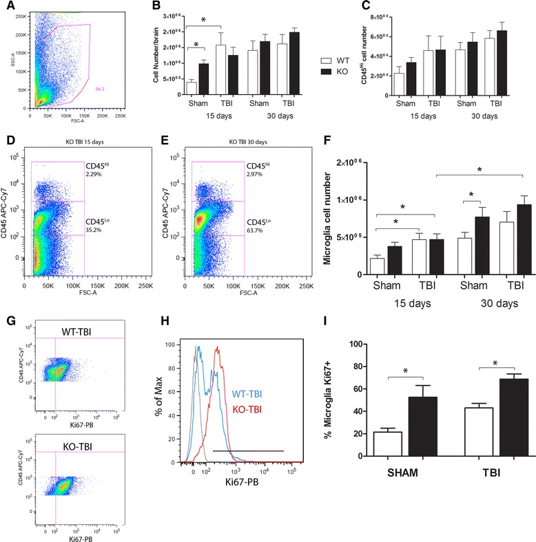
“Because TBI involves infiltration of peripheral blood monocytes into the brain [44], we first isolated mononuclear cells from WT and CX3CR1 mice at 15 or 30 days post-TBI and analyzed them with flow cytometry (Fig.”
Combadière, C., Potteaux, S., Rodero, M. P., Simon, T., Pézard, A., Esposito, B., Merval, R., Proudfoot, A. E. I., Tedgui, A., & Mallat, Z. (2008) · Circulation · doi:10.1161/circulationaha.107.745091 · Cited by 696 · cited 1×
“Conversely, monocytes of the subset Ly6C/CX3CR1 are CCR2 deficient and are dependent on CX3CR1 for recruitment [51–53].”
Chhor, V., Charpentier, T. L., Lebon, S., Oré, M. V., Lara‐Celador, I., Josserand, J., Degos, V., Jacotot, E. D., Hagberg, H., Sävman, K., et al. (2013) · Brain Behavior and Immunity · doi:10.1016/j.bbi.2013.02.005 · Cited by 676 · cited 1×
“M1 microglia are generally considered pro-inflammatory whereas, M2 microglia are generally anti-inflammatory [29 – 34].”
Neurotactin, a membrane-anchored chemokine upregulated in brain inflammation
Pan, Y., Lloyd, C. M., Zhou, H., Dolich, S., Deeds, J., Gonzalo, J.-Á., Vath, J., Gosselin, M., Ma, J., Dussault, B. J., et al. (1997) · Nature · doi:10.1038/42491 · Cited by 643 · cited 1×
“Data from some models of chronic neuronal injury suggest that CX3CR1 is protective because the absence of this receptor increases microglia activity and is associated with less favorable outcome [8, 12–16].”
Regulation of Tau Pathology by the Microglial Fractalkine Receptor
Bhaskar, K., Konerth, M., Kokiko-Cochran, O. N., Cardona, A., Ransohoff, R. M., & Lamb, B. T. (2010) · Neuron · doi:10.1016/j.neuron.2010.08.023 · Cited by 639 · cited 1×
“For instance, in a model of Alzheimer ’ s disease (AD), CX3CR1-deficient microglia from mice overexpressing hTau have increased hyperphosphorylated tau and more toxicity, as has been observed in other animal ’ s disease models [5, 17 – 20].”
Self-citationCX3CR1 deficiency leads to impairment of hippocampal cognitive function and synaptic plasticity
Rogers, J. T., Morganti, J. M., Bachstetter, A. D., Hudson, C. E., Peters, M. M., Grimmig, B. A., Weeber, E. J., Bickford, P. C., & Gemma, C. (2011) · The Journal of Neuroscience · doi:10.1523/jneurosci.3667-11.2011 · Cited by 624 · cited 4×
“Spatial learning was evaluated using the Morris water maze (MWM) task as previously described [40].”
InfluentialSelf-citationJanus-faced microglia: beneficial and detrimental consequences of microglial phagocytosis
Sierra, A., Abiega, O., Shahraz, A., & Neumann, H. (2013) · Frontiers in Cellular Neuroscience · doi:10.3389/fncel.2013.00006 · Cited by 574 · cited 10×
“Antibodies and dilutions were as follows: rat CD11b (Serotec, Raleigh, NC, USA; 1:500); mouse CD68 (Serotec; 1:200); rabbit YM1 (Stem cell technology, Vancouver, BC, Canada; 1:400); rat Marco (Serotec; 1:500); mouse NeuN (Millipore, Temecula, CA, USA; 1:1000).”
InfluentialMicroglial Cx3cr1 knockout prevents neuron loss in a mouse model of Alzheimer's disease
Fuhrmann, M., Bittner, T., Jung, C. K. E., Burgold, S., Page, R. M., Mitteregger, G., Haass, C., LaFerla, F. M., Kretzschmar, H., & Herms, J. (2010) · Nature Neuroscience · doi:10.1038/nn.2511 · Cited by 562 · cited 1×
“On the other hand, in 3x-Tg AD mice, which reproduces both Aβ and tau pathologies, deleting CX3CR1 prevents neuronal loss and microglial migration without affecting amyloid deposition [21].”
Lee, S., Varvel, N. H., Konerth, M. E., Xu, G., Cardona, A. E., Ransohoff, R. M., & Lamb, B. T. (2010) · The American Journal of Pathology · doi:10.2353/ajpath.2010.100265 · Cited by 522 · cited 1×
“Finally, other reports indicate that CX3CR1 deficiency attenuates amyloid deposition in AD mouse models characterized by extensive plaque deposition [18, 22].”
Self-citationDecreased atherosclerosis in CX3CR1–/– mice reveals a role for fractalkine in atherogenesis
Lesnik, P., Haskell, C. A., & Charo, I. F. (2003) · Journal of Clinical Investigation · doi:10.1172/jci15555 · Cited by 487 · cited 1×
“In experimental models of spinal cord injury and focal cerebral ischemia, tissue damage is attenuated and function recovers earlier following injury in CX3CR1-deficient mice, effects attributed to reduced recruitment and/or activation of microglia/macrophages [23, 24, 57, 58].”
Alzheimer's disease: synaptic dysfunction and Aβ
Shankar, G. M., & Walsh, D. M. (2009) · Molecular Neurodegeneration · doi:10.1186/1750-1326-4-48 · Cited by 486 · cited 1×
“Both beneficial and detrimental effects of CX3CR1 deficiency are associated with microglia activation [9 – 11].”
Microglia/Macrophage Polarization Dynamics in White Matter after Traumatic Brain Injury
Wang, G., Zhang, J., Hu, X., Zhang, L., Mao, L., Jiang, X., Liou, A. K.-F., Leak, R. K., Gao, Y., & Chen, J. (2013) · Journal of Cerebral Blood Flow & Metabolism · doi:10.1038/jcbfm.2013.146 · Cited by 473 · cited 1×
“Although a shift in microglia phenotype between the acute and chronic phase of TBI has recently been reported [49], we extend these observations by identifying CX3CR1 as a possible regulator of this microglia/macrophage phenotype switch following mild TBI.”
Inflammation, microglia, and alzheimer's disease
Cameron, B., & Landreth, G. E. (2009) · Neurobiology of Disease · doi:10.1016/j.nbd.2009.10.006 · Cited by 452 · cited 1×
“Both beneficial and detrimental effects of CX3CR1 deficiency are associated with microglia activation [9 – 11].”
Expression profiles for macrophage alternative activation genes in AD and in mouse models of AD.
(2006) · Journal of Neuroinflammation · doi:10.1186/1742-2094-3-27 · Cited by 393 · cited 1×
“M1 microglia are generally considered pro-inflammatory whereas, M2 microglia are generally anti-inflammatory [29–34].”
Expression of CX 3 CR1 chemokine receptors on neurons and their role in neuronal survival
Meucci, O., Fatatis, A., Simen, A. A., & Miller, R. J. (2000) · Proceedings of the National Academy of Sciences · doi:10.1073/pnas.090017497 · Cited by 385 · cited 1×
“CX3CL1 also protects against excitotoxicity through the activation of the ERK1/2 and PI3K/Akt pathways [7, 8].”
Nishiyori, A., Minami, M., Ohtani, Y., Takami, S., Yamamoto, J., Kawaguchi, N., Kume, T., Akaike, A., & Satoh, M. (1998) · FEBS Letters · doi:10.1016/s0014-5793(98)00583-3 · Cited by 365 · cited 1×
“Data from some models of chronic neuronal injury suggest that CX3CR1 is protective because the absence of this receptor increases microglia activity and is associated with less favorable outcome [8, 12–16].”
Mizutani, M., Pino, P. A., Saederup, N., Charo, I. F., Ransohoff, R. M., & Cardona, A. E. (2012) · The Journal of Immunology · doi:10.4049/jimmunol.1100421 · Cited by 346 · cited 1×
“[41, 42], and cellular pellets resuspended in cell staining buffer (Biolegend, San Diego, CA, USA).”
Self-citationProduction and neuroprotective functions of fractalkine in the central nervous system
Mizuno, T., Kawanokuchi, J., Numata, K., & Suzumura, A. (2003) · Brain Research · doi:10.1016/s0006-8993(03)02867-1 · Cited by 342 · cited 1×
“In addition, CX3CR1 microglia exacerbate cell-autonomous microglial neurotoxicity induced by lipopolysaccharide (LPS), suggesting that fractalkine signaling is important for limiting microglia toxicity [5, 6].”
Lin, S., Castaño, A. P., Nowlin, B. T., Lupher, M., & Duffield, J. S. (2009) · The Journal of Immunology · doi:10.4049/jimmunol.0901473 · Cited by 331 · cited 1×
“Ly6C monocytes are proteolitic, degrade injured tissue, and give rise to M1 macrophages in vivo [35, 56].”
Microglia Activation as a Biomarker for Traumatic Brain Injury
Hernandez‐Ontiveros, D. G., Tajiri, N., Acosta, S., Giunta, B. N., Tan, J., & Borlongan, C. V. (2013) · Frontiers in Neurology · doi:10.3389/fneur.2013.00030 · Cited by 292 · cited 1×
“A consensus based on recent evidence considers neuroinflammation as an important secondary mechanism that plays a role in delaying injury after TBI [1 – 3].”
Dénes, Á., Ferenczi, S., Halász, J., Környei, Z., & Kovács, K. J. (2008) · Journal of Cerebral Blood Flow & Metabolism · doi:10.1038/jcbfm.2008.64 · Cited by 276 · cited 2×
“In experimental models of spinal cord injury and focal cerebral ischemia, tissue damage is attenuated and function recovers earlier following injury in CX3CR1-deficient mice, effects attributed to reduced recruitment and/or activation of microglia/macrophages [23, 24, 57, 58].”
Liu, Z., Condello, C., Schain, A., Harb, R., & Grutzendler, J. (2010) · The Journal of Neuroscience · doi:10.1523/jneurosci.4403-10.2010 · Cited by 275
Cho, S.-H., Sun, B., Zhou, Y., Kauppinen, T. M., Halabisky, B., Wes, P., Ransohoff, R. M., & Gan, L. (2011) · Journal of Biological Chemistry · doi:10.1074/jbc.m111.254268 · Cited by 268 · cited 1×
“For instance, in a model of Alzheimer ’ s disease (AD), CX3CR1-deficient microglia from mice overexpressing hTau have increased hyperphosphorylated tau and more toxicity, as has been observed in other animal ’ s disease models [5, 17 – 20].”
The multifaceted profile of activated microglia
Lynch, M. A. (2009) · Molecular Neurobiology · doi:10.1007/s12035-009-8077-9 · Cited by 268 · cited 1×
“M1 microglia are generally considered pro-inflammatory whereas, M2 microglia are generally anti-inflammatory [29–34].”
Morganti, J. M., Jopson, T. D., Liu, S., Riparip, L.-K., Guandique, C. K., Gupta, N., Ferguson, A. R., & Rosi, S. (2015) · The Journal of Neuroscience · doi:10.1523/jneurosci.2405-14.2015 · Cited by 253 · cited 1×
“[50] using CX3CR1 CCR2 reporter mice have demonstrated a broad spectrum of M1-M2 polarization mRNA gene expression following TBI, with changes occurring primarily during the first 48 h.”
Fractalkine and fractalkine receptors in human neurons and glial cells
Hatori, K., Nagai, A., Heisel, R., Ryu, J. K., & Kim, S. U. (2002) · Journal of Neuroscience Research · doi:10.1002/jnr.10304 · Cited by 239 · cited 1×
“Data from some models of chronic neuronal injury suggest that CX3CR1 is protective because the absence of this receptor increases microglia activity and is associated with less favorable outcome [8, 12 – 16].”
Washington, P. M., Forcelli, P. A., Wilkins, T., Zapple, D. N., Parsadanian, M., & Burns, M. P. (2012) · Journal of Neurotrauma · doi:10.1089/neu.2012.2456 · Cited by 228 · cited 1×
“Previous studies have reported that these parameters produce a mild injury [36, 37].”
Donnelly, D. J., Longbrake, E. E., Shawler, T. M., Kigerl, K. A., Lai, W., Tovar, C. A., Ransohoff, R. M., & Popovich, P. G. (2011) · Journal of Neuroscience · doi:10.1523/jneurosci.2114-11.2011 · Cited by 219 · cited 3×
“[23] showed that abolishing CX3CR1 reduces accumulation of CD11b/Ly6C/iNOS monocytes, which confers neuroprotection and promotes recovery of function at early time points after spinal cord injury.”
Tang, Z., Gan, Y. X., Liu, Q., Yin, J., Liu, Q., Shi, J., & Shi, F. (2014) · Journal of Neuroinflammation · doi:10.1186/1742-2094-11-26 · Cited by 195 · cited 2×
“In experimental models of spinal cord injury and focal cerebral ischemia, tissue damage is attenuated and function recovers earlier following injury in CX3CR1-deficient mice, effects attributed to reduced recruitment and/or activation of microglia/macrophages [23, 24, 57, 58].”
CX3CR1 deficiency induces an early protective inflammatory environment in ischemic mice
Fumagalli, S., Perego, C., Ortolano, F., & Simoni, M. G. D. (2013) · Glia · doi:10.1002/glia.22474 · Cited by 189 · cited 1×
“Similarly, CX3CR1 mice are protected from the middle cerebral artery occlusion, with smaller infarct size at early time points after injury, which is associated with an M2 microglial phenotype [26].”
Bolkvadze, T., & Pitkänen, A. (2012) · Journal of Neurotrauma · doi:10.1089/neu.2011.1954 · Cited by 183 · cited 1×
“Previous studies have reported that these parameters produce a mild injury [36, 37].”
Kubová, H., Druga, R., Lukasiuk, K., Suchomelova, L., Haugvicová, R., Jirmanová, I., & Pitkänen, A. (2001) · Journal of Neuroscience · doi:10.1523/jneurosci.21-10-03593.2001 · Cited by 177 · cited 1×
“Fluoro-Jade B staining was performed according to Kubova et al. [38].”
CX3CL1 Is Neuroprotective in Permanent Focal Cerebral Ischemia in Rodents
Cipriani, R., Villa, P., Chece, G., Lauro, C., Paladini, A., Micotti, E., Perego, C., De Simoni, M.-G., Fredholm, B. B., Eusebi, F., et al. (2011) · The Journal of Neuroscience · doi:10.1523/jneurosci.3611-11.2011 · Cited by 166 · cited 1×
“For instance, in a model of cerebral ischemia and spinal cord injury, CX3CR1 mice showed neuroprotection and improved functional recovery [23–26].”
Chemokines and Chemokine Receptors: Multipurpose Players in Neuroinflammation
Ransohoff, R. M., Liu, L., & Cardona, A. E. (2007) · International review of neurobiology · doi:10.1016/s0074-7742(07)82010-1 · Cited by 163 · cited 1×
“For instance, in a model of Alzheimer’s disease (AD), CX3CR1-deficient microglia from mice overexpressing hTau have increased hyperphosphorylated tau and more toxicity, as has been observed in other animal’s disease models [5, 17–20].”
Self-citationBone Marrow Mesenchymal Stromal Cells Drive Protective M2 Microglia Polarization After Brain Trauma
Zanier, E. R., Pischiutta, F., Riganti, L., Marchesi, F., Turola, E., Fumagalli, S., Perego, C., Parotto, E., Vinci, P., Veglianese, P., et al. (2014) · Neurotherapeutics · doi:10.1007/s13311-014-0277-y · Cited by 160 · cited 1×
“Intraventricular infusion of mesenchymal stem cells after TBI induces early and lasting acquisition of a protective M2 microglial phenotype that is associated with reduced CD68 immunoreactivity [46].”
Primary Phagocytosis of Neurons by Inflamed Microglia: Potential Roles in Neurodegeneration
Neher, J. J., Neniškytė, U., & Brown, G. C. (2012) · Frontiers in Pharmacology · doi:10.3389/fphar.2012.00027 · Cited by 157 · cited 1×
“Although microglial phagocytosis of dead or dying neurons can be beneficial by preventing the release of pro-inflammatory molecules, under some conditions, such as inflammation, microglia also phagocytize viable neurons, thus contributing to cell death [45].”
Chemokine CX3CL1 protects rat hippocampal neurons against glutamate-mediated excitotoxicity
Limatola, C., Lauro, C., Catalano, M., Ciotti, M. T., Bertollini, C., Di Angelantonio, S., Ragozzino, D., & Eusebi, F. (2005) · Journal of Neuroimmunology · doi:10.1016/j.jneuroim.2005.03.023 · Cited by 153 · cited 1×
“CX3CL1 also protects against excitotoxicity through the activation of the ERK1/2 and PI3K/Akt pathways [7, 8].”
Isolation of brain and spinal cord mononuclear cells using percoll gradients.
Pino, P., & Cardona, A. (2011) · Journal of Visualized Experiments · doi:10.3791/2348 · Cited by 142 · cited 1×
“[41, 42], and cellular pellets resuspended in cell staining buffer (Biolegend, San Diego, CA, USA).”
Self-citationThe immunology of traumatic brain injury: a prime target for Alzheimer’s disease prevention
Giunta, B., Obregon, D., Velisetty, R., Sanberg, P. R., Borlongan, C. V., & Tan, J. (2012) · Journal of Neuroinflammation · doi:10.1186/1742-2094-9-185 · Cited by 112 · cited 1×
“A consensus based on recent evidence considers neuroinflammation as an important secondary mechanism that plays a role in delaying injury after TBI [1–3].”
Yoshida, H., Imaizumi, T., Fujimoto, K., Matsuo, N., Kimura, K., Cui, X., Matsumiya, T., Tanji, K., Shibata, T., Tamo, W., et al. (2001) · Neuroscience Letters · doi:10.1016/s0304-3940(01)01699-8 · Cited by 96 · cited 1×
“Data from some models of chronic neuronal injury suggest that CX3CR1 is protective because the absence of this receptor increases microglia activity and is associated with less favorable outcome [8, 12–16].”
Regulation of Adaptive Immunity by the Fractalkine Receptor during Autoimmune Inflammation
Garcia, J. A., Pino, P. A., Mizutani, M., Cardona, S. M., Charo, I. F., Ransohoff, R. M., Forsthuber, T. G., & Cardona, A. E. (2013) · The Journal of Immunology · doi:10.4049/jimmunol.1300040 · Cited by 89 · cited 3×
“Cells were then stained with Ki67-V450 (Clone B56) in permeabilization buffer for 20 min, washed, resuspended in 2 % PFA, and acquired on an LSR-II [43].”
InfluentialSelf-citationThawer, S. G., Mawhinney, L., Chadwick, K., de Chickera, S. N., Weaver, L. C., Brown, A. M., & Dekaban, G. A. (2013) · Journal of Neuroimmunology · doi:10.1016/j.jneuroim.2013.04.008 · Cited by 62 · cited 1×
“CX3CR1 monocytes may differentiate from Ly6C monocytes in tissue and thus may not require CX3CR1 for recruitment [54, 55].”
Rancan, M., Bye, N., Otto, V. I., Trentz, O., Kossmann, T., Frentzel, S., & Morganti-Kossmann, M. C. (2004) · Journal of Cerebral Blood Flow & Metabolism · doi:10.1097/01.wcb.0000133470.91843.72 · Cited by 54 · cited 1×
“CX3CL1 levels increase in the cerebrospinal fluid and in the brain of patients with head trauma, and increased CX3CL1/CX3CR1 is associated with better clinical outcome [27, 28].”
Research works printed but not linked: 2
- Frank-Cannon TC, Alto LT, McAlpine FE, Tansey MG. Does neuroinflammation fan the flame in neurodegenerative diseases? Mol Neurodegener. 2009;4:47. · DOI · Paper page
- Gaetani P, Pisano P, Solinas G, Colombo P, Destro A, Levi D, et al. Immunohistohemical expression of the chemokine fractalkine and its receptor in the human brain cortex after severe traumatic brain injury and brain hemorrhage. J Neurosurg Sci. 2013;57(1):55–62.
Cited by
Sorted by citations
Microglia in Pain: Detrimental and Protective Roles in Pathogenesis and Resolution of Pain
Chen, G., Zhang, Y., Qadri, Y. J., Serhan, C. N., & Ji, R. (2018) · Neuron · doi:10.1016/j.neuron.2018.11.009 · Cited by 801
Bidirectional Microglia–Neuron Communication in Health and Disease
Szepesi, Z., Manouchehrian, O., Bachiller, S., & Deierborg, T. (2018) · Frontiers in Cellular Neuroscience · doi:10.3389/fncel.2018.00323 · Cited by 459
“The activity of macrophages appears to be a potential therapeutic target in different TBI settings in adult, but not young, rodents (Febinger et al., 2015; Erturk et al., 2016; Hanlon et al., 2016; Zanier et al., 2016; Chhor et al., 2017; Saber et al., 2017).”
BackgroundResultInfluentialMicroglial Activation in Traumatic Brain Injury
Donat, C. K., Scott, G., Gentleman, S., & Sastre, M. (2017) · Frontiers in Aging Neuroscience · doi:10.3389/fnagi.2017.00208 · Cited by 512
“…2014 Mouse C57Bl/6 ♂ CCI, moderate 3 mm dpi: 3, 7 M1: CD11b, TNF-α, CD68, CD86, IL-1β M2a/c: Ym1, Arg-1, CD206, SOCS3, IL-10 Mixed expression of M1 and M2 marker mRNA M1: N at 3 + 7 dpi M2: N Ym1, Arg-1, SOCS3 at 3 dpi Febinger et al., 2015 Mouse CX3CR1−/− CX3CR1+/+ ♂ CCI, mild 3 mm dpi: 7, 15,…”
Commonalities in epileptogenic processes from different acute brain insults: Do they translate?
Klein, P., Dingledine, R. J., Aronica, E., Bernard, C., Blümcke, I., Boison, D., Brodie, M. J., Brooks‐Kayal, A. R., Engel, J., Forcelli, P. A., et al. (2017) · Epilepsia · doi:10.1111/epi.13965 · Cited by 300
Shaping Neuronal Fate: Functional Heterogeneity of Direct Microglia-Neuron Interactions
Cserép, C., Pósfai, B., & Dénes, Á. (2021) · Neuron · doi:10.1016/j.neuron.2020.11.007 · Cited by 256
“, 2016), whereas time-dependent effects were found concerning cognitive dysfunction after mild brain trauma (Febinger et al., 2015).”
Metadata
- Authors
- Febinger, H. Y., Thomasy, H. E., Pavlova, M. N., Ringgold, K. M., Barf, P. R., George, A. M., Grillo, J. N., Bachstetter, A. D., Garcia, J. A., Cardona, A. E., Opp, M. R., & Gemma, C.
- Affiliations
- University of Washington (US)
- University of Kentucky (US)
- The University of Texas at San Antonio (US)
- Corresponding author
- Gemma, Carmelina
- Venue
- Journal of Neuroinflammation
- Published
- 2015-09-02
- Volume
- 12
- Issue
- 1
- Pages
- 154-154
- ISSN
- 1742-2094 (ISSN-L)
- PMC
- PMC4557842
- OpenAlex
- W1944546979
- Cited by
- 110
- References
- 58
- Citations by year
- 2026: 10 · 2025: 8 · 2024: 5 · 2023: 10 · 2022: 6 · 2021: 15 · 2020: 10 · 2019: 13 · 2018: 16 · 2017: 7 · 2016: 9 · 2015: 1
- FWCI
- 7.76
- Type
- journal article
- Language
- en
- Open access
- gold
- doi.org
- Locations
- Journal of Neuroinflammation (publishedVersion, cc-by)
- Europe PMC (PubMed Central) (publishedVersion, cc-by)
- PubMed Central (publishedVersion, cc-by)
- PubMed (publishedVersion)
- UKnowledge (University of Kentucky) (submittedVersion, cc-by)
- Funders
- National Institutes of Health (SC1GM095426)
- Topics
- Health Sciences › Medicine › Clinical Neurology › Traumatic Brain Injury and Neurovascular Disturbances
- Life Sciences › Neuroscience › Neurology › Neuroinflammation and Neurodegeneration Mechanisms
- Health Sciences › Medicine › Clinical Neurology › Traumatic Brain Injury Research
- Keywords
- CX3CR1, fractalkine, mild traumatic brain injury, microglial activation, M1/M2 microglial polarization, neuroinflammation, controlled cortical impact, neuronal cell death, cognitive impairment, CX3CR1 knockout mice
- MeSH terms
- CX3C Chemokine Receptor 1, Analysis of Variance, Animals, Brain, Brain Injuries, Disease Models, Animal, Exploratory Behavior, Flow Cytometry, Fluoresceins, Leukocytes, Mononuclear, Macrophage Activation, Male, Mice, Transgenic, Neurons, Psychomotor Disorders, Time Factors, Maze Learning, Receptors, Chemokine, Rotarod Performance Test, Green Fluorescent Proteins, Mice
About these checks
Registry, retraction, Cites and statistics results are computed, not judged, with one exception: a printed reference that no index lists is read once by a model before it counts as a citation error. Claims are extracted by a model and kept only when their quote appears verbatim in the text; each cited source is then checked, with a model’s help, for whether it says what the claim relies on. Those verdicts can be wrong — read the claim’s quote and the cited source before relying on one.
Statistics and arithmetic are recomputed, not judged.
Confidence bands are shown only for fields calibrated on our annotated test set. None is calibrated yet, so no band is shown.
What counts as a claim ›How we measure confidence ›How we check this ›
Are you an author, or do you see a problem with this page? Request a review
