

The DOI matches its Crossref record; 104 of the 104 references it cites were found in a registry or an index.
Neurons derived from sporadic Alzheimer’s disease iPSCs reveal elevated TAU hyperphosphorylation, increased amyloid levels, and GSK3B activation
Open accessAbstract
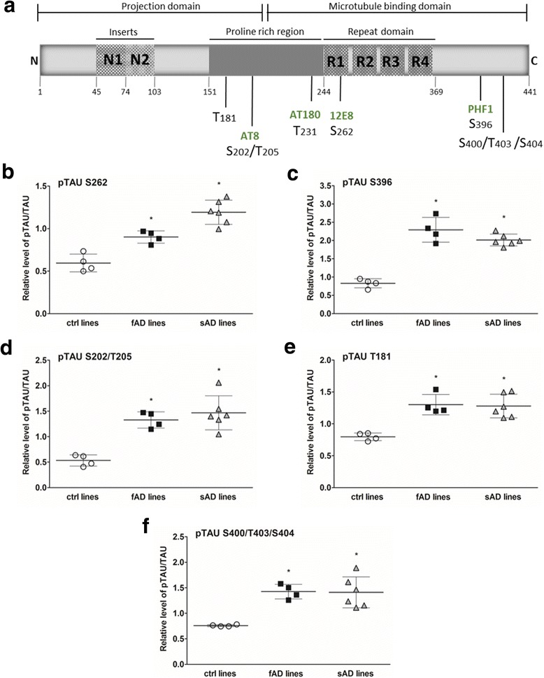
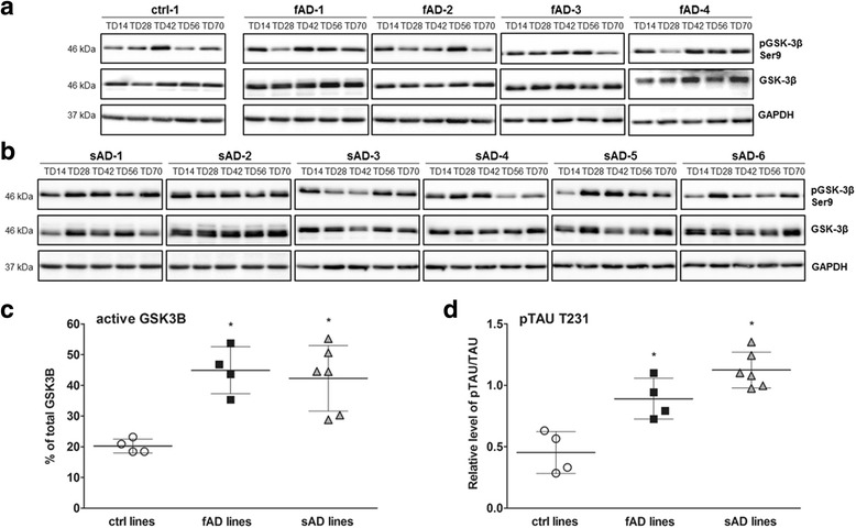
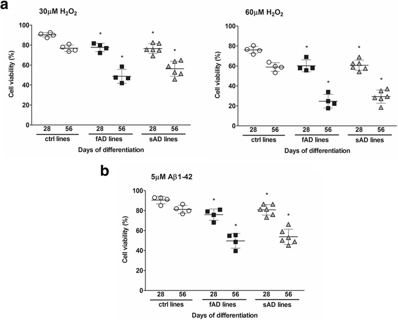
Alzheimer’s disease (AD) is the most common type of dementia, affecting one in eight adults over 65 years of age. The majority of AD cases are sporadic, with unknown etiology, and only 5% of all patients with AD present the familial monogenic form of the disease. In the present study, our aim was to establish an in vitro cell model based on patient-specific human neurons to study the pathomechanism of sporadic AD. We compared neurons derived from induced pluripotent stem cell (iPSC) lines of patients with early-onset familial Alzheimer’s disease (fAD), all caused by mutations in the PSEN1 gene; patients with late-onset sporadic Alzheimer’s disease (sAD); and three control individuals without dementia. The iPSC lines were differentiated toward mature cortical neurons, and AD pathological hallmarks were analyzed by RT-qPCR, enzyme-linked immunosorbent assay, and Western blotting methods. Neurons from patients with fAD and patients with sAD showed increased phosphorylation of TAU protein at all investigated phosphorylation sites. Relative to the control neurons, neurons derived from patients with fAD and patients with sAD exhibited higher levels of extracellular amyloid-β 1–40 (Aβ 1–40 ) and amyloid-β 1–42 (Aβ 1–42 ). However, significantly increased Aβ 1–42 /Aβ 1–40 ratios, which is one of the pathological markers of fAD, were observed only in samples of patients with fAD. Additionally, we detected increased levels of active glycogen synthase kinase 3 β, a physiological kinase of TAU, in neurons derived from AD iPSCs, as well as significant upregulation of amyloid precursor protein (APP) synthesis and APP carboxy-terminal fragment cleavage. Moreover, elevated sensitivity to oxidative stress, as induced by amyloid oligomers or peroxide, was detected in both fAD- and sAD-derived neurons. On the basis of the experiments we performed, we can conclude there is no evident difference except secreted Aβ 1–40 levels in phenotype between fAD and sAD samples. To our knowledge, this is the first study in which the hyperphosphorylation of TAU protein has been compared in fAD and sAD iPSC-derived neurons. Our findings demonstrate that iPSC technology is suitable to model both fAD and sAD and may provide a platform for developing new treatment strategies for these conditions.
Overview
The claims and statistics of this paper haven’t been checked yet.
Run the check — free, no account needed. It reads the open-access text and checks the sources it cites.
Integrity signals
Registry
Cites
Statistics
Figures
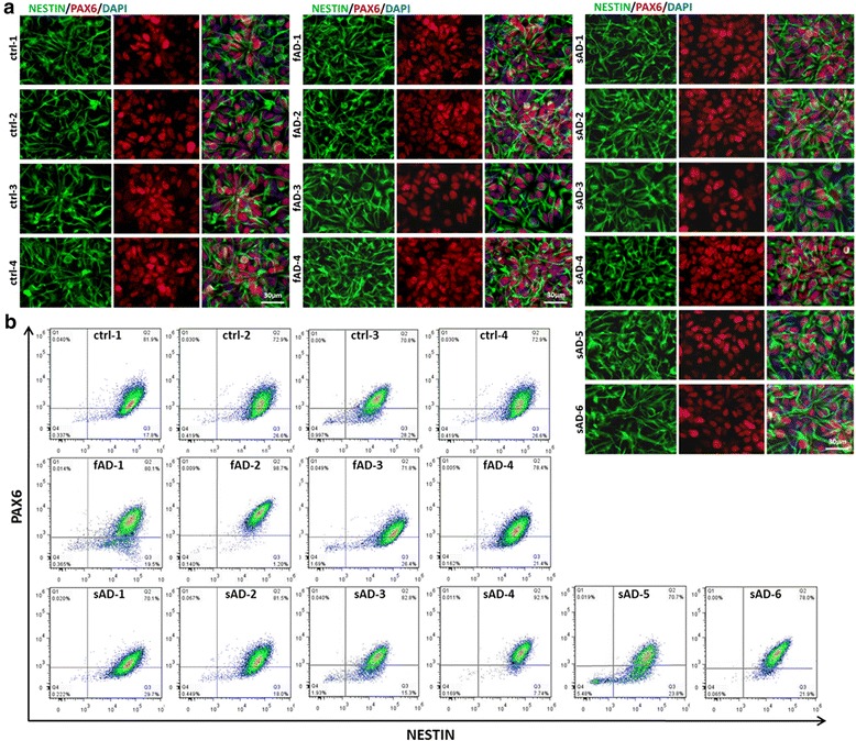
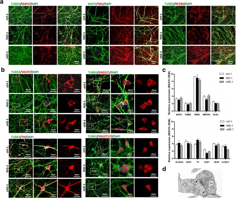
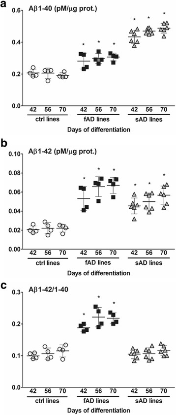
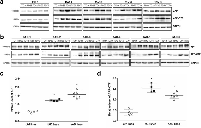
Verification
Claims the paper makes
The claims and statistics of this paper haven’t been checked yet.
Run the check — free, no account needed. It reads the open-access text and checks the sources it cites.
Claims other papers make about this one
Each is checked against this paper’s own text: does it say what the citing paper says it does?
The source backs part of the claim, not all of it — or backs it for a different population, condition or comparison.
In the citing paper
“iPSC-derived neurons from individuals with AD exhibit increased production of Aβ42, active GSK-3β, phosphorylated tau, endosomal abnormalities, and oxidative stress (12–17)”
From Modeling late-onset Alzheimer’s disease neuropathology via direct neuronal reprogramming (2024) · cites this paper as [15]
Experimental — accuracy not yet measuredThe cited source says what the claim attributes to it.
In the citing paper
“neurons derived from iPSCs obtained from sporadic AD cases also show elevated Tau hyperphosphorylation, increased Aβ levels and GSK3β activation44.”
From PLCG2 downregulation impairs synaptic function and increases Alzheimer’s disease hallmarks in neuronal cultures (2026) · cites this paper as [44]
The check wasn’t confident either way, so it gives no verdict — never disputed.
In the citing paper
“but late-stage neuropathological features, such as formation of insoluble tau and neurodegeneration have not been reported.”
From Modeling late-onset Alzheimer’s disease neuropathology via direct neuronal reprogramming (2024) · cites this paper as [15]
Experimental — accuracy not yet measured
Self-check
Percentages, sums and reported differences re-computed from the paper’s own numbers.
The self-check needs the paper’s full text.
Citations
Cites
Fast and accurate short read alignment with Burrows-Wheeler transform
Li, H., & Durbin, R. (2009) · Bioinformatics · doi:10.1093/bioinformatics/btp324 · Cited by 58,607
Richards, S., Aziz, N., Bale, S., Bick, D., Das, S., Gastier-Foster, J., Grody, W. W., Hegde, M., Lyon, E., Spector, E., et al. (2015) · Genetics in Medicine · doi:10.1038/gim.2015.30 · Cited by 32,594
The Genome Analysis Toolkit: A MapReduce framework for analyzing next-generation DNA sequencing data
McKenna, A., Hanna, M., Banks, E., Sivachenko, A., Cibulskis, K., Kernytsky, A., Garimella, K., Altshuler, D., Gabriel, S., Daly, M., et al. (2010) · Genome Research · doi:10.1101/gr.107524.110 · Cited by 27,895
Amyloid oligomers exacerbate tau pathology in a mouse model of tauopathy
Selenica, M.-L. B., Brownlow, M., Jimenez, J. P., Lee, D. C., Pena, G., Dickey, C. A., Gordon, M. N., & Morgan, D. (2013) · Neurodegenerative Diseases · doi:10.1159/000337230 · Cited by 10,359
The amyloid hypothesis of Alzheimer's disease at 25 years
Selkoe, D. J., & Hardy, J. (2016) · EMBO Molecular Medicine · doi:10.15252/emmm.201606210 · Cited by 6,164
Walsh, D. M., Klyubin, I., Fadeeva, J. V., Cullen, W. K., Anwyl, R., Wolfe, M. S., Rowan, M. J., & Selkoe, D. J. (2002) · Nature · doi:10.1038/416535a · Cited by 4,291
Grundke-Iqbal, I., Iqbal, K., Tung, Y. C., Quinlan, M., Wisniewski, H. M., & Binder, L. I. (1986) · Proceedings of the National Academy of Sciences · doi:10.1073/pnas.83.13.4913 · Cited by 3,676
Highly efficient neural conversion of human ES and iPS cells by dual inhibition of SMAD signaling
Chambers, S. M., Fasano, C. A., Papapetrou, E. P., Tomishima, M., Sadelain, M., & Studer, L. (2009) · Nature Biotechnology · doi:10.1038/nbt.1529 · Cited by 3,620
Apolipoprotein E and Alzheimer disease: risk, mechanisms and therapy
Liu, C.-C., Kanekiyo, T., Xu, H., & Bu, G. (2013) · Nature Reviews Neurology · doi:10.1038/nrneurol.2012.263 · Cited by 3,459
Uttara, B., Singh, A., Zamboni, P., & Mahajan, R. (2009) · Current Neuropharmacology · doi:10.2174/157015909787602823 · Cited by 3,378
Alzheimer's Disease and the Amyloid-β Peptide
Murphy, M. P., & LeVine, H. (2010) · Journal of Alzheimer's Disease · doi:10.3233/jad-2010-1221 · Cited by 1,750
Oxidative Stress, Synaptic Dysfunction, and Alzheimer’s Disease
Tönnies, E., & Trushina, E. (2017) · Journal of Alzheimer s Disease · doi:10.3233/jad-161088 · Cited by 1,750
Increased amyloid-β42(43) in brains of mice expressing mutant presenilin 1
Duff, K., Eckman, C., Zehr, C., Yu, X., Prada, C.-M., Pérez‐Tur, J., Hutton, M., Buée, L., Harigaya, Y., Yager, D., et al. (1996) · Nature · doi:10.1038/383710a0 · Cited by 1,486
Probing sporadic and familial Alzheimer’s disease using induced pluripotent stem cells
Israel, M. A., Yuan, S. H., Bardy, C., Reyna, S. M., Mu, Y., Herrera, C., Hefferan, M. P., Van Gorp, S., Nazor, K. L., Boscolo, F. S., et al. (2012) · Nature · doi:10.1038/nature10821 · Cited by 1,253
GSK-3α regulates production of Alzheimer's disease amyloid-β peptides
Phiel, C. J., Wilson, C., Lee, V. M. ‐., & Klein, P. S. (2003) · Nature · doi:10.1038/nature01640 · Cited by 1,211
The GSK3 hypothesis of Alzheimer’s disease
Hooper, C., Killick, R., & Lovestone, S. (2008) · Journal of Neurochemistry · doi:10.1111/j.1471-4159.2007.05194.x · Cited by 1,191
Bang, J., Spina, S., & Miller, B. L. (2015) · The Lancet · doi:10.1016/s0140-6736(15)00461-4 · Cited by 1,144
The structure and function of proline-rich regions in proteins
Williamson, M. P. (1994) · Biochemical Journal · doi:10.1042/bj2970249 · Cited by 999
Augustinack, J. C., Schneider, A., Mandelkow, E.-M., & Hyman, B. T. (2002) · Acta Neuropathologica · doi:10.1007/s004010100423 · Cited by 998
Drewes, G., Ebneth, A., Preuss, U., Mandelkow, E.-M., & Mandelkow, E. (1997) · Cell · doi:10.1016/s0092-8674(00)80208-1 · Cited by 878
(2016) · London School of Economics and Political Science Research Online (London School of Economics and Political Science) · Cited by 874
Genomic variant annotation and prioritization with ANNOVAR and wANNOVAR
Yang, H., & Wang, K. (2015) · Nature Protocols · doi:10.1038/nprot.2015.105 · Cited by 846
Wang, X., Su, B., Siedlak, S. L., Moreira, P. I., Fujioka, H., Wang, Y., Casadesus, G., & Zhu, X. (2008) · Proceedings of the National Academy of Sciences · doi:10.1073/pnas.0804871105 · Cited by 835
Biernat, J., Gustke, N., Drewes, G., Mandelkow, E., & Mandelkow, E. (1993) · Neuron · doi:10.1016/0896-6273(93)90279-z · Cited by 796
Kondo, T., Asai, M., Tsukita, K., Kutoku, Y., Ohsawa, Y., Sunada, Y., Imamura, K., Egawa, N., Yahata, N., Okita, K., et al. (2013) · Cell Stem Cell · doi:10.1016/j.stem.2013.01.009 · Cited by 741
Roher, A. E., Lowenson, J. D., Clarke, S., Woods, A. S., Cotter, R. J., Gowing, E., & Ball, M. J. (1993) · Proceedings of the National Academy of Sciences · doi:10.1073/pnas.90.22.10836 · Cited by 740
Role of abnormally phosphorylated tau in the breakdown of microtubules in Alzheimer disease.
Alonso, A. C., Zaidi, T., Grundke-Iqbal, I., & Iqbal, K. (1994) · Proceedings of the National Academy of Sciences · doi:10.1073/pnas.91.12.5562 · Cited by 738
Li, S., Jin, M., Koeglsperger, T., Shepardson, N. E., Shankar, G. M., & Selkoe, D. J. (2011) · Journal of Neuroscience · doi:10.1523/jneurosci.0203-11.2011 · Cited by 703
Zempel, H., Thies, E., Mandelkow, E., & Mandelkow, E.-M. (2010) · The Journal of Neuroscience · doi:10.1523/jneurosci.2357-10.2010 · Cited by 686
Crystal Structure of Glycogen Synthase Kinase 3β
Dajani, R., Fraser, E., Roe, S. M., Young, N., Good, V. M., Dale, T. C., & Pearl, L. H. (2001) · Cell · doi:10.1016/s0092-8674(01)00374-9 · Cited by 680
Tau phosphorylation in neuronal cell function and dysfunction
Johnson, G. V. W., & Stoothoff, W. H. (2004) · Journal of Cell Science · doi:10.1242/jcs.01558 · Cited by 624
Aβ induces astrocytic glutamate release, extrasynaptic NMDA receptor activation, and synaptic loss
Talantova, M., Sanz-Blasco, S., Zhang, X., Xia, P., Akhtar, M. W., Okamoto, S.-I., Dziewczapolski, G., Nakamura, T., Cao, G., Pratt, A. E., et al. (2013) · Proceedings of the National Academy of Sciences · doi:10.1073/pnas.1306832110 · Cited by 600
Modeling familial Alzheimer's disease with induced pluripotent stem cells
Yagi, T., Ito, D., Okada, Y., Akamatsu, W., Nihei, Y., Yoshizaki, T., Yamanaka, S., Okano, H., & Suzuki, N. (2011) · Human Molecular Genetics · doi:10.1093/hmg/ddr394 · Cited by 599
Wang, H.-W., Pasternak, J. F., Kuo, H., Ristic, H., Lambert, M. P., Chromy, B., Viola, K. L., Klein, W. L., Stine, W. B., Krafft, G. A., et al. (2002) · Brain Research · doi:10.1016/s0006-8993(01)03058-x · Cited by 527
Structure and Pathology of Tau Protein in Alzheimer Disease
Kolarova, M., García‐Sierra, F., Bartoš, A., Říčný, J., & Řípová, D. (2012) · International Journal of Alzheimer s Disease · doi:10.1155/2012/731526 · Cited by 526
Proteases and Proteolysis in Alzheimer Disease: A Multifactorial View on the Disease Process
De Strooper, B. (2010) · Physiological Reviews · doi:10.1152/physrev.00023.2009 · Cited by 455
wANNOVAR: annotating genetic variants for personal genomes via the web
Chang, X., & Wang, K. (2012) · Journal of Medical Genetics · doi:10.1136/jmedgenet-2012-100918 · Cited by 442
Post-translational modifications of tau protein in Alzheimer’s disease
Gong, C.-X., Liu, F., Grundke-Iqbal, I., & Iqbal, K. (2005) · Journal of Neural Transmission · doi:10.1007/s00702-004-0221-0 · Cited by 437
Muratore, C. R., Rice, H. C., Srikanth, P., Callahan, D. G., Shin, T., Benjamin, L. N. P., Walsh, D. M., Selkoe, D. J., & Young-Pearse, T. L. (2014) · Human Molecular Genetics · doi:10.1093/hmg/ddu064 · Cited by 379
Ferreira, S. T., Lourenco, M. V., Oliveira, M. M., & De Felice, F. G. (2015) · Frontiers in Cellular Neuroscience · doi:10.3389/fncel.2015.00191 · Cited by 373
Loss‐of‐function presenilin mutations in Alzheimer disease
Strooper, B. D. (2007) · EMBO Reports · doi:10.1038/sj.embor.7400897 · Cited by 363
Sengupta, A., Kabat, J., Novak, M., Wu, Q., Grundke-Iqbal, I., & Iqbal, K. (1998) · Archives of Biochemistry and Biophysics · doi:10.1006/abbi.1998.0813 · Cited by 363
Tau protein as a differential biomarker of tauopathies
Sergeant, N., Delacourte, A., & Buée, L. (2004) · Biochimica et Biophysica Acta (BBA) - Molecular Basis of Disease · doi:10.1016/j.bbadis.2004.06.020 · Cited by 362
Alzheimer's Aβ42 and Aβ40 peptides form interlaced amyloid fibrils
Gu, L., & Guo, Z. (2013) · Journal of Neurochemistry · doi:10.1111/jnc.12202 · Cited by 346
C-terminal inhibition of tau assembly in vitro and in Alzheimer’s disease
Abraha, A., Ghoshal, N., Gamblin, T. C., Cryns, V. L., Berry, R. W., Kuret, J., & Binder, L. I. (2000) · Journal of Cell Science · doi:10.1242/jcs.113.21.3737 · Cited by 326
Cho, J., & Johnson, G. V. W. (2004) · Journal of Neurochemistry · doi:10.1111/j.1471-4159.2004.02155.x · Cited by 282
Protein Kinase MARK/PAR-1 Is Required for Neurite Outgrowth and Establishment of Neuronal Polarity
Biernat, J., Wu, Y.-Z., Timm, T., Zheng-Fischhöfer, Q., Mandelkow, E., Meijer, L., & Mandelkow, E.-M. (2002) · Molecular Biology of the Cell · doi:10.1091/mbc.02-03-0046 · Cited by 279
Glycogen Synthase Kinase 3β Phosphorylates Tau at Both Primed and Unprimed Sites
Cho, J.-H., & Johnson, G. V. W. (2003) · Journal of Biological Chemistry · doi:10.1074/jbc.m206236200 · Cited by 266
Engel, T., Hernández, F., Ávila, J., & Lucas, J. J. (2006) · Journal of Neuroscience · doi:10.1523/jneurosci.0604-06.2006 · Cited by 262
Schmidt, M., Sachse, C., Richter, W., Chen, X., Fändrich, M., & Grigorieff, N. (2009) · Proceedings of the National Academy of Sciences · doi:10.1073/pnas.0905007106 · Cited by 261
Early-Life Risk Factors for Alzheimer Disease
Borenstein, A. R., Copenhaver, C. I., & Mortimer, J. A. (2006) · Alzheimer Disease & Associated Disorders · doi:10.1097/01.wad.0000201854.62116.d7 · Cited by 260
Wagner, U., Utton, M. A., Gallo, J., & Miller, C. C. J. (1996) · Journal of Cell Science · doi:10.1242/jcs.109.6.1537 · Cited by 258
LaPointe, N. E., Morfini, G. A., Pigino, G. F., Gaisina, I. N., Kozikowski, A. P., Binder, L. I., & Brady, S. T. (2008) · Journal of Neuroscience Research · doi:10.1002/jnr.21850 · Cited by 251
Iwatsubo, T., Saido, T. C., Mann, D. M. A., Lee, V. M.-Y., & Trojanowski, J. Q. (1996) · Europe PMC (PubMed Central) · Cited by 248
Tau phosphorylation by GSK-3β promotes tangle-like filament morphology
Rankin, C. A., Sun, Q., & Gamblin, T. C. (2007) · Molecular Neurodegeneration · doi:10.1186/1750-1326-2-12 · Cited by 220
Pseudophosphorylation of tau protein alters its ability for self‐aggregation
Haase‐Kohn, C., Stieler, J., Arendt, T., & Holzer, M. (2004) · Journal of Neurochemistry · doi:10.1046/j.1471-4159.2003.02287.x · Cited by 180
The Role of Tau Phosphorylation in the Pathogenesis of Alzheimers Disease
Mi, K., & Johnson, G. V. W. (2006) · Current Alzheimer Research · doi:10.2174/156720506779025279 · Cited by 166
Ryan, K. A., & Pimplikar, S. W. (2005) · The Journal of Cell Biology · doi:10.1083/jcb.200505078 · Cited by 166
Sproul, A. A., Jacob, S., Pre, D., Kim, S. H., Nestor, M. W., Navarro-Sobrino, M., Santa-Maria, I., Zimmer, M., Aubry, S., Steele, J. W., et al. (2014) · PLoS ONE · doi:10.1371/journal.pone.0084547 · Cited by 162
Zhang, R., Hu, X., Khant, H. A., Ludtke, S. J., Chiu, W., Schmid, M. F., Frieden, C., & Lee, J. (2009) · Proceedings of the National Academy of Sciences · doi:10.1073/pnas.0901085106 · Cited by 157
EVANS, D. B., Rank, K. B., Bhattacharya, K., Thomsen, D. R., Gurney, M. E., & Sharma, S. K. (2000) · Journal of Biological Chemistry · doi:10.1074/jbc.m000808200 · Cited by 155
Butterfield, D. A. (2003) · Current Medicinal Chemistry · doi:10.2174/0929867033456422 · Cited by 149
Boissart, C., Poulet, A., Georges, P., Darville, H., Julita, E., Delorme, R., Bourgeron, T., Peschanski, M., & Benchoua, A. (2013) · Translational Psychiatry · doi:10.1038/tp.2013.71 · Cited by 139
Hossini, A. M., Megges, M., Prigione, A., Lichtner, B., Toliat, M. R., Wruck, W., Schröter, F., Nüernberg, P., Kroll, H., Makrantonaki, E., et al. (2015) · BMC Genomics · doi:10.1186/s12864-015-1262-5 · Cited by 139
Wallon, D., The collaborators of the GMAJ project, Rousseau, S., Rovelet‐Lecrux, A., Quillard‐Muraine, M., Guyant‐Maréchal, L., Martinaud, O., Pariente, J., Puel, M., Rollin‐Sillaire, A., et al. (2012) · Journal of Alzheimer s Disease · doi:10.3233/jad-2012-120172 · Cited by 134
Llorens-Martı́n, M., Fuster-Matanzo, A., Teixeira, C. M., Jurado‐Arjona, J., Ulloa, F., DeFelipe, J., Rábano, A., Hernández, F., Soriano, E., & Ávila, J. (2013) · Molecular Psychiatry · doi:10.1038/mp.2013.4 · Cited by 133
Nishitsuji, K., Tomiyama, T., Ishibashi, K., Ito, K., Teraoka, R., Lambert, M. P., Klein, W. L., & Mori, H. (2009) · The American Journal of Pathology · doi:10.2353/ajpath.2009.080480 · Cited by 127
Amyloid-β-Derived Diffusible Ligands Cause Impaired Axonal Transport of Mitochondria in Neurons
Wang, X., Perry, G. L., Smith, M. A., & Zhu, X. (2010) · Neurodegenerative Diseases · doi:10.1159/000283484 · Cited by 126
Amyloid-beta-induced neurotoxicity is reduced by inhibition of glycogen synthase kinase-3
Koh, S., Noh, M., & Kim, S. H. (2007) · Brain Research · doi:10.1016/j.brainres.2007.10.064 · Cited by 126
Zhou, L., Brouwers, N., Benilova, I., Vandersteen, A., Mercken, M. H., Laere, K. V., Damme, P. V., Demedts, D., LEUVEN, F. V., Sleegers, K., et al. (2011) · EMBO Molecular Medicine · doi:10.1002/emmm.201100138 · Cited by 122
Neurofibrillary Pathology in Transgenic Mice Overexpressing V717F β-Amyloid Precursor Protein
Masliah, E., Sisk, A., Mallory, M. E., & Games, D. (2001) · Journal of Neuropathology & Experimental Neurology · doi:10.1093/jnen/60.4.357 · Cited by 121
The therapeutics of Alzheimer's disease: Where we stand and where we are heading
Selkoe, D. J. (2013) · Annals of Neurology · doi:10.1002/ana.24001 · Cited by 115
Mailly, F., Marin, P., Israël, M., Glowinski, J., & Prémont, J. (1999) · Journal of Neurochemistry · doi:10.1046/j.1471-4159.1999.0731181.x · Cited by 109
A chemical mechanism for tissue staining by osmium tetroxide-ferrocyanide mixtures
White, D. L., Mazurkiewicz, J. E., & Barrnett, R. J. (1979) · Journal of Histochemistry & Cytochemistry · doi:10.1177/27.7.89155 · Cited by 108
Kang, D. E., Yoon, I., Repetto, E., Busse, T., Yermian, N., Ie, L., & Koo, E. H. (2005) · Journal of Biological Chemistry · doi:10.1074/jbc.m500833200 · Cited by 107
Aging sensitizes toward ROS formation and lipid peroxidation in PS1M146L transgenic mice
Schüssel, K., Frey, C., Jourdan, C., Keil, U., Weber, C. C., Müller-Spahn, F., Müller, W. E., & Eckert, A. (2005) · Free Radical Biology and Medicine · doi:10.1016/j.freeradbiomed.2005.10.041 · Cited by 94
Foley, A. M., Ammar, Z., Lee, R. H., & Mitchell, C. S. (2015) · Journal of Alzheimer s Disease · doi:10.3233/jad-142208 · Cited by 90
Tochigi, M., Inoué, T., Suzuki‐Karasaki, M., Ochiai, T., Ra, C., & Suzuki‐Karasaki, Y. (2013) · International Journal of Oncology · doi:10.3892/ijo.2013.1769 · Cited by 72
Hydrogen Peroxide-Induced Cell Death in a Human Retinal Pigment Epithelial Cell Line, ARPE-19
Kim, M. H., Chung, J., Yang, J. W., Chung, S. M., Kwak, N. H., & Yoo, J. S. (2003) · Korean Journal of Ophthalmology · doi:10.3341/kjo.2003.17.1.19 · Cited by 72
Shi, X.-P., Tugusheva, K., Bruce, J. E., Lucka, A., Wu, G.-X., Chen-Dodson, E., Price, E., Li, Y., Xu, M., Huang, Q., et al. (2003) · Journal of Biological Chemistry · doi:10.1074/jbc.m209859200 · Cited by 70
Abdul, H. M., Wenk, G. L., Gramling, M., Hauss‐Wegrzyniak, B., & Butterfield, D. A. (2004) · Neuroscience Letters · doi:10.1016/j.neulet.2004.06.077 · Cited by 68
Tau Oligomers as Potential Targets for Alzheimer’s Diagnosis and Novel Drugs
Guzmán‐Martínez, L., Farias, G. A., & Maccioni, R. B. (2013) · Frontiers in Neurology · doi:10.3389/fneur.2013.00167 · Cited by 62
Auffret, A., Gautheron, V., Repici, M., Kraftsik, R., Mount, H. T. J., Mariani, J., & Rovira, C. (2009) · Journal of Neuroscience · doi:10.1523/jneurosci.1856-09.2009 · Cited by 62
Progranulin Reduction Is Associated With Increased Tau Phosphorylation in P301L Tau Transgenic Mice
Hosokawa, M., Arai, T., Masuda‐Suzukake, M., Kondo, H., Matsuwaki, T., Nishihara, M., Hasegawa, M., & Akiyama, H. (2015) · Journal of Neuropathology & Experimental Neurology · doi:10.1097/nen.0000000000000158 · Cited by 58
Wada, Y., Ishiguro, K., Itoh, T. J., Uchida, T., Hotani, H., Saito, T., Kishimoto, T., & Hisanaga, S.-I. (1998) · Journal of Biochemistry · doi:10.1093/oxfordjournals.jbchem.a022174 · Cited by 58
Modulation of synaptic plasticity and Tau phosphorylation by wild-type and mutant presenilin1
Dewachter, I., Ris, L., Croes, S., Borghgraef, P., Devijver, H., Voets, T., Nilius, B., Godaux, E., & LEUVEN, F. V. (2007) · Neurobiology of Aging · doi:10.1016/j.neurobiolaging.2006.11.019 · Cited by 56
Queralt, R. (2002) · Journal of Neurology Neurosurgery & Psychiatry · doi:10.1136/jnnp.72.2.266 · Cited by 53
β-Amyloid impairs the regulation of N-methyl-D-aspartate receptors by glycogen synthase kinase 3
Deng, Y., Zhe, X., Chen, P., Wei, J., Chen, S., & Yan, Z. (2013) · Neurobiology of Aging · doi:10.1016/j.neurobiolaging.2013.08.031 · Cited by 53
Bozsó, Z., Penke, B., Simon, D., Laczkó, I., Juhász, G., Szegedi, V., Kasza, Á., Soós, K., Hetényi, A., Wéber, E., et al. (2009) · Peptides · doi:10.1016/j.peptides.2009.12.001 · Cited by 48
Hydrogen peroxide induces neurite degeneration: Prevention by tocotrienols
Fukui, K., Takatsu, H., Koike, T., & Urano, S. (2011) · Free Radical Research · doi:10.3109/10715762.2011.567984 · Cited by 48
Morkūnienė, R., Čižas, P., Jankevičiūtė, S., Petrolis, R., Arandarčikaitė, O., Kriščiukaitis, A., & Borutaitė, V. (2014) · Journal of Neuroscience Research · doi:10.1002/jnr.23510 · Cited by 44
Fuster-Matanzo, A., Llorens-Martı́n, M., de Barreda, E. G., Ávila, J., & Hernández, F. (2011) · PLoS ONE · doi:10.1371/journal.pone.0027262 · Cited by 41
Hosokawa, M., Kondo, H., Serrano, G. E., Beach, T. G., Robinson, A., Mann, D. M. A., Akiyama, H., Hasegawa, M., & Arai, T. (2017) · Scientific Reports · doi:10.1038/s41598-017-01587-6 · Cited by 39
Huang, X. (2003) · Experimental Neurology · doi:10.1016/s0014-4886(03)00242-5 · Cited by 34
Li, T., Pires, C., Nielsen, T. T., Waldemar, G., Hjermind, L. E., Nielsen, J. E., Dinnyés, A., Hyttel, P., & Freude, K. (2016) · Stem Cell Research · doi:10.1016/j.scr.2016.01.002 · Cited by 33
Self-citationRedox agents modulate neuronal activity and reproduce physiological aspects of neuronal aging
Watson, S. N., Nelson, M. A., & Wildering, W. C. (2010) · Neurobiology of Aging · doi:10.1016/j.neurobiolaging.2010.01.017 · Cited by 29
H2O2-Induced Cell Death in Human Glioma Cells: Role of Lipid Peroxidation and PARP Activation
Lee, Y. W., Ha, M. S., & Kim, Y. K. (2001) · Neurochemical Research · doi:10.1023/a:1010993428770 · Cited by 28
Zhang, W., Jiao, B., Zhou, M., Zhou, T., & Shen, L. H. (2016) · Stem Cells International · doi:10.1155/2016/7828049 · Cited by 26
Tubsuwan, A., Pires, C., Rasmussen, M. A., Schmid, B., Nielsen, J. E., Hjermind, L. E., Hall, V. J., Nielsen, T. T., Waldemar, G., Hyttel, P., et al. (2015) · Stem Cell Research · doi:10.1016/j.scr.2015.12.015 · Cited by 23
Self-citationTáncos, Z., Varga, E., Kovács, E., Dinnyés, A., & Kobolák, J. (2016) · Stem Cell Research · doi:10.1016/j.scr.2016.05.013 · Cited by 22
Self-citationTáncos, Z., Varga, E., Kovács, E., Dinnyés, A., & Kobolák, J. (2016) · Stem Cell Research · doi:10.1016/j.scr.2016.05.016 · Cited by 14
Self-citationOchałek, A., Nemes, C., Varga, E., Táncos, Z., Kobolák, J., & Dinnyés, A. (2016) · Stem Cell Research · doi:10.1016/j.scr.2016.05.020 · Cited by 12
Self-citationNemes, C., Varga, E., Táncos, Z., Bock, I., Francz, B., Kobolák, J., & Dinnyés, A. (2016) · Stem Cell Research · doi:10.1016/j.scr.2016.05.019 · Cited by 11
Self-citationChandrasekaran, A., Varga, E., Nemes, C., Táncos, Z., Kobolák, J., & Dinnyés, A. (2016) · Stem Cell Research · doi:10.1016/j.scr.2016.05.014 · Cited by 8
Self-citation
Research works printed but not linked: 1
- Ferreira ST, Lourenco MV, Oliveira MM, De Felice FG. Soluble amyloid-β oligomers as synaptotoxins leading to cognitive impairment in Alzheimer’s disease. Front Cell Neurosci. 2015;9:191.
Cited by
Sorted by citations
The Amyloid-β Oligomer Hypothesis: Beginning of the Third Decade
Cline, E. N., Bicca, M. A., Viola, K. L., & Klein, W. L. (2018) · Journal of Alzheimer's Disease · doi:10.3233/jad-179941 · Cited by 870
Modeling Alzheimer’s disease with iPSC-derived brain cells
Penney, J., Ralvenius, W. T., & Tsai, L.-H. (2020) · Molecular Psychiatry · doi:10.1038/s41380-019-0468-3 · Cited by 471
Zhao, J., Fu, Y., Yamazaki, Y., Ren, Y., Davis, M. D., Liu, C.-C., Lu, W., Wang, X., Chen, K., Cherukuri, Y., et al. (2020) · Nature Communications · doi:10.1038/s41467-020-19264-0 · Cited by 374
van der Kant, R., Langness, V. F., Herrera, C. M., Williams, D. A., Fong, L. K., Leestemaker, Y., Steenvoorden, E., Rynearson, K. D., Brouwers, J. F., Helms, J. B., et al. (2019) · Cell Stem Cell · doi:10.1016/j.stem.2018.12.013 · Cited by 350
Shaping the Nrf2-ARE-related pathways in Alzheimer’s and Parkinson’s diseases
Fão, L., Mota, S. I., & Rego, A. C. (2019) · Ageing Research Reviews · doi:10.1016/j.arr.2019.100942 · Cited by 281
Metadata
- Authors
- Ochalek, A., Mihalik, B., Avci, H. X., Chandrasekaran, A., Téglási, A., Bock, I., Giudice, M. L., Táncos, Z., Molnár, K., László, L., Nielsen, J. E., Holst, B., Freude, K., Hyttel, P., Kobolák, J., & Dinnyés, A.
- Affiliations
- BioTalentum (Hungary) (HU)
- University of Szeged (HU)
- Eötvös Loránd University (HU)
- University of Copenhagen (DK)
- Bioneer (Denmark) (DK)
- Magyar Agrár- és Élettudományi Egyetem (HU)
- Corresponding author
- Dinnyés, András
- ORCID
- Venue
- Alzheimer's Research & Therapy
- Published
- 2017-12
- Volume
- 9
- Issue
- 1
- Pages
- 90-90
- ISSN
- 1758-9193 (ISSN-L)
- PMC
- PMC5709977
- OpenAlex
- W2769598455
- Cited by
- 225
- References
- 104
- Citations by year
- 2026: 16 · 2025: 27 · 2024: 28 · 2023: 19 · 2022: 29 · 2021: 25 · 2020: 38 · 2019: 29 · 2018: 13
- FWCI
- 16.19
- Type
- journal article
- Language
- en
- Open access
- gold
- doi.org
- Licence
- cc-by
- Locations
- Alzheimer s Research & Therapy (publishedVersion, cc-by)
- Europe PMC (PubMed Central) (publishedVersion, cc-by)
- PubMed Central (publishedVersion, cc-by)
- PubMed (publishedVersion)
- Research at the University of Copenhagen (University of Copenhagen) (publishedVersion, cc-by)
- DOAJ (DOAJ: Directory of Open Access Journals) (submittedVersion)
- ELTE Digital Institutional Repository (EDIT) (Eötvös Loránd University) (submittedVersion, other-oa)
- Funders
- EU FP7, STEMMAD (PIAPP-GA-2012-324451)
- EU FP7, EpiHealth (HEALTH-2012-F2-278418)
- EU FP7, EpiHealthNet (PITN-GA-2012-317146)
- Richter Gedeon Research Grant (RG-IPI-2013_TP7/026)
- Innovation Fund Denmark (BrainStem 4108-00008B, NeuroStem 4096-00001B)
- Topics
- Life Sciences › Neuroscience › Cellular and Molecular Neuroscience › Alzheimer's disease research and treatments
- Life Sciences › Biochemistry, Genetics and Molecular Biology › Molecular Biology › Pluripotent Stem Cells Research
- Health Sciences › Medicine › Psychiatry and Mental health › Dementia and Cognitive Impairment Research
- Keywords
- sporadic Alzheimer's disease, familial Alzheimer's disease, iPSC-derived neurons, tau hyperphosphorylation, amyloid-beta, amyloid precursor protein, oxidative stress, amyloid oligomers
- MeSH terms
- Glycogen Synthase Kinase 3 beta, Alzheimer Disease, Animals, Cell Differentiation, Cell Line, Transformed, Gene Expression Regulation, Humans, Hydrogen Peroxide, Mutation, Nerve Tissue Proteins, Neurons, Phosphorylation, RNA, Messenger, Time Factors, Amyloid beta-Peptides, Amyloid beta-Protein Precursor, tau Proteins, Oxidative Stress, Presenilin-1, Induced Pluripotent Stem Cells
About these checks
Registry, retraction, Cites and statistics results are computed, not judged, with one exception: a printed reference that no index lists is read once by a model before it counts as a citation error. Claims are extracted by a model and kept only when their quote appears verbatim in the text; each cited source is then checked, with a model’s help, for whether it says what the claim relies on. Those verdicts can be wrong — read the claim’s quote and the cited source before relying on one.
Statistics and arithmetic are recomputed, not judged.
Confidence bands are shown only for fields calibrated on our annotated test set. None is calibrated yet, so no band is shown.
What counts as a claim ›How we measure confidence ›How we check this ›
Are you an author, or do you see a problem with this page? Request a review
