

The DOI matches its Crossref record; 52 of the 52 references it cites were found in a registry or an index.
Predicting survival from colorectal cancer histology slides using deep learning: A retrospective multicenter study
Open accessAbstract
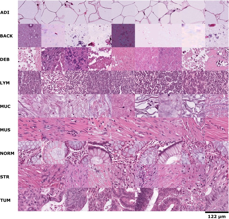
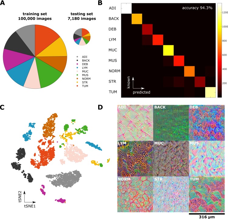
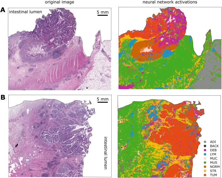
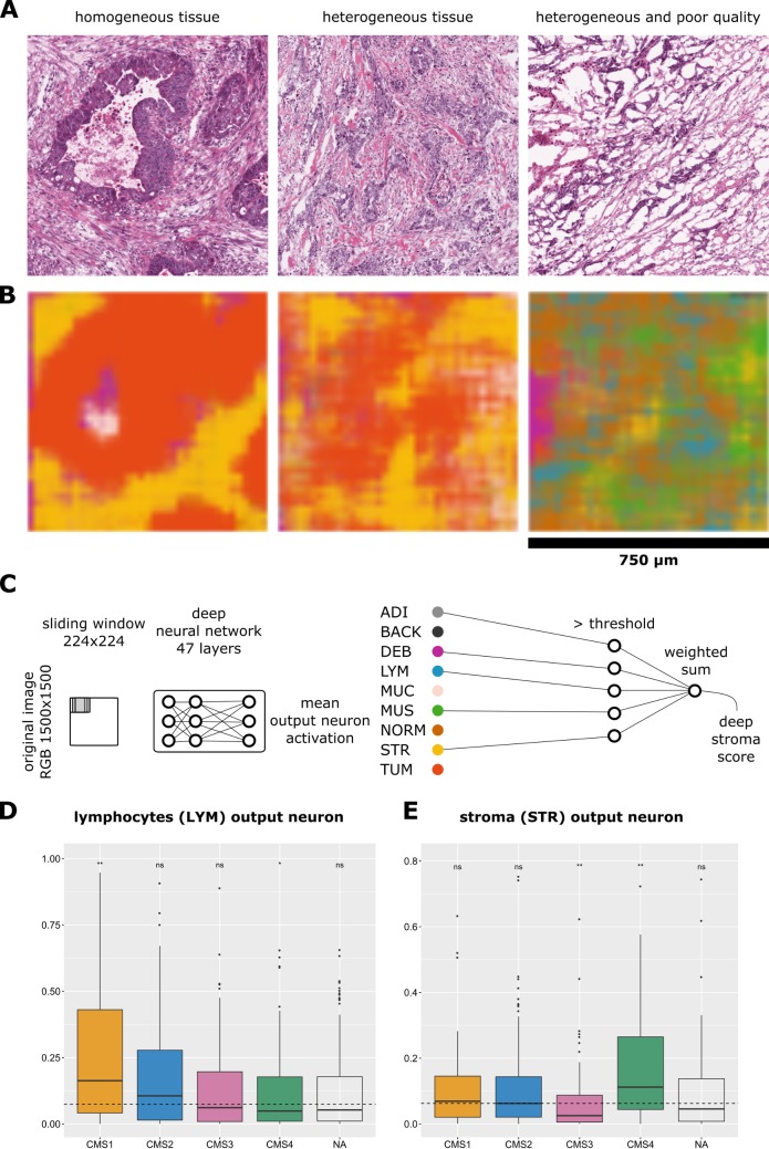
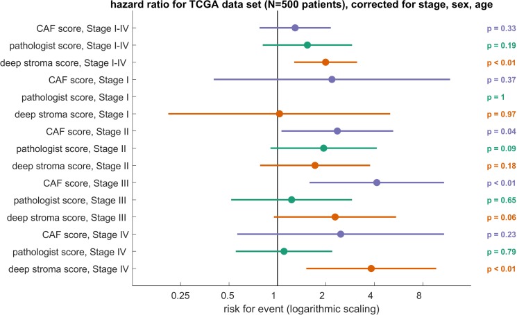
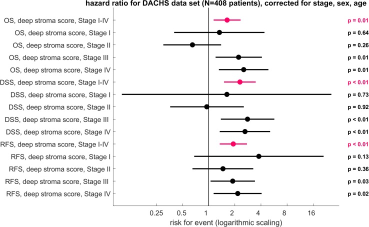
BACKGROUND: For virtually every patient with colorectal cancer (CRC), hematoxylin-eosin (HE)-stained tissue slides are available. These images contain quantitative information, which is not routinely used to objectively extract prognostic biomarkers. In the present study, we investigated whether deep convolutional neural networks (CNNs) can extract prognosticators directly from these widely available images. METHODS AND FINDINGS: We hand-delineated single-tissue regions in 86 CRC tissue slides, yielding more than 100,000 HE image patches, and used these to train a CNN by transfer learning, reaching a nine-class accuracy of >94% in an independent data set of 7,180 images from 25 CRC patients. With this tool, we performed automated tissue decomposition of representative multitissue HE images from 862 HE slides in 500 stage I-IV CRC patients in the The Cancer Genome Atlas (TCGA) cohort, a large international multicenter collection of CRC tissue. Based on the output neuron activations in the CNN, we calculated a "deep stroma score," which was an independent prognostic factor for overall survival (OS) in a multivariable Cox proportional hazard model (hazard ratio [HR] with 95% confidence interval [CI]: 1.99 [1.27-3.12], p = 0.0028), while in the same cohort, manual quantification of stromal areas and a gene expression signature of cancer-associated fibroblasts (CAFs) were only prognostic in specific tumor stages. We validated these findings in an independent cohort of 409 stage I-IV CRC patients from the "Darmkrebs: Chancen der Verhütung durch Screening" (DACHS) study who were recruited between 2003 and 2007 in multiple institutions in Germany. Again, the score was an independent prognostic factor for OS (HR 1.63 [1.14-2.33], p = 0.008), CRC-specific OS (HR 2.29 [1.5-3.48], p = 0.0004), and relapse-free survival (RFS; HR 1.92 [1.34-2.76], p = 0.0004). A prospective validation is required before this biomarker can be implemented in clinical workflows. CONCLUSIONS: In our retrospective study, we show that a CNN can assess the human tumor microenvironment and predict prognosis directly from histopathological images.
Overview
The claims and statistics of this paper haven’t been checked yet.
Run the check — free, no account needed. It reads the open-access text and checks the sources it cites.
Integrity signals
Registry
Cites
Statistics
Figures
Verification
Claims the paper makes
The claims and statistics of this paper haven’t been checked yet.
Run the check — free, no account needed. It reads the open-access text and checks the sources it cites.
Claims other papers make about this one
Each is checked against this paper’s own text: does it say what the citing paper says it does?
In the citing paper
“We applied Loki Annotate to four benchmark histopathology datasets including CRC7K41 (eight tissue types), WSSS-4LUAD42 (normal and tumor), PatchCamelyon43 (normal and tumor) and LC2500044 (benign and malignant).”
From A visual–omics foundation model to bridge histopathology with spatial transcriptomics (2025) · cites this paper as [41]
In the citing paper
“Prognostic AI models for colorectal cancer have been developed for various data modalities, including MRI (88), histopathology (89-91), and multiplex imaging (92,93).”
From Artificial Intelligence in Oncology: Current Landscape, Challenges, and Future Directions (2024) · cites this paper as [90]
Self-check
Percentages, sums and reported differences re-computed from the paper’s own numbers.
The self-check needs the paper’s full text.
Citations
Cites
Deep Residual Learning for Image Recognition
He, K., Zhang, X., Ren, S., & Sun, J. (2016) · 2016 IEEE Conference on Computer Vision and Pattern Recognition (CVPR) · doi:10.1109/cvpr.2016.90 · Cited by 235,474
Very Deep Convolutional Networks for Large-Scale Image Recognition
Simonyan, K., & Zisserman, A. (2014) · arXiv · doi:10.48550/arxiv.1409.1556 · Cited by 114,148
ImageNet classification with deep convolutional neural networks
Krizhevsky, A., Sutskever, I., & Hinton, G. E. (2017) · Communications of the ACM · doi:10.1145/3065386 · Cited by 112,155
LeCun, Y., Bengio, Y., & Hinton, G. (2015) · Nature · doi:10.1038/nature14539 · Cited by 84,126
Going deeper with convolutions
Szegedy, C., Liu, W., Jia, Y., Sermanet, P., Reed, S., Anguelov, D., Erhan, D., Vanhoucke, V., & Rabinovich, A. (2015) · 2015 IEEE Conference on Computer Vision and Pattern Recognition (CVPR) · doi:10.1109/cvpr.2015.7298594 · Cited by 48,562
(2008) · Journal of Machine Learning Research · Cited by 33,051
Mastering the game of Go with deep neural networks and tree search
Silver, D., Huang, A., Maddison, C. J., Guez, A., Sifre, L., van den Driessche, G., Schrittwieser, J., Antonoglou, I., Panneershelvam, V., Lanctot, M., et al. (2016) · Nature · doi:10.1038/nature16961 · Cited by 15,612
Dermatologist-level classification of skin cancer with deep neural networks
Esteva, A., Kuprel, B., Novoa, R. A., Ko, J., Swetter, S. M., Blau, H. M., & Thrun, S. (2017) · Nature · doi:10.1038/nature21056 · Cited by 14,362
Hinton, G., Deng, L., Yu, D., Dahl, G., Mohamed, A.-R., Jaitly, N., Senior, A., Vanhoucke, V., Nguyen, P., Sainath, T., et al. (2012) · IEEE Signal Processing Magazine · doi:10.1109/msp.2012.2205597 · Cited by 10,260 · cited 1×
“Applications of CNNsrangewidely,from speechrecognition[14,15],facerecognition[16], or traffic sign classification[17] to masteringtheJapanese gameof Go [18].”
BackgroundQuPath: Open source software for digital pathology image analysis
Bankhead, P., Loughrey, M. B., Fernández, J. A., Dombrowski, Y., McArt, D. G., Dunne, P. D., McQuaid, S., Gray, R. T., Murray, L. J., Coleman, H. G., et al. (2017) · Scientific Reports · doi:10.1038/s41598-017-17204-5 · Cited by 8,908
Cited by
Sorted by citations
Artificial intelligence in digital pathology — new tools for diagnosis and precision oncology
Bera, K., Schalper, K. A., Rimm, D. L., Velcheti, V., & Madabhushi, A. (2019) · Nature Reviews Clinical Oncology · doi:10.1038/s41571-019-0252-y · Cited by 1,698
Kather, J. N., Pearson, A. T., Halama, N., Jäger, D., Krause, J., Loosen, S. H., Marx, A., Boor, P., Tacke, F., Neumann, U. P., et al. (2019) · Nature Medicine · doi:10.1038/s41591-019-0462-y · Cited by 1,458
Self-citationTowards a general-purpose foundation model for computational pathology
Chen, R. J., Ding, T., Lu, M. Y., Williamson, D. F. K., Jaume, G., Song, A. H., Chen, B., Zhang, A., Shao, D., Shaban, M., et al. (2024) · Nature Medicine · doi:10.1038/s41591-024-02857-3 · Cited by 1,560
Targeted drug delivery strategies for precision medicines
Manzari, M. T., Shamay, Y., Kiguchi, H., Rosen, N., Scaltriti, M., & Heller, D. A. (2021) · Nature Reviews Materials · doi:10.1038/s41578-020-00269-6 · Cited by 1,183
MedMNIST v2 - A large-scale lightweight benchmark for 2D and 3D biomedical image classification
Yang, J., Shi, R., Wei, D., Liu, Z., Zhao, L., Ke, B., Pfister, H., & Ni, B. (2023) · Scientific Data · doi:10.1038/s41597-022-01721-8 · Cited by 1,043
“The PathMNIST is based on a prior study 16,17 for predicting survival from colorectal cancer histology slides, providing a dataset”
MethodTransfer learning for medical image classification: a literature review
Kim, H. E., Cosa‐Linan, A., Santhanam, N., Jannesari, M., Maros, M. E., & Ganslandt, T. (2022) · BMC Medical Imaging · doi:10.1186/s12880-022-00793-7 · Cited by 1,117
A visual-language foundation model for computational pathology
Lu, M. Y., Chen, B., Williamson, D. F. K., Chen, R. J., Liang, I., Ding, T., Jaume, G., Odintsov, I., Le, L. P., Gerber, G., et al. (2024) · Nature Medicine · doi:10.1038/s41591-024-02856-4 · Cited by 966
Deep learning in histopathology: the path to the clinic
van der Laak, J., Litjens, G., & Ciompi, F. (2021) · Nature Medicine · doi:10.1038/s41591-021-01343-4 · Cited by 902
Deep neural network models for computational histopathology: A survey
Srinidhi, C. L., Ciga, O., & Martel, A. L. (2021) · Medical Image Analysis · doi:10.1016/j.media.2020.101813 · Cited by 749
Transformer-based unsupervised contrastive learning for histopathological image classification
Wang, X., Yang, S., Zhang, J., Wang, M., Zhang, J., Yang, W., Huang, J., & Han, X. (2022) · Medical Image Analysis · doi:10.1016/j.media.2022.102559 · Cited by 751
A visual–language foundation model for pathology image analysis using medical twitter
Huang, Z., Bianchi, F., Yuksekgonul, M., Montine, T. J., & Zou, J. (2023) · Nature Medicine · doi:10.1038/s41591-023-02504-3 · Cited by 693
Pan-cancer image-based detection of clinically actionable genetic alterations
Kather, J. N., Heij, L. R., Grabsch, H. I., Loeffler, C. M. L., Echle, A., Muti, H. S., Krause, J., Niehues, J., Sommer, K. A. J., Bankhead, P., et al. (2020) · Nature Cancer · doi:10.1038/s43018-020-0087-6 · Cited by 686
Self-citationDeep learning in cancer pathology: a new generation of clinical biomarkers
Echle, A., Rindtorff, N. T., Brinker, T. J., Luedde, T., Pearson, A. T., & Kather, J. N. (2021) · British Journal of Cancer · doi:10.1038/s41416-020-01122-x · Cited by 667
Self-citationPan-cancer integrative histology-genomic analysis via multimodal deep learning
Chen, R. J., Lu, M. Y., Williamson, D. F. K., Chen, T. Y., Lipkova, J., Noor, Z., Shaban, M., Shady, M., Williams, M., Joo, B., et al. (2022) · Cancer Cell · doi:10.1016/j.ccell.2022.07.004 · Cited by 660
Molecular and histological correlations in liver cancer
Caldéraro, J., Ziol, M., Paradis, V., & Zucman‐Rossi, J. (2019) · Journal of Hepatology · doi:10.1016/j.jhep.2019.06.001 · Cited by 619
A foundation model for clinical-grade computational pathology and rare cancers detection
Vorontsov, E., Bozkurt, A., Casson, A., Shaikovski, G., Zelechowski, M., Severson, K., Zimmermann, E., Hall, J., Tenenholtz, N., Fusi, N., et al. (2024) · Nature Medicine · doi:10.1038/s41591-024-03141-0 · Cited by 572
MedViT: A robust vision transformer for generalized medical image classification
Manzari, O. N., Ahmadabadi, H., Kashiani, H., Shokouhi, S. B., & Ayatollahi, A. (2023) · Computers in Biology and Medicine · doi:10.1016/j.compbiomed.2023.106791 · Cited by 573
A deep learning model to predict RNA-Seq expression of tumours from whole-slide images
Schmauch, B., Romagnoni, A., Pronier, E., Saillard, C., Maillé, P., Calderaro, J., Kamoun, A., Sefta, M., Toldo, S., Zaslavskiy, M., et al. (2020) · Nature Communications · doi:10.1038/s41467-020-17678-4 · Cited by 533
Artificial intelligence in histopathology: enhancing cancer research and clinical oncology
Shmatko, A., Ghaffari Laleh, N., Gerstung, M., & Kather, J. N. (2022) · Nature Cancer · doi:10.1038/s43018-022-00436-4 · Cited by 446
Self-citationEmerging role of deep learning‐based artificial intelligence in tumor pathology
Jiang, Y., Yang, M., Wang, S., Li, X., & Sun, Y. K. (2020) · 癌症:英文版 · doi:10.1002/cac2.12012 · Cited by 460
MedMNIST Classification Decathlon: A Lightweight AutoML Benchmark for Medical Image Analysis
Yang, J., Shi, R., & Ni, B. (2021) · 2021 IEEE 18th International Symposium on Biomedical Imaging (ISBI) · doi:10.1109/isbi48211.2021.9434062 · Cited by 414
Li, J., Chen, J., Tang, Y., Wang, C., Landman, B. A., & Zhou, S. K. (2023) · Medical Image Analysis · doi:10.1016/j.media.2023.102762 · Cited by 454
Deep Learning Models for Histopathological Classification of Gastric and Colonic Epithelial Tumours
Iizuka, O., Kanavati, F., Kato, K., Rambeau, M., Arihiro, K., & Tsuneki, M. (2020) · Scientific Reports · doi:10.1038/s41598-020-58467-9 · Cited by 412
Yamashita, R., Long, J., Longacre, T., Peng, L., Berry, G., Martin, B., Higgins, J., Rubin, D. L., & Shen, J. (2021) · The Lancet Oncology · doi:10.1016/s1470-2045(20)30535-0 · Cited by 391
Clinical-Grade Detection of Microsatellite Instability in Colorectal Tumors by Deep Learning
Echle, A., Grabsch, H. I., Quirke, P., van den Brandt, P. A., West, N. P., Hutchins, G. G. A., Heij, L. R., Tan, X., Richman, S. D., Krause, J., et al. (2020) · Gastroenterology · doi:10.1053/j.gastro.2020.06.021 · Cited by 376
Self-citationThe Application of Deep Learning in Cancer Prognosis Prediction
Zhu, W., Xie, L., Han, J., & Guo, X. (2020) · Cancers · doi:10.3390/cancers12030603 · Cited by 398
Artificial intelligence for digital and computational pathology
Song, A. H., Jaume, G., Williamson, D. F. K., Lu, M. Y., Vaidya, A., Miller, T. R., & Mahmood, F. (2023) · Nature Reviews Bioengineering · doi:10.1038/s44222-023-00096-8 · Cited by 356
Feng, L., Liu, Z., Li, C., Li, Z., Lou, X., Shao, L., Wang, Y., Huang, Y., Chen, H., Pang, X., et al. (2022) · The Lancet Digital Health · doi:10.1016/s2589-7500(21)00215-6 · Cited by 339
Saillard, C., Schmauch, B., Laifa, O., Moarii, M., Toldo, S., Zaslavskiy, M., Pronier, E., Laurent, A., Amaddeo, G., Regnault, H., et al. (2020) · Hepatology · doi:10.1002/hep.31207 · Cited by 332
The impact of site-specific digital histology signatures on deep learning model accuracy and bias
Howard, F. M., Dolezal, J., Kochanny, S., Schulte, J., Chen, H., Heij, L., Huo, D., Nanda, R., Olopade, O. I., Kather, J. N., et al. (2021) · Nature Communications · doi:10.1038/s41467-021-24698-1 · Cited by 330
Self-citationDesigning deep learning studies in cancer diagnostics
Kleppe, A., Skrede, O.-J., Raedt, S. D., Liestøl, K., Kerr, D. J., & Danielsen, H. E. (2021) · Nature reviews. Cancer · doi:10.1038/s41568-020-00327-9 · Cited by 328
A comprehensive review of deep learning in colon cancer
Paçal, İ., Karaboğa, D., Baştürk, A., Akay, B., & Nalbantoglu, U. (2020) · Computers in Biology and Medicine · doi:10.1016/j.compbiomed.2020.104003 · Cited by 300
Deep learning-based survival prediction for multiple cancer types using histopathology images
Wulczyn, E., Steiner, D. F., Xu, Z., Sadhwani, A., Wang, H., Flament-Auvigne, I., Mermel, C. H., Chen, P.-H. C., Liu, Y., & Stumpe, M. C. (2020) · PLOS ONE · doi:10.1371/journal.pone.0233678 · Cited by 266
“While other machine learning efforts have identified prognostic significance for non-tumor elements[17,32], our observations suggest that at least for our specific models, the morphologic features of the tumor appear to be more relevant than non-tumor regions.”
BackgroundMethodDigital pathology and computational image analysis in nephropathology
Barisoni, L., Lafata, K. J., Hewitt, S. M., Madabhushi, A., & Balis, U. G. J. (2020) · Nature Reviews Nephrology · doi:10.1038/s41581-020-0321-6 · Cited by 264
A Vision-Language Foundation Model for Precision Oncology
Xiang, J., Wang, X., Zhang, X., Xi, Y., Eweje, F., Chen, Y., Li, Y., Bergstrom, C., Gopaulchan, M., Kim, T., et al. (2025) · Nature · doi:10.1038/s41586-024-08378-w · Cited by 256
Deep learning for colon cancer histopathological images analysis
Hamida, A. B., Devanne, M., Weber, J., Truntzer, C., Dérangère, V., Ghiringhelli, F., Forestier, G., & Wemmert, C. (2021) · Computers in Biology and Medicine · doi:10.1016/j.compbiomed.2021.104730 · Cited by 263
A generalized deep learning framework for whole-slide image segmentation and analysis
Khened, M., Kori, A., Rajkumar, H., Krishnamurthi, G., & Srinivasan, B. (2021) · Scientific Reports · doi:10.1038/s41598-021-90444-8 · Cited by 255
Interpretable survival prediction for colorectal cancer using deep learning
Wulczyn, E., Steiner, D. F., Moran, M., Plass, M., Reihs, R., Tan, F., Flament-Auvigne, I., Brown, T., Regitnig, P., Chen, P.-H. C., et al. (2021) · npj Digital Medicine · doi:10.1038/s41746-021-00427-2 · Cited by 231
Shi, J.-Y., Wang, X., Ding, G., Dong, Z., Han, J., Guan, Z., Ma, L., Zheng, Y., Zhang, L., Yu, G., et al. (2020) · Gut · doi:10.1136/gutjnl-2020-320930 · Cited by 198
Kuntz, S., Krieghoff‐Henning, E., Kather, J. N., Jutzi, T. B., Höhn, J., Kiehl, L., Hekler, A., Alwers, E., Kalle, C. V., Fröhling, S., et al. (2021) · European Journal of Cancer · doi:10.1016/j.ejca.2021.07.012 · Cited by 203
Self-citationArtificial Intelligence in Oncology: Current Landscape, Challenges, and Future Directions
Lotter, W., Hassett, M. J., Schultz, N., Kehl, K. L., Van Allen, E. M., & Cerami, E. (2024) · Cancer Discovery · doi:10.1158/2159-8290.cd-23-1199 · Cited by 196
Zhang, B., Yu, K., Ning, Z., Wang, K., Dong, Y., Liu, X., Liu, S., Wang, J., Zhu, C., Yu, Q., et al. (2020) · Bone · doi:10.1016/j.bone.2020.115561 · Cited by 194
Jayapandian, C. P., Chen, Y. M., Janowczyk, A. R., Palmer, M. B., Cassol, C. A., Sekulic, M., Hodgin, J. B., Zee, J., Hewitt, S. M., O’Toole, J., et al. (2020) · Kidney International · doi:10.1016/j.kint.2020.07.044 · Cited by 185
Celard, P., Iglesias, E. L., Sorribes-Fdez, J. M., Romero, R., Vieira, A. S., & Borrajo, L. (2023) · Neural Computing and Applications · doi:10.1007/s00521-022-07953-4 · Cited by 188
High-accuracy prostate cancer pathology using deep learning
Tolkach, Y., Dohmgörgen, T., Toma, M. I., & Kristiansen, G. (2020) · Nature Machine Intelligence · doi:10.1038/s42256-020-0200-7 · Cited by 185
TransPath: Transformer-Based Self-supervised Learning for Histopathological Image Classification
Wang, X., Yang, S., Zhang, J., Wang, M., Zhang, J., Huang, J., Yang, W., & Han, X. (2021) · Lecture Notes in Computer Science · doi:10.1007/978-3-030-87237-3_18 · Cited by 178
Accurate diagnosis of colorectal cancer based on histopathology images using artificial intelligence
Wang, K., Yu, G., Xu, C., Meng, X., Zhou, J., Zheng, C., Deng, Z., Shang, L., Liu, R., Su, S., et al. (2021) · BMC Medicine · doi:10.1186/s12916-021-01942-5 · Cited by 196
Xu, Y., Su, G.-H., Ma, D., Xiao, Y., Shao, Z.-M., & Jiang, Y.-Z. (2021) · Signal Transduction and Targeted Therapy · doi:10.1038/s41392-021-00729-7 · Cited by 163
“201 Furthermore, predictive models that incorporate the immune cell composition and spatial organization correlate with cancer prognosis, which several groups have proven in colorectal cancer, HCC, and melanoma 202 – 205 (Fig.”
BackgroundWoerl, A.-C., Eckstein, M., Geiger, J., Wagner, D., Daher, T., Stenzel, P., Fernandez, A., Hartmann, A., Wand, M., Roth, W., et al. (2020) · European Urology · doi:10.1016/j.eururo.2020.04.023 · Cited by 177
Cellular community detection for tissue phenotyping in colorectal cancer histology images
Javed, S., Mahmood, A., Fraz, M. M., Koohbanani, N. A., Benes, K., Tsang, Y., Hewitt, K. J., Epstein, D., Snead, D. R. J., & Rajpoot, N. (2020) · Medical Image Analysis · doi:10.1016/j.media.2020.101696 · Cited by 175
Jiang, Y., Jin, C., Yu, H., Wu, J., Chen, C., Yuan, Q., Huang, W., Hu, Y., Xu, Y., Zhou, Z., et al. (2021) · Annals of Surgery · doi:10.1097/sla.0000000000003778 · Cited by 169
Lin, J., Chen, Y.-A., Campton, D. E., Cooper, J., Coy, S., Yapp, C., Tefft, J. B., McCarty, E. F., Ligon, K. L., Rodig, S. J., et al. (2023) · Nature Cancer · doi:10.1038/s43018-023-00576-1 · Cited by 180
Artificial intelligence in gastroenterology: A state-of-the-art review
Kröner, P. T., Engels, M. M. L., Glicksberg, B. S., Johnson, K. W., Mzaik, O., van Hooft, J. E., Wallace, M. B., El‐Serag, H. B., & Krittanawong, C. (2021) · World Journal of Gastroenterology · doi:10.3748/wjg.v27.i40.6794 · Cited by 175
In-context learning enables multimodal large language models to classify cancer pathology images
Ferber, D., Wölflein, G., Wiest, I. C., Ligero, M., Sainath, S., Laleh, N. G., Nahhas, O. S. M. E., Müller‐Franzes, G. A., Jäger, D., Truhn, D., et al. (2024) · Nature Communications · doi:10.1038/s41467-024-51465-9 · Cited by 182
Self-citationJaume, G., Vaidya, A. J., Chen, R. J., Williamson, D. F. K., Liang, P. P., & Mahmood, F. (2024) · IEEE Computer Society Conference on Computer Vision and Pattern Recognition/Proceedings - IEEE Computer Society Conference on Computer Vision and Pattern Recognition/Proceedings · doi:10.1109/cvpr52733.2024.01100 · Cited by 184
HistoSegNet: Semantic Segmentation of Histological Tissue Type in Whole Slide Images
Chan, L., Hosseini, M. S., Rowsell, C. H., Plataniotis, K. N., & Damaskinos, S. (2019) · IEEE/CVF International Conference on Computer Vision (ICCV) · doi:10.1109/iccv.2019.01076 · Cited by 152
Zhao, K., Li, Z., Yao, S., Wang, Y., Wu, X., Xu, Z., Wu, L., Huang, Y., Liang, C., & Liu, Z. (2020) · EBioMedicine · doi:10.1016/j.ebiom.2020.103054 · Cited by 161
Han, C., Lin, J., Mai, J., Wang, Y., Zhang, Q., Zhao, B., Chen, X., Pan, X., Shi, Z., Xu, Z., et al. (2022) · Medical Image Analysis · doi:10.1016/j.media.2022.102487 · Cited by 172
Graham, S., Vu, Q. D., Jahanifar, M., Raza, S. E. A., Minhas, F., Snead, D. R. J., & Rajpoot, N. (2022) · Medical Image Analysis · doi:10.1016/j.media.2022.102685 · Cited by 151
Quality control stress test for deep learning-based diagnostic model in digital pathology
Schömig-Markiefka, B., Pryalukhin, A., Hulla, W., Bychkov, A., Fukuoka, J., Madabhushi, A., Achter, V., Nieroda, L., Büttner, R., Quaas, A., et al. (2021) · Modern Pathology · doi:10.1038/s41379-021-00859-x · Cited by 146
Accurate recognition of colorectal cancer with semi-supervised deep learning on pathological images
Yu, G., Sun, K., Xu, C., Shi, X.-H., Wu, C., Xie, T., Meng, R.-Q., Meng, X.-H., Wang, K.-S., Xiao, H.-M., et al. (2021) · Nature Communications · doi:10.1038/s41467-021-26643-8 · Cited by 147
Tufail, A. B., Ma, Y., Kaabar, M. K. A., Martínez, F., Junejo, A. R., Ullah, I., & Khan, R. (2021) · Computational and Mathematical Methods in Medicine · doi:10.1155/2021/9025470 · Cited by 151
Imbalanced Breast Cancer Classification Using Transfer Learning
Singh, R., Ahmed, T., Kumar, A., Singh, A. K., Pandey, A. K., & Singh, S. K. (2020) · IEEE Transactions on Computational Biology and Bioinformatics · doi:10.1109/tcbb.2020.2980831 · Cited by 139
Deep Learning on Histopathological Images for Colorectal Cancer Diagnosis: A Systematic Review
Davri, A. S., Birbas, E., Kanavos, T., Ntritsos, G., Γιαννακέας, Ν., Tzallas, A. T., & Batistatou, A. K. (2022) · Diagnostics · doi:10.3390/diagnostics12040837 · Cited by 144
Chen, H., Li, C., Li, X., Rahaman, M. M., Hu, W., Li, Y., Liu, W., Sun, C., Sun, H., Huang, X., et al. (2022) · Computers in Biology and Medicine · doi:10.1016/j.compbiomed.2022.105265 · Cited by 141
Molecular subtypes and precision treatment of triple-negative breast cancer
Zhao, S., Zuo, W., Shao, Z., & Jiang, Y. (2020) · Annals of Translational Medicine · doi:10.21037/atm.2020.03.194 · Cited by 140
A comprehensive survey on deep active learning in medical image analysis
Wang, H., Jin, Q., Li, S., Liu, S., Wang, M., & Song, Z. (2024) · Medical Image Analysis · doi:10.1016/j.media.2024.103201 · Cited by 157
Liu, X., Zhang, D., Liu, Z., Li, Z., Xie, P., Sun, K., Wei, W., Dai, W., Tang, Z., Ding, Y., et al. (2021) · EBioMedicine · doi:10.1016/j.ebiom.2021.103442 · Cited by 127
A promising deep learning-assistive algorithm for histopathological screening of colorectal cancer
Ho, C., Zhao, Z., Chen, X. F., Sauer, J., Saraf, S. A., Jialdasani, R., Taghipour, K., Sathe, A. R., Khor, L.-Y., Lim, K., et al. (2022) · Scientific Reports · doi:10.1038/s41598-022-06264-x · Cited by 132
TIAToolbox as an end-to-end library for advanced tissue image analytics
Pocock, J., Graham, S., Vu, Q. D., Jahanifar, M., Deshpande, S., Hadjigeorghiou, G., Shephard, A., Bashir, R. M. S., Bilal, M., Lu, W., et al. (2022) · Communications Medicine · doi:10.1038/s43856-022-00186-5 · Cited by 144
Cheng, N., Ren, Y., Zhou, J., Zhang, Y., Wang, D., Zhang, X., Chen, B., Liu, F., Lv, J., Cao, Q., et al. (2022) · Gastroenterology · doi:10.1053/j.gastro.2022.02.025 · Cited by 121
Fast and scalable search of whole-slide images via self-supervised deep learning
Chen, C., Lu, M. Y., Williamson, D. F. K., Chen, T. Y., Schaumberg, A. J., & Mahmood, F. (2022) · Nature Biomedical Engineering · doi:10.1038/s41551-022-00929-8 · Cited by 130
Application of Artificial Intelligence in Pathology: Trends and Challenges
Kim, I., Kang, K., Song, Y., & Kim, T. J. (2022) · Diagnostics · doi:10.3390/diagnostics12112794 · Cited by 137
Maassen, O., Fritsch, S., Palm, J., Deffge, S., Kunze, J., Marx, G., Riedel, M., Schuppert, A., & Bickenbach, J. (2021) · Journal of Medical Internet Research · doi:10.2196/26646 · Cited by 125
Colorectal Histology Tumor Detection Using Ensemble Deep Neural Network
Ghosh, S., Bandyopadhyay, A., Sahay, S., Ghosh, R., Kundu, I., & Santosh, K. (2021) · Engineering Applications of Artificial Intelligence · doi:10.1016/j.engappai.2021.104202 · Cited by 129
Recent progress in transformer-based medical image analysis
Liu, Z., Lv, Q., Yang, Z., Li, Y., Lee, C. H., & Shen, L. (2023) · Computers in Biology and Medicine · doi:10.1016/j.compbiomed.2023.107268 · Cited by 133
Deep learning in cancer genomics and histopathology
Unger, M., & Kather, J. N. (2024) · Genome Medicine · doi:10.1186/s13073-024-01315-6 · Cited by 135
Self-citationReichling, C., Taı̈eb, J., Dérangère, V., Klopfenstein, Q., Malicot, K. L., Gornet, J., Bécheur, H., Fein, F., Cojocarasu, O., Kaminsky, M. C., et al. (2019) · Gut · doi:10.1136/gutjnl-2019-319292 · Cited by 124
Gehrung, M., Crispin‐Ortuzar, M., Berman, A. G., O’Donovan, M., Fitzgerald, R. C., & Markowetz, F. M. (2021) · Nature Medicine · doi:10.1038/s41591-021-01287-9 · Cited by 127
Eloy, C., Marques, A., Pinto, J. C., Pinheiro, J., Campelos, S., Curado, M., Vale, J., & Polónia, A. (2023) · Archiv für Pathologische Anatomie und Physiologie und für Klinische Medicin · doi:10.1007/s00428-023-03518-5 · Cited by 118
Computational pathology: A survey review and the way forward
Hosseini, M. S., Bejnordi, B. E., Trinh, V. Q., Chan, L., Hasan, D., Li, X., Yang, S. C., Kim, T., Zhang, H., Wu, T., et al. (2024) · Journal of Pathology Informatics · doi:10.1016/j.jpi.2023.100357 · Cited by 142
A Comprehensive Analysis of Weakly-Supervised Semantic Segmentation in Different Image Domains
Chan, L., Hosseini, M. S., & Plataniotis, K. N. (2020) · International Journal of Computer Vision · doi:10.1007/s11263-020-01373-4 · Cited by 118
Self-supervised driven consistency training for annotation efficient histopathology image analysis
Srinidhi, C. L., Kim, S. W., Chen, F.-D., & Martel, A. L. (2022) · Medical Image Analysis · doi:10.1016/j.media.2021.102256 · Cited by 116
Framework for Detecting Breast Cancer Risk Presence Using Deep Learning
Humayun, M., Khalil, M. I., Almuayqil, S. N., & Jhanjhi, N. Z. (2023) · Electronics · doi:10.3390/electronics12020403 · Cited by 118
Li, F., Yang, Y., Wei, Y., He, P., Chen, J., Zheng, Z., & Bu, H. (2021) · Journal of Translational Medicine · doi:10.1186/s12967-021-03020-z · Cited by 113
Kumar, A., Vishwakarma, A., & Bajaj, V. (2022) · Biomedical Signal Processing and Control · doi:10.1016/j.bspc.2022.104172 · Cited by 117
A visual–omics foundation model to bridge histopathology with spatial transcriptomics
Chen, W., Zhang, P., Tran, T. N., Xiao, Y., Li, S., Shah, V. V., Cheng, H., Brannan, K. W., Youker, K., Lai, L., et al. (2025) · Nature Methods · doi:10.1038/s41592-025-02707-1 · Cited by 118
“We used OmiCLIP to encode tissue patches from a WSI and the tissue-specific bulk RNA-seq data, then calculated the cosine similarity between the encoded embeddings (Fig.”
Nahhas, O. S. M. E., van Treeck, M., Wölflein, G., Unger, M., Ligero, M., Lenz, T., Wagner, S. J., Hewitt, K. J., Khader, F., Foersch, S., et al. (2024) · Nature Protocols · doi:10.1038/s41596-024-01047-2 · Cited by 121
Self-citationArtificial intelligence-based pathology for gastrointestinal and hepatobiliary cancers
Caldéraro, J., & Kather, J. N. (2020) · Gut · doi:10.1136/gutjnl-2020-322880 · Cited by 108
Self-citationDeep learned tissue “fingerprints” classify breast cancers by ER/PR/Her2 status from H&E images
Rawat, R. R., Ortega, I., Roy, P., Sha, F., Shibata, D., Ruderman, D., & Agus, D. B. (2020) · Scientific Reports · doi:10.1038/s41598-020-64156-4 · Cited by 100
Yin, Z., Yao, C., Zhang, L., & Qi, S. (2023) · Frontiers in Medicine · doi:10.3389/fmed.2023.1128084 · Cited by 102
Deep learning supported discovery of biomarkers for clinical prognosis of liver cancer
Liang, J., Zhang, W., Yang, J., Wu, M., Dai, Q., Yin, H., Xiao, Y., & Kong, L. (2023) · Nature Machine Intelligence · doi:10.1038/s42256-023-00635-3 · Cited by 98
Marra, A., Morganti, S., Pareja, F. G., Campanella, G. S. V., Bibeau, F., Fuchs, T. J., Loda, M. F., Parwani, A. V., Scarpa, A., Reis-Filho, J. S., et al. (2025) · Annals of Oncology · doi:10.1016/j.annonc.2025.03.006 · Cited by 114
Self-citationLi, C., Lin, X., Mao, Y., Lin, W. Y., Qi, Q., Ding, X., Huang, Y., Liang, D., & Yu, Y. (2021) · Computers in Biology and Medicine · doi:10.1016/j.compbiomed.2021.105144 · Cited by 98
Sharma, P., Nayak, D. R., Balabantaray, B. K., Tanveer, M., & Nayak, R. (2023) · Neural Networks · doi:10.1016/j.neunet.2023.11.006 · Cited by 104
AI in medical diagnosis: AI prediction & human judgment
Göndöcs, D., & Dörfler, V. (2024) · Artificial Intelligence in Medicine · doi:10.1016/j.artmed.2024.102769 · Cited by 108
Artificial Intelligence and Mechanistic Modeling for Clinical Decision Making in Oncology
Benzekry, S. (2020) · Clinical Pharmacology & Therapeutics · doi:10.1002/cpt.1951 · Cited by 96
Shao, L., Liu, Z., Feng, L., Lou, X., Li, Z., Zhang, X.-Y., Wan, X., Zhou, X., Sun, K., Zhang, D.-F., et al. (2020) · Annals of Surgical Oncology · doi:10.1245/s10434-020-08659-4 · Cited by 92
Thakur, N., Yoon, H., & Chong, Y. (2020) · Cancers · doi:10.3390/cancers12071884 · Cited by 91
Williams, C. J. M., Peddle, A. M., Kasi, P. M., Seligmann, J. F., Roxburgh, C. S. D., Middleton, G., & Tejpar, S. (2024) · Nature Reviews Clinical Oncology · doi:10.1038/s41571-024-00943-6 · Cited by 99
Metadata
- Authors
- Kather, J. N., Krisam, J., Charoentong, P., Luedde, T., Herpel, E., Weis, C.-A., Gaiser, T., Marx, A., Valous, N. A., Ferber, D., Jansen, L., Reyes-Aldasoro, C. C., Zörnig, I., Jäger, D., Brenner, H., Chang-Claude, J., Hoffmeister, M., & Halama, N.
- Affiliations
- German Cancer Research Center (DE)
- University Hospital Heidelberg (DE)
- Universitätsklinikum Aachen (DE)
- Heidelberg University (DE)
- University Medical Centre Mannheim (DE)
- Corresponding author
- Kather, Jakob Nikolas
- ORCID
- Venue
- PLOS Medicine
- Publisher
- Public Library of Science (PLoS)
- Published
- 2019-01-24
- Volume
- 16
- Issue
- 1
- Pages
- e1002730
- ISSN
- 1549-1277 (ISSN-L)
- 1549-1676
- PMC
- PMC6345440
- OpenAlex
- W2914568698
- Cited by
- 1,082
- References
- 52
- Citations by year
- 2026: 158 · 2025: 164 · 2024: 180 · 2023: 170 · 2022: 175 · 2021: 129 · 2020: 87 · 2019: 17
- FWCI
- 46.54
- Type
- journal article
- Language
- en
- Open access
- gold
- doi.org
- Licence
- Locations
- PLoS Medicine (publishedVersion, cc-by)
- Europe PMC (PubMed Central) (publishedVersion, cc-by)
- PubMed Central (publishedVersion, cc-by)
- PubMed (publishedVersion)
- City Research Online (City University London) (acceptedVersion)
- RWTH Publications (RWTH Aachen) (submittedVersion, cc-by)
- DOAJ (DOAJ: Directory of Open Access Journals) (submittedVersion, cc-by-sa)
- Figshare (submittedVersion, cc-by)
- RWTH Publications (RWTH Aachen)
- Funders
- Deutsches Krebsforschungszentrum (Heidelberg School of Oncology)
- Bundesministerium für Bildung und Forschung (13GW0091E, 01ER0814, 01ER0815, 01ER1505A, 01ER1505B)
- Deutsche Forschungsgemeinschaft (BR 1704/6-1, BR 1704/6-3, BR 1704/6-4, BR 1704/17-1, CH 117/1-1, and HO 5117/2-1)
- Topics
- Physical Sciences › Computer Science › Artificial Intelligence › AI in cancer detection
- Health Sciences › Medicine › Radiology, Nuclear Medicine and Imaging › Radiomics and Machine Learning in Medical Imaging
- Health Sciences › Medicine › Oncology › Colorectal Cancer Screening and Detection
- Keywords
- colorectal cancer, deep convolutional neural networks, tumor stroma, prognostic factors, overall survival, cancer-associated fibroblasts, cancer of unknown primary, relapse-free survival, TCGA, transfer learning, histopathology image analysis, digital pathology
- MeSH terms
- Deep Learning, Colon, Coloring Agents, Eosine Yellowish-(YS), Female, Hematoxylin, Humans, Image Interpretation, Computer-Assisted, Male, Prognosis, Rectum, Retrospective Studies, Colorectal Neoplasms
About these checks
Registry, retraction, Cites and statistics results are computed, not judged, with one exception: a printed reference that no index lists is read once by a model before it counts as a citation error. Claims are extracted by a model and kept only when their quote appears verbatim in the text; each cited source is then checked, with a model’s help, for whether it says what the claim relies on. Those verdicts can be wrong — read the claim’s quote and the cited source before relying on one.
Statistics and arithmetic are recomputed, not judged.
Confidence bands are shown only for fields calibrated on our annotated test set. None is calibrated yet, so no band is shown.
What counts as a claim ›How we measure confidence ›How we check this ›
Are you an author, or do you see a problem with this page? Request a review
