

The DOI matches its Crossref record; 48 of the 48 references it cites were found in a registry or an index.
A Specific and Rapid Neural Signature for Parental Instinct
Open accessAbstract
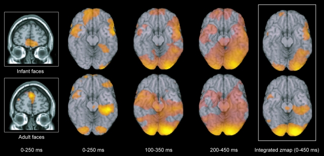
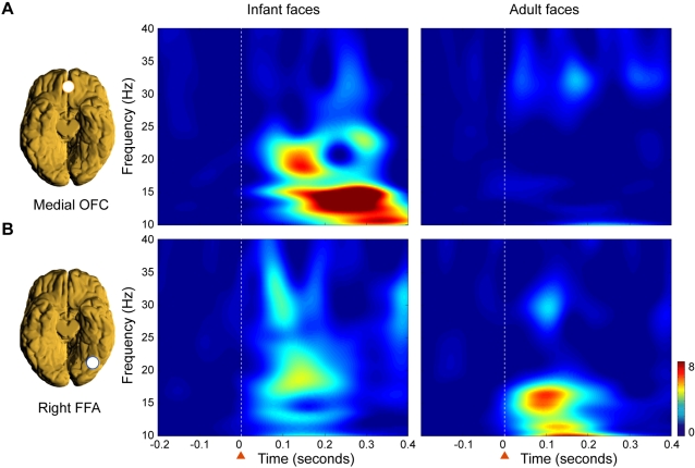
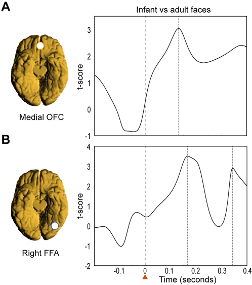
Darwin originally pointed out that there is something about infants which prompts adults to respond to and care for them, in order to increase individual fitness, i.e. reproductive success, via increased survivorship of one's own offspring. Lorenz proposed that it is the specific structure of the infant face that serves to elicit these parental responses, but the biological basis for this remains elusive. Here, we investigated whether adults show specific brain responses to unfamiliar infant faces compared to adult faces, where the infant and adult faces had been carefully matched across the two groups for emotional valence and arousal, as well as size and luminosity. The faces also matched closely in terms of attractiveness. Using magnetoencephalography (MEG) in adults, we found that highly specific brain activity occurred within a seventh of a second in response to unfamiliar infant faces but not to adult faces. This activity occurred in the medial orbitofrontal cortex (mOFC), an area implicated in reward behaviour, suggesting for the first time a neural basis for this vital evolutionary process. We found a peak in activity first in mOFC and then in the right fusiform face area (FFA). In mOFC the first significant peak (p<0.001) in differences in power between infant and adult faces was found at around 130 ms in the 10-15 Hz band. These early differences were not found in the FFA. In contrast, differences in power were found later, at around 165 ms, in a different band (20-25 Hz) in the right FFA, suggesting a feedback effect from mOFC. These findings provide evidence in humans of a potential brain basis for the "innate releasing mechanisms" described by Lorenz for affection and nurturing of young infants. This has potentially important clinical applications in relation to postnatal depression, and could provide opportunities for early identification of families at risk.
Overview
The claims and statistics of this paper haven’t been checked yet.
Run the check — free, no account needed. It reads the open-access text and checks the sources it cites.
Integrity signals
Registry
Cites
Statistics
Figures
Verification
Claims the paper makes
The claims and statistics of this paper haven’t been checked yet.
Run the check — free, no account needed. It reads the open-access text and checks the sources it cites.
Claims other papers make about this one
Each is checked against this paper’s own text: does it say what the citing paper says it does?
In the citing paper
“Again, coordinated responses in drive (amygdala) and regulation (cinglulate) figure prominently in these papers.”
From The human parental brain: In vivo neuroimaging (2011) · cites this paper as Kringelbach et al., 2008
In the citing paper
“Not only is facial recognition of one’s infant and their emotional state critical to their survival, but evidence supports human preference for the exaggerated cute/infantile features of baby faces which appear to activate reward mechanisms within the caregiver and motivate/promote mothers to caregiving, bonding and attachment.”
From Approaching the biology of human parental attachment: Brain imaging, oxytocin and coordinated assessments of mothers and fathers (2014) · cites this paper as Kringelbach et al., 2008
In the citing paper
“In addition to activation of nucleus accumbens (NAcc) in response to baby schema (Glocker et al., 2009) and medial orbitofrontal cortex (OFC) response to infant faces relative to adults (Kringelbach et al., 2008), higher levels of infantile features are preferred (Parsons et al., 2011).”
From Approaching the biology of human parental attachment: Brain imaging, oxytocin and coordinated assessments of mothers and fathers (2014) · cites this paper as Kringelbach et al., 2008
Self-check
Percentages, sums and reported differences re-computed from the paper’s own numbers.
The self-check needs the paper’s full text.
Citations
Cites
The expression of the emotions in man and animals.
Darwin, C. D. (1872) · J. Murray eBooks · doi:10.1037/10001-000 · Cited by 12,120
Attachment and Loss. Vol. 1. Attachment
Rever, G. W. (1972) · Psychosomatic Medicine · doi:10.1097/00006842-197211000-00011 · Cited by 8,446
The fusiform face area: a module in human extrastriate cortex specialized for face perception
Kanwisher, N., McDermott, J., & Chun, M. M. (1997) · The Journal of Neuroscience · doi:10.1523/jneurosci.17-11-04302.1997 · Cited by 7,738 · cited 2×
“It is still, however, controversial whether the activity in FFA represents a localized or distributed coding of faces [8,11,12].”
BackgroundA global optimisation method for robust affine registration of brain images
Jenkinson, M., & Smith, S. M. (2001) · Medical Image Analysis · doi:10.1016/s1361-8415(01)00036-6 · Cited by 7,018 · cited 2×
“The normalisation parameters were obtained using FLIRT (FMRIB’s Linear Image Registration Tool) [44] to reslice each individual’s anatomical MRI to the same orientation and position as the SAM functional volume and finding the transformation matrix from this functional space into the standard MNI space.”
MethodAttachment and Loss. Vol. I. Attachment.
Jones, N. B., & Bowlby, J. (1970) · Man · doi:10.2307/2798963 · Cited by 4,208 · cited 1×
“It is argued that these ‘‘babyish’’ features increase the infant’s chance of survival by evoking parental responses [4,5].”
BackgroundDeep Brain Stimulation for Treatment-Resistant Depression
Mayberg, H. S., Lozano, A. M., Voon, V., McNeely, H. E., Seminowicz, D., Hamani, C., Schwalb, J. M., & Kennedy, S. H. (2005) · Neuron · doi:10.1016/j.neuron.2005.02.014 · Cited by 4,059 · cited 1×
“Also, there is now evidence from deep brain stimulation linking depression to the nearby subgenual cingulate cortex which is strongly connected with the medial orbitofrontal cortex [35].”
BackgroundDistributed and overlapping representations of faces and objects in ventral temporal cortex
Haxby, J. V., Gobbini, M. I., Furey, M. L., Ishai, A., Schouten, J. L., & Pietrini, P. (2001) · Science · doi:10.1126/science.1063736 · Cited by 3,940 · cited 1×
“It is still, however, controversial whether the activity in FFA represents a localized or distributed coding of faces [8,11,12].”
BackgroundEkman, P., & Friesen, W. V. (1975) · Cited by 3,569
Speed of processing in the human visual system
Thorpe, S., Fize, D., & Marlot, C. (1996) · Nature · doi:10.1038/381520a0 · Cited by 3,538
Automatic 3D intersubject registration of MR volumetric data in standardized Talairach space
Collins, D. L., Neelin, P., Peters, T. M., & Evans, A. C. (1994) · Journal of Computer Assisted Tomography · doi:10.1097/00004728-199403000-00005 · Cited by 3,431 · cited 1×
“Group statistical maps for each frequency band were generated by first normalising the SAM functional volumes to MNI standard space [43] and then combining these volumes across participants for each time window and frequency band.”
MethodRates and risk of postpartum depression – a meta-analysis
O'hara, M. W., & Swain, A. M. (1996) · International Review of Psychiatry · doi:10.3109/09540269609037816 · Cited by 3,116 · cited 1×
“depression is common, occurring in approximately 13% of mothers after birth [29] and often within six weeks [30].”
BackgroundInternational Affective Picture System (IAPS) : Technical Manual and Affective Ratings
Lang, P. J. (1995) · Medical Entomology and Zoology · Cited by 2,870
Van Veen, B. D., Van Drongelen, W., Yuchtman, M., & Suzuki, A. (1997) · IEEE Transactions on Biomedical Engineering · doi:10.1109/10.623056 · Cited by 2,561
The human orbitofrontal cortex: linking reward to hedonic experience
Kringelbach, M. L. (2005) · Nature Reviews Neuroscience · doi:10.1038/nrn1747 · Cited by 2,180 · cited 1×
“). We were particularly interested in the role of the orbitofrontal cortex, which has previously been implicated in reward and hedonic processing”
BackgroundSelf-citationKRINGELBACH, M. (2004) · Progress in Neurobiology · doi:10.1016/j.pneurobio.2004.03.006 · Cited by 1,977 · cited 1×
“The evidence from human neuroimaging and neuropsychology studies suggests that there are medial-lateral and posterior-anterior distinctions within the human orbitofrontal cortex [26].”
BackgroundSelf-citationStimulus Specificity of Phase-Locked and Non-Phase-Locked 40 Hz Visual Responses in Human
Tallon‐Baudry, C., Bertrand, O., Delpuech, C., & Pernier, J. (1996) · Journal of Neuroscience · doi:10.1523/jneurosci.16-13-04240.1996 · Cited by 1,526 · cited 1×
“[46].”
Top-down facilitation of visual recognition
Bar, M., Kassam, K. S., Ghuman, A. S., Boshyan, J., Schmid, A. M., Dale, A. M., Hämäläinen, M. S., Marinkovic, K., Schacter, D. L., Rosen, B. R., et al. (2006) · Proceedings of the National Academy of Sciences · doi:10.1073/pnas.0507062103 · Cited by 1,489
Murray, L., Fiori-Cowley, A., Hooper, R., & Cooper, P. (1996) · Child Development · doi:10.2307/1131637 · Cited by 1,311
Murray, L., Fiori-Cowley, A., Hooper, R., & Cooper, P. (1996) · Child Development · doi:10.1111/j.1467-8624.1996.tb01871.x · Cited by 1,302 · cited 1×
“There is increasing evidence that certain features of the behaviour of depressed mothers are associated with adverse outcome, in particular their lack of responsiveness to the infant, the reduced ability to perceive their infant’s signals and less mimetic behavior [31,32] with a resultant lack of contingency between t…”
BackgroundA Cortical Region Consisting Entirely of Face-Selective Cells
Tsao, D. Y., Freiwald, W. A., Tootell, R. B. H., & Livingstone, M. S. (2006) · Science · doi:10.1126/science.1119983 · Cited by 1,131
Face-specific processing in the human fusiform gyrus
McCarthy, G., Puce, A., Gore, J. C., & Allison, T. (1997) · Journal of Cognitive Neuroscience · doi:10.1162/jocn.1997.9.5.605 · Cited by 1,127 · cited 1×
“Also, human fMRI experiments have found activity specific to faces in an area of right posterior fusiform cortex corresponding to the fusiform face area (FFA) [8,9].”
BackgroundDie angeborenen Formen möglicher Erfahrung
Lorenz, K. (2010) · Zeitschrift für Tierpsychologie · doi:10.1111/j.1439-0310.1943.tb00655.x · Cited by 1,122
Beautiful Faces Have Variable Reward Value
Aharon, I., Etcoff, N., Ariely, D., Chabris, C. F., O'Connor, E., & Breiter, H. C. (2001) · Neuron · doi:10.1016/s0896-6273(01)00491-3 · Cited by 1,087 · cited 1×
“A number of functional MRI studies have found a correlation between facial attractiveness and changes in the BOLD signal in the medial orbitofrontal cortex [19,20].”
BackgroundDissociated neural representations of intensity and valence in human olfaction
Anderson, A. K., Christoff, K., Stappen, I., Panitz, D., Ghahremani, D. G., Glover, G., Gabrieli, J. D. E., & Sobel, N. (2003) · Nature Neuroscience · doi:10.1038/nn1001 · Cited by 1,054 · cited 1×
“However, there are also several fMRI studies, which have found that the BOLD signal in the medial orbitofrontal cortex is correlated with the subjective ratings of many different stimuli in different modalities such as olfaction [21], gustation [22], somatosensory [23] and multimodal [24].”
BackgroundCross-language analysis of phonetic units in language addressed to infants
Kuhl, P. K., Andruski, J. E., Chistovich, I. A., Chistovich, L. A., Kozhevnikova, E. V., Ryskina, V. L., Stolyarova, E. I., Sundberg, U., & Lacerda, F. (1997) · Science · doi:10.1126/science.277.5326.684 · Cited by 1,041 · cited 1×
“In addition, given the evidence from various behavioral studies showing that many cute infantile things appear to invoke Lorenz’s ‘‘innate releasing mechanisms’’ [27,28].”
BackgroundBeauty in a smile: the role of medial orbitofrontal cortex in facial attractiveness
O’Doherty, J., Winston, J., Critchley, H., Perrett, D., Burt, D. M., & Dolan, R. J. (2003) · Neuropsychologia · doi:10.1016/s0028-3932(02)00145-8 · Cited by 898
Organization and functions of cells responsive to faces in the temporal cortex
Perrett, D. I., Hietanen, J. K., Oram, M. W., & Benson, P. J. (1992) · Philosophical Transactions of the Royal Society of London. Series B: Biological Sciences · doi:10.1098/rstb.1992.0003 · Cited by 882 · cited 1×
“Face processing in general has been studied extensively in macaques, with single neuron recordings and neuroimaging using fMRI [6,7].”
MethodDissociation of Neural Representation of Intensity and Affective Valuation in Human Gustation
Small, D. M., Gregory, M. D., Mak, Y. E., Gitelman, D. R., Mesulam, M. M., & Parrish, T. B. (2003) · Neuron · doi:10.1016/s0896-6273(03)00467-7 · Cited by 812 · cited 1×
“However, there are also several fMRI studies, which have found that the BOLD signal in the medial orbitofrontal cortex is correlated with the subjective ratings of many different stimuli in different modalities such as olfaction [21], gustation [22], somatosensory [23] and multimodal [24].”
BackgroundDe Araujo, I. E. T., Rolls, E. T., Kringelbach, M. L., McGlone, F., & Phillips, N. (2003) · European Journal of Neuroscience · doi:10.1046/j.1460-9568.2003.02915.x · Cited by 642 · cited 1×
“However, there are also several fMRI studies, which have found that the BOLD signal in the medial orbitofrontal cortex is correlated with the subjective ratings of many different stimuli in different modalities such as olfaction [21], gustation [22], somatosensory [23] and multimodal [24].”
Background(1999) · Medical Entomology and Zoology · Cited by 587
Facial expressions, their communicatory functions and neuro–cognitive substrates
Blair, J. (2003) · Philosophical Transactions of the Royal Society B Biological Sciences · doi:10.1098/rstb.2002.1220 · Cited by 564 · cited 1×
“Consistent with previous findings [17], we found that face processing of both adult and infant faces elicits similar waves of activity starting in the striate cortices and spreading along ventral and dorsal pathways (see Figure 1 and Table 1).”
ResultKalanit Grill-spector, A., & Kanwisher, N. (2005) · Psychological Science · doi:10.1111/j.0956-7976.2005.00796.x · Cited by 541
Three-Month-Old Infants' Reaction to Simulated Maternal Depression
Cohn, J. F., & Tronick, E. Z. (1983) · Child Development · doi:10.2307/1129876 · Cited by 535 · cited 1×
“There is increasing evidence that certain features of the behaviour of depressed mothers are associated with adverse outcome, in particular their lack of responsiveness to the infant, the reduced ability to perceive their infant’s signals and less mimetic behavior [31,32] with a resultant lack of contingency between t…”
BackgroundRepresentations of Pleasant and Painful Touch in the Human Orbitofrontal and Cingulate Cortices
Rolls, E. T. (2003) · Cerebral Cortex · doi:10.1093/cercor/13.3.308 · Cited by 535 · cited 1×
“However, there are also several fMRI studies, which have found that the BOLD signal in the medial orbitofrontal cortex is correlated with the subjective ratings of many different stimuli in different modalities such as olfaction [21], gustation [22], somatosensory [23] and multimodal [24].”
BackgroundStudies in animal and human behaviour
Lorenz, K. Z. (1970) · Virtual Defense Library (Ministerio de Defensa) · Cited by 502
Swain, J. E., Lorberbaum, J. P., Kose, S., & Strathearn, L. (2007) · Journal of Child Psychology and Psychiatry · doi:10.1111/j.1469-7610.2007.01731.x · Cited by 499 · cited 3×
“The present paradigm could eventually provide opportunities for early identification of families at risk [13].”
BackgroundWhat's new, pussycat? On talking to babies and animals
Burnham, D., Kitamura, C., & Vollmer-Conna, U. (2002) · Science · doi:10.1126/science.1069587 · Cited by 474 · cited 1×
“In addition, given the evidence from various behavioral studies showing that many cute infantile things appear to invoke Lorenz’s ‘‘innate releasing mechanisms’’ [27,28].”
BackgroundThat baby caught my eye... Attention capture by infant faces.
Brosch, T., Sander, D., & Scherer, K. R. (2007) · Emotion · doi:10.1037/1528-3542.7.3.685 · Cited by 363 · cited 1×
“In the context of the present experiment, where we have demonstrated that early activity in the medial orbitofrontal cortex is linked to the presentation of salient non-masked infant faces but not to adult faces, these findings would suggest that the structural configuration of infant faces might act as a heightened a…”
ResultCooper, P. J., & Murray, L. (1998) · BMJ · doi:10.1136/bmj.316.7148.1884 · Cited by 334
Singh, K. D., Barnes, G. R., Hillebrand, A., Forde, E. M. E., & Williams, A. L. (2002) · NeuroImage · doi:10.1006/nimg.2001.1050 · Cited by 286 · cited 1×
“to reslice each individual's anatomical MRI to the same orientation and position as the SAM functional volume and finding the transformation matrix from this functional space into the standard MNI space. This transformation matrix was then applied to each of the functional SAM volumes, in each time window and frequenc…”
MethodSpontaneous confabulators fail to suppress currently irrelevant memory traces
Schnider, A., & Ptak, R. (1999) · Nature Neuroscience · doi:10.1038/10236 · Cited by 229 · cited 1×
“The suggested monitoring process of the medial orbitofrontal cortex is consistent with intriguing findings in spontaneously confabulating patients with lesions to the medial orbitofrontal cortex [25].”
ResultTarkiainen, A., Cornelissen, P. L., & Salmelin, R. (2002) · Brain · doi:10.1093/brain/awf112 · Cited by 207 · cited 1×
“. MEG studies using dipole modelling found early focal occipotemporal activity to faces relative to words”
Interactive Regulation of Affect in Postpartum Depressed Mothers and Their Infants: An Overview
Reck, C., Hunt, A., Fuchs, T., Weiss, R., Noon, A., Moehler, E., Downing, G., Tronick, E. Z., & Mundt, C. (2004) · Psychopathology · doi:10.1159/000081983 · Cited by 203 · cited 2×
“There is increasing evidence that certain features of the behaviour of depressed mothers are associated with adverse outcome, in particular their lack of responsiveness to the infant, the reduced ability to perceive their infant’s signals and less mimetic behavior [31,32] with a resultant lack of contingency between t…”
BackgroundStatistical flattening of MEG beamformer images
Barnes, G. R., & Hillebrand, A. (2002) · Human Brain Mapping · doi:10.1002/hbm.10072 · Cited by 168 · cited 1×
“This output is also called a ‘virtual electrode’ which has the same millisecond temporal resolution as the initial MEG recordings [42].”
MethodThe neural basis of maternal responsiveness to infants: an fMRI study
Ranote, S., Elliott, R., Abel, K. M., Mitchell, R., Deakin, J. F. W., & Appleby, L. (2004) · NeuroReport · doi:10.1097/01.wnr.0000137078.64128.6a · Cited by 167 · cited 1×
“Most compare parental responses to their own infants and other infants, and show stronger activity to own infants in striate and extrastriate visual areas and in reward-related areas such as the nucleus accumbens, anterior cingulate and amygdala [13,14].”
BackgroundMutuality in Mother-Infant Interaction
Tronick, E., Als, H., & Brazelton, T. B. (1977) · Journal of Communication · doi:10.1111/j.1460-2466.1977.tb01829.x · Cited by 164 · cited 1×
“Furthermore, it has been shown experimentally that infants respond adversely with distress, crying, increased arousal and then avoidance to an unresponsive maternal face (the still face paradigm) [31,34].”
BackgroundWhat constrains the organization of the ventral temporal cortex?
Gauthier, L. (2000) · Trends in Cognitive Sciences · doi:10.1016/s1364-6613(99)01416-3 · Cited by 101 · cited 1×
“It is still, however, controversial whether the activity in FFA represents a localized or distributed coding of faces [8,11,12].”
BackgroundSYMPOSIUM ON THE CONTRIBUTION OF CURRENT THEORIES TO AN UNDERSTANDING OF CHILD DEVELOPMENT
Bowlby, J. (1957) · British Journal of Medical Psychology · doi:10.1111/j.2044-8341.1957.tb01202.x · Cited by 76
Cited by
Sorted by citations
Towards a functional neuroanatomy of pleasure and happiness
Kringelbach, M. L., & Berridge, K. C. (2009) · Trends in Cognitive Sciences · doi:10.1016/j.tics.2009.08.006 · Cited by 564
“likely to be extremely important for the survival of the species [ 98 ].”
The adaptive human parental brain: implications for children's social development
Feldman, R. (2015) · Trends in Neurosciences · doi:10.1016/j.tins.2015.04.004 · Cited by 419
Tuning the developing brain to social signals of emotions
Leppänen, J. M., & Nelson, C. A. (2008) · Nature reviews. Neuroscience · doi:10.1038/nrn2554 · Cited by 457
“Consistent with such a neuromodulatory role, recent studies have provided evidence for an early response in the orbitofrontal cortex (130 ms after stimulus onset) that precedes activity in occipitotemporal perceptual-representation areas (165 ms post-stimulus”
The origins of altruism in offspring care.
Preston, S. D. (2013) · Psychological Bulletin · doi:10.1037/a0031755 · Cited by 368
Baby schema modulates the brain reward system in nulliparous women
Glocker, M. L., Langleben, D. D., Ruparel, K., Loughead, J. W., Valdez, J. N., Griffin, M. D., Sachser, N., & Gur, R. C. (2009) · Proceedings of the National Academy of Sciences · doi:10.1073/pnas.0811620106 · Cited by 365
Metadata
- Authors
- Kringelbach, M. L., Lehtonen, A., Squire, S., Harvey, A. G., Craske, M. G., Holliday, I. E., Green, A. L., Aziz, T. Z., Hansen, P. C., Cornelissen, P. L., & Stein, A.
- Affiliations
- Aarhus University (DK)
- University of Oxford (GB)
- University of California, Berkeley (US)
- University of California, Los Angeles (US)
- Aston University (GB)
- John Radcliffe Hospital (GB)
- University of Birmingham (GB)
- University of York (GB)
- Corresponding author
- Kringelbach, Morten L.
- ORCID
- Venue
- PLoS ONE
- Publisher
- Public Library of Science (PLoS)
- Published
- 2008-02-27
- Volume
- 3
- Issue
- 2
- Pages
- e1664
- ISSN
- 1932-6203 (ISSN-L)
- PMC
- PMC2244707
- OpenAlex
- W2123848474
- Cited by
- 262
- References
- 47
- Citations by year
- 2026: 7 · 2025: 9 · 2024: 17 · 2023: 11 · 2022: 24 · 2021: 12 · 2020: 13 · 2019: 10 · 2018: 12 · 2017: 22 · 2016: 18 · 2015: 20 · 2014: 20 · 2013: 11 · 2012: 16
- FWCI
- 6.74
- Type
- journal article
- Language
- en
- Open access
- gold
- doi.org
- Locations
- PLoS ONE (publishedVersion, cc-by)
- Europe PMC (PubMed Central) (publishedVersion, cc-by)
- PubMed Central (publishedVersion, cc-by)
- PubMed (publishedVersion)
- Northumbria Research Link (Northumbria University) (acceptedVersion)
- Aston Publications Explorer (Aston University) (submittedVersion)
- DOAJ (DOAJ: Directory of Open Access Journals) (submittedVersion)
- Research Explorer (The University of Manchester) (submittedVersion, cc-by)
- University of Birmingham Research Portal (University of Birmingham) (submittedVersion, other-oa)
- Funders
- Wellcome Trust (017571, 071571)
- Lord Dowding Fund
- TrygFonden
- Topics
- Life Sciences › Neuroscience › Cognitive Neuroscience › Face Recognition and Perception
- Social Sciences › Psychology › Experimental and Cognitive Psychology › Evolutionary Psychology and Human Behavior
- Life Sciences › Neuroscience › Endocrine and Autonomic Systems › Neuroendocrine regulation and behavior
- Keywords
- infant face perception, face processing, medial orbitofrontal cortex, fusiform face area, Lorenz, infant care, reward processing, postpartum depression, magnetoencephalography
- MeSH terms
- Adult, Brain Mapping, Face, Frontal Lobe, Humans, Infant, Newborn, Pattern Recognition, Visual, Magnetoencephalography, Parenting
About these checks
Registry, retraction, Cites and statistics results are computed, not judged, with one exception: a printed reference that no index lists is read once by a model before it counts as a citation error. Claims are extracted by a model and kept only when their quote appears verbatim in the text; each cited source is then checked, with a model’s help, for whether it says what the claim relies on. Those verdicts can be wrong — read the claim’s quote and the cited source before relying on one.
Statistics and arithmetic are recomputed, not judged.
Confidence bands are shown only for fields calibrated on our annotated test set. None is calibrated yet, so no band is shown.
What counts as a claim ›How we measure confidence ›How we check this ›
Are you an author, or do you see a problem with this page? Request a review
