

The DOI matches its Crossref record; 48 of the 48 references it cites were found in a registry or an index.
A Specific and Rapid Neural Signature for Parental Instinct
Open accessAbstract
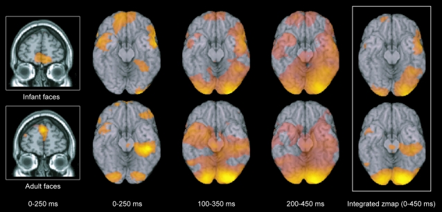
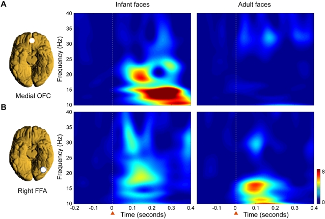
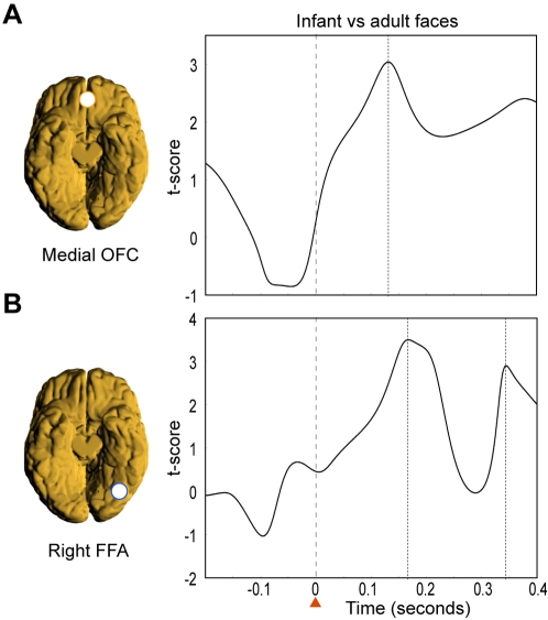
Darwin originally pointed out that there is something about infants which prompts adults to respond to and care for them, in order to increase individual fitness, i.e. reproductive success, via increased survivorship of one's own offspring. Lorenz proposed that it is the specific structure of the infant face that serves to elicit these parental responses, but the biological basis for this remains elusive. Here, we investigated whether adults show specific brain responses to unfamiliar infant faces compared to adult faces, where the infant and adult faces had been carefully matched across the two groups for emotional valence and arousal, as well as size and luminosity. The faces also matched closely in terms of attractiveness. Using magnetoencephalography (MEG) in adults, we found that highly specific brain activity occurred within a seventh of a second in response to unfamiliar infant faces but not to adult faces. This activity occurred in the medial orbitofrontal cortex (mOFC), an area implicated in reward behaviour, suggesting for the first time a neural basis for this vital evolutionary process. We found a peak in activity first in mOFC and then in the right fusiform face area (FFA). In mOFC the first significant peak (p<0.001) in differences in power between infant and adult faces was found at around 130 ms in the 10-15 Hz band. These early differences were not found in the FFA. In contrast, differences in power were found later, at around 165 ms, in a different band (20-25 Hz) in the right FFA, suggesting a feedback effect from mOFC. These findings provide evidence in humans of a potential brain basis for the "innate releasing mechanisms" described by Lorenz for affection and nurturing of young infants. This has potentially important clinical applications in relation to postnatal depression, and could provide opportunities for early identification of families at risk.
Overview
The claims and statistics of this paper haven’t been checked yet.
Run the check — free, no account needed. It reads the open-access text and checks the sources it cites.
Integrity signals
Registry
Cites
Statistics
Figures
Verification
Claims the paper makes
The claims and statistics of this paper haven’t been checked yet.
Run the check — free, no account needed. It reads the open-access text and checks the sources it cites.
Claims other papers make about this one
Each is checked against this paper’s own text: does it say what the citing paper says it does?
In the citing paper
“Again, coordinated responses in drive (amygdala) and regulation (cinglulate) figure prominently in these papers.”
From The human parental brain: In vivo neuroimaging (2011) · cites this paper as Kringelbach et al., 2008
In the citing paper
“Not only is facial recognition of one’s infant and their emotional state critical to their survival, but evidence supports human preference for the exaggerated cute/infantile features of baby faces which appear to activate reward mechanisms within the caregiver and motivate/promote mothers to caregiving, bonding and attachment.”
From Approaching the biology of human parental attachment: Brain imaging, oxytocin and coordinated assessments of mothers and fathers (2014) · cites this paper as Kringelbach et al., 2008
In the citing paper
“In addition to activation of nucleus accumbens (NAcc) in response to baby schema (Glocker et al., 2009) and medial orbitofrontal cortex (OFC) response to infant faces relative to adults (Kringelbach et al., 2008), higher levels of infantile features are preferred (Parsons et al., 2011).”
From Approaching the biology of human parental attachment: Brain imaging, oxytocin and coordinated assessments of mothers and fathers (2014) · cites this paper as Kringelbach et al., 2008
Self-check
Percentages, sums and reported differences re-computed from the paper’s own numbers.
The self-check needs the paper’s full text.
Citations
Cites
The expression of the emotions in man and animals.
Darwin, C. D. (1872) · J. Murray eBooks · doi:10.1037/10001-000 · Cited by 12,120
Attachment and Loss. Vol. 1. Attachment
Rever, G. W. (1972) · Psychosomatic Medicine · doi:10.1097/00006842-197211000-00011 · Cited by 8,446
The fusiform face area: a module in human extrastriate cortex specialized for face perception
Kanwisher, N., McDermott, J., & Chun, M. M. (1997) · The Journal of Neuroscience · doi:10.1523/jneurosci.17-11-04302.1997 · Cited by 7,738 · cited 2×
“It is still, however, controversial whether the activity in FFA represents a localized or distributed coding of faces [8,11,12].”
BackgroundA global optimisation method for robust affine registration of brain images
Jenkinson, M., & Smith, S. M. (2001) · Medical Image Analysis · doi:10.1016/s1361-8415(01)00036-6 · Cited by 7,018 · cited 2×
“The normalisation parameters were obtained using FLIRT (FMRIB’s Linear Image Registration Tool) [44] to reslice each individual’s anatomical MRI to the same orientation and position as the SAM functional volume and finding the transformation matrix from this functional space into the standard MNI space.”
MethodAttachment and Loss. Vol. I. Attachment.
Jones, N. B., & Bowlby, J. (1970) · Man · doi:10.2307/2798963 · Cited by 4,208 · cited 1×
“It is argued that these ‘‘babyish’’ features increase the infant’s chance of survival by evoking parental responses [4,5].”
BackgroundDeep Brain Stimulation for Treatment-Resistant Depression
Mayberg, H. S., Lozano, A. M., Voon, V., McNeely, H. E., Seminowicz, D., Hamani, C., Schwalb, J. M., & Kennedy, S. H. (2005) · Neuron · doi:10.1016/j.neuron.2005.02.014 · Cited by 4,059 · cited 1×
“Also, there is now evidence from deep brain stimulation linking depression to the nearby subgenual cingulate cortex which is strongly connected with the medial orbitofrontal cortex [35].”
BackgroundDistributed and overlapping representations of faces and objects in ventral temporal cortex
Haxby, J. V., Gobbini, M. I., Furey, M. L., Ishai, A., Schouten, J. L., & Pietrini, P. (2001) · Science · doi:10.1126/science.1063736 · Cited by 3,940 · cited 1×
“It is still, however, controversial whether the activity in FFA represents a localized or distributed coding of faces [8,11,12].”
BackgroundEkman, P., & Friesen, W. V. (1975) · Cited by 3,569
Speed of processing in the human visual system
Thorpe, S., Fize, D., & Marlot, C. (1996) · Nature · doi:10.1038/381520a0 · Cited by 3,538
Automatic 3D intersubject registration of MR volumetric data in standardized Talairach space
Collins, D. L., Neelin, P., Peters, T. M., & Evans, A. C. (1994) · Journal of Computer Assisted Tomography · doi:10.1097/00004728-199403000-00005 · Cited by 3,431 · cited 1×
“Group statistical maps for each frequency band were generated by first normalising the SAM functional volumes to MNI standard space [43] and then combining these volumes across participants for each time window and frequency band.”
Method
Cited by
Sorted by citations
Towards a functional neuroanatomy of pleasure and happiness
Kringelbach, M. L., & Berridge, K. C. (2009) · Trends in Cognitive Sciences · doi:10.1016/j.tics.2009.08.006 · Cited by 564
“likely to be extremely important for the survival of the species [ 98 ].”
The adaptive human parental brain: implications for children's social development
Feldman, R. (2015) · Trends in Neurosciences · doi:10.1016/j.tins.2015.04.004 · Cited by 419
Tuning the developing brain to social signals of emotions
Leppänen, J. M., & Nelson, C. A. (2008) · Nature reviews. Neuroscience · doi:10.1038/nrn2554 · Cited by 457
“Consistent with such a neuromodulatory role, recent studies have provided evidence for an early response in the orbitofrontal cortex (130 ms after stimulus onset) that precedes activity in occipitotemporal perceptual-representation areas (165 ms post-stimulus”
The origins of altruism in offspring care.
Preston, S. D. (2013) · Psychological Bulletin · doi:10.1037/a0031755 · Cited by 368
Baby schema modulates the brain reward system in nulliparous women
Glocker, M. L., Langleben, D. D., Ruparel, K., Loughead, J. W., Valdez, J. N., Griffin, M. D., Sachser, N., & Gur, R. C. (2009) · Proceedings of the National Academy of Sciences · doi:10.1073/pnas.0811620106 · Cited by 365
The Power of Kawaii: Viewing Cute Images Promotes a Careful Behavior and Narrows Attentional Focus
Nittono, H., Fukushima, M., Yano, A., & Moriya, H. (2012) · PLoS ONE · doi:10.1371/journal.pone.0046362 · Cited by 374
“Baby schema modulates perception and attention at early stages of visual processing [14] and activates the reward system of the brain [15].”
BackgroundOn Cuteness: Unlocking the Parental Brain and Beyond
Kringelbach, M. L., Stark, E. A., Alexander, C., Bornstein, M. H., & Stein, A. L. (2016) · Trends in Cognitive Sciences · doi:10.1016/j.tics.2016.05.003 · Cited by 326
“Magnetoencephalography (MEG) has shown that men and women, parents and nonparents all have rapid (within 140 ms) selective neural responses to infant faces [15] (Figure 2A).”
InfluentialSwain, J. E., Kim, P., Spicer, J., Ho, S. S., Dayton, C. J., Elmadih, A., & Abel, K. M. (2014) · Brain Research · doi:10.1016/j.brainres.2014.03.007 · Cited by 290
“, 2009) and medial orbitofrontal cortex (OFC) response to infant faces relative to adults (Kringelbach et al., 2008), higher levels of infantile features are preferred (Parsons et al.”
The human parental brain: In vivo neuroimaging
Swain, J. E. (2011) · Progress in Neuro-Psychopharmacology and Biological Psychiatry · doi:10.1016/j.pnpbp.2010.10.017 · Cited by 234
“Related work has used baby face stimuli to underline the role of limbic systems in humans that may be critical for care giving thoughts and behaviors even in non-parents (Glocker et al., 2009; Kringelbach et al., 2008).”
BackgroundBuilding a neuroscience of pleasure and well-being
Berridge, K. C., & Kringelbach, M. L. (2011) · Psychology of Well-Being: Theory, Research and Practice · doi:10.1186/2211-1522-1-3 · Cited by 229
The neural and hormonal bases of human parentalcare
Rilling, J. K. (2013) · Neuropsychologia · doi:10.1016/j.neuropsychologia.2012.12.017 · Cited by 230
Thomsen, K. R., Whybrow, P. C., & Kringelbach, M. L. (2015) · Frontiers in Behavioral Neuroscience · doi:10.3389/fnbeh.2015.00049 · Cited by 256
“…with e.g., conditioned place preferences (Mayo et al., 2013; Mayo and De Wit, 2015) as well as more direct brain manipulations such as transcranial magnetic stimulation, transcranial direct current stimulation and deep brain stimulation (Kringelbach et al., 2007, 2011; Lozano and Lipsman, 2013).”
BackgroundBornstein, M. H. (2015) · Wiley eBooks · doi:10.1002/9781118963418.childpsy403 · Cited by 229
The Right Brain Implicit Self Lies at the Core of Psychoanalysis
Schore, A. N. (2011) · Psychoanalytic Dialogues · doi:10.1080/10481885.2011.545329 · Cited by 219
“Similarly, using magnetoencephalography Kringlebach et al. (2008) showed that the orbitofrontal cortex and right fusiform gyrus act as “a specific neural signature for parental instinct,” responding to the face of an infant in 130 milliseconds.”
MethodRiem, M. M. E., van IJzendoorn, M. H., Tops, M., Boksem, M. A. S., Rombouts, S. A. R. B., & Bakermans-Kranenburg, M. J. (2012) · Neuropsychopharmacology · doi:10.1038/npp.2011.313 · Cited by 191
“…examined oxytocin effects on the ventral striatum, the ACC, and the OFC because of the suggested significance of these regions in mother–infant bonding and reward processing (Berridge and Kringelbach, 2008; Kringelbach, 2005; Kringelbach et al, 2008; Seifritz et al, 2003; Strathearn et al, 2009).”
BackgroundInfluentialKringelbach, M. L., & Berridge, K. C. (2017) · Emotion Review · doi:10.1177/1754073916684558 · Cited by 199
“…of infants has been shown to elicit significant activity already after only 50 ms in the brainstem (Parsons et al., 2014) while infant facial and auditory features elicit activity in the orbitofrontal cortex within around 130 ms (Kringelbach et al., 2008; Young, Parsons, Stevner, et al., 2016).”
BackgroundBrown, S. L., & Brown, R. M. (2015) · Neuroscience & Biobehavioral Reviews · doi:10.1016/j.neubiorev.2015.04.004 · Cited by 212
Neurobiology of culturally common maternal responses to infant cry
Bornstein, M. H., Putnick, D. L., Rigo, P., Esposito, G., Swain, J. E., Suwalsky, J. T. D., Su, X., Du, X., Zhang, K., Cote, L. R., et al. (2017) · Proceedings of the National Academy of Sciences · doi:10.1073/pnas.1712022114 · Cited by 168
“Indeed, mothers react neurobiologically (32-39), autonomically (28, 39, 40), and hormonally (41-44) as well as behaviorally (45-48) to their babies’ cries.”
BackgroundThe Rediscovery of Slowness: Exploring the Timing of Cognition
Kringelbach, M. L., McIntosh, A. R., Ritter, P., Jirsa, V., & Deco, G. (2015) · Trends in Cognitive Sciences · doi:10.1016/j.tics.2015.07.011 · Cited by 159
“Caregiving behavior depends partly on very fast processing of infant cues, exemplified by the fact that infant sounds elicit significant activity already after only 50 ms in the brainstem [69], and that infant facial features elicit activity in the orbitofrontal cortex within around 130 ms [70].”
BackgroundPet Face: Mechanisms Underlying Human-Animal Relationships
Borgi, M., & Cirulli, F. (2016) · Frontiers in Psychology · doi:10.3389/fpsyg.2016.00298 · Cited by 129
“…studies support the idea of infantile faces as highly biologically relevant stimuli rapidly and unconsciously capturing attention and eliciting positive emotions (Brosch et al., 2007; Kringelbach et al., 2008; Caria et al., 2012; Senese et al., 2013; Borgi et al., 2014; Esposito et al., 2014).”
BackgroundResultThe functional neuroanatomy of pleasure and happiness.
Kringelbach, M. L., & Berridge, K. C. (2010) · PubMed · Cited by 156
The Motivational Salience of Infant Faces Is Similar for Men and Women
Parsons, C. E., Young, K. S., Kumari, N., Stein, A. L., & Kringelbach, M. L. (2011) · PLoS ONE · doi:10.1371/journal.pone.0020632 · Cited by 156
“We have recently identified a putative neural signature of this ‘parental instinct’ [6].”
BackgroundMethodSimpson, E. A., Murray, L., Paukner, A., & Ferrari, P. F. (2014) · Philosophical Transactions of the Royal Society B: Biological Sciences · doi:10.1098/rstb.2013.0289 · Cited by 150
“There is a fundamental motivation 131 on the part of both the parents and newborns to be in social engagement with each other, 132 reflected in their preferential responses to faces and eye contact [67-73].”
Lonstein, J. S., Lévy, F., & Fleming, A. S. (2015) · Hormones and Behavior · doi:10.1016/j.yhbeh.2015.06.011 · Cited by 158
“Infants and their sensory cues are rewarding Human infants can be rewarding and activate regions of the adult brain that are associated with reward and pleasure (e.g., Kringelbach et al., 2008).”
The functional neuroanatomy of the evolving parent–infant relationship
Parsons, C. E., Young, K. S., Murray, L., Stein, A. L., & Kringelbach, M. L. (2010) · Progress in Neurobiology · doi:10.1016/j.pneurobio.2010.03.001 · Cited by 149
“…when ked average group responses to baby and adult faces from the virtual electrodes show z band from around 130 ms, and not present to adult faces (Kringelbach et al., 2008). formal comparisons, this effect was evident in both adult males and females, and both parents and non-parents, possibly…”
BackgroundCurrent challenges to research on animal-assisted interventions
Serpell, J. A., McCune, S., Gee, N. R., & Griffin, J. A. (2017) · Applied Developmental Science · doi:10.1080/10888691.2016.1262775 · Cited by 162
“Brain imaging studies further suggest that this type of response may be hard-wired in the sense that it can be localized to a specific region of the brain—the medial orbitofrontal cortex (Kringelbach et al., 2008).”
BackgroundChild gender influences paternal behavior, language, and brain function.
Mascaro, J. S., Rentscher, K. E., Hackett, P. D., Mehl, M. R., & Rilling, J. K. (2017) · Behavioral Neuroscience · doi:10.1037/bne0000199 · Cited by 149
“This region may be integral for coding the hedonic pleasure associated with a stimulus (Kringelbach & Berridge, 2009), and intriguingly, it is this region that appears to respond early and selectively in response to infant faces (Kringelbach et al., 2008).”
Rutherford, H. J. V., Williams, S. K., Moy, S., Mayes, L. C., & Johns, J. M. (2011) · Frontiers in Psychiatry · doi:10.3389/fpsyt.2011.00037 · Cited by 140
“Therefore we turn our attention to the neural circuitry of the stress system, its involvement in MB, and modulation by cocaine.”
BackgroundSpecies-specific response to human infant faces in the premotor cortex
Caria, A., de Falco, S., Venuti, P., Lee, S., Esposito, G., Rigo, P., Birbaumer, N., & Bornstein, M. H. (2012) · NeuroImage · doi:10.1016/j.neuroimage.2011.12.068 · Cited by 135
“These regions have previously been associated with parental attachment (Glocker et al., 2009; Kringelbach et al., 2008; Ranote et al., 2004; Swain et al., 2007) and linked to “baby schema” features in artificially manipulated infant faces (Glocker et al., 2009).”
InfluentialNeural and behavioral responses to attractiveness in adult and infant faces
Hahn, A. C., & Perrett, D. I. (2014) · Neuroscience & Biobehavioral Reviews · doi:10.1016/j.neubiorev.2014.08.015 · Cited by 167
“Kringelbach et al. (2008) did not find any evidence of a babyspecific response in the neural regions of the core system, however other studies have indicated that viewing infant faces correlates with increased activation in the FG and IFG (Bartels & Zeki, 2004; Caria et al., 2012; Glocker et al.,…”
InfluentialRiem, M. M. E., Bakermans-Kranenburg, M. J., van IJzendoorn, M. H., Out, D., & Rombouts, S. A. R. B. (2012) · Attachment & Human Development · doi:10.1080/14616734.2012.727252 · Cited by 134
“For example, it exhibits a very rapid and specific response to an infant face and it has been suggested that this might be the brain basis for the ‘‘innate releasing mechanism’’ described by Lorenz (Kringelbach et al., 2008).”
BackgroundPearson, R. M., Melotti, R., Heron, J., Joinson, C., Stein, A., Ramchandani, P. G., & Evans, J. (2012) · Infant Behavior and Development · doi:10.1016/j.infbeh.2012.07.020 · Cited by 128
Self-citationDepressive symptoms in early pregnancy disrupt attentional processing of infant emotion
Pearson, R. M., Cooper, R. M., Penton‐Voak, I. S., Lightman, S. L., & Evans, J. (2009) · Psychological Medicine · doi:10.1017/s0033291709990961 · Cited by 128
“For example, adults are quicker to detect a cue that follows a neutral infant versus a neutral adult face on a dot probe task (Brosch et al. 2007) and brain activity in reward centres is seen within 150 ms of presentation of infant but not adult faces (Kringelbach et al. 2008).”
BackgroundThe effects of postnatal maternal depression and anxiety on the processing of infant faces
Arteche, A. X., Joormann, J., Harvey, A. G., Craske, M., Gotlib, I. H., Lehtonen, A., Counsell, N., & Stein, A. L. (2011) · Journal of Affective Disorders · doi:10.1016/j.jad.2011.04.015 · Cited by 138
Sakaki, M., Niki, K., & Mather, M. (2011) · Cognitive Affective & Behavioral Neuroscience · doi:10.3758/s13415-011-0062-x · Cited by 126
“Likewise, infants’ faces more quickly induced strong activity in reward-related regions in the brain than did adults’ faces (Kringelbach et al., 2008; Nitschke et al., 2004).”
Sex differences in social cognition: the case of face processing
Proverbio, A. M. (2017) · Journal of Neuroscience Research · doi:10.1002/jnr.23817 · Cited by 110
“…stimuli inducing a greater arousal in the percipient would be subjected to a prioritized processing (Harmon-Jones et al., 2011) because of their biological relevance; for example, infant faces would trigger an instinctive parental response (Kringelbach et al., 2008; Parson et al., 2013).”
BackgroundThe Neuroscience of Happiness and Pleasure
Kringelbach, M. L., & Berridge, K. C. (2010) · Social research · doi:10.1353/sor.2010.0049 · Cited by 110
Piallini, G., Palo, F. D., & Simonelli, A. (2015) · Frontiers in Psychology · doi:10.3389/fpsyg.2015.01625 · Cited by 106
Gender differences in the incentive salience of adult and infant faces
Hahn, A. C., Xiao, D., Sprengelmeyer, R., & Perrett, D. I. (2012) · Quarterly Journal of Experimental Psychology · doi:10.1080/17470218.2012.705860 · Cited by 102
“Vol. 66, No. 1, 200–208, http://dx.doi.org/10.1080/17470218.2012.705860 200 # 2013 The Experimental Psychology Society unfamiliar infants are also capable of eliciting neural responses in equivalent brain regions, regardless of participant gender or parental status (Kringelbach et al., 2008).”
BackgroundInfluentialHrdy, S. B. (2015) · Hormones and Behavior · doi:10.1016/j.yhbeh.2015.10.016 · Cited by 101
Esposito, G., Nakazawa, J., Ogawa, S., Stival, R., Kawashima, A., Putnick, D. L., & Bornstein, M. H. (2014) · PLoS ONE · doi:10.1371/journal.pone.0106705 · Cited by 99
“[11] found that observing unfamiliar infant faces is associated with activity in the orbitofrontal cortex (OFC) and enhanced responsiveness in the fusiform face area that is not present when observing adult faces.”
BackgroundHuman Infant Faces Provoke Implicit Positive Affective Responses in Parents and Non-Parents Alike
Senese, V. P., De Falco, S., Bornstein, M. H., Caria, A., Buffolino, S., & Venuti, P. (2013) · PLoS ONE · doi:10.1371/journal.pone.0080379 · Cited by 96
“However, in accord with other studies, no gender or parental status differences were expected in adults’ implicit reactions to infant faces [48],[50].”
BackgroundResultInfluentialRare long-range cortical connections enhance human information processing
Deco, G., Perl, Y. S., Vuust, P., Tagliazucchi, E., Kennedy, H., & Kringelbach, M. L. (2021) · Current Biology · doi:10.1016/j.cub.2021.07.064 · Cited by 95
“As an example, MEG studies of infant and adult faces have shown activity in the FFA for both stimuli but with simultaneous activity in the orbitofrontal cortex (OFC) at around 130 ms only for the infant faces.(52) Interestingly, even small deviations from the infant face template, such as cleft lip, lead to much dimin…”
BackgroundEvolutionary neuromarketing: darwinizing the neuroimaging paradigm for consumer behavior
Garcia, J. R., & Saad, G. (2008) · Journal of Consumer Behaviour · doi:10.1002/cb.259 · Cited by 111
“On a related note, adults display preferential rew^ard-associated medial orbitofrontal cortex activation to images of unfamiliar infant faces over unfamiliar adult faces, suggesting an evolutionarily rooted positive response to infants (Kringelbach et al., 2008).”
BackgroundIs child death the crucible of human evolution?
Volk, T., & Atkinson, J. (2008) · Journal of Social Evolutionary and Cultural Psychology · doi:10.1037/h0099341 · Cited by 91
Interpretation of infant facial expression in the context of maternal postnatal depression
Stein, A. L., Arteche, A. X., Lehtonen, A., Craske, M. G., Harvey, A. G., Counsell, N., & Murray, L. (2010) · Infant Behavior and Development · doi:10.1016/j.infbeh.2010.03.002 · Cited by 91
“Images were drawn from a database of digital photographs of 27 infants who were filmed at home (see Kringelbach et al., 2008 for details).”
MethodWebb, R., & Ayers, S. (2014) · Cognition & Emotion · doi:10.1080/02699931.2014.977849 · Cited by 89
Understanding the human parental brain: A critical role of the orbitofrontal cortex
Parsons, C. E., Stark, E. A., Young, K. S., Stein, A. L., & Kringelbach, M. L. (2013) · Social Neuroscience · doi:10.1080/17470919.2013.842610 · Cited by 91
Sex differences in the social brain and in social cognition
Proverbio, A. M. (2021) · Journal of Neuroscience Research · doi:10.1002/jnr.24787 · Cited by 88
Is It a Baby? Perceived Age Affects Brain Processing of Faces Differently in Women and Men
Proverbio, A. M., Riva, F., Zani, A., & Martin, E. (2011) · Journal of Cognitive Neuroscience · doi:10.1162/jocn_a_00041 · Cited by 83
“Unlike the MEG study by Kringelbach et al. (2008), which found an medial orbito-frontal (mOBF; 130 msec) and then an FFA (165 msec) increase in response to infant faces, we are unable to establish whether the neural circuit implies an mOBF modulation of FFA cortex.”
BackgroundResultInfluentialMascaro, J. S., Hackett, P. D., & Rilling, J. K. (2014) · Psychoneuroendocrinology · doi:10.1016/j.psyneuen.2014.04.014 · Cited by 84
“Fathers also activate the vmPFC/OFC to a greater extent than non-fathers, a finding consistent with many recent studies highlighting the importance of the OFC within the ‘‘parental brain’’ (Kringelbach et al., 2008; Parsons et al., 2013).”
Colonnello, V., Iacobucci, P., Fuchs, T. A. E., Newberry, R. C., & Panksepp, J. (2011) · Neuroscience & Biobehavioral Reviews · doi:10.1016/j.neubiorev.2011.03.014 · Cited by 84
Neural systems and hormones mediating attraction to infant and child faces
Luo, L. (2015) · Frontiers in Psychology · doi:10.3389/fpsyg.2015.00970 · Cited by 91
Adults’ responsiveness to children’s facial expressions
Aradhye, C., Vonk, J. M., & Arida, D. (2015) · Journal of Experimental Child Psychology · doi:10.1016/j.jecp.2015.02.006 · Cited by 79
Peltola, M. J., Yrttiaho, S., Puura, K., Proverbio, A. M., Mononen, N., Lehtimäki, T. J., & Leppänen, J. M. (2014) · Emotion · doi:10.1037/a0035959 · Cited by 76
Specific maternal brain responses to their own child’s face: An fMRI meta-analysis
Rigo, P., Kim, P., Esposito, G., Putnick, D. L., Venuti, P., & Bornstein, M. H. (2018) · Developmental Review · doi:10.1016/j.dr.2018.12.001 · Cited by 74
“Infant faces are a special class of salient social stimuli and elicit feelings of care and empathic approach in adults (Bowlby, 1969; Brosch et al., 2007; Caria et al., 2012; Glocker et al. 2009; Kringelbach et al., 2008; Lorenz, 1971; Leibenluft et al., 2004; Kringelbach et al., 2016; Senese et al., 2013, 2016).”
BackgroundYamamoto, R. T., Dan Ariely, C., Chi, W., Langleben, D. D., & Elman, I. (2009) · PLoS ONE · doi:10.1371/journal.pone.0006042 · Cited by 71
“, regardless of the gender and/or of parental status of the participants, as parents and non-parents of both genders activated brain motivational regions when exposed to such stimuli [9].”
BackgroundThe Application of Electroencephalography to Investigate the Neural Bases of Parenting: A Review
Maupin, A. N., Hayes, N. J., Mayes, L. C., & Rutherford, H. J. V. (2015) · Parenting · doi:10.1080/15295192.2015.992735 · Cited by 72
“…brain regions are recruited during processing of infant stimuli may be better served by techniques with either higher spatial resolution (e.g., fMRI) or with strengths in spatial as well as temporal resolution (e.g., magnetoencephalography or MEG; Kringelbach et al., 2008; Parsons et al., 2013).”
BackgroundMethodYoung, K. S., Parsons, C. E., Stein, A. L., Vuust, P., Craske, M. G., & Kringelbach, M. L. (2016) · Behavioural Brain Research · doi:10.1016/j.bbr.2016.09.012 · Cited by 70
“id differentiation of infant and adult facial stimuli [93].”
BackgroundA review of infants’ and children’s facial cues’ influence on adults’ perceptions and behaviors.
Franklin, P., & Volk, A. A. (2018) · Evolutionary Behavioral Sciences · doi:10.1037/ebs0000108 · Cited by 66
“At the neurological level, adults displayed stronger activation in brain regions related to facial processing (e.g., fusiform gyrus and middle occipital gyrus; Kringelbach et al., 2008), attention processing (e.”
The Evolution of Human Infancy: Why It Helps to Be Helpless
Rosenberg, K. E. (2021) · Annual Review of Anthropology · doi:10.1146/annurev-anthro-111819-105454 · Cited by 70
Technologically-assisted communication attenuates inter-brain synchrony
Schwartz, L., Lévy, J., Endevelt–Shapira, Y., Djalovski, A., Hayut, O., Dumas, G., & Feldman, R. (2022) · NeuroImage · doi:10.1016/j.neuroimage.2022.119677 · Cited by 66
Women are better at seeing faces where there are none: an ERP study of face pareidolia
Proverbio, A. M., & Galli, J. (2016) · Social Cognitive and Affective Neuroscience · doi:10.1093/scan/nsw064 · Cited by 71
“…over the right hemisphere. at U IC L ibrary, C ollections D evelopm ent on June 6, 2016 http://scan.oxfordjournals.org/ D ow nloaded from response to the faces of infants, or ‘baby schema’ response (Kringelbach et al., 2008; Glocker et al., 2009; Proverbio et al., 2011b; Luo et al., 2015).”
BackgroundParental reflective functioning and the neural correlates of processing infant affective cues.
Rutherford, H. J. V., Maupin, A. N., Landi, N., Potenza, M. N., & Mayes, L. C. (2017) · Social Neuroscience · doi:10.1080/17470919.2016.1193559 · Cited by 65
“In the brain, infant faces are rapidly differentiated from adult faces, and the processing of these infant faces may have a modulating effect across neural circuits recruited in face perception (Kringelbach et al., 2008).”
BackgroundAttentional bias toward infant faces – Review of the adaptive and clinical relevance
Lucion, M. K., Oliveira, V. F., Bizarro, L., Bischoff, A. R., Silveira, P. P., & Kauer‐Sant’Anna, M. (2017) · International Journal of Psychophysiology · doi:10.1016/j.ijpsycho.2017.01.008 · Cited by 66
Kringelbach, M. L., Vuust, P., & Geake, J. G. (2008) · Interdisciplinary Science Reviews · doi:10.1179/174327908x392889 · Cited by 64
The pleasure of food: Underlying brain mechanisms of eating and other pleasures
Kringelbach, M. L. (2015) · Flavour · doi:10.1186/s13411-014-0029-2 · Cited by 62
“Even studies on the much shorter timescales of milliseconds have found activity in the medial OFC related to the reward of images of cute babies [71] (Figure 5D).”
BackgroundSelf-citationFace cells in orbitofrontal cortex represent social categories
Barat, E., Wirth, S., & Duhamel, J.-R. (2018) · Proceedings of the National Academy of Sciences · doi:10.1073/pnas.1806165115 · Cited by 58
“For example, fMRI studies in healthy adults observe activity in OFC for viewing infants (50, 51) and attractive female faces (52– 54).”
BackgroundPratt, M., Zeev‐Wolf, M., Goldstein, A., & Feldman, R. (2019) · Progress in Neuro-Psychopharmacology and Biological Psychiatry · doi:10.1016/j.pnpbp.2019.03.005 · Cited by 54
Thompson‐Booth, C., Viding, E. M., Mayes, L. C., Rutherford, H. J. V., Hodsoll, S., & McCrory, E. J. P. (2014) · PLoS ONE · doi:10.1371/journal.pone.0109362 · Cited by 52
“Compared to adult faces, infant faces elicit enhanced activation in a distributed network implicated in face perception, reward processing and attentional processing [34,35].”
BackgroundYoung, K. S., Parsons, C. E., Elmholdt, E.-M. J., Woolrich, M. W., van Hartevelt, T. J., Stevner, A. B. A., Stein, A. L., & Kringelbach, M. L. (2015) · Cerebral Cortex · doi:10.1093/cercor/bhv306 · Cited by 58
“In support, there is mounting evidence that swift propagation of sensory information to the OFC (140 ms) from primary sensory regions may aid preattentive recognition (Bar et al. 2006; Kringelbach et al. 2008; D’Hondt et al. 2013; Parsons, Young, et al. 2013).”
BackgroundResultInfluentialLévy, J., Yirmiya, K., Goldstein, A., & Feldman, R. (2019) · Developmental Cognitive Neuroscience · doi:10.1016/j.dcn.2019.100658 · Cited by 52
“This, however, is not completely at odds with the literature: while some studies found that trauma and stress did impact child prosociality (Bergmann et al., 2016; Daud et al., 2008), others showed no differences (Flouri et al., 2010; Küenzlen et al., 2016).”
The maternal brain: Neural responses to infants in mothers with and without mood disorder
Bjertrup, A. J., Friis, N. K., & Miskowiak, K. W. (2019) · Neuroscience & Biobehavioral Reviews · doi:10.1016/j.neubiorev.2019.09.011 · Cited by 50
Parsons, C. E., Young, K. S., Mohseni, H., Woolrich, M. W., Thomsen, K. R., Joensson, M., Murray, L., Goodacre, T., Stein, A., & Kringelbach, M. L. (2013) · Social Neuroscience · doi:10.1080/17470919.2013.795189 · Cited by 48
“Differential activity apparent in the right OFC in response to infant faces confirms our previous finding (Kringelbach et al., 2008) and is likely to reflect early, positive affective processing of “infant schema.”
BackgroundMethodResultInfluentialSelf-citationIntuitive parenting: understanding the neural mechanisms of parents’ adaptive responses to infants
Parsons, C. E., Young, K. S., Stein, A. L., & Kringelbach, M. L. (2017) · Current Opinion in Psychology · doi:10.1016/j.copsyc.2017.02.010 · Cited by 50
Parsons, C. E., Young, K. S., Bhandari, R., van IJzendoorn, M. H., Bakermans‐Kranenburg, M. J., Stein, A. L., & Kringelbach, M. L. (2013) · Developmental Science · doi:10.1111/desc.12112 · Cited by 47
“In order to reduce any confounding effects of stimulus gender, we had an independent sample of adult females (n = 40) rate faces from a larger set of 13 (Kringelbach et al., 2008) as ‘male’, ‘female’, or ‘cannot tell’.”
BackgroundElsegood, K. J., & Duff, S. C. (2010) · Sexual Abuse · doi:10.1177/1079063209359926 · Cited by 49
“A recent neuroimaging study supports this possibility, showing that the perception of infant faces, but not adult faces, activates the medial orbitofrontal cortex (Kringelbach et al., 2008).”
BackgroundReady for action: a role for the human midbrain in responding to infant vocalizations
Parsons, C. E., Young, K. S., Joensson, M., Brattico, E., Hyam, J. A., Stein, A. L., Green, A. L., Aziz, T. Z., & Kringelbach, M. L. (2013) · Social Cognitive and Affective Neuroscience · doi:10.1093/scan/nst076 · Cited by 45
“Recent neuroimaging studies have suggested that salient visual cues from the infant may be subject to specialized processing, compared with the same cues from adults (Kringelbach et al., 2008; Parsons et al., 2013).”
Oxytocin promotes face-sensitive neural responses to infant and adult faces in mothers
Peltola, M. J., Strathearn, L., & Puura, K. (2018) · Psychoneuroendocrinology · doi:10.1016/j.psyneuen.2018.02.012 · Cited by 43
“, 2007) and imaging studies showing that infant faces are potent stimuli for activating the brain’s dopaminergic circuits (Glocker et al., 2009; Kringelbach et al., 2008) through which OT may exert its effects (Shamay-Tsoory and Abu-Akel, 2016; Strathearn, 2011).”
InfluentialNeural and Developmental Bases of the Ability to Recognize Social Signals of Emotions
Leppänen, J. M. (2011) · Emotion Review · doi:10.1177/1754073910387942 · Cited by 49
“…suggested that structures such as the amygdala and orbitofrontal cortex are important in rapid assignment of affective value to external stimuli (Kringelbach et al., 2008; Rothstein et al., 2010; Tsuchiya, Moradi, Felsen, Yamazaki, & Adolphs, 2009), in modulation and enhancement of activity in…”
BackgroundNo Race Effect (ORE) in the Automatic Orienting toward Baby Faces: When Ethnic Group does not matter
Proverbio, A. M., Gabriele, V. D., Manfredi, M., & Adorni, R. (2011) · Psychology · doi:10.4236/psych.2011.29140 · Cited by 44
“The response to infant faces seems independent of attention and is thought to be at the basis of parental instinct in humans (Kringelbach et al., 2008).”
BackgroundAttentional Prioritization of Infant Faces Is Limited to Own-Race Infants
Hodsoll, J. P., Quinn, K. A., & Hodsoll, S. (2010) · PLoS ONE · doi:10.1371/journal.pone.0012509 · Cited by 43
“Indeed, stimuli that conform to this baby schema are likely to elicit a positive behavioral response [3], [4], and the perception of infant features activates brain structures associated with the reward system [5], [6].”
BackgroundBornstein, M. H., Arterberry, M. E., & Mash, C. (2013) · Developmental Neuropsychology · doi:10.1080/87565641.2013.804923 · Cited by 42
“Using MEG, Kringelbach et al. (2008) found that activity in the medial orbitofrontal cortex, an area implicated in reward, occurred within a seventh of a second in response to generic unfamiliar infant faces.”
MethodFasano, M. C., Cabral, J., Stevner, A. B. A., Vuust, P., Cantou, P., Brattico, E., & Kringelbach, M. L. (2022) · Human Brain Mapping · doi:10.1002/hbm.26060 · Cited by 48
“plays a significant role on many timescales within the pleasure cycle (Kringelbach et al. 2008; Parsons et al. 2013; Young et al. 2016), this region has been proposed to be part of the top-down neural route to conscious liking of music, providing a modulation of the sensory musical pleasure by means of reward value at…”
BackgroundThe Effect of Cleft Lip on Adults' Responses to Faces: Cross-Species Findings
Parsons, C. E., Young, K. S., Parsons, E., Dean, A., Murray, L., Goodacre, T., Dalton, L. J., Stein, A. L., & Kringelbach, M. L. (2011) · PLoS ONE · doi:10.1371/journal.pone.0025897 · Cited by 40
Child brain exhibits a multi-rhythmic response to attachment cues
Pratt, M., Goldstein, A., & Feldman, R. (2018) · Social Cognitive and Affective Neuroscience · doi:10.1093/scan/nsy062 · Cited by 44
“Infant faces elicited higher beta oscillations as compared to adult faces in the medial orbitofrontal cortex of non-parents (Kringelbach et al., 2008), and the authors interpreted the findings as pointing to the existence of an intrinsic neural system for attachment in humans.”
BackgroundResultYoung, K. S., Parsons, C. E., Stein, A., & Kringelbach, M. L. (2015) · Frontiers in Behavioral Neuroscience · doi:10.3389/fnbeh.2015.00026 · Cited by 38
“I., Sirota, L., and Weller, A. (2002). Comparison of skinto-skin (kangaroo) and traditional care: parenting outcomes and preterm infant”
BackgroundInfluentialHahn, A. C., Symons, L. A., Kredel, T., Hanson, K., Hodgson, L., Schiavone, L., & Jantzen, K. J. (2015) · Social Neuroscience · doi:10.1080/17470919.2015.1059361 · Cited by 41
“…neuroimaging studies have provided evidence of a “baby-specific” neural response to infant stimuli (e.g., Caria et al., 2012; Grasso, Moser, Dozier, & Simons, 2009; Kringelbach et al., 2008; Parsons et al., 2014; Proverbio, Riva, Martin, & Zani, 2010a; Proverbio, Riva, Zani, & Martin, 2011b).”
BackgroundMethodResultInfluentialAttachment and Developmental Psychopathology
Fearon, P. R. M., Groh, A. M., Bakermans‐Kranenburg, M. J., van IJzendoorn, M. H., & Roisman, G. I. (2016) · Wiley eBooks · doi:10.1002/9781119125556.devpsy108 · Cited by 41
Antenatal and postnatal depressive symptoms: Association with quality of mother–infant interaction
Binda, V., Figueroa-Leigh, F., & Olhaberry, M. (2019) · Infant Behavior and Development · doi:10.1016/j.infbeh.2019.101386 · Cited by 47
The Power of Smiling: The Adult Brain Networks Underlying Learned Infant Emotionality
Stark, E. A., Cabral, J., Riem, M. M. E., van IJzendoorn, M. H., Stein, A. L., & Kringelbach, M. L. (2019) · Cerebral Cortex · doi:10.1093/cercor/bhz219 · Cited by 33
Neurognosis, the Development of Neural Models, and the Study of the Ancient Mind
Laughlin, C. D., & Loubser, J. H. N. (2010) · Time and Mind · doi:10.2752/175169610x12632240392712 · Cited by 34
Interpreting infant vocal distress: The ameliorative effect of musical training in depression.
Young, K. S., Parsons, C. E., Stein, A. L., & Kringelbach, M. L. (2012) · Emotion · doi:10.1037/a0028705 · Cited by 34
“Adults have been shown to be remarkably attuned to infant facial configuration, and emerging evidence suggests that the specialized processing of infant faces may be supported by affective neural circuits (Kringelbach et al., 2008; Parsons, Young, Murray, Stein, & Kringelbach, 2010).”
BackgroundBhandari, R., Bakermans-Kranenburg, M. J., van der Veen, R., Parsons, C. E., Young, K. S., Grewen, K. M., Stein, A., Kringelbach, M. L., & van IJzendoorn, M. H. (2014) · Physiology & Behavior · doi:10.1016/j.physbeh.2014.04.028 · Cited by 33
“p E D P R O O F images were obtained from a standardized database (for detailed description of the stimuli see [30]).”
Emotions Are at the Core of Individual Social Performance
Kotrschal, K. M. (2012) · The science of the mind · doi:10.1007/978-4-431-54123-3_1 · Cited by 33
Effects of Infant Cleft Lip on Adult Gaze and Perceptions of “Cuteness”
Rayson, H., Christine, E. P., Katherine, S. Y., Goodacre, T., Kringelbach, M. L., Bonaiuto, J. J., McSorley, E., & Murray, L. (2016) · The Cleft Palate-Craniofacial Journal · doi:10.1597/16-015 · Cited by 33
“, 2010), with specific patterns of brain activity occurring in response to unfamiliar infant, but not adult, faces, implying a biological basis for this attunement (Kringelbach et al., 2008).”
BackgroundMethodInfluentialOxytocin and Human Sensitive and Protective Parenting
Bakermans‐Kranenburg, M. J., & van IJzendoorn, M. H. (2017) · Current topics in behavioral neurosciences · doi:10.1007/7854_2017_23 · Cited by 36
Interpreting Infant Emotional Expressions: Parenthood has Differential Effects on Men and Women
Parsons, C. E., Young, K. S., Elmholdt, E.-M. J., Stein, A. L., & Kringelbach, M. L. (2016) · Quarterly Journal of Experimental Psychology · doi:10.1080/17470218.2016.1141967 · Cited by 36
“…found that females are more responsive than males when viewing unfamiliar infant faces (e.g., Proverbio, Zani, & Adorni, 2008), other work has found effects in the opposite direction (e.g., Weisman, Feldman, & Goldstein, 2012), and some has reported no difference (e.g., Kringelbach et al., 2008).”
InfluentialUsing infrared thermography to assess emotional responses to infants
Esposito, G., Nakazawa, J., Ogawa, S., Stival, R., Putnick, D. L., & Bornstein, M. H. (2014) · Early Child Development and Care · doi:10.1080/03004430.2014.932153 · Cited by 35
“…supported Lorenz’s hypothesis that infant schema tend to release a set of care behaviours in conspecifics that are immediate, automatic, unconscious (Kringelbach et al., 2008; Senese et al., 2013), and evolutionarily vital for survival (Caria et al., 2012; Glocker et al., 2009a, 2009b; Parsons et…”
Generation WhatsApp: inter-brain synchrony during face-to-face and texting communication
Schwartz, L., Lévy, J., Hayut, O., Netzer, O., Endevelt–Shapira, Y., & Feldman, R. (2024) · Scientific Reports · doi:10.1038/s41598-024-52587-2 · Cited by 37
Metadata
- Authors
- Kringelbach, M. L., Lehtonen, A., Squire, S., Harvey, A. G., Craske, M. G., Holliday, I. E., Green, A. L., Aziz, T. Z., Hansen, P. C., Cornelissen, P. L., & Stein, A.
- Affiliations
- Aarhus University (DK)
- University of Oxford (GB)
- University of California, Berkeley (US)
- University of California, Los Angeles (US)
- Aston University (GB)
- John Radcliffe Hospital (GB)
- University of Birmingham (GB)
- University of York (GB)
- Corresponding author
- Kringelbach, Morten L.
- ORCID
- Venue
- PLoS ONE
- Publisher
- Public Library of Science (PLoS)
- Published
- 2008-02-27
- Volume
- 3
- Issue
- 2
- Pages
- e1664
- ISSN
- 1932-6203 (ISSN-L)
- PMC
- PMC2244707
- OpenAlex
- W2123848474
- Cited by
- 262
- References
- 47
- Citations by year
- 2026: 7 · 2025: 9 · 2024: 17 · 2023: 11 · 2022: 24 · 2021: 12 · 2020: 13 · 2019: 10 · 2018: 12 · 2017: 22 · 2016: 18 · 2015: 20 · 2014: 20 · 2013: 11 · 2012: 16
- FWCI
- 6.74
- Type
- journal article
- Language
- en
- Open access
- gold
- doi.org
- Locations
- PLoS ONE (publishedVersion, cc-by)
- Europe PMC (PubMed Central) (publishedVersion, cc-by)
- PubMed Central (publishedVersion, cc-by)
- PubMed (publishedVersion)
- Northumbria Research Link (Northumbria University) (acceptedVersion)
- Aston Publications Explorer (Aston University) (submittedVersion)
- DOAJ (DOAJ: Directory of Open Access Journals) (submittedVersion)
- Research Explorer (The University of Manchester) (submittedVersion, cc-by)
- University of Birmingham Research Portal (University of Birmingham) (submittedVersion, other-oa)
- Funders
- Wellcome Trust (017571, 071571)
- Lord Dowding Fund
- TrygFonden
- Topics
- Life Sciences › Neuroscience › Cognitive Neuroscience › Face Recognition and Perception
- Social Sciences › Psychology › Experimental and Cognitive Psychology › Evolutionary Psychology and Human Behavior
- Life Sciences › Neuroscience › Endocrine and Autonomic Systems › Neuroendocrine regulation and behavior
- Keywords
- infant face perception, face processing, medial orbitofrontal cortex, fusiform face area, Lorenz, infant care, reward processing, postpartum depression, magnetoencephalography
- MeSH terms
- Adult, Brain Mapping, Face, Frontal Lobe, Humans, Infant, Newborn, Pattern Recognition, Visual, Magnetoencephalography, Parenting
About these checks
Registry, retraction, Cites and statistics results are computed, not judged, with one exception: a printed reference that no index lists is read once by a model before it counts as a citation error. Claims are extracted by a model and kept only when their quote appears verbatim in the text; each cited source is then checked, with a model’s help, for whether it says what the claim relies on. Those verdicts can be wrong — read the claim’s quote and the cited source before relying on one.
Statistics and arithmetic are recomputed, not judged.
Confidence bands are shown only for fields calibrated on our annotated test set. None is calibrated yet, so no band is shown.
What counts as a claim ›How we measure confidence ›How we check this ›
Are you an author, or do you see a problem with this page? Request a review
