

The DOI matches its Crossref record; 54 of the 55 references it cites were found in a registry or an index.
NLRP3 Inflammasome Is Expressed and Functional in Mouse Brain Microglia but Not in Astrocytes
Open accessAbstract
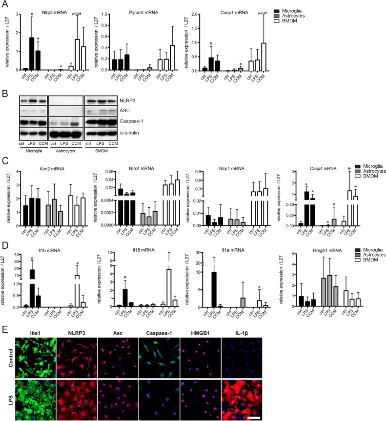
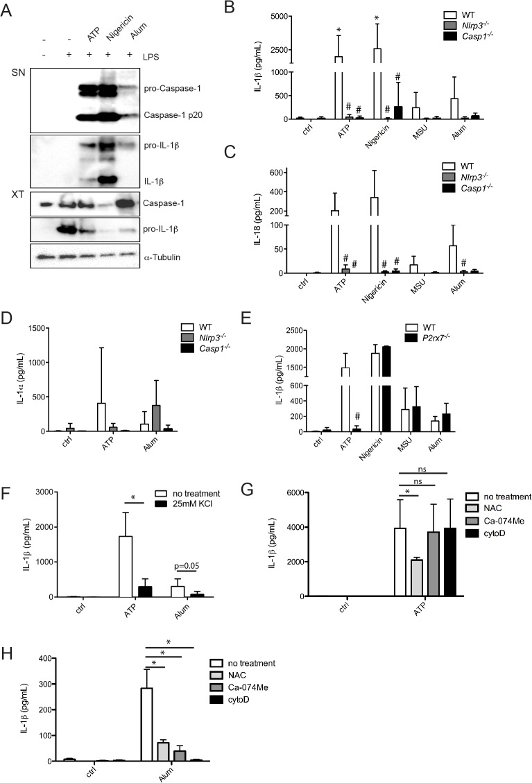
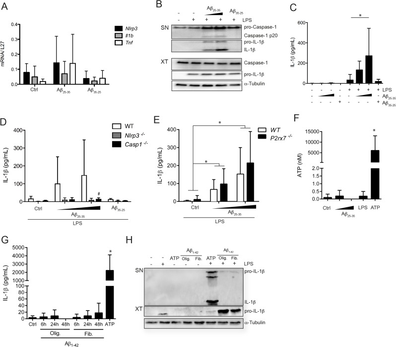
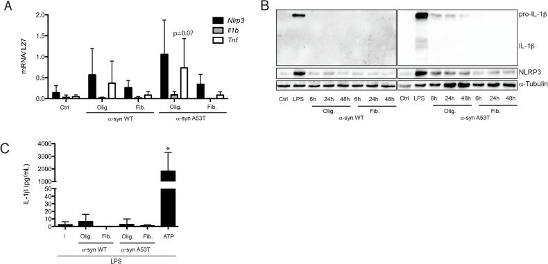
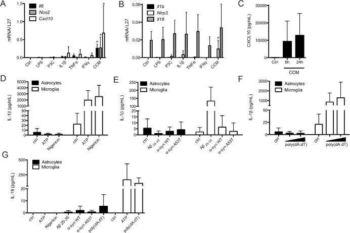
Neuroinflammation is the local reaction of the brain to infection, trauma, toxic molecules or protein aggregates. The brain resident macrophages, microglia, are able to trigger an appropriate response involving secretion of cytokines and chemokines, resulting in the activation of astrocytes and recruitment of peripheral immune cells. IL-1β plays an important role in this response; yet its production and mode of action in the brain are not fully understood and its precise implication in neurodegenerative diseases needs further characterization. Our results indicate that the capacity to form a functional NLRP3 inflammasome and secretion of IL-1β is limited to the microglial compartment in the mouse brain. We were not able to observe IL-1β secretion from astrocytes, nor do they express all NLRP3 inflammasome components. Microglia were able to produce IL-1β in response to different classical inflammasome activators, such as ATP, Nigericin or Alum. Similarly, microglia secreted IL-18 and IL-1α, two other inflammasome-linked pro-inflammatory factors. Cell stimulation with α-synuclein, a neurodegenerative disease-related peptide, did not result in the release of active IL-1β by microglia, despite a weak pro-inflammatory effect. Amyloid-β peptides were able to activate the NLRP3 inflammasome in microglia and IL-1β secretion occurred in a P2X7 receptor-independent manner. Thus microglia-dependent inflammasome activation can play an important role in the brain and especially in neuroinflammatory conditions.
Overview
The claims and statistics of this paper haven’t been checked yet.
Run the check — free, no account needed. It reads the open-access text and checks the sources it cites.
Integrity signals
Registry
Cites
Statistics
Figures
Verification
Claims the paper makes
The claims and statistics of this paper haven’t been checked yet.
Run the check — free, no account needed. It reads the open-access text and checks the sources it cites.
Claims other papers make about this one
Each is checked against this paper’s own text: does it say what the citing paper says it does?
In the citing paper
“Inflammasome signaling in the CNS has also been reported in the microglia, the brain’s critical innate immune cells (206), in astrocytes (207), perivascular brain-resident macrophages (208), oligodendrocytes (209), endothelial cells (210), as well as in neurons (211).”
From The gut-brain axis: how microbiota and host inflammasome influence brain physiology and pathology (2020) · cites this paper as [206]
In the citing paper
“stimulation of microglia cell culture with ATP enhance the expression of NLRP3, ASC protein and caspase-1 along with enhanced secretion of IL-1β and IL-18 in a NLRP3 inflammasome-dependent manner.”
From P2X7 Receptor Signaling in Stress and Depression (2019) · cites this paper as [153]
In the citing paper
“The same NLRP3 inflammasome components (ASC, caspase-1 and cytokines) were not observed in astrocytes culture cell, indicating a cell-type specific NLRP3 expression [153].”
From P2X7 Receptor Signaling in Stress and Depression (2019) · cites this paper as [153]
In the citing paper
“Additionally, the effects of ATP in microglia NRLP3 activation were blocked by pre-treatment with potassium chloride (KCl), preventing the efflux of K+, suggesting requirement of low concentrations of this ion in the intracellular environment for the activation of NLRP3 inflammasome [153].”
From P2X7 Receptor Signaling in Stress and Depression (2019) · cites this paper as [153]
Self-check
Percentages, sums and reported differences re-computed from the paper’s own numbers.
The self-check needs the paper’s full text.
Citations
Cites
Martinon, F., Burns, K., & Tschopp, J. (2002) · Molecular Cell · doi:10.1016/s1097-2765(02)00599-3 · Cited by 6,066
Gout-associated uric acid crystals activate the NALP3 inflammasome
Martinon, F., Pétrilli, V., Mayor, A., Tardivel, A., & Tschopp, J. (2006) · Nature · doi:10.1038/nature04516 · Cited by 5,326
Self-citationAstrocytes: Biology and pathology
Sofroniew, M. V., & Vinters, H. V. (2010) · Acta Neuropathologica · doi:10.1007/s00401-009-0619-8 · Cited by 5,196
Mechanisms underlying inflammation in neurodegeneration
Glass, C. K., Saijo, K., Winner, B., Marchetto, M. C., & Gage, F. H. (2010) · Cell · doi:10.1016/j.cell.2010.02.016 · Cited by 3,580
Cryopyrin activates the inflammasome in response to toxins and ATP
Mariathasan, S., Weiss, D. S., Newton, K., McBride, J., O'Rourke, K., Roose-Girma, M., Lee, W. P., Weinrauch, Y., Monack, D. M., & Dixit, V. M. (2006) · Nature · doi:10.1038/nature04515 · Cited by 3,080
NLRP3 is activated in Alzheimer’s disease and contributes to pathology in APP/PS1 mice
Heneka, M. T., Kummer, M. P., Stutz, A., Delekate, A., Schwartz, S., Vieira-Saecker, A., Griep, A., Axt, D., Remus, A., Tzeng, T.-C., et al. (2013) · Nature · doi:10.1038/nature11729 · Cited by 2,898
Activation and regulation of the inflammasomes
Latz, E., Xiao, T. S., & Stutz, A. (2013) · Nature Reviews Immunology · doi:10.1038/nri3452 · Cited by 2,884
Identification of a unique TGF-β–dependent molecular and functional signature in microglia
Butovsky, O., Jedrychowski, M. P., Moore, C. S., Cialic, R., Lanser, A. J., Gabriely, G., Koeglsperger, T., Dake, B., Wu, P. M., Doykan, C. E., et al. (2014) · Nature Neuroscience · doi:10.1038/nn.3599 · Cited by 2,543
The NALP3 inflammasome is involved in the innate immune response to amyloid-β
Halle, A., Hornung, V., Petzold, G. C., Stewart, C. R., Monks, B. G., Reinheckel, T., Fitzgerald, K. A., Latz, E., Moore, K. J., & Golenbock, D. T. (2008) · Nature Immunology · doi:10.1038/ni.1636 · Cited by 2,441
Innate immune activation in neurodegenerative disease
Heneka, M. T., Kummer, M. P., & Latz, E. (2014) · Nature Reviews Immunology · doi:10.1038/nri3705 · Cited by 1,437
Cited by
Sorted by citations
Oxidative Stress in Neurodegenerative Diseases: From Molecular Mechanisms to Clinical Applications
Liu, Z., Zhou, T., Ziegler, A. C., Dimitrion, P., & Zuo, L. (2017) · Oxidative Medicine and Cellular Longevity · doi:10.1155/2017/2525967 · Cited by 810
The gut-brain axis: how microbiota and host inflammasome influence brain physiology and pathology
Rutsch, A., Kantsjö, J. B., & Ronchi, F. (2020) · Frontiers in Immunology · doi:10.3389/fimmu.2020.604179 · Cited by 821
Inflammasomes in neuroinflammatory and neurodegenerative diseases
Voet, S., Srinivasan, S., Lamkanfi, M., & van Loo, G. (2019) · EMBO Molecular Medicine · doi:10.15252/emmm.201810248 · Cited by 761
Glycolysis – a key player in the inflammatory response
Soto‐Heredero, G., Heras, M. M. G. D. L., Gabandé‐Rodríguez, E., Oller, J., & Mittelbrunn, M. (2020) · FEBS Journal · doi:10.1111/febs.15327 · Cited by 660
Neuroinflammation in the central nervous system: Symphony of glial cells
Yang, Q., & Zhou, J. (2018) · Glia · doi:10.1002/glia.23571 · Cited by 539
The P2X7 receptor: A main player in inflammation
Adinolfi, E., Giuliani, A. L., De Marchi, E., Pegoraro, A., Orioli, E., & Di Virgilio, F. (2018) · Biochemical Pharmacology · doi:10.1016/j.bcp.2017.12.021 · Cited by 385
Mitochondria-associated membranes (MAMs) and inflammation
Missiroli, S., Patergnani, S., Caroccia, N., Pedriali, G., Perrone, M., Previati, M., Wieckowski, M. R., & Giorgi, C. (2018) · Cell Death and Disease · doi:10.1038/s41419-017-0027-2 · Cited by 398
Tang, T., Lang, X., Xu, C., Wang, X., Gong, T., Yang, Y., Cui, J., Bai, L., Wang, J., Jiang, W., et al. (2017) · Nature Communications · doi:10.1038/s41467-017-00227-x · Cited by 375
Inflammation in Alzheimer’s disease: Lessons learned from microglia-depletion models
Spangenberg, E. E., & Green, K. N. (2017) · Brain, Behavior, and Immunity · doi:10.1016/j.bbi.2016.07.003 · Cited by 352
Fiery Cell Death: Pyroptosis in the Central Nervous System
McKenzie, B., Dixit, V. M., & Power, C. (2019) · Trends in Neurosciences · doi:10.1016/j.tins.2019.11.005 · Cited by 341
Yue, N., Huang, H., Zhu, X., Han, Q., Wang, Y., Li, B., Liu, Q., Wu, G., Zhang, Y., & Yu, J. (2017) · Journal of Neuroinflammation · doi:10.1186/s12974-017-0865-y · Cited by 334
Innate Immunity and Neurodegeneration
Labzin, L. I., Heneka, M. T., & Latz, E. (2018) · Annual Review of Medicine · doi:10.1146/annurev-med-050715-104343 · Cited by 319
Bioenergetic regulation of microglia
Ghosh, S., Castillo, E., Frias, E. S., & Swanson, R. A. (2017) · Glia · doi:10.1002/glia.23271 · Cited by 298
Gong, Z., Pan, J., Shen, Q., Li, M., & Peng, Y. (2018) · Journal of Neuroinflammation · doi:10.1186/s12974-018-1282-6 · Cited by 289
Sarkar, S., Malovic, E., Harischandra, D. S., Ghaisas, S., Panicker, N., Charli, A., Palanisamy, B. N., Rokad, D., Jin, H., Anantharam, V., et al. (2017) · npj Parkinson s Disease · doi:10.1038/s41531-017-0032-2 · Cited by 288
Switching of the Microglial Activation Phenotype Is a Possible Treatment for Depression Disorder
Zhang, L., Zhang, J., & You, Z. (2018) · Frontiers in Cellular Neuroscience · doi:10.3389/fncel.2018.00306 · Cited by 284
Emerging Roles for the Immune System in Traumatic Brain Injury
McKee, C. A., & Lukens, J. R. (2016) · Frontiers in Immunology · doi:10.3389/fimmu.2016.00556 · Cited by 277
Fyn kinase regulates misfolded α-synuclein uptake and NLRP3 inflammasome activation in microglia
Panicker, N., Sarkar, S., Harischandra, D. S., Neal, M. P., Kam, T., Jin, H., Saminathan, H., Langley, M. R., Charli, A., Samidurai, M., et al. (2019) · The Journal of Experimental Medicine · doi:10.1084/jem.20182191 · Cited by 257
Inflammasomes in Tissue Damages and Immune Disorders After Trauma
Bortolotti, P., Faure, E., & Kipnis, É. (2018) · Frontiers in Immunology · doi:10.3389/fimmu.2018.01900 · Cited by 268
Inflammasome: Its role in traumatic brain and spinal cord injury
Mortezaee, K., Khanlarkhani, N., Beyer, C., & Zendedel, A. (2017) · Journal of Cellular Physiology · doi:10.1002/jcp.26287 · Cited by 255
The role of inflammasomes in human diseases and their potential as therapeutic targets
Yao, J., Sterling, K., Wang, Z., Zhang, Y., & Song, W. (2024) · Signal Transduction and Targeted Therapy · doi:10.1038/s41392-023-01687-y · Cited by 260
Fu, Q., Wu, J., Zhou, X.-Y., Ji, M.-H., Mao, Q.-H., Li, Q., Zong, M.-M., Zhou, Z.-Q., & Yang, J.-J. (2019) · Inflammation · doi:10.1007/s10753-018-0894-4 · Cited by 232
Microglia: Housekeeper of the Central Nervous System
Kabba, J. A., Xu, Y., Christian, H., Ruan, W., Kitchen, C., Xiang, Y., Zhang, L., Saavedra, J. M., & Pang, T. (2017) · Cellular and Molecular Neurobiology · doi:10.1007/s10571-017-0504-2 · Cited by 265
Targeting the Microglial NLRP3 Inflammasome and Its Role in Parkinson's Disease
Haque, M. E., Akther, M., Jakaria, M., Kim, I., Azam, S., & Choi, D. (2019) · Movement Disorders · doi:10.1002/mds.27874 · Cited by 251
The microglial NLRP3 inflammasome is activated by amyotrophic lateral sclerosis proteins
Deora, V., Lee, J. D., Albornoz, E. A., McAlary, L., Jagaraj, C. J., Robertson, A. A. B., Atkin, J. D., Cooper, M. A., Schroder, K., Yerbury, J. J., et al. (2020) · Glia · doi:10.1002/glia.23728 · Cited by 245
Retinal microglia – A key player in healthy and diseased retina
Rathnasamy, G., Foulds, W. S., Ling, E., & Kaur, C. (2018) · Progress in Neurobiology · doi:10.1016/j.pneurobio.2018.05.006 · Cited by 250
Xu, X. J., Yin, D., Ren, H., Gao, W., Li, F., Sun, D., Wu, Y., Zhou, S., Lyu, L., Yang, M., et al. (2018) · Neurobiology of Disease · doi:10.1016/j.nbd.2018.05.016 · Cited by 240
The Role of Microglia and the Nlrp3 Inflammasome in Alzheimer's Disease
Hanslik, K. L., & Ulland, T. K. (2020) · Frontiers in Neurology · doi:10.3389/fneur.2020.570711 · Cited by 236
Role of pyroptosis in spinal cord injury and its therapeutic implications
Mamun, A. A., Wu, Y., Monalisa, I., Jia, C., Zhou, K., Munir, F., & Xiao, J. (2020) · Journal of Advanced Research · doi:10.1016/j.jare.2020.08.004 · Cited by 232
Glial Cells Shape Pathology and Repair After Spinal Cord Injury
Gaudet, A. D., & Fonken, L. K. (2018) · Neurotherapeutics · doi:10.1007/s13311-018-0630-7 · Cited by 242
Metabolic Dysregulation Contributes to the Progression of Alzheimer’s Disease
Yan, X., Hu, Y., Wang, B., Wang, S., & Zhang, X. (2020) · Frontiers in Neuroscience · doi:10.3389/fnins.2020.530219 · Cited by 230
NLRP3 gene knockout blocks NF-κB and MAPK signaling pathway in CUMS-induced depression mouse model
Su, W., Zhang, Y., Chen, Y., Gong, H., Lian, Y., Wei, P., Liu, Y., Wang, Y., You, Z., Feng, S.-J., et al. (2017) · Behavioural Brain Research · doi:10.1016/j.bbr.2017.01.018 · Cited by 215
Microglia and CNS Interleukin-1: Beyond Immunological Concepts
Liu, X., & Quan, N. (2018) · Frontiers in Neurology · doi:10.3389/fneur.2018.00008 · Cited by 221
Wang, Z., Meng, S., Cao, L., Chen, Y., Zuo, Z., & Peng, S. (2018) · Journal of Neuroinflammation · doi:10.1186/s12974-018-1137-1 · Cited by 209
Wu, A., Zhou, X., Qiao, G., Yu, L., Tang, Y., Yan, L., Qiu, W., Pan, R., Yu, C., Law, B. Y. K., et al. (2020) · Ageing Research Reviews · doi:10.1016/j.arr.2020.101202 · Cited by 196
miRNA-23a/CXCR4 regulates neuropathic pain via directly targeting TXNIP/NLRP3 inflammasome axis
Pan, Z., Shan, Q., Gu, P., Wang, X. M., Tai, W. L., Sun, M.-L., Luo, X., Sun, L., & Cheung, C. W. (2018) · Journal of Neuroinflammation · doi:10.1186/s12974-018-1073-0 · Cited by 188
Depression, dementia and immune dysregulation
Hayley, S., Hakim, A. M., & Albert, P. R. (2021) · Brain · doi:10.1093/brain/awaa405 · Cited by 186
Potential therapeutic targets for intracerebral hemorrhage-associated inflammation: An update
Ren, H., Han, R., Chen, X., Liu, X., Wan, J., Wang, L., Yang, X., & Wang, J. (2020) · Journal of Cerebral Blood Flow & Metabolism · doi:10.1177/0271678x20923551 · Cited by 180
Reactive Glia Inflammatory Signaling Pathways and Epilepsy
Sanz, P., & García-Gimeno, M. A. (2020) · International Journal of Molecular Sciences · doi:10.3390/ijms21114096 · Cited by 179
Impaired insulin signalling and allostatic load in Alzheimer disease
Felice, F. G. D., Gonçalves, R. A., & Ferreira, S. T. (2022) · Nature reviews. Neuroscience · doi:10.1038/s41583-022-00558-9 · Cited by 183
Jang, J., Park, S., Jin Hur, H., Cho, H.-J., Hwang, I., Pyo Kang, Y., Im, I., Lee, H., Lee, E., Yang, W., et al. (2016) · Nature Communications · doi:10.1038/ncomms13129 · Cited by 166
Zeller, M. V., Dias, D., Sebastião, A. M., & Valente, C. A. (2021) · Journal of Alzheimer s Disease · doi:10.3233/jad-210268 · Cited by 179
Inflammasomes link vascular disease with neuroinflammation and brain disorders
Lénárt, N., Brough, D., & Dénes, Á. (2016) · Journal of Cerebral Blood Flow & Metabolism · doi:10.1177/0271678x16662043 · Cited by 170
The P2X7 Receptor: Central Hub of Brain Diseases
Andrejew, R., Oliveira-Giacomelli, Á., Ribeiro, D. E., Glaser, T., Arnaud-Sampaio, V. F., Lameu, C., & Ulrich, H. (2020) · Frontiers in Molecular Neuroscience · doi:10.3389/fnmol.2020.00124 · Cited by 162
Alzheimer’s Disease Pathogenesis: Role of Autophagy and Mitophagy Focusing in Microglia
Eshraghi, M., Adlimoghaddam, A., Mahmoodzadeh, A., Sharifzad, F., Yasavoli-Sharahi, H., Lorzadeh, S., Albensi, B. C., & Ghavami, S. (2021) · International Journal of Molecular Sciences · doi:10.3390/ijms22073330 · Cited by 158
Mohanty, A., Tiwari-Pandey, R., & Pandey, N. R. (2019) · Journal of Cell Communication and Signaling · doi:10.1007/s12079-019-00507-9 · Cited by 164
Jie, F., Yang, X., Yang, B., Liu, Y., Wu, L., & Lu, B. (2022) · Biomedicine & Pharmacotherapy · doi:10.1016/j.biopha.2022.113317 · Cited by 161
Exploring New Inflammatory Biomarkers and Pathways during LPS-Induced M1 Polarization
Cunha, C., Gomes, C., Vaz, A. R., & Brites, D. (2016) · Mediators of Inflammation · doi:10.1155/2016/6986175 · Cited by 153
Crowley, T., Cryan, J. F., Downer, E. J., & O’Leary, O. F. (2016) · Brain, Behavior, and Immunity · doi:10.1016/j.bbi.2016.02.001 · Cited by 150
NLRP3 inflammasome: a promising target in ischemic stroke
Gao, L., Dong, Q., Song, Z., Shen, F., Shi, J., & Li, Y. (2016) · Inflammation Research · doi:10.1007/s00011-016-0981-7 · Cited by 150
Hyperactivation of P2X7 receptors as a culprit of COVID-19 neuropathology
Ribeiro, D. E., Oliveira-Giacomelli, Á., Glaser, T., Arnaud-Sampaio, V. F., Andrejew, R., Dieckmann, L., Baranova, J., Lameu, C., Ratajczak, M. Z., & Ulrich, H. (2021) · Molecular Psychiatry · doi:10.1038/s41380-020-00965-3 · Cited by 145
Activation of the NLRP 3 inflammasome in microglia: the role of ceramide
Scheiblich, H., Schlütter, A., Golenbock, D. T., Latz, E., Martinez‐Martinez, P., & Heneka, M. T. (2017) · Journal of Neurochemistry · doi:10.1111/jnc.14225 · Cited by 147
Microglial priming of antigen presentation and adaptive stimulation in Alzheimer’s disease
Das, R., & Chinnathambi, S. (2019) · Cellular and Molecular Life Sciences · doi:10.1007/s00018-019-03132-2 · Cited by 147
Neuroinflammatory responses in Alzheimer’s disease
Dansokho, C., & Heneka, M. T. (2017) · Journal of Neural Transmission · doi:10.1007/s00702-017-1831-7 · Cited by 146
P2X7 Receptors Amplify CNS Damage in Neurodegenerative Diseases
Illés, P. (2020) · International Journal of Molecular Sciences · doi:10.3390/ijms21175996 · Cited by 145
NLRP3 inflammasome in neurodegenerative disease
Anderson, F. L., Biggs, K. E., Rankin, B. E., & Havrda, M. C. (2022) · Translational research · doi:10.1016/j.trsl.2022.08.006 · Cited by 154
NLRP3 inflammasome as a potential treatment in ischemic stroke concomitant with diabetes
Hong, P., Gu, R., Li, F., Xiong, X., Liang, W., You, Z.-J., & Zhang, H. (2019) · Journal of Neuroinflammation · doi:10.1186/s12974-019-1498-0 · Cited by 139
Mitochondria as a centrally positioned hub in the innate immune response
Sandhir, R., Halder, A., & Sunkaria, A. (2017) · Biochimica et Biophysica Acta (BBA) - Molecular Basis of Disease · doi:10.1016/j.bbadis.2016.10.020 · Cited by 135
Fan, Z., Liang, Z., Yang, H. M., Pan, Y., Zheng, Y., & Wang, X. (2017) · Journal of Neuroinflammation · doi:10.1186/s12974-017-1036-x · Cited by 136
Cathepsin B links oxidative stress to the activation of NLRP3 inflammasome
Bai, H., Yang, B., Yu, W., Xiao, Y. C., Yu, D., & Zhang, Q. (2017) · Experimental Cell Research · doi:10.1016/j.yexcr.2017.11.015 · Cited by 138
Innate Immune Response in Brain, NF-Kappa B Signaling and Cystatins
Kopitar‐Jerala, N. (2015) · Frontiers in Molecular Neuroscience · doi:10.3389/fnmol.2015.00073 · Cited by 132
Zhao, H., Pan, P., Yang, Y., Ge, H., Chen, W., Qu, J., Shi, J., Cui, G., Liu, X., Feng, H., et al. (2017) · Journal of Neuroinflammation · doi:10.1186/s12974-017-0940-4 · Cited by 134
Microglia in Alzheimer’s Disease: A Target for Therapeutic Intervention
Zhang, G., Wang, Z., Hu, H., Zhao, M., & Sun, L. (2021) · Frontiers in Cellular Neuroscience · doi:10.3389/fncel.2021.749587 · Cited by 137
P2X7 Receptor Signaling in Stress and Depression
Ribeiro, D. E., Roncalho, A. L., Glaser, T., Ulrich, H., Wegener, G., & Joca, S. (2019) · International Journal of Molecular Sciences · doi:10.3390/ijms20112778 · Cited by 129
Tonkin, R. S., Bowles, C., Perera, C. J., Keating, B. A., Makker, P. G. S., Duffy, S. S., Lees, J. G., Tran, C., Don, A. S., Fath, T., et al. (2017) · Experimental Neurology · doi:10.1016/j.expneurol.2017.10.016 · Cited by 124
Cheng, Y., Song, Y., Chen, H., Li, Q., Gao, Y. Z., Lu, G., & Luo, C. (2021) · Oxidative Medicine and Cellular Longevity · doi:10.1155/2021/5005136 · Cited by 124
Estrogen Attenuates Local Inflammasome Expression and Activation after Spinal Cord Injury
Zendedel, A., Mönnink, F., Hassanzadeh, G., Zaminy, A., Ansar, M. M., Habib, P., Slowik, A., Kipp, M., & Beyer, C. (2017) · Molecular Neurobiology · doi:10.1007/s12035-017-0400-2 · Cited by 119
Xu, Q., Zhao, B., Ye, Y., Li, Y., Zhang, Y., Xiong, X., & Gu, L. (2021) · Journal of Neuroinflammation · doi:10.1186/s12974-021-02137-8 · Cited by 119
Molecular mechanisms of aluminum neurotoxicity: Update on adverse effects and therapeutic strategies
Skalny, A. V., Aschner, M., Jiang, Y., Gluhcheva, Y. G., Tizabi, Y., Łobiński, R., & Tinkov, A. A. (2021) · Advances in neurotoxicology · doi:10.1016/bs.ant.2020.12.001 · Cited by 123
The neuroprotective potential of carotenoids in vitro and in vivo
Manochkumar, J., Doss, C. G. P., El-Seedi, H. R., Efferth, T., & Ramamoorthy, S. (2021) · Phytomedicine · doi:10.1016/j.phymed.2021.153676 · Cited by 117
Zhao, Z., Wang, Y., Zhou, R., Li, Y., Gao, Y., Tu, D., Wilson, B., Song, S., Feng, J., Hong, J.-S., et al. (2020) · Journal of Neuroinflammation · doi:10.1186/s12974-020-1728-5 · Cited by 109
Cai, Y., Kong, H., Pan, Y., Jiang, L., Pan, X.-X., Hu, L., Qian, Y., Jiang, C., & Liu, W. (2016) · Journal of Neuroinflammation · doi:10.1186/s12974-016-0520-z · Cited by 107
Li, T., Lu, L., Pember, E., Li, X., Zhang, B., & Zhu, Z. (2022) · Cells · doi:10.3390/cells11121925 · Cited by 109
Yue, N., Li, B., Yang, L., Han, Q.-Q., Huang, H.-J., Wang, Y.-L., Wang, J., Yu, R., Wu, G.-C., Liu, Q., et al. (2018) · Frontiers in Molecular Neuroscience · doi:10.3389/fnmol.2018.00149 · Cited by 100
Dodd, W., Noda, I., Martinez, M., Hosaka, K., & Hoh, B. L. (2021) · Journal of Neuroinflammation · doi:10.1186/s12974-021-02207-x · Cited by 98
Couturier, J., Stancu, I., Schakman, O., Pierrot, N., Huaux, F., Kienlen‐Campard, P., Dewachter, I., & Octave, J. (2016) · Journal of Neuroinflammation · doi:10.1186/s12974-016-0477-y · Cited by 95
Orioli, E., Marchi, E. D., Giuliani, A. L., & Adinolfi, E. (2017) · Current Medicinal Chemistry · doi:10.2174/0929867324666170303161659 · Cited by 99
Bibliometric Analysis of the Inflammasome and Pyroptosis in Brain
Chen, Y., Li, Y., Guo, L., Hong, J. Y., Zhao, W., Hu, X., Chang, C., Liu, W., & Xiong, K. (2021) · Frontiers in Pharmacology · doi:10.3389/fphar.2020.626502 · Cited by 94
Gullotta, G. S., Costantino, G., Sortino, M. A., & Spampinato, S. F. (2023) · International Journal of Molecular Sciences · doi:10.3390/ijms24119144 · Cited by 97
Yang, F.-X., Zhao, K., Zhang, X., Zhang, J., & Xu, B.-N. (2016) · Journal of Neural Transplantation and Plasticity · doi:10.1155/2016/8928530 · Cited by 89
The NLRP3 Inflammasome: Role and Therapeutic Potential in Pain Treatment
Starobova, H., Nadar, E. I., & Vetter, I. (2020) · Frontiers in Physiology · doi:10.3389/fphys.2020.01016 · Cited by 88
Meningeal lymphatics-microglia axis regulates synaptic physiology
Kim, K., Abramishvili, D., Du, S., Papadopoulos, Z., Cao, J., Herz, J., Smirnov, I., Thomas, J., Colonna, M., & Kipnis, J. (2025) · Cell · doi:10.1016/j.cell.2025.02.022 · Cited by 86
Inhibition of NLRP3 Inflammasome: A Prospective Target for the Treatment of Ischemic Stroke
Feng, Y.-S., Tan, Z., Wang, M., Xing, Y., Dong, F., & Zhang, F. (2020) · Frontiers in Cellular Neuroscience · doi:10.3389/fncel.2020.00155 · Cited by 85
Plasma-borne indicators of inflammasome activity in Parkinson’s disease patients
Anderson, F. L., von Herrmann, K. M., Andrew, A. S., Kuras, Y. I., Young, A. L., Scherzer, C. R., Hickey, W. F., Lee, S., & Havrda, M. C. (2021) · npj Parkinson s Disease · doi:10.1038/s41531-020-00147-6 · Cited by 89
Serdar, M., Kempe, K., Rizazad, M., Herz, J., Bendix, I., Felderhoff‐Müser, U., & Sabir, H. (2019) · Frontiers in Cellular Neuroscience · doi:10.3389/fncel.2019.00237 · Cited by 79
Redox Signaling, Neuroinflammation, and Neurodegeneration
Aguilera, G., Colín-González, A. L., Rangel‐López, E., Chavarría, A., & Santamaría, A. (2017) · Antioxidants and Redox Signaling · doi:10.1089/ars.2017.7099 · Cited by 81
Machado, V., Zöller, T., Attaai, A., & Spittau, B. (2016) · International Journal of Molecular Sciences · doi:10.3390/ijms17020151 · Cited by 76
Li, Z., Xu, H., Xu, Y., Lu, G., Peng, Q., Chen, J., Bi, R., Li, J., Chen, S., Li, H., et al. (2021) · CNS Neuroscience & Therapeutics · doi:10.1111/cns.13732 · Cited by 76
Distinct Features of Brain-Resident Macrophages: Microglia and Non-Parenchymal Brain Macrophages
Lee, E., Eo, J.-C., Lee, C., & Yu, J. (2021) · Molecules and Cells · doi:10.14348/molcells.2021.0060 · Cited by 83
Lawana, V., Singh, N., Sarkar, S., Charli, A., Jin, H., Anantharam, V., Kanthasamy, A. G., & Kanthasamy, A. (2017) · Journal of Neuroimmune Pharmacology · doi:10.1007/s11481-017-9746-5 · Cited by 73
Bellezza, I., Grottelli, S., Costanzi, E., Scarpelli, P. T., Pigna, E., Morozzi, G., Mezzasoma, L., Peirce, M. J., Moresi, V., Adamo, S., et al. (2017) · Molecular Neurobiology · doi:10.1007/s12035-017-0502-x · Cited by 73
Rezaee, D., Saadatpour, F., Akbari, N., Zoghi, A., Najafi, S., Beyranvand, P., Rarani, F. Z., Rashidi, M. A., Bagheri‐Mohammadi, S., & Bakhtiari, M. (2023) · Ageing Research Reviews · doi:10.1016/j.arr.2023.102090 · Cited by 76
Zielinski, M. R., Gerashchenko, D., Karpova, S. A., Konanki, V., McCarley, R. W., Sutterwala, F. S., Strecker, R. E., & Basheer, R. (2017) · Brain Behavior and Immunity · doi:10.1016/j.bbi.2017.01.012 · Cited by 73
NLRP3 Inflammasomes in Parkinson’s disease and their Regulation by Parkin
Yan, Y., Fang, Y., Zheng, R., Pu, J., & Zhang, B. (2020) · Neuroscience · doi:10.1016/j.neuroscience.2020.08.004 · Cited by 72
The Role of P2X7 Receptor in Alzheimer’s Disease
Francistiová, L., Bianchi, C., Lauro, C. D., Sebastián‐Serrano, Á., de Diego-García, L., Kobolák, J., Dinnyés, A., & Díaz-Hernández, M. (2020) · Frontiers in Molecular Neuroscience · doi:10.3389/fnmol.2020.00094 · Cited by 73
Gliotransmitters and cytokines in the control of blood-brain barrier permeability
Osipova, E. D., Semyachkina-Glushkovskaya, O. V., Morgun, A. V., Pisareva, N. V., Malinovskaya, N. A., Boitsova, E. B., Pozhilenkova, E. A., Belova, O. A., Salmin, V. V., Taranushenko, T. E., et al. (2018) · Reviews in the Neurosciences · doi:10.1515/revneuro-2017-0092 · Cited by 72
The Role of NF-κB Triggered Inflammation in Cerebral Ischemia
Jover‐Mengual, T., Hwang, J.-Y., Byun, H.-R., Court-Vazquez, B. L., Centeno, J. M., Burguete, M. C., & Zukin, R. S. (2021) · Frontiers in Cellular Neuroscience · doi:10.3389/fncel.2021.633610 · Cited by 69
Degechisa, S. T., Dabi, Y. T., & Gizaw, S. T. (2022) · Immunity Inflammation and Disease · doi:10.1002/iid3.647 · Cited by 69
Serdar, M., Kempe, K., Herrmann, R., Picard, D. J., Remke, M., Herz, J., Bendix, I., Felderhoff‐Müser, U., & Sabir, H. (2020) · Frontiers in Neurology · doi:10.3389/fneur.2020.540878 · Cited by 73
Neuroinflammation, Early-Life Adversity, and Brain Development
Andersen, S. L. (2022) · Harvard Review of Psychiatry · doi:10.1097/hrp.0000000000000325 · Cited by 73
Metadata
- Authors
- Gustin, A., Kirchmeyer, M., Koncina, E., Felten, P., Losciuto, S., Heurtaux, T., Tardivel, A., Heuschling, P., & Dostert, C.
- Affiliations
- University of Luxembourg (LU)
- University of Lausanne (CH)
- Corresponding author
- Dostert, Catherine
- Venue
- PLoS ONE
- Publisher
- Public Library of Science (PLoS)
- Published
- 2015-06-19
- Volume
- 10
- Issue
- 6
- Pages
- e0130624
- ISSN
- 1932-6203 (ISSN-L)
- PMC
- PMC4474809
- OpenAlex
- W582182564
- Cited by
- 369
- References
- 55
- Citations by year
- 2026: 21 · 2025: 27 · 2024: 33 · 2023: 38 · 2022: 33 · 2021: 44 · 2020: 59 · 2019: 27 · 2018: 29 · 2017: 37 · 2016: 17 · 2015: 3
- FWCI
- 12.44
- Type
- journal article
- Language
- en
- Open access
- gold
- doi.org
- Licence
- Locations
- PLoS ONE (publishedVersion, cc-by)
- PubMed Central (publishedVersion, cc-by)
- PubMed (publishedVersion)
- IRIS (submittedVersion)
- Open Repository and Bibliography (University of Luxembourg) (submittedVersion, cc-by-nc-sa)
- DOAJ (DOAJ: Directory of Open Access Journals) (submittedVersion, cc-by-sa)
- Figshare (submittedVersion, cc-by)
- SERVAL (Université de Lausanne) (submittedVersion, other-oa)
- Funders
- Université du Luxembourg
- Fonds National de la Recherche Luxembourg
- Université de Lausanne
- Topics
- Life Sciences › Biochemistry, Genetics and Molecular Biology › Molecular Biology › Inflammasome and immune disorders
- Life Sciences › Neuroscience › Neurology › Neuroinflammation and Neurodegeneration Mechanisms
- Life Sciences › Immunology and Microbiology › Immunology › Immune Response and Inflammation
- Keywords
- NLRP3 inflammasome, microglia, astrocytes, IL-1β secretion, neuroinflammation, IL-18, IL-1α, alpha-synuclein, amyloid-beta, P2X7 receptor, neurodegenerative diseases
- MeSH terms
- NLR Family, Pyrin Domain-Containing 3 Protein, Animals, Astrocytes, Brain, Carrier Proteins, Cells, Cultured, Enzyme-Linked Immunosorbent Assay, Mice, Inbred C57BL, Peptide Fragments, Amyloid beta-Peptides, Microglia, Mice, Knockout, Caspase 1, Interleukin-18, Mice, alpha-Synuclein, Interleukin-1alpha, Interleukin-1beta, Receptors, Purinergic P2X7, Inflammasomes
About these checks
Registry, retraction, Cites and statistics results are computed, not judged, with one exception: a printed reference that no index lists is read once by a model before it counts as a citation error. Claims are extracted by a model and kept only when their quote appears verbatim in the text; each cited source is then checked, with a model’s help, for whether it says what the claim relies on. Those verdicts can be wrong — read the claim’s quote and the cited source before relying on one.
Statistics and arithmetic are recomputed, not judged.
Confidence bands are shown only for fields calibrated on our annotated test set. None is calibrated yet, so no band is shown.
What counts as a claim ›How we measure confidence ›How we check this ›
Are you an author, or do you see a problem with this page? Request a review
