

The DOI matches its Crossref record; 72 of the 73 references it cites were found in a registry or an index, and 1 are not checked yet.
Brain organoid formation on decellularized porcine brain ECM hydrogels
Open accessAbstract
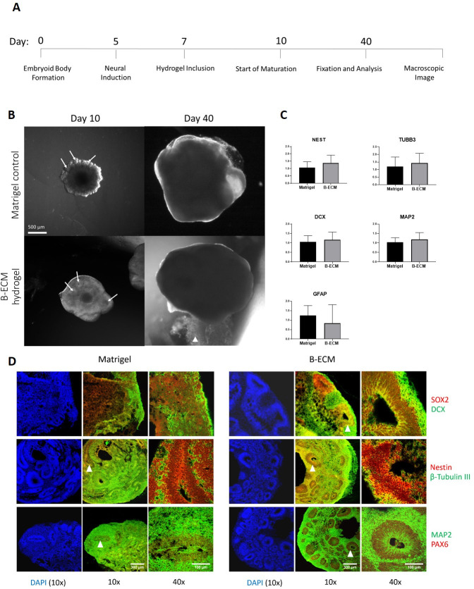
Human brain tissue models such as cerebral organoids are essential tools for developmental and biomedical research. Current methods to generate cerebral organoids often utilize Matrigel as an external scaffold to provide structure and biologically relevant signals. Matrigel however is a nonspecific hydrogel of mouse tumor origin and does not represent the complexity of the brain protein environment. In this study, we investigated the application of a decellularized adult porcine brain extracellular matrix (B-ECM) which could be processed into a hydrogel (B-ECM hydrogel) to be used as a scaffold for human embryonic stem cell (hESC)-derived brain organoids. We decellularized pig brains with a novel detergent- and enzyme-based method and analyzed the biomaterial properties, including protein composition and content, DNA content, mechanical characteristics, surface structure, and antigen presence. Then, we compared the growth of human brain organoid models with the B-ECM hydrogel or Matrigel controls in vitro. We found that the native brain source material was successfully decellularized with little remaining DNA content, while Mass Spectrometry (MS) showed the loss of several brain-specific proteins, while mainly different collagen types remained in the B-ECM. Rheological results revealed stable hydrogel formation, starting from B-ECM hydrogel concentrations of 5 mg/mL. hESCs cultured in B-ECM hydrogels showed gene expression and differentiation outcomes similar to those grown in Matrigel. These results indicate that B-ECM hydrogels can be used as an alternative scaffold for human cerebral organoid formation, and may be further optimized for improved organoid growth by further improving protein retention other than collagen after decellularization.
Overview
The claims and statistics of this paper haven’t been checked yet.
Run the check — free, no account needed. It reads the open-access text and checks the sources it cites.
Integrity signals
Registry
Cites
Statistics
Figures
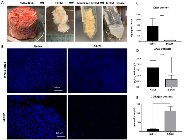
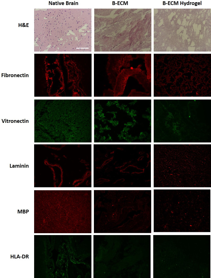
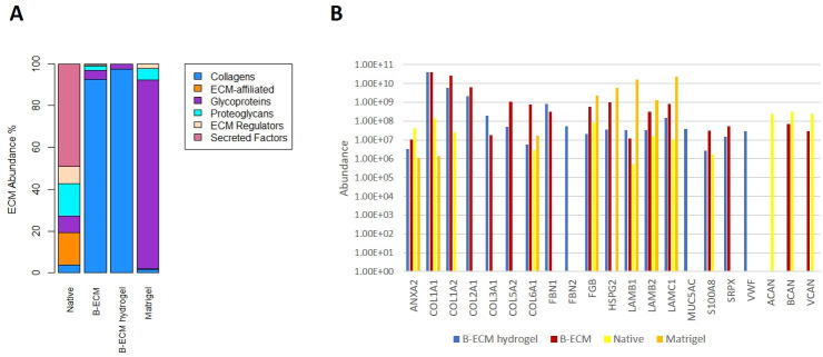
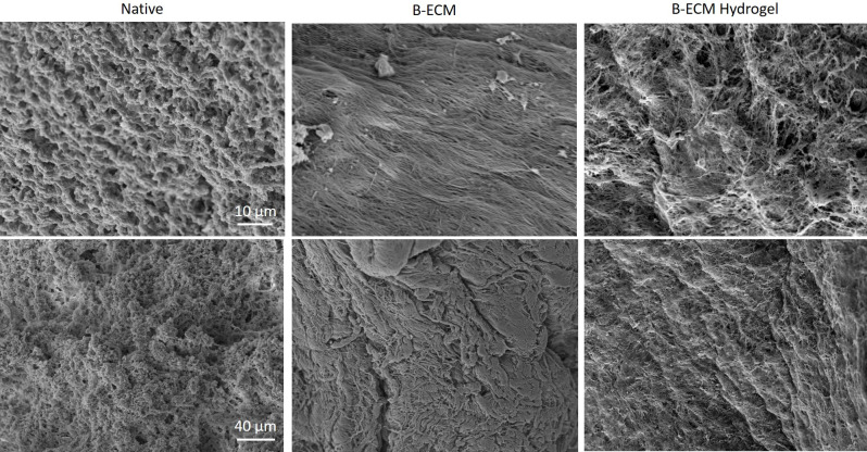
Verification
Claims the paper makes
The claims and statistics of this paper haven’t been checked yet.
Run the check — free, no account needed. It reads the open-access text and checks the sources it cites.
Claims other papers make about this one
Each is checked against this paper’s own text: does it say what the citing paper says it does?
The cited source says what the claim attributes to it.
In the citing paper
“Simsa et al. discovered that hydrogels derived from decellularized adult porcine brain ECM (B‐ECM) served as scaffolds for brain organoids derived from human embryonic stem cells (hESCs).”
From Bioactive scaffolds for tissue engineering: A review of decellularized extracellular matrix applications and innovations (2025) · cites this paper as [198]
The cited source says what the claim attributes to it.
In the citing paper
“They could be further optimized for organoid growth by enhancing the preservation of proteins beyond collagen after decellularization.[ 198 ]”
From Bioactive scaffolds for tissue engineering: A review of decellularized extracellular matrix applications and innovations (2025) · cites this paper as [198]
Self-check
Percentages, sums and reported differences re-computed from the paper’s own numbers.
The self-check needs the paper’s full text.
Citations
Cites
Ror2 signaling regulates Golgi structure and transport through IFT20 for tumor invasiveness
Nishita, M., Park, S.-Y., Nishio, T., Kamizaki, K., Wang, Z., Tamada, K., Takumi, T., Hashimoto, R., Otani, H., Pazour, G. J., et al. (2017) · Scientific Reports · doi:10.1038/s41598-016-0028-x · Cited by 15,947
Matrix elasticity directs stem cell lineage specification
Engler, A. J., Sen, S., Sweeney, H. L., & Discher, D. E. (2006) · Cell · doi:10.1016/j.cell.2006.06.044 · Cited by 13,915
Cerebral organoids model human brain development and microcephaly
Lancaster, M. A., Renner, M., Martin, C.-A., Wenzel, D., Bicknell, L. S., Hurles, M. E., Homfray, T., Penninger, J. M., Jackson, A. P., & Knoblich, J. A. (2013) · Nature · doi:10.1038/nature12517 · Cited by 5,372
Rappsilber, J., Mann, M., & Ishihama, Y. (2007) · Nature Protocols · doi:10.1038/nprot.2007.261 · Cited by 4,641
The extracellular matrix at a glance
Frantz, C., Stewart, K. M., & Weaver, V. M. (2010) · Journal of Cell Science · doi:10.1242/jcs.023820 · Cited by 4,334
Decellularization of tissues and organs
Gilbert, T. W., Sellaro, T. L., & Badylak, S. F. (2006) · Biomaterials · doi:10.1016/j.biomaterials.2006.02.014 · Cited by 2,222
Brain-Region-Specific Organoids Using Mini-bioreactors for Modeling ZIKV Exposure
Qian, X., Nguyen, H. N., Song, M. M., Hadiono, C., Ogden, S. C., Hammack, C., Yao, B., Hamersky, G. R., Jacob, F., Zhong, C., et al. (2016) · Cell · doi:10.1016/j.cell.2016.04.032 · Cited by 2,127
The basics of brain development
Stiles, J., & Jernigan, T. L. (2010) · Neuropsychology Review · doi:10.1007/s11065-010-9148-4 · Cited by 2,009
Generation of cerebral organoids from human pluripotent stem cells
Lancaster, M. A., & Knoblich, J. A. (2014) · Nature Protocols · doi:10.1038/nprot.2014.158 · Cited by 1,823
Kulak, N. A., Pichler, G., Paron, I., Nagaraj, N., & Mann, M. (2014) · Nature Methods · doi:10.1038/nmeth.2834 · Cited by 1,719
Matrigel: A complex protein mixture required for optimal growth of cell culture
Hughes, C. S., Postovit, L. M., & Lajoie, G. A. (2010) · PROTEOMICS · doi:10.1002/pmic.200900758 · Cited by 1,534
Badylak, S. F., Taylor, D., & Uygun, K. (2011) · Annual Review of Biomedical Engineering · doi:10.1146/annurev-bioeng-071910-124743 · Cited by 1,041
Extracellular space structure revealed by diffusion analysis
Nicholson, C., & Syková, E. (1998) · Trends in Neurosciences · doi:10.1016/s0166-2236(98)01261-2 · Cited by 914
Jo, J., Xiao, Y., Sun, A. X., Cukuroglu, E., Tran, H.-D., Göke, J., Tan, Z. Y., Saw, T. Y., Tan, C.-P., Lokman, H., et al. (2016) · Cell Stem Cell · doi:10.1016/j.stem.2016.07.005 · Cited by 861
Extracellular matrix hydrogels from decellularized tissues: Structure and function
Saldin, L. T., Cramer, M. C., Velankar, S., White, L. J., & Badylak, S. F. (2016) · Acta Biomaterialia · doi:10.1016/j.actbio.2016.11.068 · Cited by 841
Georges, P. C., Miller, W. J., Meaney, D. F., Sawyer, E. S., & Janmey, P. A. (2006) · Biophysical Journal · doi:10.1529/biophysj.105.073114 · Cited by 724
Methods of tissue decellularization used for preparation of biologic scaffolds and in vivo relevance
Keane, T. J., Swinehart, I. T., & Badylak, S. F. (2015) · Methods · doi:10.1016/j.ymeth.2015.03.005 · Cited by 670
Edgar, D., Timpl, R., & Thoenen, H. (1984) · The EMBO Journal · doi:10.1002/j.1460-2075.1984.tb01997.x · Cited by 631
Antoine, E. E., Vlachos, P. P., & Rylander, M. N. (2014) · Tissue Engineering Part B: Reviews · doi:10.1089/ten.teb.2014.0086 · Cited by 611
Raja, W. K., Mungenast, A. E., Lin, Y.-T., Ko, T., Abdurrob, F., Seo, J., & Tsai, L.-H. (2016) · PLOS ONE · doi:10.1371/journal.pone.0161969 · Cited by 569
Preparation and rheological characterization of a gel form of the porcine urinary bladder matrix
Freytes, D. O., Martin, J. D., Velankar, S., Lee, A. S., & Badylak, S. F. (2008) · Biomaterials · doi:10.1016/j.biomaterials.2007.12.014 · Cited by 531
Ruoslahti, E. (1996) · Glycobiology · doi:10.1093/glycob/6.5.489 · Cited by 467
Derivation of Human Midbrain-Specific Organoids from Neuroepithelial Stem Cells
Monzel, A. S., Smits, L. M., Hemmer, K., Hachi, S., Moreno, E. L., van Wuellen, T., Jarazo, J., Walter, J., Brüggemann, I., Boussaad, I., et al. (2017) · Stem Cell Reports · doi:10.1016/j.stemcr.2017.03.010 · Cited by 435
A protocol for rheological characterization of hydrogels for tissue engineering strategies
Zuidema, J. M., Rivet, C. J., Gilbert, R. J., & Morrison, F. A. (2013) · Journal of Biomedical Materials Research Part B Applied Biomaterials · doi:10.1002/jbm.b.33088 · Cited by 395
Brain Extracellular Matrix in Neurodegeneration
Bonneh‐Barkay, D., & Wiley, C. A. (2008) · Brain Pathology · doi:10.1111/j.1750-3639.2008.00195.x · Cited by 313
Hydrogels derived from central nervous system extracellular matrix
Medberry, C. J., Crapo, P. M., Siu, B. F., Carruthers, C. A., Wolf, M. T., Nagarkar, S. P., Agrawal, V., Jones, K. E., Kelly, J., Johnson, S. A., et al. (2012) · Biomaterials · doi:10.1016/j.biomaterials.2012.10.062 · Cited by 278
Sackett, S. D., Tremmel, D. M., Ma, F., Feeney, A. K., Maguire, R. M., Brown, M. E., Zhou, Y., Li, X., O’Brien, C., Li, L., et al. (2018) · Scientific Reports · doi:10.1038/s41598-018-28857-1 · Cited by 269
The impact of detergents on the tissue decellularization process: A ToF-SIMS study
White, L. J., Taylor, A. J., Faulk, D. M., Keane, T. J., Saldin, L. T., Reing, J. E., Swinehart, I. T., Turner, N. J., Ratner, B. D., & Badylak, S. F. (2017) · Acta Biomaterialia · doi:10.1016/j.actbio.2016.12.033 · Cited by 268
Perfusion decellularization of human and porcine lungs: Bringing the matrix to clinical scale
Gilpin, S. E., Guyette, J. P., Gonzalez, G., Ren, X., Asara, J. M., Mathisen, D. J., Vacanti, J. P., & Ott, H. C. (2014) · The Journal of Heart and Lung Transplantation · doi:10.1016/j.healun.2013.10.030 · Cited by 263
Biologic scaffolds composed of central nervous system extracellular matrix
Crapo, P. M., Medberry, C. J., Reing, J. E., Tottey, S., van der Merwe, Y., Jones, K. E., & Badylak, S. F. (2012) · Biomaterials · doi:10.1016/j.biomaterials.2012.01.044 · Cited by 263
The pig model in brain imaging and neurosurgery
Sauleau, P., Lapouble, E., Val‐Laillet, D., & Malbert, C. (2009) · animal · doi:10.1017/s1751731109004649 · Cited by 253
Reassessment of brain elasticity for analysis of biomechanisms of hydrocephalus
Taylor, Z. A., & Miller, K. (2004) · Journal of Biomechanics · doi:10.1016/j.jbiomech.2003.11.027 · Cited by 235
Decellularized Porcine Brain Matrix for Cell Culture and Tissue Engineering Scaffolds
DeQuach, J. A., Yuan, S. H., Goldstein, L. S. B., & Christman, K. L. (2011) · Tissue Engineering Part A · doi:10.1089/ten.tea.2010.0724 · Cited by 234
The effect of detergents on the basement membrane complex of a biologic scaffold material
Faulk, D. M., Carruthers, C. A., Warner, H. J., Kramer, C. R., Reing, J. E., Zhang, L., D’Amore, A., & Badylak, S. F. (2013) · Acta Biomaterialia · doi:10.1016/j.actbio.2013.09.006 · Cited by 227
Lam, D., Enright, H. A., Cadena, J., Peters, S. K. G., Sales, A. P., Osburn, J. J., Soscia, D. A., Kulp, K. S., Wheeler, E. K., & Fischer, N. O. (2019) · Scientific Reports · doi:10.1038/s41598-019-40128-1 · Cited by 213
Baiguera, S., Gaudio, C. D., Lucatelli, E., Kuevda, E. V., Boieri, M., Mazzanti, B., Bianco, A., & Macchiarini, P. (2013) · Biomaterials · doi:10.1016/j.biomaterials.2013.10.060 · Cited by 211
Recellularization of Well-Preserved Acellular Kidney Scaffold Using Embryonic Stem Cells
Bonandrini, B., Figliuzzi, M., Papadimou, E., Morigi, M., Perico, N., Casiraghi, F., Dipl, C., Sangalli, F., Conti, S., Benigni, A., et al. (2014) · Tissue Engineering Part A · doi:10.1089/ten.tea.2013.0269 · Cited by 195
Hoshiba, T., Chen, G., Endo, C., Maruyama, H., Wakui, M., Nemoto, E., Kawazoe, N., & Tanaka, M. (2015) · Stem Cells International · doi:10.1155/2016/6397820 · Cited by 187
Stachowiak, E. K., Benson, C. A., Narla, S. T., Dimitri, A., Chuye, L. E. B., Dhiman, S., Harikrishnan, K., Elahi, S., Freedman, D., Brennand, K. J., et al. (2017) · Translational Psychiatry · doi:10.1038/s41398-017-0054-x · Cited by 179
Koh, I., Cha, J., Park, J., Choi, J., Kang, S., & Kim, P. (2018) · Scientific Reports · doi:10.1038/s41598-018-22681-3 · Cited by 163
Massensini, A. R., Ghuman, H., Saldin, L. T., Medberry, C. J., Keane, T. J., Nicholls, F. J., Velankar, S., Badylak, S. F., & Modo, M. (2015) · Acta Biomaterialia · doi:10.1016/j.actbio.2015.08.040 · Cited by 152
Hwang, J., San, B. H., Turner, N. J., White, L. J., Faulk, D. M., Badylak, S. F., Li, Y., & Yu, S. M. (2017) · Acta Biomaterialia · doi:10.1016/j.actbio.2017.01.079 · Cited by 147
Sood, D., Chwalek, K., Stuntz, E., Pouli, D., Du, C., Tang‐Schomer, M. D., Georgakoudi, I., Black, L. D., & Kaplan, D. L. (2015) · ACS Biomaterials Science & Engineering · doi:10.1021/acsbiomaterials.5b00446 · Cited by 123
Self-citationTailoring material properties of a nanofibrous extracellular matrix derived hydrogel
Johnson, T. D., Lin, S. Y., & Christman, K. L. (2011) · Nanotechnology · doi:10.1088/0957-4484/22/49/494015 · Cited by 116
Systematic in vitro comparison of decellularization protocols for blood vessels
Simsa, R., Padma, A. M., Heher, P., Hellström, M., Teuschl, A., Jenndahl, L., Bergh, N., & Fogelstrand, P. (2018) · PLOS ONE · doi:10.1371/journal.pone.0209269 · Cited by 115
Self-citationAnalysis of oxygen transport in a diffusion‐limited model of engineered heart tissue
Brown, D. A., MacLellan, W. R., Laks, H., Dunn, J., Wu, B. M., & Beygui, R. E. (2006) · Biotechnology and Bioengineering · doi:10.1002/bit.21295 · Cited by 102
Shellswell, G. B., Restall, D. J., Duance, V. C., & Bailey, A. J. (1979) · FEBS Letters · doi:10.1016/0014-5793(79)80520-7 · Cited by 94
Physical and chemical modifications of collagen gels: Impact on diffusion
Erikson, A., Andersen, H. N., Næss, S. N., Sikorski, P., & Davies, C. D. L. (2007) · Biopolymers · doi:10.1002/bip.20874 · Cited by 91
Modulation of Material Properties of a Decellularized Myocardial Matrix Scaffold
Singelyn, J. M., & Christman, K. L. (2011) · Macromolecular Bioscience · doi:10.1002/mabi.201000423 · Cited by 90
Brain matrix: structure, turnover and necessity
Rauch, U. (2007) · Biochemical Society Transactions · doi:10.1042/bst0350656 · Cited by 88
The impact of decellularization agents on renal tissue extracellular matrix
Poornejad, N., Schaumann, L. B., Buckmiller, E. M., Momtahan, N., Gassman, J. R., Ma, H. H., Roeder, B. L., Reynolds, P. R., & Cook, A. D. (2016) · Journal of Biomaterials Applications · doi:10.1177/0885328216656099 · Cited by 78
Fibronectin supports neurite outgrowth and axonal regeneration of adult brain neurons in vitro
Tonge, D. A., de Burgh, H. T., Docherty, R. J., Humphries, M. J., Craig, S. E., & Pizzey, J. A. (2012) · Brain Research · doi:10.1016/j.brainres.2012.03.024 · Cited by 78
Building Biocompatible Hydrogels for Tissue Engineering of the Brain and Spinal Cord
Aurand, E. R., Wagner, J. L., Lanning, C. J., & Bjugstad, K. B. (2012) · Journal of Functional Biomaterials · doi:10.3390/jfb3040839 · Cited by 77
Wu, Y., Wang, J., Shi, Y., Pu, H., Leak, R. K., Liou, A. K. F., Badylak, S. F., Liu, Z., Zhang, J., Chen, J., et al. (2017) · Cell Transplantation · doi:10.1177/0963689717714090 · Cited by 74
Ribatti, D., Conconi, M. T., Nico, B., Baiguera, S., Corsi, P., Parnigotto, P. P., & Nussdorfer, G. G. (2003) · Brain Research · doi:10.1016/s0006-8993(03)03225-6 · Cited by 68
Kuljanin, M., Brown, C. F. C., Raleigh, M. J., Lajoie, G., & Flynn, L. E. (2017) · Biomaterials · doi:10.1016/j.biomaterials.2017.08.012 · Cited by 63
Oksdath, M., Perrin, S. L., Bardy, C., Hilder, E. F., DeForest, C. A., Arrua, R. D., & Gomez, G. A. (2018) · APL Bioengineering · doi:10.1063/1.5045124 · Cited by 62
The matrix reloaded: the evolution of regenerative hydrogels
Jabbari, E., Leijten, J., Xu, Q., & Khademhosseini, A. (2015) · Materials Today · doi:10.1016/j.mattod.2015.10.005 · Cited by 60
The effects of nonignorable missing data on label-free mass spectrometry proteomics experiments
O’Brien, J. J., Gunawardena, H. P., Paulo, J. A., Chen, X., Ibrahim, J. G., Gygi, S. P., & Qaqish, B. F. (2018) · The Annals of Applied Statistics · doi:10.1214/18-aoas1144 · Cited by 54
Intact Telopeptides Enhance Interactions between Collagens
Shayegan, M., Altindal, T., Kiefl, E., & Forde, N. R. (2016) · Biophysical Journal · doi:10.1016/j.bpj.2016.10.039 · Cited by 52
Effect of fluid dynamics on decellularization efficacy and mechanical properties of blood vessels
Simsa, R., Vila, X. M., Salzer, E., Teuschl, A., Jenndahl, L. E., Bergh, N., & Fogelstrand, P. (2019) · PLoS ONE · doi:10.1371/journal.pone.0220743 · Cited by 43
Self-citationNondestructive Methods for Monitoring Cell Removal During Rat Liver Decellularization
Geerts, S., Özer, S., Jaramillo, M., Yarmush, M. L., & Uygun, B. E. (2016) · Tissue Engineering Part C Methods · doi:10.1089/ten.tec.2015.0571 · Cited by 42
Physical Properties of the Extracellular Matrix of Decellularized Porcine Liver
Ijima, H., Nakamura, S., Bual, R. P., Shirakigawa, N., & Tanoue, S. (2018) · Gels · doi:10.3390/gels4020039 · Cited by 38
Decellularized Whole Heart for Bioartificial Heart
Aubin, H., Kranz, A., Hülsmann, J., Lichtenberg, A., & Akhyari, P. (2013) · Methods in molecular biology · doi:10.1007/978-1-62703-511-8_14 · Cited by 37
Freytes, D. O., O’Neill, J. D., Duan-Arnold, Y., Wrona, E. A., & Vunjak‐Novakovic, G. (2014) · Methods in molecular biology · doi:10.1007/978-1-4939-1047-2_7 · Cited by 36
Lin, Q., Wong, H. L., Tian, F., Huang, Y., Xu, J., Yang, J., Chen, P.-P., Fan, Z.-L., Lu, C., & Zhao, Y. (2016) · International Journal of Pharmaceutics · doi:10.1016/j.ijpharm.2016.12.028 · Cited by 34
Wang, J., Li, L., Chen, T., Ma, J., Zhu, Y., Zhuang, J., & Chang, C. (2017) · Scientific Reports · doi:10.1038/s41598-017-03650-8 · Cited by 31
An In Vitro System to Evaluate the Effects of Ischemia on Survival of Cells Used for Cell Therapy
Davis, B. H., Schroeder, T., Yarmolenko, P., Guilak, F., Dewhirst, M. W., & Taylor, D. A. (2007) · Annals of Biomedical Engineering · doi:10.1007/s10439-007-9301-2 · Cited by 30
Caught in the matrix: how vitronectin controls neuronal differentiation
Wechsler‐Reya, R. J. (2001) · Trends in Neurosciences · doi:10.1016/s0166-2236(00)02058-0 · Cited by 30
Fibronectin, Collagen, Fibrin - Components of Extracellular Matrix for Nerve Regeneration
Alovskaya, A., Alekseeva, T., Phillips, J. B., King, K., & Brown, R. A. (2007) · Open Research Online (The Open University) · Cited by 24
Extracellular matrix heparin induces alteration of the cell adhesion during brain development
Ушакова, Г. А., Nikonenko, I., Nikonenko, A. G., & Skibo, G. G. (2002) · Neurochemistry International · doi:10.1016/s0197-0186(01)00028-6 · Cited by 7
Rothenbücher, T. S. P., & Martı́nez-Serrano, A. (2019) · Future Neurology · doi:10.2217/fnl-2018-0043 · Cited by 6
Self-citationCollagen in the Nervous System
Kucharz, E. J. (1992) · Springer eBooks · doi:10.1007/978-3-642-76197-3_19 · Cited by 5
Research works printed but not linked: 24
- TSP Rothenbücher, 2019, Future Neurol, FNL3
- EK Stachowiak, 2017, Transl Psychiatry, 6
- AS Monzel, 2017, Stem Cell Reports, 1144
- M Oksdath, 2018, APL Bioeng, 041501
- C Frantz, 2010, J Cell Sci, 4195
- A Alovskaya, 2007, Top tissue Eng
- R Simsa, 2018, PLoS One, e0209269
- R Simsa, 2019, PLoS One, e0220743
- D Lam, 2019, Sci Rep, 4159
- M Shayegan, 2016, Biophys J, 2404
- SD Sackett, 2018, Sci Rep, 10452
- M Kuljanin, 2017, Biomaterials, 130
- JJ O’brien, 2018, Ann Appl Stat, 2075
- J Wang, 2017, Sci Rep, 1
- DA Tonge, 2012, Brain Res, 8
- EE Antoine, 2014, Tissue Eng Part B Rev, 683
- A Erikson, 2008, Biopolymers, 135
- TJ Keane, 2015, Methods, 25
- S Geerts, 2016, Tissue Eng—Part C Methods, 671
- ER Aurand, 2012, J Funct Biomater, 839
- BH Davis, 2007, Ann Biomed Eng, 1414
- DA Brown, 2007, Biotechnol Bioeng, 962
- E Jabbari, 2016, Mater Today, 190
- J Stiles, 2010, Neuropsychol Rev, 327
Cited by
Sorted by citations
Brown, M., Li, J., Moraes, C., Tabrizian, M., & Li-Jessen, N. Y. K. (2022) · Biomaterials · doi:10.1016/j.biomaterials.2022.121786 · Cited by 282
Organoids: The current status and biomedical applications
Yang, S., Hu, H., Kung, H., Zou, R.-Q., Dai, Y.-S., Hu, Y.-F., Wang, T., Lv, T., Yu, J., & Li, F. (2023) · MedComm · doi:10.1002/mco2.274 · Cited by 300
Liu, J., Song, Q., Yin, W., Li, C., An, N., Le, Y., Wang, Q., Feng, Y., Hu, Y., & Wang, Y. (2025) · Exploration · doi:10.1002/exp.20230078 · Cited by 140
Advances in Spheroids and Organoids on a Chip
Fang, G., Chen, Y., Lu, H., & Jin, D. (2023) · Advanced Functional Materials · doi:10.1002/adfm.202215043 · Cited by 135
Engineering the Extracellular Matrix for Organoid Culture
Heo, J. H., Kang, D., Seo, S. J., & Jin, Y. (2022) · International Journal of Stem Cells · doi:10.15283/ijsc21190 · Cited by 116
Metadata
- Authors
- Simsa, R., Rothenbücher, T., Gürbüz, H., Ghosheh, N., Emneus, J., Jenndahl, L., Kaplan, D. L., Bergh, N., Serrano, A. M., & Fogelstrand, P.
- Affiliations
- Tufts University (US)
- Centro de Biología Molecular Severo Ochoa (ES)
- Technical University of Denmark (DK)
- University of Skövde (SE)
- Sahlgrenska University Hospital (SE)
- Corresponding author
- Simsa, Robin
- Rothenbücher, Theresa
- Gürbüz, Hakan
- Ghosheh, Nidal
- Jenndahl, Lachmi
- Kaplan, David L.
- Bergh, Niklas
- Fogelstrand, Per
- ORCID
- Venue
- PLOS ONE
- Publisher
- Public Library of Science (PLoS)
- Published
- 2021-01-28
- Volume
- 16
- Issue
- 1
- Pages
- e0245685
- ISSN
- 1932-6203 (ISSN-L)
- PMC
- PMC7842896
- OpenAlex
- W3124422938
- Cited by
- 114
- References
- 72
- Citations by year
- 2026: 10 · 2025: 26 · 2024: 24 · 2023: 27 · 2022: 21 · 2021: 6
- FWCI
- 18.32
- Type
- journal article
- Language
- en
- Open access
- gold
- doi.org
- Licence
- Locations
- PLoS ONE (publishedVersion, cc-by)
- PubMed Central (publishedVersion, cc-by)
- PubMed (publishedVersion)
- Diva portal (Dalarna University Library) (submittedVersion, other-oa)
- DIGITAL.CSIC (Spanish National Research Council (CSIC)) (submittedVersion, cc-by)
- DOAJ (DOAJ: Directory of Open Access Journals) (submittedVersion, cc-by-sa)
- Technical University of Denmark, DTU Orbit (Technical University of Denmark, DTU) (submittedVersion, other-oa)
- Funders
- Horizon 2020 (722779)
- MINECO SAF (2017-83241-R)
- ISC-III RETICS TerCel (RD16/0011/0032)
- ASCTN-Training (813851)
- National Institutes of Health (P41EB027062, R01NS092847)
- Topics
- Health Sciences › Medicine › Surgery › Tissue Engineering and Regenerative Medicine
- Physical Sciences › Engineering › Biomedical Engineering › 3D Printing in Biomedical Research
- Physical Sciences › Materials Science › Biomaterials › Hydrogels: synthesis, properties, applications
- Keywords
- brain organoids, porcine brain, hydrogels, scaffold materials, human embryonic stem cells, Matrigel alternative, decellularization, collagen, protein retention, hydrogel rheology, organoid differentiation
- MeSH terms
- Human Embryonic Stem Cells, Animals, Brain, Brain Chemistry, Cell Line, Extracellular Matrix, Humans, Organoids, Swine, Hydrogels
About these checks
Registry, retraction, Cites and statistics results are computed, not judged, with one exception: a printed reference that no index lists is read once by a model before it counts as a citation error. Claims are extracted by a model and kept only when their quote appears verbatim in the text; each cited source is then checked, with a model’s help, for whether it says what the claim relies on. Those verdicts can be wrong — read the claim’s quote and the cited source before relying on one.
Statistics and arithmetic are recomputed, not judged.
Confidence bands are shown only for fields calibrated on our annotated test set. None is calibrated yet, so no band is shown.
What counts as a claim ›How we measure confidence ›How we check this ›
Are you an author, or do you see a problem with this page? Request a review
