Cited by 50

The DOI matches its Crossref record; 5 of the 5 references it cites were found in a registry or an index.

Interaction of the Human Papillomavirus E6 Oncoprotein with Sorting Nexin 27 Modulates Endocytic Cargo Transport Pathways

Ganti, Ketaki, Massimi, Paola, Manzo-Merino, Joaquin, Tomaić, Vjekoslav, Pim, David, Playford, Martin P., Lizano, Marcela, Roberts, Sally, Kranjec, Christian, Doorbar, John, Banks, Lawrence·2016·PLOS Pathogens

Overview

The claims and statistics of this paper haven’t been checked yet.

Run the check — free, no account needed. It reads the open-access text and checks the sources it cites.

A few free checks a day · papers already verified open instantly · results are public

Integrity signals

Registry

Crossref · 10.1371/journal.ppat.1005854

Cites

5 of 5 references verified0 citation errors · 0 not checkable

Statistics

Figures

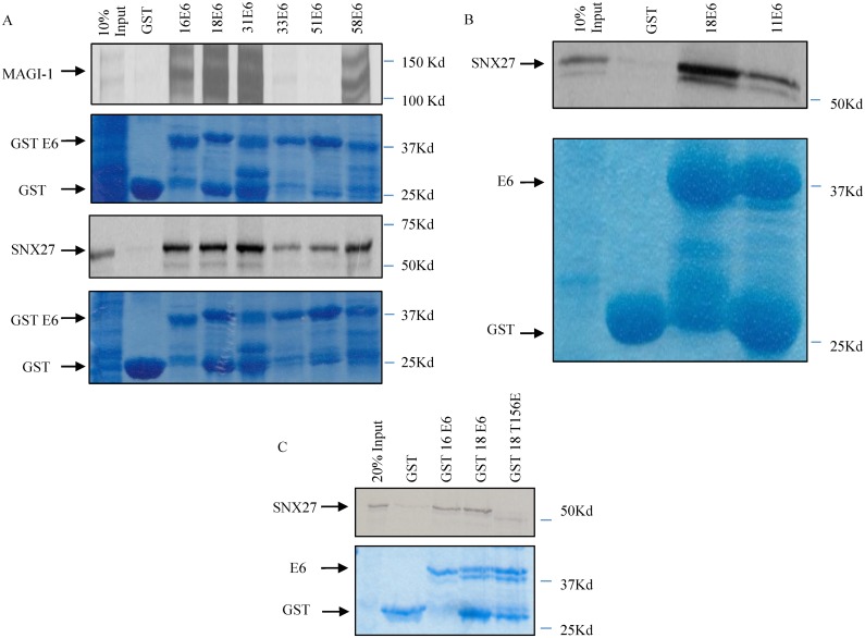
Fig 1 SNX27 interacts with multiple high-risk HPV E6 proteins. Panel A: SNX27 and MAGI-1 were translated and radio-labelled in vitro using the TNT Coupled Reticulocyte Lysate System (Promega). The translated proteins were then incubated with purified GST-tagged HPV E6s as indicated. The arrows indicate the position of MAGI-1 and SNX27 on the autoradiograms, which show the levels of bound and input proteins. The Coomassie Blue staining shows loading of the purified GST proteins, with the arrows indicating the position of GST and the GST tagged E6s. Panel B: SNX27 was translated and radio-labelled in vitro using the TNT Coupled Reticulocyte Lysate System (Promega). The translated proteins were then incubated with purified GST alone, GST-HPV18 E6 or GST-HPV11 E6 as indicated, with the upper panel showing the autoradiogram of bound and input SNX27. The Coomassie Blue staining shows loading of the purified GST proteins. Arrows indicate the position of GST and the GST-tagged E6s. Panel C: SNX27 was translated and radio-labelled in vitro using the TNT Coupled Reticulocyte Lysate System (Promega). The translated proteins were then incubated with purified GST alone, GST-HPV16 E6, GST-HPV18 E6 or the GST-HPV18 E6 T156E mutant as indicated. The upper panel shows the autoradiogram of bound and input proteins with the arrows indicating the position of SNX27. The Coomassie Blue staining shows loading of the purified GST proteins, with the arrows indicating the position of GST and the GST-tagged E6s.
Fig 1 SNX27 interacts with multiple high-risk HPV E6 proteins. Panel A: SNX27 and MAGI-1 were translated and radio-labelled in vitro using the TNT Coupled Reticulocyte Lysate System (Promega). The translated proteins were then incubated with purified GST-tagged HPV E6s as indicated. The arrows indicate the position of MAGI-1 and SNX27 on the autoradiograms, which show the levels of bound and input proteins. The Coomassie Blue staining shows loading of the purified GST proteins, with the arrows indicating the position of GST and the GST tagged E6s. Panel B: SNX27 was translated and radio-labelled in vitro using the TNT Coupled Reticulocyte Lysate System (Promega). The translated proteins were then incubated with purified GST alone, GST-HPV18 E6 or GST-HPV11 E6 as indicated, with the upper panel showing the autoradiogram of bound and input SNX27. The Coomassie Blue staining shows loading of the purified GST proteins. Arrows indicate the position of GST and the GST-tagged E6s. Panel C: SNX27 was translated and radio-labelled in vitro using the TNT Coupled Reticulocyte Lysate System (Promega). The translated proteins were then incubated with purified GST alone, GST-HPV16 E6, GST-HPV18 E6 or the GST-HPV18 E6 T156E mutant as indicated. The upper panel shows the autoradiogram of bound and input proteins with the arrows indicating the position of SNX27. The Coomassie Blue staining shows loading of the purified GST proteins, with the arrows indicating the position of GST and the GST-tagged E6s.CC0 · PubMed Central
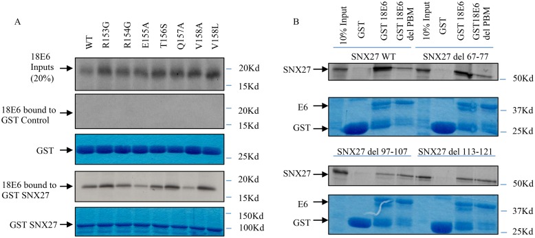
Fig 2 Interaction between HPV-18 E6 and SNX27 involves PBM-PDZ recognition. Panel A: The C-terminal HPV 18E6 mutants were translated and radio-labelled in vitro using the TNT Coupled Reticulocyte Lysate System (Promega). The translated proteins were then incubated with purified GST alone or GST-tagged SNX27 as indicated. The top panel shows input levels of the different HPV E6 mutants. The lower pairs of panels show the Coomassie stained gels of input GST and GST-SNX27 (lower panel in each pair) and the autoradiograms of the bound E6 (upper panel in each pair). Panel B: Wild type SNX27 and the various SNX27 deletion mutants was translated in vitro in rabbit reticulocyte lysate and radio labelled using the TNT Coupled Reticulocyte Lysate System (Promega). The translated proteins were then incubated with purified GST alone, GST-HPV18 E6 or the GST-HPV18 E6ΔPBM mutant as indicated. The arrows indicate the position of SNX27 on the autoradiograms, showing the levels of input and bound proteins. The Coomaasie Blue staining shows loading of the purified GST proteins.
Fig 2 Interaction between HPV-18 E6 and SNX27 involves PBM-PDZ recognition. Panel A: The C-terminal HPV 18E6 mutants were translated and radio-labelled in vitro using the TNT Coupled Reticulocyte Lysate System (Promega). The translated proteins were then incubated with purified GST alone or GST-tagged SNX27 as indicated. The top panel shows input levels of the different HPV E6 mutants. The lower pairs of panels show the Coomassie stained gels of input GST and GST-SNX27 (lower panel in each pair) and the autoradiograms of the bound E6 (upper panel in each pair). Panel B: Wild type SNX27 and the various SNX27 deletion mutants was translated in vitro in rabbit reticulocyte lysate and radio labelled using the TNT Coupled Reticulocyte Lysate System (Promega). The translated proteins were then incubated with purified GST alone, GST-HPV18 E6 or the GST-HPV18 E6ΔPBM mutant as indicated. The arrows indicate the position of SNX27 on the autoradiograms, showing the levels of input and bound proteins. The Coomaasie Blue staining shows loading of the purified GST proteins.CC0 · PubMed Central
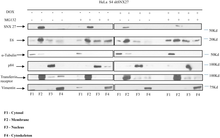
Fig 3 HPV-18 E6 resides primarily within the membrane fraction of HeLa cells. The stably transfected HeLa S4 shSNX27 cells were treated with Doxycycline (DOX) or DMSO for 72 hours. The cells were then treated with the proteosome inhibitor MG132 or control DMSO, as indicated, for a further 3 hours. The cells were then harvested and cytoplasmic, membrane, nuclear and cytoskeletal fractions were prepared using the ProteoExtract Cell Fractionation Kit (Calbiochem). The fractions were run on SDS polyacrylamide gels and HPV-18 E6 and SNX27 detected by western blotting. The arrows indicate the position of SNX27, HPV-18 E6, and the markers of the different fractions: α-Tubulin (cytoplasmic), Transferrin Receptor (membrane), p84 (nuclear) and Vimentin (cytoskeletal).
Fig 3 HPV-18 E6 resides primarily within the membrane fraction of HeLa cells. The stably transfected HeLa S4 shSNX27 cells were treated with Doxycycline (DOX) or DMSO for 72 hours. The cells were then treated with the proteosome inhibitor MG132 or control DMSO, as indicated, for a further 3 hours. The cells were then harvested and cytoplasmic, membrane, nuclear and cytoskeletal fractions were prepared using the ProteoExtract Cell Fractionation Kit (Calbiochem). The fractions were run on SDS polyacrylamide gels and HPV-18 E6 and SNX27 detected by western blotting. The arrows indicate the position of SNX27, HPV-18 E6, and the markers of the different fractions: α-Tubulin (cytoplasmic), Transferrin Receptor (membrane), p84 (nuclear) and Vimentin (cytoskeletal).CC0 · PubMed Central
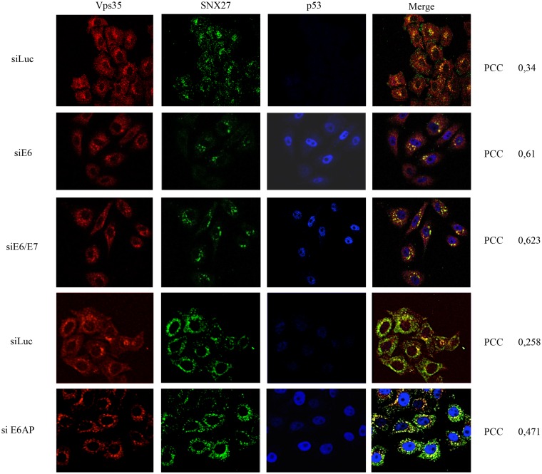
Fig 4 SNX27 accumulates in close proximity to retromer components in the absence of E6. HeLa cells were transfected with siRNA against Luciferase (siLuc), HPV-18 E6 (siE6), HPV-18 E6/E7 (siE6/E7) or E6AP (siE6AP) for 72 hours and the cells were then fixed using 4% Paraformaldehyde followed by permeabilization with PBS containing 0.1% Triton X-100. The cells were then stained for Vps35 (red) SNX27 (green) and p53 (blue) by indirect immunofluorescence. The images were acquired using the LSM 510 META Confocal Microscope (Carl Zeiss) and co-localisation between Vps35 and SNX27 was quantified by Velocity Software, with PCC values shown at the right hand side. Note the marked increase in co-localisation between SNX27 and Vps35 (given as Pearson’s Correlation Coefficient (PCC)) following ablation of E6, E6/E7 and E6AP.
Fig 4 SNX27 accumulates in close proximity to retromer components in the absence of E6. HeLa cells were transfected with siRNA against Luciferase (siLuc), HPV-18 E6 (siE6), HPV-18 E6/E7 (siE6/E7) or E6AP (siE6AP) for 72 hours and the cells were then fixed using 4% Paraformaldehyde followed by permeabilization with PBS containing 0.1% Triton X-100. The cells were then stained for Vps35 (red) SNX27 (green) and p53 (blue) by indirect immunofluorescence. The images were acquired using the LSM 510 META Confocal Microscope (Carl Zeiss) and co-localisation between Vps35 and SNX27 was quantified by Velocity Software, with PCC values shown at the right hand side. Note the marked increase in co-localisation between SNX27 and Vps35 (given as Pearson’s Correlation Coefficient (PCC)) following ablation of E6, E6/E7 and E6AP.CC0 · PubMed Central
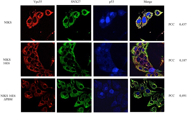
Fig 5 HPV-16 E6 modulation of SNX27 localisation is PBM-dependent. Control NIKS, NIKS expressing HPV-16 E6 and NIKS expressing the HPV-16 E6 ΔPBM mutant were fixed using 4% Paraformaldehyde followed by permeabilization with PBS containing 0.1% Triton X-100. The cells were then stained for Vps35 (red), SNX27 (green) and p53 (red) by indirect immunofluorescence. The images were acquired using the LSM 510 META Confocal Microscope (Carl Zeiss) and co-localisation between Vps35 and SNX27 quantified by Velocity Software, with PCC values shown at the right hand side. Note the marked increase in co-localisation between SNX27 and Vps35 in the control NIKS and NIKS containing E6ΔPBM.
Fig 5 HPV-16 E6 modulation of SNX27 localisation is PBM-dependent. Control NIKS, NIKS expressing HPV-16 E6 and NIKS expressing the HPV-16 E6 ΔPBM mutant were fixed using 4% Paraformaldehyde followed by permeabilization with PBS containing 0.1% Triton X-100. The cells were then stained for Vps35 (red), SNX27 (green) and p53 (red) by indirect immunofluorescence. The images were acquired using the LSM 510 META Confocal Microscope (Carl Zeiss) and co-localisation between Vps35 and SNX27 quantified by Velocity Software, with PCC values shown at the right hand side. Note the marked increase in co-localisation between SNX27 and Vps35 in the control NIKS and NIKS containing E6ΔPBM.CC0 · PubMed Central
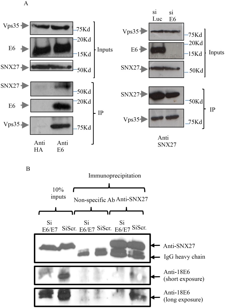
Fig 6 HPV-18 E6 exists in a complex with SNX27 and the retromer in HeLa cells. Panel A Left. HeLa cell extracts were incubated with anti-HPV-18 E6 monoclonal antibody or anti-HA monoclonal antibody as a negative control and the immune complexes were precipitated using Protein A Sepharose beads. The beads were washed and then loaded onto SDS polyacrylamide gels and co-immunoprecipitating proteins detected by SDS-PAGE followed by western blotting. The arrows indicate the position of SNX27, E6 and Vps35, with the upper three panels showing the levels of input proteins and the lower panels showing the immunoprecipitates. Panel A Right. HeLa cells were treated with siRNA against Luciferase as control or against HPV-18 E6 for 72 hours. Membrane fractions were prepared using the ProteoExtract Cell Fractionation Kit (Calbiochem) and then incubated with anti-SNX27 antibody and immune complexes were precipitated using Protein A Sepharose beads. The beads were washed, loaded onto SDS polyacrylamide gels and immunoprecipitating proteins detected by western blotting. The arrows indicate the position of SNX27 andVps35, with the upper two panels showing protein inputs and the two lower panels showing the immunoprecipitation. Panel B. HeLa cells were transfected with siE6/E7 or scrambled siRNA (siScr) and after 72hrs cells were harvested and immunoprecipitated using non-specific antibody or anti-SNX27. Complexes were then analysed by western blotting for HPV-18 E6 (anti-18 E6) and SNX27. Arrows indicate the positions of SNX27 and E6 together with the IgG heavy chain and a short and long exposure of the anti-18E6 western blot is provided for further clarity. Input proteins are also shown.
Fig 6 HPV-18 E6 exists in a complex with SNX27 and the retromer in HeLa cells. Panel A Left. HeLa cell extracts were incubated with anti-HPV-18 E6 monoclonal antibody or anti-HA monoclonal antibody as a negative control and the immune complexes were precipitated using Protein A Sepharose beads. The beads were washed and then loaded onto SDS polyacrylamide gels and co-immunoprecipitating proteins detected by SDS-PAGE followed by western blotting. The arrows indicate the position of SNX27, E6 and Vps35, with the upper three panels showing the levels of input proteins and the lower panels showing the immunoprecipitates. Panel A Right. HeLa cells were treated with siRNA against Luciferase as control or against HPV-18 E6 for 72 hours. Membrane fractions were prepared using the ProteoExtract Cell Fractionation Kit (Calbiochem) and then incubated with anti-SNX27 antibody and immune complexes were precipitated using Protein A Sepharose beads. The beads were washed, loaded onto SDS polyacrylamide gels and immunoprecipitating proteins detected by western blotting. The arrows indicate the position of SNX27 andVps35, with the upper two panels showing protein inputs and the two lower panels showing the immunoprecipitation. Panel B. HeLa cells were transfected with siE6/E7 or scrambled siRNA (siScr) and after 72hrs cells were harvested and immunoprecipitated using non-specific antibody or anti-SNX27. Complexes were then analysed by western blotting for HPV-18 E6 (anti-18 E6) and SNX27. Arrows indicate the positions of SNX27 and E6 together with the IgG heavy chain and a short and long exposure of the anti-18E6 western blot is provided for further clarity. Input proteins are also shown.CC0 · PubMed Central
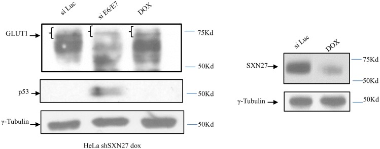
Fig 7 Loss of HPV-18 E6 alters GLUT-1 levels in HeLa cells. HeLa S4 cells were treated with siRNA against Luciferase, HPV-18 E6/E7 or DOX to induce SNX27 shRNA for 72 hours. The cells were then harvested and levels of GLUT-1 expression analysed by western blotting. Slower migrating forms of GLUT-1 are indicated by the brackets. Knock down of E6 and SNX27 were verified by western blotting for p53 and SNX27 respectively. The lower panel shows γ-tubulin as the loading control.
Fig 7 Loss of HPV-18 E6 alters GLUT-1 levels in HeLa cells. HeLa S4 cells were treated with siRNA against Luciferase, HPV-18 E6/E7 or DOX to induce SNX27 shRNA for 72 hours. The cells were then harvested and levels of GLUT-1 expression analysed by western blotting. Slower migrating forms of GLUT-1 are indicated by the brackets. Knock down of E6 and SNX27 were verified by western blotting for p53 and SNX27 respectively. The lower panel shows γ-tubulin as the loading control.CC0 · PubMed Central
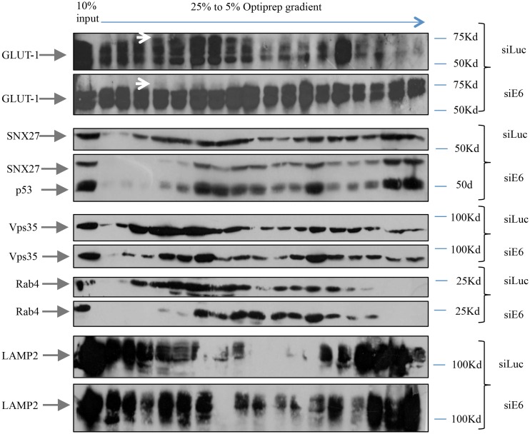
Fig 8 The loss of HPV-18 E6 in HeLa cells leads to an altered distribution of SNX27 and GLUT-1 cargo within endocytic compartments. HeLa cells were treated with siRNA against Luciferase or E6 for 72 hours and post nuclear extracts were loaded onto 5–25% OptiPrep gradients. The collected fractions were loaded on 12% SDS polyacrylamide gels followed by Western Blotting. The arrows indicate the position of GLUT-1, SNX27, p53 as a control for E6 knock down, but which was undetectable in the siLuc transfected cells, Rab4, Vps35 and LAMP2. The upper white arrows indicates the higher molecular weight forms of GLUT-1 that are absent following the loss of E6.
Fig 8 The loss of HPV-18 E6 in HeLa cells leads to an altered distribution of SNX27 and GLUT-1 cargo within endocytic compartments. HeLa cells were treated with siRNA against Luciferase or E6 for 72 hours and post nuclear extracts were loaded onto 5–25% OptiPrep gradients. The collected fractions were loaded on 12% SDS polyacrylamide gels followed by Western Blotting. The arrows indicate the position of GLUT-1, SNX27, p53 as a control for E6 knock down, but which was undetectable in the siLuc transfected cells, Rab4, Vps35 and LAMP2. The upper white arrows indicates the higher molecular weight forms of GLUT-1 that are absent following the loss of E6.CC0 · PubMed Central
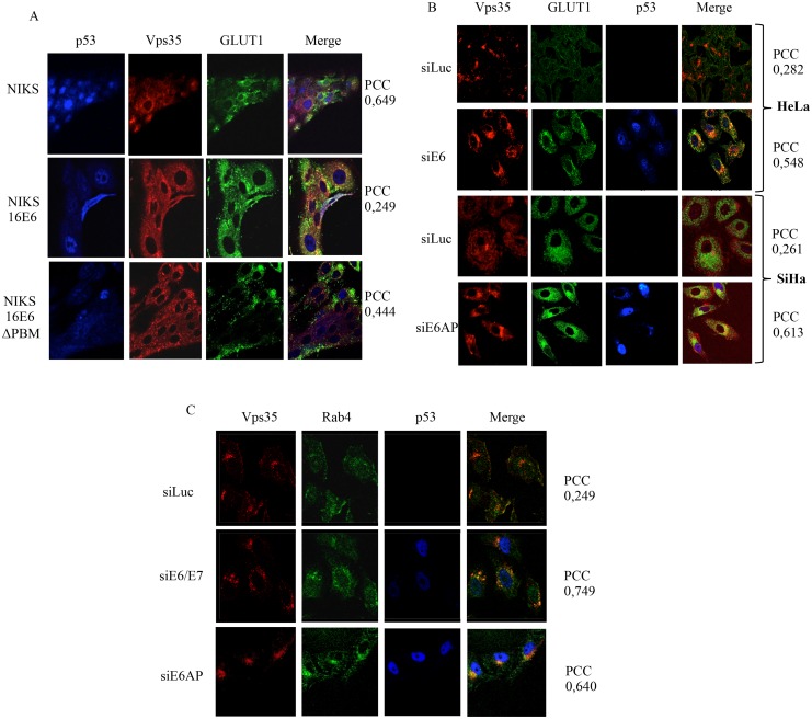
Fig 9 GLUT-1 distribution is modulated by E6 in a PBM-dependent manner. Panel A. Control NIKS, NIKS expressing HPV-16 E6 and NIKS expressing the HPV-16 E6ΔPBM mutant were fixed using 4% paraformaldehyde followed by permeabilization with PBS containing 0.1% Triton X-100. The cells were then stained for p53 (blue), Vps35 (red) and GLUT-1 (green) by indirect immunofluorescence. Co-localisation between Vps35 and GLUT-1 was measured using Velocity software and POC values are shown at the right. Panel B: HeLa and SiHa cells were transfected with siRNA against Luciferase, HPV-18 E6 or E6AP as indicated for 72 hours and the cells were then fixed using 4% paraformaldehyde followed by permeabilization with PBS containing 0.1% Triton X-100. The cells were then stained for Vps35 (red), GLUT-1 (green) and p53 (blue) by indirect immunofluorescence. The images were acquired using the LSM 510 META Confocal Microscope (Carl Zeiss). Co-localisation between GLUT1 and Vps35 was measured using Velocity software and PCC values are shown at the right hand side. Panel C: HeLa cells were transfected with siRNA against Luciferase or HPV-18 E6/E7 or E6AP for 72 hours and the cells were then fixed using 4% Paraformaldehyde followed by permeabilization with PBS containing 0.1% Triton X-100. The cells were then stained for Vps35 (red), Rab4 (green) and p53 (blue) by indirect immunofluorescence. The images were acquired using the LSM 510 META Confocal Microscope (Carl Zeiss). Co-localisation between GLUT-1 and Rab4 was measured using Velocity software and PCC values are shown at the right hand side.
Fig 9 GLUT-1 distribution is modulated by E6 in a PBM-dependent manner. Panel A. Control NIKS, NIKS expressing HPV-16 E6 and NIKS expressing the HPV-16 E6ΔPBM mutant were fixed using 4% paraformaldehyde followed by permeabilization with PBS containing 0.1% Triton X-100. The cells were then stained for p53 (blue), Vps35 (red) and GLUT-1 (green) by indirect immunofluorescence. Co-localisation between Vps35 and GLUT-1 was measured using Velocity software and POC values are shown at the right. Panel B: HeLa and SiHa cells were transfected with siRNA against Luciferase, HPV-18 E6 or E6AP as indicated for 72 hours and the cells were then fixed using 4% paraformaldehyde followed by permeabilization with PBS containing 0.1% Triton X-100. The cells were then stained for Vps35 (red), GLUT-1 (green) and p53 (blue) by indirect immunofluorescence. The images were acquired using the LSM 510 META Confocal Microscope (Carl Zeiss). Co-localisation between GLUT1 and Vps35 was measured using Velocity software and PCC values are shown at the right hand side. Panel C: HeLa cells were transfected with siRNA against Luciferase or HPV-18 E6/E7 or E6AP for 72 hours and the cells were then fixed using 4% Paraformaldehyde followed by permeabilization with PBS containing 0.1% Triton X-100. The cells were then stained for Vps35 (red), Rab4 (green) and p53 (blue) by indirect immunofluorescence. The images were acquired using the LSM 510 META Confocal Microscope (Carl Zeiss). Co-localisation between GLUT-1 and Rab4 was measured using Velocity software and PCC values are shown at the right hand side.CC0 · PubMed Central
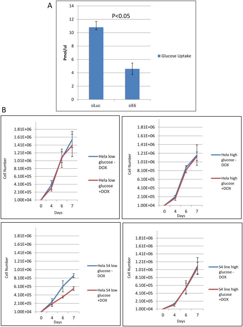
Fig 10 Modulation of glucose uptake and cell proliferation by E6 and SNX27. Panel A. HeLa cells were treated with siRNA against Luciferase or HPV-18 E6 for 72 hours and glucose uptake was analyzed using the glucose analog 2-deoxyglucose (2-DG) using the Colorimetric Glucose Uptake Assay Kit (Abcam) according to the manufacturer’s instructions. The graph shows the cumulative results of three independent experiments, with 2-DG uptake measured in pmol/μl. Error bars represent standard deviations with a p-value < 0.05. Panel B. Control HeLa cells and HeLa S4 DOX inducible shRNA SNX27 cells were plated in medium containing low or high glucose in the presence and absence of DOX as indicated. Cells were then counted over a period of 4 days and the numbers represent the means from three independent assays. Standard deviations are shown.
Fig 10 Modulation of glucose uptake and cell proliferation by E6 and SNX27. Panel A. HeLa cells were treated with siRNA against Luciferase or HPV-18 E6 for 72 hours and glucose uptake was analyzed using the glucose analog 2-deoxyglucose (2-DG) using the Colorimetric Glucose Uptake Assay Kit (Abcam) according to the manufacturer’s instructions. The graph shows the cumulative results of three independent experiments, with 2-DG uptake measured in pmol/μl. Error bars represent standard deviations with a p-value < 0.05. Panel B. Control HeLa cells and HeLa S4 DOX inducible shRNA SNX27 cells were plated in medium containing low or high glucose in the presence and absence of DOX as indicated. Cells were then counted over a period of 4 days and the numbers represent the means from three independent assays. Standard deviations are shown.CC0 · PubMed Central

Verification

Claims the paper makes

The claims and statistics of this paper haven’t been checked yet.

Run the check — free, no account needed. It reads the open-access text and checks the sources it cites.

A few free checks a day · papers already verified open instantly · results are public

Claims other papers make about this one

Each is checked against this paper’s own text: does it say what the citing paper says it does?

Self-check

Percentages, sums and reported differences re-computed from the paper’s own numbers.

The self-check needs the paper’s full text.

Citations

Cites

Show all 5 references

Research works printed but not linked: 46

  1. H Zur Hausen, 2002, Papillomaviruses and cancer: from basic studies to clinical application, Nat Rev Cancer, 2, 342, 10.1038/nrc798 · DOI
  2. D Smotkin, 1986, Transcription of human papillomavirus type 16 early genes in a cervical cancer and a cancer-derived cell line and identification of the E7 protein, Proc Natl Acad Sci U S A, 83, 4680, 10.1073/pnas.83.13.4680 · DOI
  3. L Banks, 1987, Identification of human papillomavirus type 18 E6 polypeptide in cells derived from human cervical carcinomas, J Gen Virol, 68, 1351, 10.1099/0022-1317-68-5-1351 · DOI
  4. M Yoshinouchi, 2003, In vitro and in vivo growth suppression of human papillomavirus 16-positive cervical cancer cells by E6 siRNA, Mol Ther, 8, 762, 10.1016/j.ymthe.2003.08.004 · DOI · Paper page
  5. DL Jones, 1997, Destabilization of the RB tumor suppressor protein and stabilization of p53 contribute to HPV type 16 E7-induced apoptosis, Virology, 239, 97, 10.1006/viro.1997.8851 · DOI · Paper page
  6. F Noya, 2001, p21cip1 degradation in differentiated keratinocytes is abrogated by costabilization with cyclin E induced by human papillomavirus E7, J Virol, 75, 6121, 10.1128/JVI.75.13.6121-6134.2001 · DOI · Paper page
  7. M Scheffner, 1990, The E6 oncoprotein encoded by human papillomavirus types 16 and 18 promotes the degradation of p53, Cell, 63, 1129, 10.1016/0092-8674(90)90409-8 · DOI · Paper page
  8. M Thomas, 1998, Inhibition of Bak-induced apoptosis by HPV-18 E6, Oncogene, 17, 2943, 10.1038/sj.onc.1202223 · DOI · Paper page
  9. T Kiyono, 1997, Binding of high-risk human papillomavirus E6 oncoproteins to the human homologue of the Drosophila discs large tumor suppressor protein, Proc Natl Acad Sci U S A, 94, 11612, 10.1073/pnas.94.21.11612 · DOI · Paper page
  10. RA Watson, 2003, Activity of the human papillomavirus E6 PDZ-binding motif correlates with an enhanced morphological transformation of immortalized human keratinocytes, J Cell Sci, 116, 4925, 10.1242/jcs.00809 · DOI
  11. ML Nguyen, 2003, The PDZ ligand domain of the human papillomavirus type 16 E6 protein is required for E6’s induction of epithelial hyperplasia in vivo, J Virol, 77, 6957, 10.1128/JVI.77.12.6957-6964.2003 · DOI
  12. C Lee, 2004, Role of the PDZ domain-binding motif of the oncoprotein E6 in the pathogenesis of Human Papillomavirus Type 31, J Virol, 78, 12366, 10.1128/JVI.78.22.12366-12377.2004 · DOI · Paper page
  13. C Delury, 2013, The role of protein kinase A regulation of the E6 PDZ-binding domain during the differentiation-dependent life cycle of human papillomavirus type 18, J Virol, 87, 9463, 10.1128/JVI.01234-13 · DOI
  14. F Facciuto, 2012, Differential expression of PDZ domain-containing proteins in human diseases—Challenging topics and novel issues, FEBS J, 279, 3538, 10.1111/j.1742-4658.2012.08699.x · DOI
  15. S Nakagawa, 2000, Human scribble (Vartul) is targeted for ubiquitin-mediated degradation by the high-risk papillomavirus E6 proteins and the E6AP ubiquitin-protein ligase, Mol Cell Biol, 20, 8244, 10.1128/MCB.20.21.8244-8253.2000 · DOI · Paper page
  16. S Roberts, 2012, The PDZ protein discs-large (DLG): The “Jekyll and Hyde” of the epithelial polarity proteins, FEBS J, 279, 3549, 10.1111/j.1742-4658.2012.08729.x · DOI
  17. C Kranjec, 2011, A systematic analysis of human papillomavirus (HPV) E6 PDZ substrates identifies MAGI-1 as a major target of HPV type 16 (HPV-16) and HPV-18 whose loss accompanies disruption of tight junctions, J Virol, 85, 1757, 10.1128/JVI.01756-10 · DOI
  18. E Belotti, 2013, The human PDZome: a gateway to PSD95-Disc large-zonula occludens (PDZ)-mediated functions, Mol Cell Proteomics, 12, 2587, 10.1074/mcp.O112.021022 · DOI
  19. A Suzuki, 2006, The PAR-aPKC system: lessons in polarity, J Cell Sci, 119, 979, 10.1242/jcs.02898 · DOI
  20. AI Ivanov, 2005, Endocytosis of the apical junctional complex: Mechanisms and possible roles in regulation of epithelial barriers, BioEssays, 27, 356, 10.1002/bies.20203 · DOI
  21. JM Shivas, 2010, Polarity and endocytosis: reciprocal regulation, Trends Cell Biol, 20, 445, 10.1016/j.tcb.2010.04.003 · DOI
  22. L Johannes, 2008, Tracing the retrograde route in protein trafficking, Cell, 135, 1175, 10.1016/j.cell.2008.12.009 · DOI · Paper page
  23. J Carlton, 2005, Sorting nexins—unifying trends and new perspectives, Traffic, 6, 75, 10.1111/j.1600-0854.2005.00260.x · DOI
  24. PJ Cullen, 2008, Endosomal sorting and signalling: an emerging role for sorting nexins, Nat Rev Mol Cell Biol, 9, 574, 10.1038/nrm2427 · DOI · Paper page
  25. JRT Van Weering, 2010, SNX-BAR proteins in phosphoinositide-mediated, tubular-based endosomal sorting, Semin Cell Dev Biol, 21, 371, 10.1016/j.semcdb.2009.11.009 · DOI
  26. CA Worby, 2002, Sorting out the cellular functions of sorting nexins, Nat Rev Mol Cell Biol, 3, 919, 10.1038/nrm974 · DOI · Paper page
  27. R Ghai, 2011, PX-FERM proteins: A link between endosomal trafficking and signaling?, Small GTPases, 2, 259, 10.4161/sgtp.2.5.17276 · DOI
  28. Y Kajii, 2003, A developmentally regulated and psychostimulant-inducible novel rat gene mrt1 encoding PDZ-PX proteins isolated in the neocortex, Mol Psychiatry, 8, 434, 10.1038/sj.mp.4001258 · DOI
  29. L Cai, 2011, Deficiency of sorting nexin 27 (SNX27) leads to growth retardation and elevated levels of N-methyl-D-aspartate receptor 2C (NR2C), Mol Cell Biol, 31, 1734, 10.1128/MCB.01044-10 · DOI
  30. BEL Lauffer, 2010, SNX27 mediates PDZ-directed sorting from endosomes to the plasma membrane, J Cell Biol, 190, 565, 10.1083/jcb.201004060 · DOI · Paper page
  31. L Joubert, 2004, New sorting nexin (SNX27) and NHERF specifically interact with the 5-HT4a receptor splice variant: roles in receptor targeting, J Cell Sci, 117, 5367, 10.1242/jcs.01379 · DOI
  32. JL Valdes, 2011, Sorting nexin 27 protein regulates trafficking of a p21-activated kinase (PAK) interacting exchange factor (β-Pix)-G protein-coupled receptor kinase interacting protein (GIT) complex via a PDZ domain interaction, J Biol Chem, 286, 39403, 10.1074/jbc.M111.260802 · DOI
  33. P Temkin, 2011, SNX27 mediates retromer tubule entry and endosome-to-plasma membrane trafficking of signalling receptors, Nat Cell Biol, 13, 715, 10.1038/ncb2252 · DOI · Paper page
  34. F Steinberg, 2013, A global analysis of SNX27-retromer assembly and cargo specificity reveals a function in glucose and metal ion transport, Nat Cell Biol, 15, 461, 10.1038/ncb2721 · DOI · Paper page
  35. BA Glaunsinger, 2000, Interactions of the PDZ-protein MAGI-1 with adenovirus E4-ORF1 and high-risk papillomavirus E6 oncoproteins, Oncogene, 19, 5270, 10.1038/sj.onc.1203906 · DOI · Paper page
  36. M Thomas, 2008, Analysis of specificity determinants in the interactions of different HPV E6 proteins with their PDZ domain-containing substrates, Virology, 376, 371, 10.1016/j.virol.2008.03.021 · DOI
  37. Y Zhang, 2007, Structures of a human papillomavirus (HPV) E6 polypeptide bound to MAGUK proteins: mechanisms of targeting tumor suppressors by a high-risk HPV oncoprotein, J Virol, 81, 3618, 10.1128/JVI.02044-06 · DOI
  38. M Gallon, 2014, A unique PDZ domain and arrestin-like fold interaction reveals mechanistic details of endocytic recycling by SNX27-retromer, Proc Natl Acad Sci U S A
  39. V Tomaic, 2009, The stability of the human papillomavirus E6 oncoprotein is E6AP dependent, Virology, 393, 7, 10.1016/j.virol.2009.07.029 · DOI · Paper page
  40. T Asano, 1991, The role of N-glycosylation of GLUT1 for glucose transport activity, J. Biol Chem, 266, 24632, 10.1016/S0021-9258(18)54276-9 · DOI
  41. MW Chien, 2015, Glucosamine modulates T cell differentiation through downregulating N-linked glycosylation of CD25, J. Biol. Chem, 290, 29329, 10.1074/jbc.M115.674671 · DOI
  42. DR Sheff, 1999, The receptors recycling pathway contains two distinct populations of early endosomes with different sorting functions, J Cell Biol, 145, 123, 10.1083/jcb.145.1.123 · DOI · Paper page
  43. R Vincentelli, 2015, Quantifying domain-ligand affinities and specificities by high-throughput holdup assay, Nat Methods
  44. B Allen-Hoffmann, 2000, Normal growth and differentiation in a spontaneously immortalised near-diploid human keratinocyte cell line, NIKS, J Inv Dermatol, 114, 444, 10.1046/j.1523-1747.2000.00869.x · DOI
  45. I Nicolaides, 2011, Stabilization of HPV E6 protein by PDZ proteins, and potential implications for genome maintenance, Virology, 414, 137, 10.1016/j.virol.2011.03.017 · DOI · Paper page
  46. SS Boon, 2012, High-Risk Human Papillomavirus E6 Oncoproteins interact with 14-3-3 in a PDZ Binding Motif-Dependent manner, J Virol, 87, 1586, 10.1128/JVI.02074-12 · DOI

Cited by

Metadata

Authors
Ganti, K., Massimi, P., Manzo-Merino, J., Tomaić, V., Pim, D., Playford, M. P., Lizano, M., Roberts, S., Kranjec, C., Doorbar, J., & Banks, L.
Venue
PLOS Pathogens
Published
2016-09-20
Volume
12
Issue
9
Pages
e1005854
ISSN
1553-7374
Cited by
50
References
5
Language
en
Funders
  • Associazione Italiana per la Ricerca sul Cancro (0923450/Z10/Z)
  • Wellcome Trust (09345/B/10/Z)

About these checks

Registry, retraction, Cites and statistics results are computed, not judged, with one exception: a printed reference that no index lists is read once by a model before it counts as a citation error. Claims are extracted by a model and kept only when their quote appears verbatim in the text; each cited source is then checked, with a model’s help, for whether it says what the claim relies on. Those verdicts can be wrong — read the claim’s quote and the cited source before relying on one.

Statistics and arithmetic are recomputed, not judged.

Confidence bands are shown only for fields calibrated on our annotated test set. None is calibrated yet, so no band is shown.

What counts as a claim ›How we measure confidence ›How we check this ›

Are you an author, or do you see a problem with this page? Request a review