

The DOI matches its Crossref record; 9 of the 9 references it cites were found in a registry or an index.
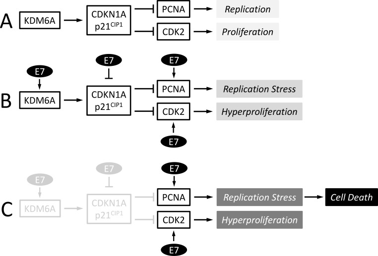
KDM6A addiction of cervical carcinoma cell lines is triggered by E7 and mediated by p21CIP1 suppression of replication stress
Overview
The claims and statistics of this paper haven’t been checked yet.
Run the check — free, no account needed. It reads the open-access text and checks the sources it cites.
Integrity signals
Registry
Cites
Statistics
Figures
Verification
Claims the paper makes
The claims and statistics of this paper haven’t been checked yet.
Run the check — free, no account needed. It reads the open-access text and checks the sources it cites.
Claims other papers make about this one
Each is checked against this paper’s own text: does it say what the citing paper says it does?
In the citing paper
“increased KDM6A in response to high-risk HPV E7 expression was shown to cause de-repression of the cell cycle and DNA replication inhibitor p21CIP1 and this activity was shown to be required in high-risk E7 expressing cells for p21CIP1’s ability to inhibit DNA replication through PCNA binding [335]”
From The Not-So-Good, the Bad and the Ugly: HPV E5, E6 and E7 Oncoproteins in the Orchestration of Carcinogenesis (2021) · cites this paper as [335]
Self-check
Percentages, sums and reported differences re-computed from the paper’s own numbers.
The self-check needs the paper’s full text.
Citations
Cites
PCNA, the Maestro of the Replication Fork
Moldovan, G.-L., Pfander, B., & Jentsch, S. (2007) · Cell · doi:10.1016/j.cell.2007.05.003 · Cited by 1,796
Senescence in premalignant tumours
Collado, M., Gil, J., Efeyan, A., Guerra, C., Schuhmacher, A. J., Barradas, M., Benguría, A., Zaballos, A., Flores, J. M., Barbacid, M., et al. (2005) · Nature · doi:10.1038/436642a · Cited by 1,482
Myc signaling via the ARF tumor suppressor regulates p53-dependent apoptosis and immortalization
Zindy, F., Eischen, C. M., Randle, D. H., Kamijo, T., Cleveland, J. L., Sherr, C. J., & Roussel, M. F. (1998) · Genes & Development · doi:10.1101/gad.12.15.2424 · Cited by 1,245
De Santa, F., Totaro, M. G., Prosperini, E., Notarbartolo, S., Testa, G., & Natoli, G. (2007) · Cell · doi:10.1016/j.cell.2007.08.019 · Cited by 965
Huibregtse, J. M., Scheffner, M., & Howley, P. M. (1991) · The EMBO Journal · doi:10.1002/j.1460-2075.1991.tb04990.x · Cited by 879
Epigenetic reprogramming in cancer
Suvà, M. L., Riggi, N., & Bernstein, B. E. (2013) · Science · doi:10.1126/science.1230184 · Cited by 706
Cell-cycle inhibition by independent CDK and PCNA binding domains in p21Cip1
Luo, Y., Hurwitz, J., & Massagué, J. (1995) · Nature · doi:10.1038/375159a0 · Cited by 549
Separate domains of p21 involved in the inhibition of Cdk kinase and PCNA
Chen, J., Jackson, P. K., Kirschner, M. W., & Dutta, A. (1995) · Nature · doi:10.1038/374386a0 · Cited by 548 · cited 2×
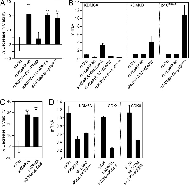
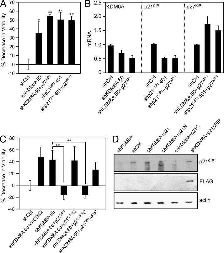
“In contrast, ectopic expression of the carboxyl terminal p21 domain, which contains the PCNA binding site and is sufficient to inhibit PCNA [42], efficiently rescued the decrease in viability upon KDM6A depletion.”
BackgroundHistone demethylase JMJD3 contributes to epigenetic control of INK4a/ARF by oncogenic RAS
Barradas, M., Anderton, E., Acosta, J. C., Li, S., Banito, A., Rodriguez-Niedenführ, M., Maertens, G., Banck, M., Zhou, M.-M., Walsh, M. J., et al. (2009) · Genes & Development · doi:10.1101/gad.511109 · Cited by 352
Research works printed but not linked: 86
- J Ferlay, 2010, Estimates of worldwide burden of cancer in 2008: GLOBOCAN 2008, Int J Cancer, 127, 2893, 10.1002/ijc.25516 · DOI
- DM Parkin, 2006, The global health burden of infection-associated cancers in the year 2002, Int J Cancer, 118, 3030, 10.1002/ijc.21731 · DOI
- M Schiffman, 2007, Human papillomavirus and cervical cancer, Lancet, 370, 890, 10.1016/S0140-6736(07)61416-0 · DOI
- ML Gillison, 2000, Evidence for a causal association between human papillomavirus and a subset of head and neck cancers, J Natl Cancer Inst, 92, 709, 10.1093/jnci/92.9.709 · DOI
- IH Frazer, 2004, Prevention of cervical cancer through papillomavirus vaccination, Nat Rev Immunol, 4, 46, 10.1038/nri1260 · DOI
- N Dyson, 1989, The human papillomavirus-16 E7 oncoprotein is able to bind to the retinoblastoma gene product, Science, 243, 934, 10.1126/science.2537532 · DOI · Paper page
- BA Werness, 1990, Association of human papillomavirus types 16 and 18 E6 proteins with p53, Science, 248, 76, 10.1126/science.2157286 · DOI · Paper page
- A Brehm, 1999, The E7 oncoprotein associates with Mi2 and histone deacetylase activity to promote cell growth, EMBO J, 18, 2449, 10.1093/emboj/18.9.2449 · DOI · Paper page
- MS Longworth, 2004, The binding of histone deacetylases and the integrity of zinc finger-like motifs of the E7 protein are essential for the life cycle of human papillomavirus type 31, J Virol, 78, 3533, 10.1128/JVI.78.7.3533-3541.2004 · DOI
- N Avvakumov, 2003, Interaction of the HPV E7 proteins with the pCAF acetyltransferase, Oncogene, 22, 3833, 10.1038/sj.onc.1206562 · DOI · Paper page
- A Bernat, 2003, Interaction between the HPV E7 oncoprotein and the transcriptional coactivator p300, Oncogene, 22, 7871, 10.1038/sj.onc.1206896 · DOI
- A Baldwin, 2006, Human papillomavirus E7 oncoprotein dysregulates steroid receptor coactivator 1 localization and function, J Virol, 80, 6669, 10.1128/JVI.02497-05 · DOI
- SM Huang, 2002, Down regulation of the interleukin-8 promoter by human papillomavirus type 16 E6 and E7 through effects on CREB binding protein/p300 and P/CAF, J Virol, 76, 8710, 10.1128/JVI.76.17.8710-8721.2002 · DOI · Paper page
- ME McLaughlin-Drubin, 2013, Tumor suppressor p16INK4A is necessary for survival of cervical carcinoma cell lines, Proc Natl Acad Sci U S A, 110, 16175, 10.1073/pnas.1310432110 · DOI · Paper page
- ME McLaughlin-Drubin, 2011, Human papillomavirus E7 oncoprotein induces KDM6A and KDM6B histone demethylase expression and causes epigenetic reprogramming, Proc Natl Acad Sci U S A, 108, 2130, 10.1073/pnas.1009933108 · DOI · Paper page
- ME McLaughlin-Drubin, 2008, Human papillomavirus type 16 E7 oncoprotein associates with E2F6, Journal of virology, 82, 8695, 10.1128/JVI.00579-08 · DOI · Paper page
- PL Hyland, 2011, Evidence for alteration of EZH2, BMI1, and KDM6A and epigenetic reprogramming in human papillomavirus type 16 E6/E7-expressing keratinocytes, J Virol, 85, 10999, 10.1128/JVI.00160-11 · DOI
- YB Schwartz, 2007, Polycomb silencing mechanisms and the management of genomic programmes, Nat Rev Genet, 8, 9, 10.1038/nrg1981 · DOI · Paper page
- B Tolhuis, 2006, Genome-wide profiling of PRC1 and PRC2 Polycomb chromatin binding in Drosophila melanogaster, Nat Genet, 38, 694, 10.1038/ng1792 · DOI
- K Plath, 2003, Role of histone H3 lysine 27 methylation in X inactivation, Science, 300, 131, 10.1126/science.1084274 · DOI
- B Schuettengruber, 2009, Recruitment of polycomb group complexes and their role in the dynamic regulation of cell fate choice, Development, 136, 3531, 10.1242/dev.033902 · DOI
- JA Simon, 2008, Roles of the EZH2 histone methyltransferase in cancer epigenetics, Mutation research, 647, 21, 10.1016/j.mrfmmm.2008.07.010 · DOI
- K Agger, 2007, UTX and JMJD3 are histone H3K27 demethylases involved in HOX gene regulation and development, Nature, 449, 731, 10.1038/nature06145 · DOI
- K Jepsen, 2007, SMRT-mediated repression of an H3K27 demethylase in progression from neural stem cell to neuron, Nature, 450, 415, 10.1038/nature06270 · DOI
- F Lan, 2007, A histone H3 lysine 27 demethylase regulates animal posterior development, Nature, 449, 689, 10.1038/nature06192 · DOI
- MG Lee, 2007, Demethylation of H3K27 regulates polycomb recruitment and H2A ubiquitination, Science, 318, 447, 10.1126/science.1149042 · DOI
- DF Jarrard, 2002, Alterations in the p16/pRb cell cycle checkpoint occur commonly in primary and metastatic human prostate cancer, Cancer Lett, 185, 191, 10.1016/S0304-3835(02)00282-3 · DOI
- G van Haaften, 2009, Somatic mutations of the histone H3K27 demethylase gene UTX in human cancer, Nat Genet, 41, 521, 10.1038/ng.349 · DOI · Paper page
- ML Nickerson, 2014, Concurrent alterations in TERT, KDM6A, and the BRCA pathway in bladder cancer, Clin Cancer Res, 20, 4935, 10.1158/1078-0432.CCR-14-0330 · DOI
- C Curtis, 2012, The genomic and transcriptomic architecture of 2,000 breast tumours reveals novel subgroups, Nature, 486, 346, 10.1038/nature10983 · DOI · Paper page
- JH Kim, 2014, UTX and MLL4 coordinately regulate transcriptional programs for cell proliferation and invasiveness in breast cancer cells, Cancer Res, 74, 1705, 10.1158/0008-5472.CAN-13-1896 · DOI
- SF Gameiro, 2017, Human papillomavirus dysregulates the cellular apparatus controlling the methylation status of H3K27 in different human cancers to consistently alter gene expression regardless of tissue of origin, Oncotarget
- K Agger, 2009, The H3K27me3 demethylase JMJD3 contributes to the activation of the INK4A-ARF locus in response to oncogene- and stress-induced senescence, Genes Dev, 23, 1171, 10.1101/gad.510809 · DOI · Paper page
- IB Weinstein, 2006, Mechanisms of disease: Oncogene addiction—a rationale for molecular targeting in cancer therapy, Nat Clin Pract Oncol, 3, 448, 10.1038/ncponc0558 · DOI
- DH Kim, 2013, Histone H3K27 trimethylation inhibits H3 binding and function of SET1-like H3K4 methyltransferase complexes, Mol Cell Biol, 33, 4936, 10.1128/MCB.00601-13 · DOI
- DL Jones, 1997, The human papillomavirus E7 oncoprotein can uncouple cellular differentiation and proliferation in human keratinocytes by abrogating p21Cip1-mediated inhibition of cdk2, Genes & Development, 11, 2101, 10.1101/gad.11.16.2101 · DOI · Paper page
- JO Funk, 1997, Inhibition of CDK activity and PCNA-dependent DNA replication by p21 is blocked by interaction with the HPV-16 E7 oncoprotein, Genes & Development, 11, 2090, 10.1101/gad.11.16.2090 · DOI · Paper page
- M Nakanishi, 1995, Identification of the active region of the DNA synthesis inhibitory gene p21Sdi1/CIP1/WAF1, EMBO Journal, 14, 555, 10.1002/j.1460-2075.1995.tb07031.x · DOI
- E Warbrick, 1995, A small peptide inhibitor of DNA replication defines the site of interaction between the cyclin-dependent kinase inhibitor p21WAF1 and proliferating cell nuclear antigen, Curr Biol, 5, 275, 10.1016/S0960-9822(95)00058-3 · DOI
- JM Gulbis, 1996, Structure of the C-terminal region of p21(WAF1/CIP1) complexed with human PCNA, Cell, 87, 297, 10.1016/S0092-8674(00)81347-1 · DOI
- MK Shin, 2009, Human papillomavirus E7 oncoprotein overrides the tumor suppressor activity of p21Cip1 in cervical carcinogenesis, Cancer Res, 69, 5656, 10.1158/0008-5472.CAN-08-3711 · DOI
- S van den Heuvel, 1993, Distinct roles for cyclin-dependent kinases in cell cycle control, Science, 262, 2050, 10.1126/science.8266103 · DOI
- T Abbas, 2008, PCNA-dependent regulation of p21 ubiquitylation and degradation via the CRL4Cdt2 ubiquitin ligase complex, Genes Dev, 22, 2496, 10.1101/gad.1676108 · DOI
- KN Choe, 2017, Forging Ahead through Darkness: PCNA, Still the Principal Conductor at the Replication Fork, Mol Cell, 65, 380, 10.1016/j.molcel.2016.12.020 · DOI · Paper page
- B Stillman, 1996, Cell cycle control of DNA replication, Science, 274, 1659, 10.1126/science.274.5293.1659 · DOI · Paper page
- A Dutta, 1997, Initiation of DNA replication in eukaryotic cells, Annu Rev Cell Dev Biol, 13, 293, 10.1146/annurev.cellbio.13.1.293 · DOI
- S Hong, 2015, STAT-5 Regulates Transcription of the Topoisomerase IIbeta-Binding Protein 1 (TopBP1) Gene To Activate the ATR Pathway and Promote Human Papillomavirus Replication, MBio, 6, e02006, 10.1128/mBio.02006-15 · DOI
- CA Moody, 2009, Human papillomaviruses activate the ATM DNA damage pathway for viral genome amplification upon differentiation, PLoS Pathog, 5, e1000605, 10.1371/journal.ppat.1000605 · DOI · Paper page
- T Reinson, 2013, Engagement of the ATR-dependent DNA damage response at the human papillomavirus 18 replication centers during the initial amplification, J Virol, 87, 951, 10.1128/JVI.01943-12 · DOI
- N Sakakibara, 2011, The papillomavirus E1 helicase activates a cellular DNA damage response in viral replication foci, J Virol, 85, 8981, 10.1128/JVI.00541-11 · DOI
- N Spardy, 2008, HPV-16 E7 reveals a link between DNA replication stress, fanconi anemia D2 protein, and alternative lengthening of telomere-associated promyelocytic leukemia bodies, Cancer Res, 68, 9954, 10.1158/0008-5472.CAN-08-0224 · DOI
- RA Burrell, 2013, Replication stress links structural and numerical cancer chromosomal instability, Nature, 494, 492, 10.1038/nature11935 · DOI
- AC Bester, 2011, Nucleotide deficiency promotes genomic instability in early stages of cancer development, Cell, 145, 435, 10.1016/j.cell.2011.03.044 · DOI · Paper page
- L Hu, 2013, Replication stress and mitotic dysfunction in cells expressing simian virus 40 large T antigen, J Virol, 87, 13179, 10.1128/JVI.02224-13 · DOI
- C Lukas, 2011, 53BP1 nuclear bodies form around DNA lesions generated by mitotic transmission of chromosomes under replication stress, Nat Cell Biol, 13, 243, 10.1038/ncb2201 · DOI
- AH Wyllie, 1987, Rodent fibroblast tumours expressing human myc and ras genes: growth, metastasis and endogenous oncogene expression, Br J Cancer, 56, 251, 10.1038/bjc.1987.186 · DOI
- DS Askew, 1991, Constitutive c-myc expression in an IL-3-dependent myeloid cell line suppresses cell cycle arrest and accelerates apoptosis, Oncogene, 6, 1915
- GI Evan, 1992, Induction of apoptosis in fibroblasts by c-myc protein, Cell, 69, 119, 10.1016/0092-8674(92)90123-T · DOI
- M Scheffner, 1990, The E6 oncoprotein encoded by human papillomavirus types 16 and 18 promotes the degradation of p53, Cell, 63, 1129, 10.1016/0092-8674(90)90409-8 · DOI · Paper page
- N Dyson, 1992, Homologous sequences in adenovirus E1A and human papillomavirus E7 proteins mediate interaction with the same set of cellular proteins, Journal of Virology, 66, 6893, 10.1128/JVI.66.12.6893-6902.1992 · DOI · Paper page
- K Münger, 1989, Complex formation of human papillomavirus E7 proteins with the retinoblastoma tumor suppressor gene product, The EMBO Journal, 8, 4099, 10.1002/j.1460-2075.1989.tb08594.x · DOI · Paper page
- SN Boyer, 1996, E7 protein of human papilloma virus-16 induces degradation of retinoblastoma protein through the ubiquitin-proteasome pathway, Cancer Res, 56, 4620
- DL Jones, 1997, Destabilization of the RB tumor suppressor and stabilization of p53 contribute to HPV type 16 E7-induced apoptosis, Virology, 239, 97, 10.1006/viro.1997.8851 · DOI · Paper page
- JK Wang, 2010, The histone demethylase UTX enables RB-dependent cell fate control, Genes Dev, 24, 327, 10.1101/gad.1882610 · DOI
- M Terashima, 2010, The tumor suppressor Rb and its related Rbl2 genes are regulated by Utx histone demethylase, Biochem Biophys Res Commun, 399, 238, 10.1016/j.bbrc.2010.07.061 · DOI
- L Rocha-Viegas, 2014, Role of UTX in retinoic acid receptor-mediated gene regulation in leukemia, Mol Cell Biol, 34, 3765, 10.1128/MCB.00839-14 · DOI
- S Chen, 2012, The histone H3 Lys 27 demethylase JMJD3 regulates gene expression by impacting transcriptional elongation, Genes Dev, 26, 1364, 10.1101/gad.186056.111 · DOI
- C Estaras, 2012, Genome-wide analysis reveals that Smad3 and JMJD3 HDM co-activate the neural developmental program, Development, 139, 2681, 10.1242/dev.078345 · DOI
- RM Alani, 1998, Alterations in cyclin-dependent kinase 2 function during differentiation of primary human keratinocytes, Molecular carcinogenesis, 23, 226, 10.1002/(SICI)1098-2744(199812)23:4<226::AID-MC5>3.0.CO;2-N · DOI
- C Missero, 1996, The absence of p21Cip1/WAF1 alters keratinocyte growth and differentiation and promotes ras-tumor progression, Genes Dev, 10, 3065, 10.1101/gad.10.23.3065 · DOI
- G Maga, 2003, Proliferating cell nuclear antigen (PCNA): a dancer with many partners, J Cell Sci, 116, 3051, 10.1242/jcs.00653 · DOI
- F Noya, 2001, p21cip1 Degradation in differentiated keratinocytes is abrogated by costabilization with cyclin E induced by human papillomavirus E7, J Virol, 75, 6121, 10.1128/JVI.75.13.6121-6134.2001 · DOI · Paper page
- DL Jones, 1999, Expression of the HPV E7 oncoprotein mimics but does not evoke a p53- dependent cellular DNA damage response pathway, Virology, 258, 406, 10.1006/viro.1999.9733 · DOI · Paper page
- Y Jian, 1998, Post-transcriptional induction of p21cip1 protein by human papillomavirus E7 inhibits unscheduled DNA synthesis reactivated in differentiated keratinocytes, Oncogene, 17, 2027, 10.1038/sj.onc.1202142 · DOI · Paper page
- SG Hwang, 2002, Human papillomavirus type 16 E7 binds to E2F1 and activates E2F1-driven transcription in a retinoblastoma protein-independent manner, J Biol Chem, 277, 2923, 10.1074/jbc.M109113200 · DOI · Paper page
- C Vaziri, 2003, A p53-dependent checkpoint pathway prevents rereplication, Mol Cell, 11, 997, 10.1016/S1097-2765(03)00099-6 · DOI
- E Liu, 2007, The ATR-mediated S phase checkpoint prevents rereplication in mammalian cells when licensing control is disrupted, J Cell Biol, 179, 643, 10.1083/jcb.200704138 · DOI
- S Duensing, 2002, The human papillomavirus type 16 E6 and E7 oncoproteins independently induce numerical and structural chromosome instability, Cancer research, 62, 7075
- N Korzeniewski, 2011, Genomic instability and cancer: lessons learned from human papillomaviruses, Cancer Lett, 305, 113, 10.1016/j.canlet.2010.10.013 · DOI
- DC Anacker, 2016, HPV31 utilizes the ATR-Chk1 pathway to maintain elevated RRM2 levels and a replication-competent environment in differentiating Keratinocytes, Virology, 499, 383, 10.1016/j.virol.2016.09.028 · DOI
- DC Anacker, 2014, Productive replication of human papillomavirus 31 requires DNA repair factor Nbs1, J Virol, 88, 8528, 10.1128/JVI.00517-14 · DOI
- TG Edwards, 2013, Human papillomavirus episome stability is reduced by aphidicolin and controlled by DNA damage response pathways, J Virol, 87, 3979, 10.1128/JVI.03473-12 · DOI
- CL Halbert, 1991, The E7 gene of human papillomavirus type 16 is sufficient for immortalization of human epithelial cells, Journal of Virology, 65, 473, 10.1128/JVI.65.1.473-478.1991 · DOI · Paper page
- PA Longo, 2013, Transient mammalian cell transfection with polyethylenimine (PEI), Methods Enzymol, 529, 227, 10.1016/B978-0-12-418687-3.00018-5 · DOI
- X Sun, 2005, Frequent somatic mutations of the transcription factor ATBF1 in human prostate cancer, Nat Genet, 37, 407, 10.1038/ng1528 · DOI
- RA Schwab, 2011, Visualization of DNA replication in the vertebrate model system DT40 using the DNA fiber technique, J Vis Exp, e3255
Cited by
Basukala, O., & Banks, L. (2021) · Viruses · doi:10.3390/v13101892 · Cited by 137
KDM6A regulates cell plasticity and pancreatic cancer progression by noncanonical activin pathway
Yi, Z., Wei, S., Jin, L., Jeyarajan, S., Yang, J., Gu, Y., Kim, H. S., Schechter, S., Lu, S., Paulsen, M. T., et al. (2022) · Cellular and Molecular Gastroenterology and Hepatology · doi:10.1016/j.jcmgh.2021.09.014 · Cited by 41
Metadata
- Authors
- Soto, D. R., Barton, C., Munger, K., & McLaughlin-Drubin, M. E.
- Venue
- PLOS Pathogens
- Publisher
- Public Library of Science (PLoS)
- Published
- 2017-10-02
- Volume
- 13
- Issue
- 10
- Pages
- e1006661
- ISSN
- 1553-7374
- PMC
- PMC5638616
- Cited by
- 39
- References
- 9
- Language
- en
- Update notices
- new_version (2017-10-12)
- Funders
- American Cancer Society (126540-RSG-14-203-01-MPC)
- National Cancer Institute (CA066980)
About these checks
Registry, retraction, Cites and statistics results are computed, not judged, with one exception: a printed reference that no index lists is read once by a model before it counts as a citation error. Claims are extracted by a model and kept only when their quote appears verbatim in the text; each cited source is then checked, with a model’s help, for whether it says what the claim relies on. Those verdicts can be wrong — read the claim’s quote and the cited source before relying on one.
Statistics and arithmetic are recomputed, not judged.
Confidence bands are shown only for fields calibrated on our annotated test set. None is calibrated yet, so no band is shown.
What counts as a claim ›How we measure confidence ›How we check this ›
Are you an author, or do you see a problem with this page? Request a review
