

The DOI matches its Crossref record; 11 of the 11 references it cites were found in a registry or an index.
Dimeric Amyloid β Protein Rapidly Accumulates in Lipid Rafts followed by Apolipoprotein E and Phosphorylated Tau Accumulation in the Tg2576 Mouse Model of Alzheimer's Disease
Abstract
To investigate lipid rafts as a site where amyloid β protein (Aβ) oligomers might accumulate and cause toxicity in Alzheimer's disease (AD), we analyzed Aβ in the Tg2576 transgenic mouse model of AD. Aβ was highly concentrated in lipid rafts, which comprise a small fraction of brain volume but contain 27% of brain Aβ42 and 24% of Aβ40 in young mice. In the Tg2576 model, memory impairment begins at 6 months before amyloid plaques are visible. Here we show that Aβ dimers appear in lipid rafts at 6 months and that raft Aβ, which is primarily dimeric, rapidly accumulates reaching levels >500× those in young mice by 24–28 months. A similar large accumulation of dimeric Aβ was observed in lipid rafts from AD brain. In contrast to extracellular amyloid fibrils, which are SDS-insoluble, virtually all Aβ in lipid rafts is SDS soluble. Coupled with recent studies showing that synthetic and naturally occurring Aβ oligomers can inhibit hippocampal long-term potentiation, the in vivo age-dependent accumulation of SDS-soluble Aβ dimers in lipid rafts at the time when memory impairment begins in Tg2576 mice provides strong evidence linking Aβ oligomers to memory impairment. After dimeric Aβ began to accumulate in lipid rafts of the Tg2576 brain, apolipoprotein E (ApoE) and then phosphorylated tau accumulated. A similar increase in ApoE and a large increase in phosphorylated tau was observed in lipid rafts from AD brain. These findings suggest that lipid rafts may be an important site for interaction between dimeric Aβ, ApoE, and tau.
Overview
The claims and statistics of this paper haven’t been checked yet.
Run the check — free, no account needed. It reads the open-access text and checks the sources it cites.
Integrity signals
Registry
Cites
Statistics
Figures
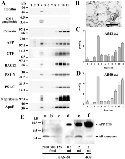
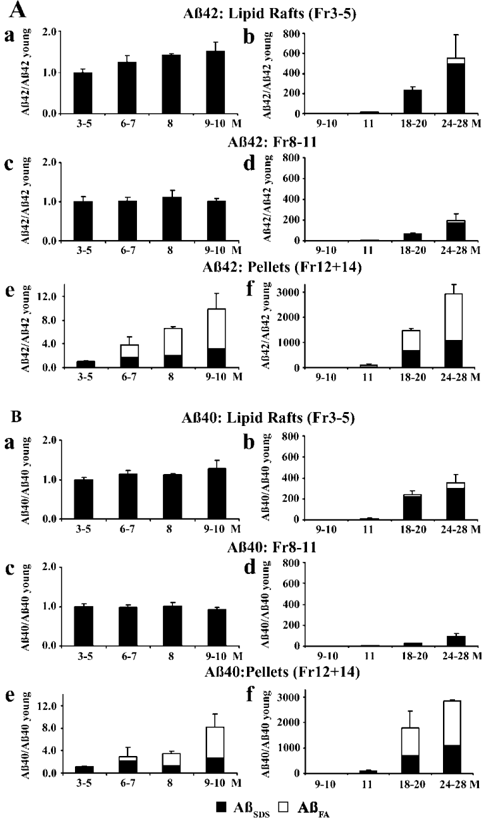
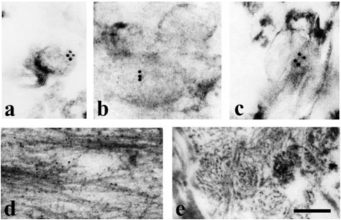
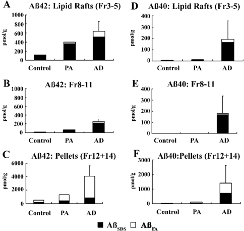
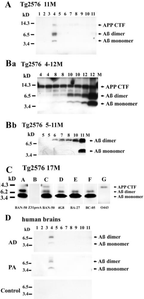
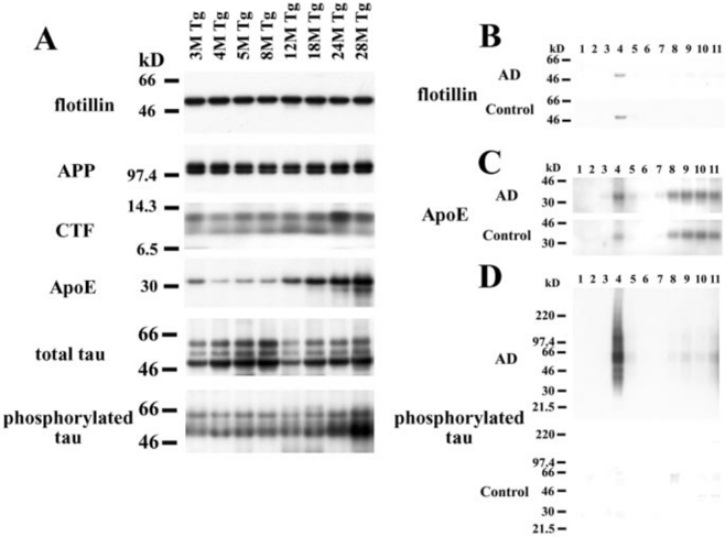
Verification
Claims the paper makes
The claims and statistics of this paper haven’t been checked yet.
Run the check — free, no account needed. It reads the open-access text and checks the sources it cites.
Claims other papers make about this one
Each is checked against this paper’s own text: does it say what the citing paper says it does?
In the citing paper
“This is in good accordance with a recent article showing that ApoE, a LRP ligand, is co-localized with APP and BACE in lipid rafts (46).”
From The Low Density Lipoprotein Receptor-related Protein (LRP) Is a Novel β-Secretase (BACE1) Substrate (2005) · cites this paper as [46]
Self-check
Percentages, sums and reported differences re-computed from the paper’s own numbers.
The self-check needs the paper’s full text.
Citations
Cites
Amyloid oligomers exacerbate tau pathology in a mouse model of tauopathy
Selenica, M.-L. B., Brownlow, M., Jimenez, J. P., Lee, D. C., Pena, G., Dickey, C. A., Gordon, M. N., & Morgan, D. (2013) · Neurodegenerative Diseases · doi:10.1159/000337230 · Cited by 10,359
Functional rafts in cell membranes
Simons, K., & Ikonen, E. (1997) · Nature · doi:10.1038/42408 · Cited by 9,757
Diffusible, nonfibrillar ligands derived from Aβ 1–42 are potent central nervous system neurotoxins
Lambert, M. P., Barlow, A. K., Chromy, B. A., Edwards, C., Freed, R., Liosatos, M., Morgan, T. E., Rozovsky, I., Trommer, B., Viola, K. L., et al. (1998) · Proceedings of the National Academy of Sciences · doi:10.1073/pnas.95.11.6448 · Cited by 3,506
Plaque-independent disruption of neural circuits in Alzheimer’s disease mouse models
Hsia, A. Y., Masliah, E., McConlogue, L., Yu, G.-Q., Tatsuno, G., Hu, K., Kholodenko, D., Malenka, R. C., Nicoll, R. A., & Mucke, L. (1999) · Proceedings of the National Academy of Sciences · doi:10.1073/pnas.96.6.3228 · Cited by 1,123
Kawarabayashi, T., Younkin, L. H., Saido, T. C., Shoji, M., Ashe, K. H., & Younkin, S. G. (2001) · The Journal of Neuroscience · doi:10.1523/jneurosci.21-02-00372.2001 · Cited by 1,053 · cited 8×
“, 2002), whereas amyloid plaques first appear at 8 months (Kawarabayashi et al., 2001).”
BackgroundResultInfluentialSelf-citationMa, J., Yee, A., Brewer, H. B., Das, S., & Potter, H. (1994) · Nature · doi:10.1038/372092a0 · Cited by 871
Lack of apolipoprotein E dramatically reduces amyloid β-peptide deposition
Bales, K. R., Verina, T., Dodel, R. C., Du, Y., Altstiel, L., Bender, M., Hyslop, P., Johnstone, E. M., Little, S. P., Cummins, D. J., et al. (1997) · Nature Genetics · doi:10.1038/ng1197-263 · Cited by 830
Holtzman, D. M., Bales, K. R., Wu, S., Bhat, P., Parsadanian, M., Fagan, A. M., Chang, L. K., Sun, Y., & Paul, S. M. (1999) · Journal of Clinical Investigation · doi:10.1172/jci6179 · Cited by 349
Fagan, A. M., Watson, M., Parsadanian, M., Bales, K. R., Paul, S. M., & Holtzman, D. M. (2002) · Neurobiology of Disease · doi:10.1006/nbdi.2002.0483 · Cited by 290
Maas, T., Eidenmüller, J., & Brandt, R. (2000) · Journal of Biological Chemistry · doi:10.1074/jbc.m000389200 · Cited by 226
Research works printed but not linked: 45
- 1997, J Neurochem, 69, 1746, 10.1046/j.1471-4159.1997.69041746.x · DOI
- 10.1074/jbc.272.21.13793 · DOI
- 10.1083/jcb.131.5.1327 · DOI
- 10.1146/annurev.cellbio.14.1.111 · DOI
- 1995, Biochem J, 306, 599, 10.1042/bj3060599 · DOI · Paper page
- 10.1074/jbc.272.37.22987 · DOI
- 10.1016/0197-4580(95)00013-5 · DOI · Paper page
- 10.1083/jcb.200207113 · DOI · Paper page
- 10.1073/pnas.92.3.763 · DOI · Paper page
- 10.1126/science.1062097 · DOI · Paper page
- 10.1073/pnas.87.15.5827 · DOI
- 10.1006/bbrc.1995.1912 · DOI
- 10.1002/1531-8249(200006)47:6<739::AID-ANA6>3.0.CO;2-8 · DOI
- 10.1073/pnas.050004797 · DOI · Paper page
- 10.1080/096876899294607 · DOI
- 10.1126/science.274.5284.99 · DOI · Paper page
- 10.1038/72237 · DOI · Paper page
- 10.1016/0006-8993(91)91558-I · DOI
- 1997, J Neurochem, 69, 2026, 10.1046/j.1471-4159.1997.69052026.x · DOI
- 2002, J Neurosci, 22, 698, 10.1523/JNEUROSCI.22-03-00698.2002 · DOI · Paper page
- 10.1038/nm0698-730 · DOI
- 10.1126/science.1058189 · DOI · Paper page
- 10.1016/S0002-9440(10)65184-X · DOI · Paper page
- 10.1002/1531-8249(199912)46:6<860::AID-ANA8>3.0.CO;2-M · DOI
- 10.1021/bi981843u · DOI
- 2000, J Neurosci, 20, 4050, 10.1523/JNEUROSCI.20-11-04050.2000 · DOI · Paper page
- 2001, Am J Pathol, 158, 2209, 10.1016/S0002-9440(10)64693-7 · DOI
- 10.1177/42.2.8288861 · DOI
- 10.1074/jbc.M005968200 · DOI
- 10.1074/jbc.270.16.9564 · DOI
- 10.1016/S0960-9822(01)00394-3 · DOI · Paper page
- 10.1083/jcb.122.4.789 · DOI
- 2001, Physiol Rev, 81, 741, 10.1152/physrev.2001.81.2.741 · DOI · Paper page
- 10.1172/JCI200216783 · DOI
- 1991, Biochim Biophys Acta, 1090, 91, 10.1016/0167-4781(91)90041-J · DOI
- 10.1002/ana.410420612 · DOI
- 10.1038/35036052 · DOI · Paper page
- 10.1126/science.8191290 · DOI · Paper page
- 10.1006/nbdi.2001.0470 · DOI · Paper page
- 10.1021/bi001048s · DOI · Paper page
- 10.1042/BST0300552 · DOI
- 2002, J Neurosci, 22, 1858, 10.1523/JNEUROSCI.22-05-01858.2002 · DOI
- 10.1074/jbc.272.12.7977 · DOI
- 2000, J Neuropathol Exp Neurol, 59, 723, 10.1093/jnen/59.8.723 · DOI
- 10.1038/nm1095-1062 · DOI
Cited by
Sorted by citations
Soluble protein oligomers in neurodegeneration: lessons from the Alzheimer's amyloid β-peptide
Haass, C., & Selkoe, D. J. (2007) · Nature Reviews Molecular Cell Biology · doi:10.1038/nrm2101 · Cited by 4,744
Intracellular amyloid-β in Alzheimer's disease
LaFerla, F. M., Green, K. N., & Oddo, S. (2007) · Nature Reviews Neuroscience · doi:10.1038/nrn2168 · Cited by 1,942
Shankar, G. M., Bloodgood, B. L., Townsend, M., Walsh, D. M., Selkoe, D. J., & Sabatini, B. L. (2007) · The Journal of Neuroscience · doi:10.1523/jneurosci.4970-06.2007 · Cited by 1,592
Deciphering the Molecular Basis of Memory Failure in Alzheimer's Disease
Walsh, D. M., & Selkoe, D. J. (2004) · Neuron · doi:10.1016/j.neuron.2004.09.010 · Cited by 1,207
Soluble oligomers of the amyloid β-protein impair synaptic plasticity and behavior
Selkoe, D. J. (2008) · Behavioural Brain Research · doi:10.1016/j.bbr.2008.02.016 · Cited by 1,038
Linking lipids to Alzheimer's disease: cholesterol and beyond
Di Paolo, G., & Kim, T.-W. (2011) · Nature Reviews Neuroscience · doi:10.1038/nrn3012 · Cited by 972
Lee, S. J. C., Nam, E., Lee, H. J., Savelieff, M. G., & Lim, M. H. (2017) · Chemical Society Reviews · doi:10.1039/c6cs00731g · Cited by 576
Lipids and Alzheimer’s Disease
Kao, Y.-C., Ho, P.-C., Tu, Y.-K., Jou, I.-M., & Tsai, K.-J. (2020) · International Journal of Molecular Sciences · doi:10.3390/ijms21041505 · Cited by 485
Association of γ-Secretase with Lipid Rafts in Post-Golgi and Endosome Membranes
Vetrivel, K. S., Cheng, H., Lin, W., Sakurai, T., Li, T., Nukina, N., Wong, P. C., Xu, H., & Thinakaran, G. (2004) · Journal of Biological Chemistry · doi:10.1074/jbc.m407986200 · Cited by 413
Self-citationTemporal Profile of Amyloid-β (Aβ) Oligomerization in an in Vivo Model of Alzheimer Disease
Oddo, S., Caccamo, A., Tran, L., Lambert, M. P., Glabe, C. G., Klein, W. L., & LaFerla, F. M. (2006) · Journal of Biological Chemistry · doi:10.1074/jbc.m507892200 · Cited by 379
Tau Aggregation and Its Interplay with Amyloid-β
Nisbet, R. M., Polanco, J.-C., Ittner, L. M., & Götz, J. (2015) · Acta Neuropathologica · doi:10.1007/s00401-014-1371-2 · Cited by 359
“The aggregation of Aβ at the cell surface is facilitated by membrane microdomains, termed lipid rafts, which are enriched in cholesterol and sphingolipids [37, 46].”
BackgroundSoluble Aβ oligomer production and toxicity
Larson, M. E., & Lesné, S. E. (2012) · Journal of Neurochemistry · doi:10.1111/j.1471-4159.2011.07478.x · Cited by 356
Soluble Aβ Inhibits Specific Signal Transduction Cascades Common to the Insulin Receptor Pathway
Townsend, M., Mehta, T., & Selkoe, D. J. (2007) · Journal of Biological Chemistry · doi:10.1074/jbc.m610390200 · Cited by 318
Mechanisms of secretion and spreading of pathological tau protein
Brunello, C. A., Merezhko, M., Uronen, R.-L., & Huttunen, H. J. (2020) · Cellular and Molecular Life Sciences · doi:10.1007/s00018-019-03349-1 · Cited by 304
A Dynamic Relationship between Intracellular and Extracellular Pools of Aβ
Oddo, S., Caccamo, A., Smith, I. F., Green, K. N., & LaFerla, F. M. (2006) · The American Journal of Pathology · doi:10.2353/ajpath.2006.050593 · Cited by 261
The Low Density Lipoprotein Receptor-related Protein (LRP) Is a Novel β-Secretase (BACE1) Substrate
von Arnim, C. A. F., Kinoshita, A., Peltan, I. D., Tangredi, M. M., Herl, L., Lee, B. M., Spoelgen, R., Hshieh, T. T., Ranganathan, S., Battey, F. D., et al. (2005) · Journal of Biological Chemistry · doi:10.1074/jbc.m414248200 · Cited by 258
Synaptic Changes in Alzheimer's Disease
Gylys, K. H., Fein, J. A., Yang, F., Wiley, D. J., Miller, C. A., & Cole, G. M. (2004) · The American Journal of Pathology · doi:10.1016/s0002-9440(10)63436-0 · Cited by 253
Bates, K. A., Verdile, G., Li, Q.-X., Ames, D., Hudson, P., Masters, C. L., & Martins, R. N. (2009) · Molecular Psychiatry · doi:10.1038/mp.2008.96 · Cited by 247
Hung, L. W., Ciccotosto, G. D., Giannakis, E., Tew, D. J., Perez, K., Masters, C. L., Cappai, R., Wade, J. D., & Barnham, K. J. (2008) · The Journal of Neuroscience · doi:10.1523/jneurosci.3916-08.2008 · Cited by 210
Naert, G., & Rivest, S. (2011) · The Journal of Neuroscience · doi:10.1523/jneurosci.0299-11.2011 · Cited by 207
“In the Tg2576 model, memory impairment begins when A dimers start to accumulate (i.e., 6 months before plaque formation) (Kawarabayashi et al., 2004).”
BackgroundSubcellular Topography of Neuronal Aβ Peptide in APPxPS1 Transgenic Mice
Langui, D., Girardot, N., El Hachimi, K. H., Allinquant, B., Blanchard, V., Pradier, L., & Duyckaerts, C. (2004) · The American Journal of Pathology · doi:10.1016/s0002-9440(10)63405-0 · Cited by 165
Kitazawa, M., Cheng, D., & LaFerla, F. M. (2009) · Journal of Neurochemistry · doi:10.1111/j.1471-4159.2009.05901.x · Cited by 165
Nuclear Tau and Its Potential Role in Alzheimer’s Disease
Bukar Maina, M., Al-Hilaly, Y., & Serpell, L. (2016) · Biomolecules · doi:10.3390/biom6010009 · Cited by 161
Insulin Receptor Dysfunction Impairs Cellular Clearance of Neurotoxic Oligomeric Aβ
Zhao, W.-Q., Lacor, P. N., Chen, H., Lambert, M. P., Quon, M. J., Krafft, G. A., & Klein, W. L. (2009) · Journal of Biological Chemistry · doi:10.1074/jbc.m109.011015 · Cited by 154
Dynamic association of tau with neuronal membranes is regulated by phosphorylation
Pooler, A. M., Usardi, A., Evans, C. J., Philpott, K. L., Noble, W., & Hanger, D. P. (2012) · Neurobiology of Aging · doi:10.1016/j.neurobiolaging.2011.01.005 · Cited by 148
Alzheimer’s disease as oligomeropathy
Ono, K. (2018) · Neurochemistry International · doi:10.1016/j.neuint.2017.08.010 · Cited by 129
Internalization of β-Amyloid Peptide by Primary Neurons in the Absence of Apolipoprotein E
Saavedra, L., Mohamed, A., Ma, V., Kar, S., & de Chaves, E. P. (2007) · Journal of Biological Chemistry · doi:10.1074/jbc.m701823200 · Cited by 118
The keystone of Alzheimer pathogenesis might be sought in Aβ physiology
Puzzo, D., Gulisano, W., Arancio, O., & Palmeri, A. (2015) · Neuroscience · doi:10.1016/j.neuroscience.2015.08.039 · Cited by 116
Williams, C., Mehrian Shai, R., Wu, Y., Hsu, Y.-H., Sitzer, T., Spann, B., McCleary, C., Mo, Y., & Miller, C. A. (2009) · PLoS ONE · doi:10.1371/journal.pone.0004936 · Cited by 116
Hernandez, P., Lee, G., Sjoberg, M., & Maccioni, R. B. (2009) · Journal of Alzheimer's Disease · doi:10.3233/jad-2009-0933 · Cited by 107
Role of the Lipid Membrane and Membrane Proteins in Tau Pathology
Bok, E., Leem, E., Lee, B.-R., Lee, J. M., Yoo, C. J., Lee, E. M., & Kim, J. (2021) · Frontiers in Cell and Developmental Biology · doi:10.3389/fcell.2021.653815 · Cited by 61
Moore, B. D., Rangachari, V., Tay, W. M., Milkovic, N. M., & Rosenberry, T. L. (2009) · Biochemistry · doi:10.1021/bi901571t · Cited by 43
Tarawneh, R. (2023) · Biomolecules · doi:10.3390/biom13050830 · Cited by 29
Intracerebroventricular passive immunization with anti‐oligoAβ antibody in TgCRND8
Chauhan, N. B. (2007) · Journal of Neuroscience Research · doi:10.1002/jnr.21110 · Cited by 24
Metadata
- Authors
- Kawarabayashi, T., Shoji, M., Younkin, L. H., Wen-Lang, L., Dickson, D. W., Murakami, T., Matsubara, E., Abe, K., Ashe, K. H., & Younkin, S. G.
- Venue
- The Journal of Neuroscience
- Publisher
- Society for Neuroscience
- Published
- 2004-04-14
- Volume
- 24
- Issue
- 15
- Pages
- 3801-3809
- ISSN
- 0270-6474
- 1529-2401
- Cited by
- 320
- References
- 11
- Language
- en
About these checks
Registry, retraction, Cites and statistics results are computed, not judged, with one exception: a printed reference that no index lists is read once by a model before it counts as a citation error. Claims are extracted by a model and kept only when their quote appears verbatim in the text; each cited source is then checked, with a model’s help, for whether it says what the claim relies on. Those verdicts can be wrong — read the claim’s quote and the cited source before relying on one.
Statistics and arithmetic are recomputed, not judged.
Confidence bands are shown only for fields calibrated on our annotated test set. None is calibrated yet, so no band is shown.
What counts as a claim ›How we measure confidence ›How we check this ›
Are you an author, or do you see a problem with this page? Request a review
