

The DOI matches its Crossref record; 50 of the 54 references it cites were found in a registry or an index; none of the 62 sources its claims cite is checked for support yet.
Sustained Inflammasome Activity in Macrophages Impairs Wound Healing in Type 2 Diabetic Humans and Mice
Open accessAbstract
The hypothesis of this study was that sustained activity of the Nod-like receptor protein (NLRP)-3 inflammasome in wounds of diabetic humans and mice contributes to the persistent inflammatory response and impaired healing characteristic of these wounds. Macrophages (Mp) isolated from wounds on diabetic humans and db/db mice exhibited sustained inflammasome activity associated with low level of expression of endogenous inflammasome inhibitors. Soluble factors in the biochemical milieu of these wounds are sufficient to activate the inflammasome, as wound-conditioned medium activates caspase-1 and induces release of interleukin (IL)-1β and IL-18 in cultured Mp via a reactive oxygen species–mediated pathway. Importantly, inhibiting inflammasome activity in wounds of db/db mice using topical application of pharmacological inhibitors improved healing of these wounds, induced a switch from proinflammatory to healing-associated Mp phenotypes, and increased levels of prohealing growth factors. Furthermore, data generated from bone marrow–transfer experiments from NLRP-3 or caspase-1 knockout to db/db mice indicated that blocking inflammasome activity in bone marrow cells is sufficient to improve healing. Our findings indicate that sustained inflammasome activity in wound Mp contributes to impaired early healing responses of diabetic wounds and that the inflammasome may represent a new therapeutic target for improving healing in diabetic individuals.
Overview
AI snapshot
Written from the paper’s full text.
TL;DR
Sustained NLRP-3 inflammasome activity in wound macrophages from type 2 diabetic humans and db/db mice contributes to persistent inflammation and impaired healing. Blocking inflammasome activity pharmacologically or via NLRP-3/caspase-1–null bone marrow transfer improved early wound healing and shifted macrophages toward a prohealing phenotype.
Key findings
- Macrophages from chronic wounds of diabetic humans and db/db mice showed sustained inflammasome activity, with high NLRP-3, caspase-1, IL-1β, and IL-18, but low expression of endogenous inhibitors PI-9 and caspase-12. Active caspase-1 was detected in 9.9 ± 1.4% of db/db wound macrophages versus 4.8 ± 1.1% of db/+ wound macrophages.
- Wound-conditioned medium activated caspase-1 and induced IL-1β/IL-18 release in cultured human and mouse macrophages via an ROS-mediated pathway; active caspase-1 was detected in 10.5 ± 1.3% of conditioned-medium–stimulated cells versus 2.9 ± 0.7% of nonstimulated controls.
- Topical glyburide (200 μmol/L) or YVAD (20 μmol/L) in db/db wounds accelerated re-epithelialization, increased granulation tissue, reduced wound IL-1β/IL-18, increased IGF-1 and TGF-β, and shifted wound macrophage expression away from proinflammatory cytokines toward IGF-1, TGF-β, IL-10, and CD206.
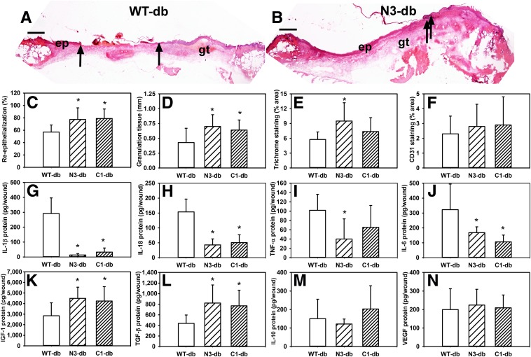
- Bone marrow transfer from NLRP-3– or caspase-1–null mice to db/db mice improved re-epithelialization and granulation tissue formation and reduced wound IL-1β and IL-18, indicating bone marrow–derived inflammasome activity is sufficient to impair healing.
Methods
- Human chronic wound biopsies were collected from five type 2 diabetic patients (2 male, 3 female) with nonhealing sacral or lower-limb wounds lasting at least 3 months; mouse experiments used db/db, db/+, C57Bl/6 wild-type, NLRP-3 knockout, caspase-1 knockout, and IL-1R1 knockout mice.
- Excisional wounds were made with an 8-mm biopsy punch; treatments were applied topically every other day starting 3 days postinjury in F-127 pluronic gel, and healing was assessed on day 10 by re-epithelialization, granulation tissue, collagen, CD31, and α-smooth-muscle actin staining.
- Macrophages were isolated by magnetic depletion and CD11b selection; inflammasome activity was assessed by real-time PCR, ELISA, flow cytometry for active caspase-1, immunofluorescence, and in vitro stimulation with wound-conditioned medium plus inhibitors; data were analyzed by ANOVA or ANOVA on ranks with Student-Newman-Keuls post hoc tests, P ≤ 0.05.
Limitations
- Stated: caspase-1–null mice were also deficient in caspase-11, so bone marrow–transfer experiments could not distinguish the roles of these caspases in impaired healing.
- Stated: topical glyburide and YVAD could block inflammasome activity in keratinocytes as well as macrophages, although the authors note macrophages were the dominant IL-1β producers and bone marrow transfer was sufficient to reduce wound IL-1β/IL-18.
- Evident: the human component is limited to five wound biopsies with no human therapeutic intervention, and macrophage phenotype changes after YVAD treatment were not measured.
Model-written summary. Check it against the paper before relying on it.
Integrity signals
Registry
Cites
Statistics
Figures
Verification
Claims the paper makes
65 Own result · 29 Own interpretation · 24 Not checked yet · 3 Own method
A result the paper reports from its own data — it cites no other source for it, so there is none to check. The quote shows it is in the paper.
In the paper
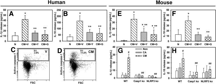
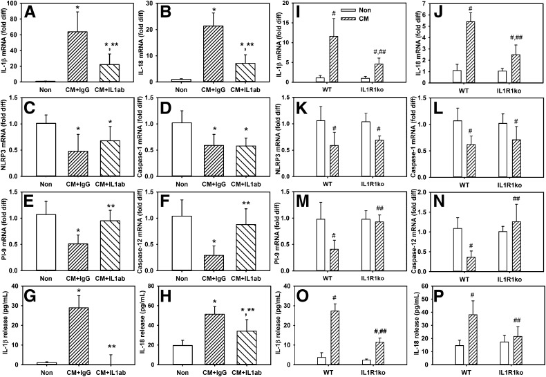
“Wound-conditioned medium increased release of IL-1β and IL-18 into the medium surrounding cultured Mp (Fig. 3A and B).”
§ Sustained Inflammasome Activity in Macrophages Impairs Wound Healing in Type 2 Diabetic Humans and Mice > Results > Diabetic Wound Environment Is Sufficient to Upregulate Inflammasome Activity
plaindirect- Asserted: passes — The authors put it forward as true.
- Checkable: passes — It can be checked against evidence.
- Load-bearing: passes — The paper’s conclusions depend on it.
- Based: passes — The paper gives a reason why it holds.
A result the paper reports from its own data — it cites no other source for it, so there is none to check. The quote shows it is in the paper.
In the paper
“active caspase-1 was detected in 2.9 ± 0.7% of nonstimulated control cells and in 10.5 ± 1.3% of conditioned medium–stimulated cells.”
§ Sustained Inflammasome Activity in Macrophages Impairs Wound Healing in Type 2 Diabetic Humans and Mice > Results > Diabetic Wound Environment Is Sufficient to Upregulate Inflammasome Activity
plain- Asserted: passes — The authors put it forward as true.
- Checkable: passes — It can be checked against evidence.
- Load-bearing: passes — The paper’s conclusions depend on it.
- Based: passes — The paper gives a reason why it holds.
A result the paper reports from its own data — it cites no other source for it, so there is none to check. The quote shows it is in the paper.
In the paper
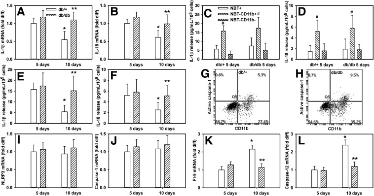
“When different wound cell populations were compared, Mp were the dominant producers of IL-1β and IL-18 after wounding in both db/+ and db/db mice (Fig. 2C and D).”
§ Sustained Inflammasome Activity in Macrophages Impairs Wound Healing in Type 2 Diabetic Humans and Mice > Results > Sustained Inflammasome Activity in Mp From Diabetic Mouse Wounds
plaincorrelational- Asserted: passes — The authors put it forward as true.
- Checkable: passes — It can be checked against evidence.
- Load-bearing: passes — The paper’s conclusions depend on it.
- Based: passes — The paper gives a reason why it holds.
A result the paper reports from its own data — it cites no other source for it, so there is none to check. The quote shows it is in the paper.
In the paper
“although no difference was observed in Mp expression of NLRP-3 or caspase-1 on days 5 and 10 postinjury in either db/+ or db/db mice (Fig. 2I and J)”
§ Sustained Inflammasome Activity in Macrophages Impairs Wound Healing in Type 2 Diabetic Humans and Mice > Results > Sustained Inflammasome Activity in Mp From Diabetic Mouse Wounds
plain- Asserted: passes — The authors put it forward as true.
- Checkable: passes — It can be checked against evidence.
- Load-bearing: passes — The paper’s conclusions depend on it.
- Based: passes — The paper gives a reason why it holds.
A result the paper reports from its own data — it cites no other source for it, so there is none to check. The quote shows it is in the paper.
In the paper
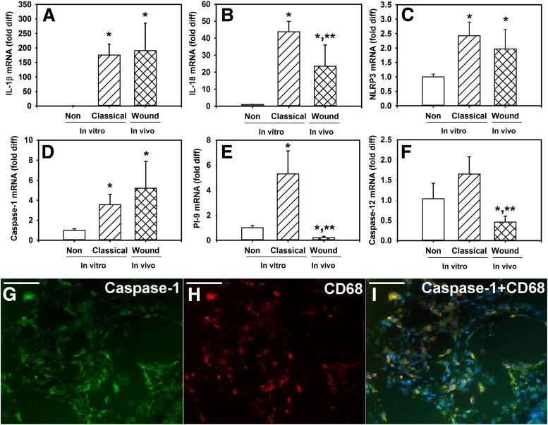
“Mp isolated from chronic wounds expressed high levels of inflammasome components NLRP-3 and caspase-1, and inflammasome targets IL-1β and IL-18, but only low levels of proteinase inhibitor-9 (PI-9) and caspase-12, which are negative regulators of inflammasome activity (Fig. 1A–F).”
§ Sustained Inflammasome Activity in Macrophages Impairs Wound Healing in Type 2 Diabetic Humans and Mice > Results > Inflammasome Components in Mp From Chronic Diabetic Human Wounds
plain- Asserted: passes — The authors put it forward as true.
- Checkable: passes — It can be checked against evidence.
- Load-bearing: passes — The paper’s conclusions depend on it.
- Based: passes — The paper gives a reason why it holds.
A result the paper reports from its own data — it cites no other source for it, so there is none to check. The quote shows it is in the paper.
In the paper
“both the caspase-1 inhibitor YVAD and the inflammasome inhibitor glyburide reduced the conditioned medium–induced release of IL-1β and IL-18.”
§ Sustained Inflammasome Activity in Macrophages Impairs Wound Healing in Type 2 Diabetic Humans and Mice > Results > Diabetic Wound Environment Is Sufficient to Upregulate Inflammasome Activity
plaindirect- Asserted: passes — The authors put it forward as true.
- Checkable: passes — It can be checked against evidence.
- Load-bearing: passes — The paper’s conclusions depend on it.
- Based: passes — The paper gives a reason why it holds.
A result the paper reports from its own data — it cites no other source for it, so there is none to check. The quote shows it is in the paper.
In the paper
“Mp isolated from chronic wounds expressed high levels of inflammasome components NLRP-3 and caspase-1, and inflammasome targets IL-1β and IL-18, but only low levels of proteinase inhibitor-9 (PI-9) and caspase-12, which are negative regulators of inflammasome activity (Fig. 1A–F).”
§ Sustained Inflammasome Activity in Macrophages Impairs Wound Healing in Type 2 Diabetic Humans and Mice > Results > Inflammasome Components in Mp From Chronic Diabetic Human Wounds
plain- Asserted: passes — The authors put it forward as true.
- Checkable: passes — It can be checked against evidence.
- Load-bearing: passes — The paper’s conclusions depend on it.
- Based: passes — The paper gives a reason why it holds.
A result the paper reports from its own data — it cites no other source for it, so there is none to check. The quote shows it is in the paper.
In the paper
“Wound-conditioned medium from db/db mice induced IL-1β and IL-18 release”
§ Sustained Inflammasome Activity in Macrophages Impairs Wound Healing in Type 2 Diabetic Humans and Mice > Results > Diabetic Wound Environment Is Sufficient to Upregulate Inflammasome Activity
plaindirect- Asserted: passes — The authors put it forward as true.
- Checkable: passes — It can be checked against evidence.
- Load-bearing: passes — The paper’s conclusions depend on it.
- Based: passes — The paper gives a reason why it holds.
A result the paper reports from its own data — it cites no other source for it, so there is none to check. The quote shows it is in the paper.
In the paper
“active caspase-1 was detected in 4.8 ± 1.1% of db/+ wound Mp and in 9.9 ± 1.4% of db/db wound Mp (mean ± SD).”
§ Sustained Inflammasome Activity in Macrophages Impairs Wound Healing in Type 2 Diabetic Humans and Mice > Results > Sustained Inflammasome Activity in Mp From Diabetic Mouse Wounds
plaincorrelational- Asserted: passes — The authors put it forward as true.
- Checkable: passes — It can be checked against evidence.
- Load-bearing: passes — The paper’s conclusions depend on it.
- Based: passes — The paper gives a reason why it holds.
A result the paper reports from its own data — it cites no other source for it, so there is none to check. The quote shows it is in the paper.
In the paper
“Interestingly, wound-conditioned medium downregulated expression of NLRP-3, caspase-1, PI-9, and caspase-12”
§ Sustained Inflammasome Activity in Macrophages Impairs Wound Healing in Type 2 Diabetic Humans and Mice > Results > Blocking IL-1β Activity Reduces Inflammasome Activity in Cultured Mp
plaindirect- Asserted: passes — The authors put it forward as true.
- Checkable: passes — It can be checked against evidence.
- Load-bearing: unclear — It is unclear whether the paper’s conclusions depend on it.
- Based: passes — The paper gives a reason why it holds.
- It may state two separate claims as one. 17
- Probably not a substantial claim. 17
A result the paper reports from its own data — it cites no other source for it, so there is none to check. The quote shows it is in the paper.
In the paper
“expression of the inflammasome inhibitors PI-9 and caspase-12 was upregulated from day 5 to day 10 postinjury in db/+ mice (Fig. 2K and L), associated with the downregulation of inflammasome activity.”
§ Sustained Inflammasome Activity in Macrophages Impairs Wound Healing in Type 2 Diabetic Humans and Mice > Results > Sustained Inflammasome Activity in Mp From Diabetic Mouse Wounds
plaincorrelational- Asserted: passes — The authors put it forward as true.
- Checkable: passes — It can be checked against evidence.
- Load-bearing: passes — The paper’s conclusions depend on it.
- Based: passes — The paper gives a reason why it holds.
It may state two separate claims as one.
A result the paper reports from its own data — it cites no other source for it, so there is none to check. The quote shows it is in the paper.
In the paper
“Mp expression and release of IL-1β and IL-18 was high on day 5 postinjury and then downregulated on day 10 postinjury in db/+ mice. In contrast, expression and release of these inflammasome targets were maintained at high levels on day 10 in db/db mice (Fig. 2A, B, E, and F).”
§ Sustained Inflammasome Activity in Macrophages Impairs Wound Healing in Type 2 Diabetic Humans and Mice > Results > Sustained Inflammasome Activity in Mp From Diabetic Mouse Wounds
plain- Asserted: passes — The authors put it forward as true.
- Checkable: passes — It can be checked against evidence.
- Load-bearing: passes — The paper’s conclusions depend on it.
- Based: passes — The paper gives a reason why it holds.
It may state two separate claims as one.
A result the paper reports from its own data — it cites no other source for it, so there is none to check. The quote shows it is in the paper.
In the paper
“In contrast, expression of these inflammasome inhibitors was maintained at low levels in db/db mice, associated with sustained high levels of inflammasome activity.”
§ Sustained Inflammasome Activity in Macrophages Impairs Wound Healing in Type 2 Diabetic Humans and Mice > Results > Sustained Inflammasome Activity in Mp From Diabetic Mouse Wounds
plaincorrelational- Asserted: passes — The authors put it forward as true.
- Checkable: passes — It can be checked against evidence.
- Load-bearing: passes — The paper’s conclusions depend on it.
- Based: passes — The paper gives a reason why it holds.
It may state two separate claims as one.
A result the paper reports from its own data — it cites no other source for it, so there is none to check. The quote shows it is in the paper.
In the paper
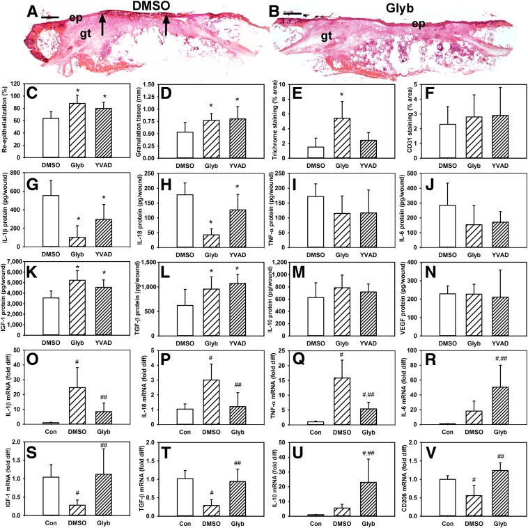
“glyburide but not YVAD treatment increased collagen deposition on day 10 postinjury (Fig. 6E).”
§ Sustained Inflammasome Activity in Macrophages Impairs Wound Healing in Type 2 Diabetic Humans and Mice > Results > Blocking Inflammasome Activity Improves Healing in db/db Mice
plaindirect- Asserted: passes — The authors put it forward as true.
- Checkable: passes — It can be checked against evidence.
- Load-bearing: passes — The paper’s conclusions depend on it.
- Based: passes — The paper gives a reason why it holds.
It may state two separate claims as one.
A result the paper reports from its own data — it cites no other source for it, so there is none to check. The quote shows it is in the paper.
In the paper
“there were nonsignificant trends of decreased TNF-α and IL-6 in glyburide- and YVAD-treated wounds (Fig. 6I and J).”
§ Sustained Inflammasome Activity in Macrophages Impairs Wound Healing in Type 2 Diabetic Humans and Mice > Results > Blocking Inflammasome Activity Improves Healing in db/db Mice
hedged- Asserted: passes — The authors put it forward as true.
- Checkable: passes — It can be checked against evidence.
- Load-bearing: fails — The paper’s conclusions may not depend on it.
- Based: passes — The paper gives a reason why it holds.
Probably not a substantial claim. The paper’s conclusions may not depend on it.
A result the paper reports from its own data — it cites no other source for it, so there is none to check. The quote shows it is in the paper.
In the paper
“there were nonsignificant trends of decreased TNF-α and IL-6 in glyburide- and YVAD-treated wounds (Fig. 6I and J).”
§ Sustained Inflammasome Activity in Macrophages Impairs Wound Healing in Type 2 Diabetic Humans and Mice > Results > Blocking Inflammasome Activity Improves Healing in db/db Mice
hedged- Asserted: passes — The authors put it forward as true.
- Checkable: passes — It can be checked against evidence.
- Load-bearing: fails — The paper’s conclusions may not depend on it.
- Based: passes — The paper gives a reason why it holds.
Probably not a substantial claim. The paper’s conclusions may not depend on it.
A result the paper reports from its own data — it cites no other source for it, so there is none to check. The quote shows it is in the paper.
In the paper
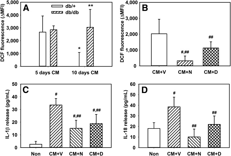
“Conditioned medium from day 10 wounds of db/+ mice did not increase DCF fluorescence, but conditioned medium from db/db mice increased DCF fluorescence to a level similar to that of day 5 conditioned medium.”
§ Sustained Inflammasome Activity in Macrophages Impairs Wound Healing in Type 2 Diabetic Humans and Mice > Results > Blocking ROS Reduces Inflammasome Activity in Cultured Mp
plaindirect- Asserted: passes — The authors put it forward as true.
- Checkable: passes — It can be checked against evidence.
- Load-bearing: passes — The paper’s conclusions depend on it.
- Based: passes — The paper gives a reason why it holds.
It may state two separate claims as one.
A result the paper reports from its own data — it cites no other source for it, so there is none to check. The quote shows it is in the paper.
In the paper
“but the blocking antibody only inhibited the downregulation of PI-9 and caspase-12 (Fig. 5C–F).”
§ Sustained Inflammasome Activity in Macrophages Impairs Wound Healing in Type 2 Diabetic Humans and Mice > Results > Blocking IL-1β Activity Reduces Inflammasome Activity in Cultured Mp
plaindirect- Asserted: passes — The authors put it forward as true.
- Checkable: passes — It can be checked against evidence.
- Load-bearing: unclear — It is unclear whether the paper’s conclusions depend on it.
- Based: passes — The paper gives a reason why it holds.
It may state two separate claims as one.
A result the paper reports from its own data — it cites no other source for it, so there is none to check. The quote shows it is in the paper.
In the paper
“both treatments led to increased α-smooth-muscle actin staining”
§ Sustained Inflammasome Activity in Macrophages Impairs Wound Healing in Type 2 Diabetic Humans and Mice > Results > Blocking Inflammasome Activity Improves Healing in db/db Mice
plaindirect- Asserted: passes — The authors put it forward as true.
- Checkable: passes — It can be checked against evidence.
- Load-bearing: unclear — It is unclear whether the paper’s conclusions depend on it.
- Based: passes — The paper gives a reason why it holds.
It may state two separate claims as one.
A conclusion the paper draws from its own results — it cites no other source for it, so there is none to check. The quote shows it is in the paper.
In the paper
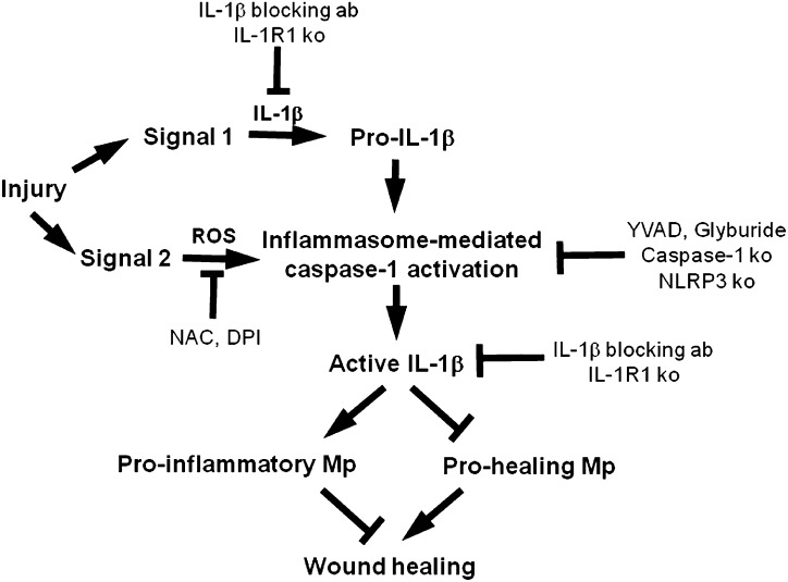
“Taken together, these data indicate that IL-1β in the diabetic wound environment may induce expression of IL-1β and IL-18 and contribute to sustained inflammasome activity in wound Mp.”
§ Sustained Inflammasome Activity in Macrophages Impairs Wound Healing in Type 2 Diabetic Humans and Mice > Results > Blocking IL-1β Activity Reduces Inflammasome Activity in Cultured Mp
hedgedconditional- Asserted: passes — The authors put it forward as true.
- Checkable: passes — It can be checked against evidence.
- Load-bearing: passes — The paper’s conclusions depend on it.
- Based: passes — The paper gives a reason why it holds.
It may state two separate claims as one.
What the paper did — it cites no source for this, so there is none to check. The quote shows it is in the paper.
In the paper
“Negative controls included nonstained slides and isotype-specific control antibodies (IgG1 and IgG2b; Biolegend) along with secondary antibodies.”
§ Sustained Inflammasome Activity in Macrophages Impairs Wound Healing in Type 2 Diabetic Humans and Mice > Research Design and Methods > Immunofluorescence
plain- Asserted: passes — The authors put it forward as true.
- Checkable: passes — It can be checked against evidence.
- Load-bearing: fails — The paper’s conclusions may not depend on it.
- Based: passes — The paper gives a reason why it holds.
Probably not a substantial claim. The paper’s conclusions may not depend on it.
What the paper did — it cites no source for this, so there is none to check. The quote shows it is in the paper.
In the paper
“Engraftment was verified in preliminary experiments using congenic mice and was found to be >85% by flow cytometry.”
§ Sustained Inflammasome Activity in Macrophages Impairs Wound Healing in Type 2 Diabetic Humans and Mice > Research Design and Methods > Bone Marrow Transfer
plain- Asserted: passes — The authors put it forward as true.
- Checkable: passes — It can be checked against evidence.
- Load-bearing: passes — The paper’s conclusions depend on it.
- Based: passes — The paper gives a reason why it holds.
Probably not a substantial claim.
None of the sources cited for this claim have been checked yet.
In the paper
“During inflammasome activation, the proform of caspase-1 is recruited to the NLRP-3 complex and cleaved to produce active caspase-1, which then cleaves and activates the potent proinflammatory cytokines IL-1β and IL-18.”
§ Sustained Inflammasome Activity in Macrophages Impairs Wound Healing in Type 2 Diabetic Humans and Mice > Introduction
What the sources say
- Not checked yetcites [16] Martinon, 2009, The inflammasomes: guardians of the body, Annu Rev Immunol source
- Not checked yetcites [17] Strowig, 2012, Inflammasomes in health and disease, Nature source
- Not checked yetcites [18] Pedra, 2009, Sensing pathogens and danger signals by the inflammasome, Curr Opin Immunol source
plaindirect- Asserted: passes — The authors put it forward as true.
- Checkable: passes — It can be checked against evidence.
- Load-bearing: passes — The paper’s conclusions depend on it.
- Based: passes — The paper gives a reason why it holds.
It may state two separate claims as one.
None of the sources cited for this claim have been checked yet.
In the paper
“During impaired healing associated with diabetes, wounds exhibit prolonged accumulation of Mp associated with elevated levels of proinflammatory cytokines and proteases and reduced levels of various growth factors (2,12,13).”
§ Sustained Inflammasome Activity in Macrophages Impairs Wound Healing in Type 2 Diabetic Humans and Mice > Introduction
What the sources say
- Not checked yetcites [2] Blakytny, 2006, The molecular biology of chronic wounds and delayed healing in diabetes, Diabet Med source
- Not checked yetcites [12] Goren, 2003, Leptin and wound inflammation in diabetic ob/ob mice: differential regulation of neutrophil and macrophage influx and a potential role for the scab as a sink for inflammatory cells and mediators, Diabetes source
- Not checked yetcites [13] Greenhalgh, 1990, PDGF and FGF stimulate wound healing in the genetically diabetic mouse, Am J Pathol
plaincorrelational- Asserted: passes — The authors put it forward as true.
- Checkable: passes — It can be checked against evidence.
- Load-bearing: passes — The paper’s conclusions depend on it.
- Based: passes — The paper gives a reason why it holds.
It may state two separate claims as one.
None of the sources cited for this claim have been checked yet.
In the paper
“Although previous studies have demonstrated that platelet-derived growth factor, fibroblast growth factor, vascular endothelial growth factor (VEGF), or erythropoietin can improve healing in diabetic mice (13,36,37)”
§ Sustained Inflammasome Activity in Macrophages Impairs Wound Healing in Type 2 Diabetic Humans and Mice > Discussion
What the sources say
- Not checked yetcites [13] Greenhalgh, 1990, PDGF and FGF stimulate wound healing in the genetically diabetic mouse, Am J Pathol
- Not checked yetcites [36] Brem, 2009, Mechanism of sustained release of vascular endothelial growth factor in accelerating experimental diabetic healing, J Invest Dermatol source
- Not checked yetcites [37] Hamed, 2010, Topical erythropoietin promotes wound repair in diabetic rats, J Invest Dermatol source
boosteddirect- Asserted: passes — The authors put it forward as true.
- Checkable: passes — It can be checked against evidence.
- Load-bearing: fails — The paper’s conclusions may not depend on it.
- Based: passes — The paper gives a reason why it holds.
Probably not a substantial claim. The paper’s conclusions may not depend on it.
A conclusion the paper draws from its own results — it cites no other source for it, so there is none to check. The quote shows it is in the paper.
In the paper
“Importantly, inhibiting the inflammasome in vivo, either using topical application of pharmacological inhibitors or via bone marrow transfer from NLRP-3– or caspase-1–null mice to db/db mice, promotes the early healing response in part by downregulating the proinflammatory phenotype characteristic of Mp in diabetic wounds and increasing production of prohealing factors.”
§ Sustained Inflammasome Activity in Macrophages Impairs Wound Healing in Type 2 Diabetic Humans and Mice > Discussion
plaindirect- Asserted: passes — The authors put it forward as true.
- Checkable: passes — It can be checked against evidence.
- Load-bearing: passes — The paper’s conclusions depend on it.
- Based: passes — The paper gives a reason why it holds.
It may state two separate claims as one.
- Rests on an assumption: Bone marrow transfer from donor mice to db/db mice specifically targets the inflammasome in macrophages.
A conclusion the paper draws from its own results — it cites no other source for it, so there is none to check. The quote shows it is in the paper.
In the paper
“which, in turn, was associated with a more prohealing wound environment and improved healing in db/db mice.”
§ Sustained Inflammasome Activity in Macrophages Impairs Wound Healing in Type 2 Diabetic Humans and Mice > Results > Blocking Inflammasome Activity Improves Healing in db/db Mice
plaincorrelational- Asserted: passes — The authors put it forward as true.
- Checkable: passes — It can be checked against evidence.
- Load-bearing: passes — The paper’s conclusions depend on it.
- Based: passes — The paper gives a reason why it holds.
It may state two separate claims as one.
None of the sources cited for this claim have been checked yet.
In the paper
“The NLRP-3 inflammasome plays a role in the pathophysiology of many inflammatory diseases, including obesity, insulin resistance, and diabetes (21,31–35).”
§ Sustained Inflammasome Activity in Macrophages Impairs Wound Healing in Type 2 Diabetic Humans and Mice > Discussion
What the sources say
- Not checked yetcites [21] Dinarello, 2009, Immunological and inflammatory functions of the interleukin-1 family, Annu Rev Immunol source
- Not checked yetcites [31] Dinarello, 2004, Therapeutic strategies to reduce IL-1 activity in treating local and systemic inflammation, Curr Opin Pharmacol source
- Not checked yetcites [32] Larsen, 2007, Interleukin-1-receptor antagonist in type 2 diabetes mellitus, N Engl J Med source
- Not checked yetcites [33] Osborn, 2008, Treatment with an Interleukin 1 beta antibody improves glycemic control in diet-induced obesity, Cytokine source
- Not checked yetcites [34] Stienstra, 2011, Inflammasome is a central player in the induction of obesity and insulin resistance, Proc Natl Acad Sci USA source
- Not checked yetcites [35] Vandanmagsar, 2011, The NLRP3 inflammasome instigates obesity-induced inflammation and insulin resistance, Nat Med source
plaindirect- Asserted: passes — The authors put it forward as true.
- Checkable: passes — It can be checked against evidence.
- Load-bearing: passes — The paper’s conclusions depend on it.
- Based: passes — The paper gives a reason why it holds.
Probably not a substantial claim.
A conclusion the paper draws from its own results — it cites no other source for it, so there is none to check. The quote shows it is in the paper.
In the paper
“These data indicate that blocking inflammasome activity downregulated Mp expression of proinflammatory cytokines measured and upregulated expression of prohealing factors”
§ Sustained Inflammasome Activity in Macrophages Impairs Wound Healing in Type 2 Diabetic Humans and Mice > Results > Blocking Inflammasome Activity Improves Healing in db/db Mice
hedgeddirect- Asserted: passes — The authors put it forward as true.
- Checkable: passes — It can be checked against evidence.
- Load-bearing: passes — The paper’s conclusions depend on it.
- Based: passes — The paper gives a reason why it holds.
It may state two separate claims as one.
None of the sources cited for this claim have been checked yet.
In the paper
“Chronic wounds are typically associated with a persistent inflammatory response that involves accumulation of Mp (19,26–28)”
§ Sustained Inflammasome Activity in Macrophages Impairs Wound Healing in Type 2 Diabetic Humans and Mice > Discussion
What the sources say
- Not checked yetcites [19] Mast, 1996, Interactions of cytokines, growth factors, and proteases in acute and chronic wounds, Wound Repair Regen source
- Not checked yetcites [26] Loots, 1998, Differences in cellular infiltrate and extracellular matrix of chronic diabetic and venous ulcers versus acute wounds, J Invest Dermatol source
- Not checked yetcites [27] Rosner, 1995, Immunohistochemical characterization of the cutaneous cellular infiltrate in different areas of chronic leg ulcers, APMIS source
- Not checked yetcites [28] Trengove, 2000, Mitogenic activity and cytokine levels in non-healing and healing chronic leg ulcers, Wound Repair Regen source
plaincorrelational- Asserted: passes — The authors put it forward as true.
- Checkable: passes — It can be checked against evidence.
- Load-bearing: fails — The paper’s conclusions may not depend on it.
- Based: passes — The paper gives a reason why it holds.
Probably not a substantial claim. The paper’s conclusions may not depend on it.
In the paper
“Glyburide is a commonly used sulfonylurea drug for the treatment of type 2 diabetes and was recently shown to inhibit the NLRP-3 inflammasome through a mechanism that appears to be different from that involved in its ability to promote insulin secretion from pancreatic β-cells (38).”
§ Sustained Inflammasome Activity in Macrophages Impairs Wound Healing in Type 2 Diabetic Humans and Mice > Discussion
What the sources say
- Not checked yetcites [38] Lamkanfi, 2009, Glyburide inhibits the Cryopyrin/Nalp3 inflammasome, J Cell Biol source
hedgeddirect- Asserted: passes — The authors put it forward as true.
- Checkable: passes — It can be checked against evidence.
- Load-bearing: passes — The paper’s conclusions depend on it.
- Based: passes — The paper gives a reason why it holds.
It may state two separate claims as one.
None of the sources cited for this claim have been checked yet.
In the paper
“In fact, either premature changes in phenotype or delayed/impaired changes in phenotype have been associated with impaired healing (14,29,49).”
§ Sustained Inflammasome Activity in Macrophages Impairs Wound Healing in Type 2 Diabetic Humans and Mice > Discussion
What the sources say
- Not checked yetcites [14] Mirza, 2011, Dysregulation of monocyte/macrophage phenotype in wounds of diabetic mice, Cytokine source
- Not checked yetcites [29] Rodero, 2013, Reduced Il17a expression distinguishes a Ly6c(lo)MHCII(hi) macrophage population promoting wound healing, J Invest Dermatol source
- Not checked yetcites [49] Perdiguero, 2011, p38/MKP-1-regulated AKT coordinates macrophage transitions and resolution of inflammation during tissue repair, J Cell Biol source
plaincorrelational- Asserted: passes — The authors put it forward as true.
- Checkable: passes — It can be checked against evidence.
- Load-bearing: passes — The paper’s conclusions depend on it.
- Based: passes — The paper gives a reason why it holds.
It may state two separate claims as one.
None of the sources cited for this claim have been checked yet.
In the paper
“For example, both murine and human keratinocytes express inflammasome components NLRP-3, ASC, caspase-1, IL-1β, and IL-18 (43,44).”
§ Sustained Inflammasome Activity in Macrophages Impairs Wound Healing in Type 2 Diabetic Humans and Mice > Discussion
What the sources say
- Not checked yetcites [43] Feldmeyer, 2007, The inflammasome mediates UVB-induced activation and secretion of interleukin-1beta by keratinocytes, Curr Biol source
- Not checked yetcites [44] Watanabe, 2007, Activation of the IL-1beta-processing inflammasome is involved in contact hypersensitivity, J Invest Dermatol source
plain- Asserted: passes — The authors put it forward as true.
- Checkable: passes — It can be checked against evidence.
- Load-bearing: fails — The paper’s conclusions may not depend on it.
- Based: passes — The paper gives a reason why it holds.
Probably not a substantial claim. The paper’s conclusions may not depend on it.
None of the sources cited for this claim have been checked yet.
In the paper
“Caspase recruitment domain–only proteins (COPs), such as Iceberg, Pseudo-ICE, and caspase-12, and pyrin domain–only proteins (POPs), such as pyrin, POP1, and POP2, are thought to inhibit inflammasome activity by blocking inflammasome assembly and subsequent caspase-1 cleavage (16,39).”
§ Sustained Inflammasome Activity in Macrophages Impairs Wound Healing in Type 2 Diabetic Humans and Mice > Discussion
What the sources say
hedgedconditional- Asserted: passes — The authors put it forward as true.
- Checkable: passes — It can be checked against evidence.
- Load-bearing: fails — The paper’s conclusions may not depend on it.
- Based: passes — The paper gives a reason why it holds.
Probably not a substantial claim. The paper’s conclusions may not depend on it.
In the paper
“Components of the inflammasome can be expressed in cell types other than Mp that are involved in wound healing (42).”
§ Sustained Inflammasome Activity in Macrophages Impairs Wound Healing in Type 2 Diabetic Humans and Mice > Discussion
What the sources say
- Not checked yetcites [42] Yazdi, 2010, The role of the inflammasome in nonmyeloid cells, J Clin Immunol source
plain- Asserted: passes — The authors put it forward as true.
- Checkable: passes — It can be checked against evidence.
- Load-bearing: fails — The paper’s conclusions may not depend on it.
- Based: passes — The paper gives a reason why it holds.
Probably not a substantial claim. The paper’s conclusions may not depend on it.
None of the sources cited for this claim have been checked yet.
In the paper
“Recent studies have shown that Mp phenotypes change over the different phases of tissue repair and that the sequential appearance of these phenotypes may be required for efficient repair (11,14,29,49,50).”
§ Sustained Inflammasome Activity in Macrophages Impairs Wound Healing in Type 2 Diabetic Humans and Mice > Discussion
What the sources say
- Not checked yetcites [11] Willenborg, 2012, CCR2 recruits an inflammatory macrophage subpopulation critical for angiogenesis in tissue repair, Blood source
- Not checked yetcites [14] Mirza, 2011, Dysregulation of monocyte/macrophage phenotype in wounds of diabetic mice, Cytokine source
- Not checked yetcites [29] Rodero, 2013, Reduced Il17a expression distinguishes a Ly6c(lo)MHCII(hi) macrophage population promoting wound healing, J Invest Dermatol source
- Not checked yetcites [49] Perdiguero, 2011, p38/MKP-1-regulated AKT coordinates macrophage transitions and resolution of inflammation during tissue repair, J Cell Biol source
- Not checked yetcites [50] Novak, 2013, Macrophage phenotypes during tissue repair, J Leukoc Biol source
hedgedconditional- Asserted: passes — The authors put it forward as true.
- Checkable: passes — It can be checked against evidence.
- Load-bearing: passes — The paper’s conclusions depend on it.
- Based: passes — The paper gives a reason why it holds.
Probably not a substantial claim. It may state two separate claims as one.
None of the sources cited for this claim have been checked yet.
In the paper
“Accumulating evidence indicates that Mp are critical orchestrators in healing of many tissues, including skin (3,4,7,48).”
§ Sustained Inflammasome Activity in Macrophages Impairs Wound Healing in Type 2 Diabetic Humans and Mice > Discussion
What the sources say
- Not checked yetcites [3] Arnold, 2007, Inflammatory monocytes recruited after skeletal muscle injury switch into antiinflammatory macrophages to support myogenesis, J Exp Med source
- Not checked yetcites [4] Mirza, 2009, Selective and specific macrophage ablation is detrimental to wound healing in mice, Am J Pathol source
- Not checked yetcites [7] Duffield, 2005, Selective depletion of macrophages reveals distinct, opposing roles during liver injury and repair, J Clin Invest source
- Not checked yetcites [48] van Amerongen, 2007, Macrophage depletion impairs wound healing and increases left ventricular remodeling after myocardial injury in mice, Am J Pathol source
hedgeddirect- Asserted: passes — The authors put it forward as true.
- Checkable: passes — It can be checked against evidence.
- Load-bearing: fails — The paper’s conclusions may not depend on it.
- Based: passes — The paper gives a reason why it holds.
Probably not a substantial claim. The paper’s conclusions may not depend on it.
A conclusion the paper draws from its own results — it cites no other source for it, so there is none to check. The quote shows it is in the paper.
In the paper
“Thus, topical application of glyburide and YVAD in our studies could block inflammasome activity in keratinocytes as well as Mp.”
§ Sustained Inflammasome Activity in Macrophages Impairs Wound Healing in Type 2 Diabetic Humans and Mice > Discussion
hedgedconditional- Asserted: passes — The authors put it forward as true.
- Checkable: passes — It can be checked against evidence.
- Load-bearing: fails — The paper’s conclusions may not depend on it.
- Based: passes — The paper gives a reason why it holds.
Probably not a substantial claim. The paper’s conclusions may not depend on it.
None of the sources cited for this claim have been checked yet.
In the paper
“Interestingly, humans express a broad array of COPs and POPs, whereas rodents only express a limited subset (16,39).”
§ Sustained Inflammasome Activity in Macrophages Impairs Wound Healing in Type 2 Diabetic Humans and Mice > Discussion
What the sources say
plain- Asserted: passes — The authors put it forward as true.
- Checkable: passes — It can be checked against evidence.
- Load-bearing: fails — The paper’s conclusions may not depend on it.
- Based: passes — The paper gives a reason why it holds.
Probably not a substantial claim. The paper’s conclusions may not depend on it. It may state two separate claims as one.
None of the sources cited for this claim have been checked yet.
In the paper
“In addition, other inhibitors such as PI-9 act by directly binding and inhibiting caspase-1 (40,41).”
§ Sustained Inflammasome Activity in Macrophages Impairs Wound Healing in Type 2 Diabetic Humans and Mice > Discussion
What the sources say
- Not checked yetcites [40] Young, 2000, The serpin proteinase inhibitor 9 is an endogenous inhibitor of interleukin 1beta-converting enzyme (caspase-1) activity in human vascular smooth muscle cells, J Exp Med source
- Not checked yetcites [41] Annand, 1999, Caspase-1 (interleukin-1beta-converting enzyme) is inhibited by the human serpin analogue proteinase inhibitor 9, Biochem J source
plaindirect- Asserted: passes — The authors put it forward as true.
- Checkable: passes — It can be checked against evidence.
- Load-bearing: fails — The paper’s conclusions may not depend on it.
- Based: passes — The paper gives a reason why it holds.
Probably not a substantial claim. The paper’s conclusions may not depend on it.
None of the sources cited for this claim have been checked yet.
In the paper
“and contact hypersensitivity appears to be mediated by the inflammasome/IL-1β pathway (44,45).”
§ Sustained Inflammasome Activity in Macrophages Impairs Wound Healing in Type 2 Diabetic Humans and Mice > Discussion
What the sources say
- Not checked yetcites [44] Watanabe, 2007, Activation of the IL-1beta-processing inflammasome is involved in contact hypersensitivity, J Invest Dermatol source
- Not checked yetcites [45] Sutterwala, 2006, Critical role for NALP3/CIAS1/Cryopyrin in innate and adaptive immunity through its regulation of caspase-1, Immunity source
hedgedconditional- Asserted: passes — The authors put it forward as true.
- Checkable: passes — It can be checked against evidence.
- Load-bearing: fails — The paper’s conclusions may not depend on it.
- Based: passes — The paper gives a reason why it holds.
Probably not a substantial claim. The paper’s conclusions may not depend on it.
In the paper
“In addition, ultraviolet irradiation–induced IL-1β secretion from human keratinocytes in vitro appears to be mediated by the NLRP-3 inflammasome (43)”
§ Sustained Inflammasome Activity in Macrophages Impairs Wound Healing in Type 2 Diabetic Humans and Mice > Discussion
What the sources say
- Not checked yetcites [43] Feldmeyer, 2007, The inflammasome mediates UVB-induced activation and secretion of interleukin-1beta by keratinocytes, Curr Biol source
hedgedconditional- Asserted: passes — The authors put it forward as true.
- Checkable: passes — It can be checked against evidence.
- Load-bearing: fails — The paper’s conclusions may not depend on it.
- Based: passes — The paper gives a reason why it holds.
Probably not a substantial claim. The paper’s conclusions may not depend on it.
The paper relies on this without support.
The db/db mouse model of type 2 diabetes is representative of human diabetic wound healing for testing inflammasome-targeted therapies.
“Diabetic db/db mice, nondiabetic db/+ controls, and C57Bl/6 wild-type controls were obtained from The Jackson Laboratory.”
“Our findings indicate that sustained inflammasome activity in wound Mp contributes to impaired early healing responses of diabetic wounds and that the inflammasome may represent a new therapeutic target for improving healing in diabetic individuals.”
The paper relies on this without support.
Bone marrow transfer from donor mice to db/db mice specifically targets the inflammasome in macrophages.
“To more specifically target the inflammasome in Mp, we used bone marrow transfer to determine the role of bone marrow cell–derived inflammasome activity in impaired healing of wounds in diabetic mice.”
Claims other papers make about this one
Each is checked against this paper’s own text: does it say what the citing paper says it does?
In the citing paper
“Separate reports using two different mouse models of nonhealing wounds108,109 have demonstrated that iron overloading and sustained inflammasome activation of macrophages results in defective transition from an inflammatory M1 to a prohealing M2 phenotype.”
From The Innate Immune System in Acute and Chronic Wounds (2016) · cites this paper as [108]
Self-check
Percentages, sums and reported differences re-computed from the paper’s own numbers.
Each contradiction between two passages listed here was confirmed by an independent check.
Rated minor: it would not touch a result — a value no result rests on, a p-value that stays on the same side of the significance level, or a conflict that is clerical, resolved elsewhere in the paper, or outside its own findings.
In the paper
§ Sustained Inflammasome Activity in Macrophages Impairs Wound Healing in Type 2 Diabetic Humans and Mice > Results > Blocking Inflammasome Activity Improves Healing in db/db Mice
“These data indicate that blocking inflammasome activity downregulated Mp expression of proinflammatory cytokines measured and upregulated expression of prohealing factors”
§ Sustained Inflammasome Activity in Macrophages Impairs Wound Healing in Type 2 Diabetic Humans and Mice > Results > Blocking Inflammasome Activity Improves Healing in db/db Mice
“although expression of IL-6 was increased (Fig. 6R)”
Citations
Cites
Singer, A. J., & Clark, R. A. F. (1999) · New England Journal of Medicine · doi:10.1056/nejm199909023411006 · Cited by 6,106
Immunological and inflammatory functions of the interleukin-1 family
Dinarello, C. A. (2009) · Annual Review of Immunology · doi:10.1146/annurev.immunol.021908.132612 · Cited by 3,466 · cited 1×
“Since IL-1b, in turn, is known to induce a proinflammatory Mp phenotype (15,21), the NLRP-3 inflammasome may be part of a positive feedback loop that sustains inflammation in chronic wounds and contributes to impaired healing.”
BackgroundThe NLRP3 inflammasome instigates obesity-induced inflammation and insulin resistance
Vandanmagsar, B., Youm, Y.-H., Ravussin, A., Galgani, J. E., Stadler, K., Mynatt, R. L., Ravussin, E., Stephens, J. M., & Dixit, V. D. (2011) · Nature Medicine · doi:10.1038/nm.2279 · Cited by 2,655
Non-canonical inflammasome activation targets caspase-11
Kayagaki, N., Warming, S., Lamkanfi, M., Walle, L. V., Louie, S., Dong, J., Newton, K., Qu, Y., Liu, J., Heldens, S., et al. (2011) · Nature · doi:10.1038/nature10558 · Cited by 2,530 · cited 1×
“A limitation of our study is that the caspase1–null mice were also found to be deficient in caspase-11 (46,47), and so our bone marrow–transfer experiments do not distinguish the role of these caspases in the impaired healing of wounds in db/db mice.”
BackgroundThe Inflammasomes: Guardians of the Body
Martinon, F., Mayor, A., & Tschopp, J. (2009) · Annual Review of Immunology · doi:10.1146/annurev.immunol.021908.132715 · Cited by 2,436 · cited 4×
“Whereas a number of different danger signals, including ATP, uric acid crystals, cholesterol crystals, and glucose, can activate the inflammasome (16,17,25), a common downstream mediator may be increased production of ROS (25).”
BackgroundInfluentialNahrendorf, M., Swirski, F. K., Aikawa, E., Stangenberg, L., Wurdinger, T., Figueiredo, J.-L., Libby, P., Weissleder, R., & Pittet, M. J. (2007) · The Journal of Experimental Medicine · doi:10.1084/jem.20070885 · Cited by 2,300
Inflammasomes in health and disease
Strowig, T., Henao-Mejia, J., Elinav, E., & Flavell, R. (2012) · Nature · doi:10.1038/nature10759 · Cited by 2,230 · cited 2×
“Whereas a number of different danger signals, including ATP, uric acid crystals, cholesterol crystals, and glucose, can activate the inflammasome (16,17,25), a common downstream mediator may be increased production of ROS (25).”
BackgroundArnold, L., Henry, A., Poron, F., Baba-Amer, Y., van Rooijen, N., Plonquet, A., Gherardi, R. K., & Chazaud, B. (2007) · The Journal of Experimental Medicine · doi:10.1084/jem.20070075 · Cited by 2,002 · cited 1×
“Accumulating evidence indicates that Mp are critical orchestrators in healing of many tissues, including skin (3,4,7,48).”
BackgroundNLRP3 inflammasome activation: the convergence of multiple signalling pathways on ROS production?
Tschopp, J., & Schroder, K. (2010) · Nature reviews. Immunology · doi:10.1038/nri2725 · Cited by 1,859 · cited 2×
“Whereas a number of different danger signals, including ATP, uric acid crystals, cholesterol crystals, and glucose, can activate the inflammasome (16,17,25), a common downstream mediator may be increased production of ROS (25).”
BackgroundInterleukin-1-receptor antagonist in type 2 diabetes mellitus
Larsen, C. M., Faulenbach, M., Vaag, A., Vølund, A., Ehses, J. A., Seifert, B., Mandrup-Poulsen, T., & Donath, M. Y. (2007) · New England Journal of Medicine · doi:10.1056/nejmoa065213 · Cited by 1,798
Cited by
Sorted by citations
The Effects of Type 2 Diabetes Mellitus on Organ Metabolism and the Immune System
Daryabor, G., Atashzar, M. R., Kabelitz, D., Meri, S., & Kalantar, K. (2020) · Frontiers in Immunology · doi:10.3389/fimmu.2020.01582 · Cited by 617
“have shown that an impaired wound healing process in animals with T2DM is due to high levels of NLRP3 inflammasome activity, which promotes the generation of IL-1β and IL-18 in macrophages (149, 150).”
BackgroundNunan, R., Harding, K. G., & Martin, P. (2014) · Disease Models & Mechanisms · doi:10.1242/dmm.016782 · Cited by 542
“However, in general, a large influx of innate immune cells into chronic wounds, and their retention there, is likely to inhibit many repair processes (Mirza et al., 2014).”
BackgroundHan, Y., Xu, X., Tang, C., Gao, P., Chen, X., Xiong, X., Yang, M., Yang, S., Zhu, X., Yuan, S., et al. (2018) · Redox Biology · doi:10.1016/j.redox.2018.02.013 · Cited by 470
“Activated NLRP3-mediated inflammation cleaves the precursors of IL-1β and IL-18, which leads to an immunoinflammatory response [48,49].”
BackgroundNlrp3-inflammasome activation in non-myeloid-derived cells aggravates diabetic nephropathy
Shahzad, K., Bock, F. M., Dong, W., Wang, H., Kopf, S., Kohli, S., Al‐Dabet, M. M., Ranjan, S., Wolter, J., Wacker, C., et al. (2014) · Kidney International · doi:10.1038/ki.2014.271 · Cited by 423
The Healing Power of Neutrophils
Phillipson, M., & Kubes, P. (2019) · Trends in Immunology · doi:10.1016/j.it.2019.05.001 · Cited by 398
Macrophage-Mediated Inflammation in Skin Wound Healing
Hassanshahi, A., Moradzad, M., Ghalamkari, S., Fadaei, M., Cowin, A. J., & Hassanshahi, M. (2022) · Cells · doi:10.3390/cells11192953 · Cited by 390
Updates in Diabetic Wound Healing, Inflammation, and Scarring
Dasari, N., Jiang, A. Y., Skochdopole, A. J., Chung, J., Reece, E. M., Vorstenbosch, J., & Winocour, S. J. (2021) · Seminars in Plastic Surgery · doi:10.1055/s-0041-1731460 · Cited by 357
Dysfunctional Wound Healing in Diabetic Foot Ulcers: New Crossroads
Davis, F. M., Kimball, A., Boniakowski, A. E., & Gallagher, K. A. (2018) · Current Diabetes Reports · doi:10.1007/s11892-018-0970-z · Cited by 334
Macrophage-based therapeutic strategies in regenerative medicine
Spiller, K. L., & Koh, T. J. (2017) · Advanced Drug Delivery Reviews · doi:10.1016/j.addr.2017.05.010 · Cited by 310
“For example, M1-like macrophages impair regeneration in some situations [13,77] and promote regeneration in others [33,63].”
BackgroundInfluentialSelf-citationThe Innate Immune System in Acute and Chronic Wounds
MacLeod, A. S., & Mansbridge, J. N. (2016) · Advances in Wound Care · doi:10.1089/wound.2014.0608 · Cited by 289
Macrophage Related Chronic Inflammation in Non-Healing Wounds
Li, M., Hou, Q., Zhong, L., Zhao, Y., & Fu, X. (2021) · Frontiers in Immunology · doi:10.3389/fimmu.2021.681710 · Cited by 307
“Both mouse and human diabetic wound conditioned media prefer to induce a proinflammatory macrophage phenotype of BM- derived macrophage in vitro, which suggests an imbalanced microenvironment (43, 44).”
BackgroundThe NLRP3 inflammasome in kidney disease and autoimmunity
Hutton, H., Ooi, J. D., Holdsworth, S. R., & Kitching, A. R. (2016) · Nephrology · doi:10.1111/nep.12785 · Cited by 237
“Several studies, including human ex vivo studies, have identified mitochondrial ROS as an activator of NLRP3-ASC in diabetes.(27,57) A comprehensive study of the NLRP3 inflammasome’s role in the pathogenesis of diabetic nephropathy was recently undertaken by Shahzad et al.”
BackgroundGuo, Y., Ding, S., Shang, C., Zhang, C., Li, M., Zhang, Q., Gu, L., Heng, B. C., Zhang, S., Mei, F., et al. (2023) · Advanced Materials · doi:10.1002/adma.202306292 · Cited by 227
Liu, D., Yang, P., Gao, M., Yu, T., Shi, Y., Zhang, M., Yao, M., Liu, Y., & Zhang, X. (2019) · Clinical Science · doi:10.1042/cs20180600 · Cited by 221
Kimball, A., Schaller, M. A., Joshi, A., Davis, F. M., denDekker, A. D., Boniakowski, A. E., Bermick, J., Obi, A. T., Moore, B. B., Henke, P. K., et al. (2018) · Arteriosclerosis Thrombosis and Vascular Biology · doi:10.1161/atvbaha.118.310703 · Cited by 216
Diabetes medications: Impact on inflammation and wound healing
Salazar, J. J., Ennis, W. J., & Koh, T. J. (2015) · Journal of Diabetes and its Complications · doi:10.1016/j.jdiacomp.2015.12.017 · Cited by 205
“Glyburide treatment also induced concurrent increased expression of pro-healing IGF-1, TGF-β, and IL-10 growth factors (Mirza et al., 2014).”
BackgroundSelf-citationExploring the contribution of pro-inflammatory cytokines to impaired wound healing in diabetes
Nirenjen, S., Narayanan, J., Tamilanban, T., Subramaniyan, V., Chitra, V., Fuloria, N. K., Wong, L. S., Ramachawolran, G., Sekar, M., Gupta, G., et al. (2023) · Frontiers in Immunology · doi:10.3389/fimmu.2023.1216321 · Cited by 225
“Diabetic human and db/db mouse wound macrophages exhibit elevated IL1b and NALP3 inflammasome components during the first ten days of healing, and inhibiting the inflammasome was associated with better wound healing in these models (38).”
BackgroundRoles of the NLRP3 inflammasome in the pathogenesis of diabetic nephropathy
Qiu, Y.-Y., & Tang, L. (2016) · Pharmacological Research · doi:10.1016/j.phrs.2016.11.004 · Cited by 198
“ROS-dependent activation of the NLRP3 inflammasome has recently been described in diabetic patients [86].”
BackgroundQing, L., Fu, J., Wu, P., Zhou, Z., Yu, F., & Tang, J. (2019) · Europe PMC (PubMed Central) · Cited by 190
Alternative Pathways of IL-1 Activation, and Its Role in Health and Disease
Pyrillou, K., Burzynski, L. C., & Clarke, M. C. H. (2020) · Frontiers in Immunology · doi:10.3389/fimmu.2020.613170 · Cited by 202
Macrophage PPARγ and impaired wound healing in type 2 diabetes
Mirza, R. E., Fang, M. M., Novak, M. L., Urao, N., Sui, A., Ennis, W. J., & Koh, T. J. (2015) · The Journal of Pathology · doi:10.1002/path.4548 · Cited by 177
“To generate cultures of human or mouse Mp, peripheral blood mononuclear cells from normal volunteers (Zen-Bio) or bone marrow cells from wild-type C57Bl/6 mice and IL1R1 KO mice were cultured as described [20,21].”
BackgroundMethodInfluentialSelf-citationMacrophage-mediated inflammation in diabetic wound repair
Wolf, S., Melvin, W. J., & Gallagher, K. A. (2021) · Seminars in Cell and Developmental Biology · doi:10.1016/j.semcdb.2021.06.013 · Cited by 190
Kimball, A., Davis, F. M., denDekker, A. D., Joshi, A., Schaller, M. A., Bermick, J., Xing, X., Burant, C., Obi, A. T., Nysz, D., et al. (2019) · Immunity · doi:10.1016/j.immuni.2019.06.015 · Cited by 172
“The finding that key inflammatory cytokines involved in wound repair, such as IL-1β and TNF-α, are increased in Setdb2-deficient wound macrophages suggests that Setdb2 Immunity.”
InfluentialMacrophage Dysregulation and Impaired Skin Wound Healing in Diabetes
Barman, P. K., & Koh, T. J. (2020) · Frontiers in Cell and Developmental Biology · doi:10.3389/fcell.2020.00528 · Cited by 175
Self-citationZhu, Y., Zhu, C., Yang, H., Deng, J., & Fan, D. (2020) · Pharmacological Research · doi:10.1016/j.phrs.2020.104746 · Cited by 156
Ponrasu, T., Kannan, R., Ramachandran, B., Ganeshkumar, M., Suguna, L., & Muthuvijayan, V. (2018) · Journal of Colloid and Interface Science · doi:10.1016/j.jcis.2018.01.110 · Cited by 147
Hozzein, W. N., Badr, G., Ghamdi, A. A. A., et al. (2015) · Cellular Physiology and Biochemistry · doi:10.1159/000430221 · Cited by 147
Lv, D., Cao, X., Li, Z. A., Dong, Y., Xu, Z., Rong, Y., Xu, H., Wang, Z., Yang, H., Yin, R. H., et al. (2023) · Cell Reports Medicine · doi:10.1016/j.xcrm.2023.101129 · Cited by 149
“New evidence suggests that elevated NLRP3 levels in people with diabetes contribute to chronic inflammation and oxidative stress, thus impairing wound healing.(12,13) Even so, further research is needed for fully understanding the regulation of NLRP3 in diabetic wounds and determining the best approaches to treat this…”
BackgroundChen, S., Wang, H., Su, Y., John, J. V., McCarthy, A. D., Wong, S. L., & Xie, J. (2020) · Acta Biomaterialia · doi:10.1016/j.actbio.2020.03.035 · Cited by 142
“M1 type macrophages play a decisive role in the sustained pro-inflammatory response and lead to elevated levels of pro-inflammatory cytokines and proteases, ultimately reducing levels of various growth factors [57].”
BackgroundHauck, S., Zager, P., Halfter, N., Wandel, E., Torregrossa, M., Kakpenova, A., Rother, S., Ordieres, M. D., Räthel, S., Berg, A., et al. (2021) · Bioactive Materials · doi:10.1016/j.bioactmat.2021.04.026 · Cited by 142
“Consistent to the increased gene expression, we detected significantly more IL-10 protein in wounds treated with sHA releasing hydrogels (Fig.”
BackgroundInfluentialNLRP3 inflammasome as a potential treatment in ischemic stroke concomitant with diabetes
Hong, P., Gu, R., Li, F., Xiong, X., Liang, W., You, Z.-J., & Zhang, H. (2019) · Journal of Neuroinflammation · doi:10.1186/s12974-019-1498-0 · Cited by 139
Effect of a Novel Macrophage-Regulating Drug on Wound Healing in Patients With Diabetic Foot Ulcers
Huang, Y., Lin, C.-W., Cheng, N., Cazzell, S. M., Chen, H., Huang, K.-F., Tung, K., Huang, H., Lin, P., Perng, C.-K., et al. (2021) · JAMA Network Open · doi:10.1001/jamanetworkopen.2021.22607 · Cited by 135
Chu, J., Shi, P., Yan, W., Fu, J., Yang, Z., He, C., Deng, X., & Liu, H. (2018) · Nanoscale · doi:10.1039/c8nr02538j · Cited by 131
Roles and crosstalks of macrophages in diabetic nephropathy
Li, H.-D., You, Y., Shao, B., Wu, W., Wang, Y.-F., Guo, J.-B., Meng, X., & Chen, H. (2022) · Frontiers in Immunology · doi:10.3389/fimmu.2022.1015142 · Cited by 134
“Macrophages exhibit sustained inflammasome activity in diabetic mice (98).”
BackgroundHuang, W., Jiao, J., Liu, J., Huang, M., Hu, Y., Ran, W., Li, Y., Xiong, Y., Mei, L., Quan, Z., et al. (2020) · Cell Death Discovery · doi:10.1038/s41420-020-00318-7 · Cited by 127
The M2a macrophage subset may be critically involved in the fibrogenesis of endometriosis in mice
Duan, J., Liu, X., Wang, H., & Guo, S.-W. (2018) · Reproductive BioMedicine Online · doi:10.1016/j.rbmo.2018.05.017 · Cited by 121
Innate Immunity in Diabetic Wound Healing: Focus on the Mastermind Hidden in Chronic Inflammatory
Geng, K., Ma, X., Jiang, Z., Huang, W. G., Gao, C., Pu, Y., Luo, L., Xu, Y., & Xu, Y. (2021) · Frontiers in Pharmacology · doi:10.3389/fphar.2021.653940 · Cited by 125
“External use of sulfonylureas (such as Glibenclamide) can inhibit the activation of NLRP3, up-regulate IL-10, IGF-1 and TGF-β, down-regulate IL-1β, IL-18, and TNF-α, and as a result down-regulate the proinflammatory M1 phenotype, up-regulate the pro-healing M2 phenotype and promote wound healing in DM mice (Mirza et a…”
BackgroundBurgess, M. O., Wicks, K., Gardasevic, M., & Mace, K. A. (2019) · ImmunoHorizons · doi:10.4049/immunohorizons.1900038 · Cited by 119
“Taken together, the data suggest that the recruited GFP diabetic macrophages were more proinflammatory, as has been previously reported for other macrophage subsets (4, 27, 34) but that the long-term skin-resident macrophages responded to injury with a mixed phenotype.”
ResultÇam, M. E., Yildiz, S., Alenezi, H., Cesur, S., Ozcan, G. S., Erdemir, G., Edirisinghe, U., Akakın, D., Kuruca, S. E., Kabasakal, L., et al. (2020) · Journal of The Royal Society Interface · doi:10.1098/rsif.2019.0712 · Cited by 114
Human αβ and γδ T Cells in Skin Immunity and Disease
Cruz, M. S., Diamond, A., Russell, A., & Jameson, J. (2018) · Frontiers in Immunology · doi:10.3389/fimmu.2018.01304 · Cited by 119
“Chronic non-healing wounds in T2DM patients are caused by a disruption in one or more stages of the normal wound healing process (158, 159).”
BackgroundHealing of Chronic Wounds: An Update of Recent Developments and Future Possibilities
Kathawala, M. H., Ng, W. L., Liu, D., Naing, M. W., Yeong, W. Y., Spiller, K. L., Dyke, M. E. V., & Ng, K. W. (2019) · Tissue Engineering Part B Reviews · doi:10.1089/ten.teb.2019.0019 · Cited by 118
Biomaterials strategies to balance inflammation and tenogenesis for tendon repair
Hou, J., Yang, R., Vuong, I., Li, F., Kong, J., & Mao, H. (2021) · Acta Biomaterialia · doi:10.1016/j.actbio.2021.05.043 · Cited by 112
Roles of Macrophages in Atherogenesis
Farahi, L., Sinha, S. K., & Lusis, A. J. (2021) · Frontiers in Pharmacology · doi:10.3389/fphar.2021.785220 · Cited by 117
Rehak, L., Giurato, L., Meloni, M., Panunzi, A., Manti, G. M., & Uccioli, L. (2022) · Journal of Clinical Medicine · doi:10.3390/jcm11030889 · Cited by 112
Nassiri, S., Zakeri, I., Weingarten, M. S., & Spiller, K. L. (2015) · Journal of Investigative Dermatology · doi:10.1038/jid.2015.30 · Cited by 105
“Taking cues from recent literature identifying abnormal macrophage behavior in diabetic ulcers (see, e.g., Miao et al., 2012; Mirza et al., 2014), we set out to determine whether this finding could be applied to identify a healing signature.”
BackgroundPereira, S. G., Moura, J., Carvalho, E., & Empadinhas, N. (2017) · Frontiers in Microbiology · doi:10.3389/fmicb.2017.01791 · Cited by 107
“This is a consequence of diabetic-related impairments that thwart an adequate immune response against pathogens (Ming et al., 2012; Mirza et al., 2014; Moura et al., 2014; Leal et al., 2015).”
BackgroundThe macrophage: a key player in the pathophysiology of peripheral neuropathies
Msheik, Z., Massry, M. E., Rovini, A. M. F., Billet, F., & Desmoulière, A. (2022) · Journal of Neuroinflammation · doi:10.1186/s12974-022-02454-6 · Cited by 108
Targeting the NLRP3 inflammasome–IL-1β pathway in type 2 diabetes and obesity
Meier, D. T., Souza, J. D. P., & Donath, M. Y. (2024) · Diabetologia · doi:10.1007/s00125-024-06306-1 · Cited by 124
Notch Regulates Macrophage-Mediated Inflammation in Diabetic Wound Healing
Kimball, A., Joshi, A., Boniakowski, A. E., Schaller, M. A., Chung, J., Allen, R., Bermick, J., Carson, W. F., Henke, P. K., Maillard, I. P., et al. (2017) · Frontiers in Immunology · doi:10.3389/fimmu.2017.00635 · Cited by 101
Kimball, A., Joshi, A., Carson, W. F., Boniakowski, A. E., Schaller, M. A., Allen, R., Bermick, J., Davis, F. M., Henke, P. K., Burant, C., et al. (2017) · Diabetes · doi:10.2337/db17-0194 · Cited by 105
“It is well established that macrophages drive increased inflammation in obesity and T2D and contribute to the chronic inflammation seen in diabetic wounds; however, the etiology of this increased inflammatory state is unclear (5,12,29,33,34).”
BackgroundHuang, Y., Zhu, Q., Yang, X., Xu, H., Sun, B., Wang, X., & Sheng, J. (2018) · The FASEB Journal · doi:10.1096/fj.201800337r · Cited by 103
“chronic ulcer (diabetic wounds and venous ulcers) (29).”
BackgroundThe Inflammasome in Chronic Complications of Diabetes and Related Metabolic Disorders
Menini, S., Iacobini, C., Vitale, M., & Pugliese, G. (2020) · Cells · doi:10.3390/cells9081812 · Cited by 97
“Multiple DM-related metabolic factors may function as NLRP3-inducing stimuli [7,22] (Figure 2).”
BackgroundMacrophage mediation in normal and diabetic wound healing responses
Ganesh, G. V., & Ramkumar, K. M. (2020) · Inflammation Research · doi:10.1007/s00011-020-01328-y · Cited by 100
Myeloid cell dysfunction and the pathogenesis of the diabetic chronic wound
Wicks, K., Torbica, T., & Mace, K. A. (2014) · Seminars in Immunology · doi:10.1016/j.smim.2014.04.006 · Cited by 93
Contributions of cell subsets to cytokine production during normal and impaired wound healing
Mirza, R. E., & Koh, T. J. (2014) · Cytokine · doi:10.1016/j.cyto.2014.09.005 · Cited by 96
Self-citationWeinheimer-Haus, E. M., Mirza, R. E., & Koh, T. J. (2015) · PLoS ONE · doi:10.1371/journal.pone.0119106 · Cited by 95
“Furthermore, the inflammasome/IL-1β pathway is involved in the pathogenesis of various inflammatory skin diseases [27–29], and we and others have previously shown that sustained NLRP-3 inflammasome activity contributes to impaired healing in diabetic wounds [25,30].”
BackgroundSelf-citationBiomimetic Approaches for Bone Tissue Engineering
Ng, J., Spiller, K. L., Bernhard, J., & Vunjak‐Novakovic, G. (2016) · Tissue Engineering Part B Reviews · doi:10.1089/ten.teb.2016.0289 · Cited by 100
Long Noncoding RNA GAS5 Regulates Macrophage Polarization and Diabetic Wound Healing
Hu, J., Zhang, L., Liechty, C., Zgheib, C., Hodges, M. M., Liechty, K. W., & Xu, J. (2020) · Journal of Investigative Dermatology · doi:10.1016/j.jid.2019.12.030 · Cited by 94
“Flow cytometry was performed as described (Mirza et al., 2014).”
Ghosh, S., Luo, D., He, W., Chen, J., Su, X., & Huang, H. (2020) · Pharmacological Research · doi:10.1016/j.phrs.2020.104861 · Cited by 94
Zhang, X., Dai, J.-Z., Li, L., Chen, H., & Chai, Y. (2017) · Journal of Diabetes Research · doi:10.1155/2017/5281358 · Cited by 91
“The sustained production of IL-1β in the diabetic wound environment acts as a positive feedback loop to sustain the proinflammatory macrophage phenotype and blocks the induction of a healing-associated macrophage phenotype [19].”
BackgroundHaque, S. T., Saha, S. K., Haque, M. E., & Biswas, N. (2021) · Biomaterials Science · doi:10.1039/d1bm01211h · Cited by 87
Dai, J., Chen, H., & Chai, Y. (2019) · Medical Science Monitor · doi:10.12659/msm.915806 · Cited by 86
“Previously, it has been shown that sustained NLRP3 inflammasome activity in wounds of diabetic humans and mice contributes to the persistent inflammatory response and impaired healing characteristic of these wounds [30].”
BackgroundThe Immunological Impact of IL-1 Family Cytokines on the Epidermal Barrier
Macleod, T., Berekméri, A., Bridgewood, C., Stacey, M. A., McGonagle, D., & Wittmann, M. (2021) · Frontiers in Immunology · doi:10.3389/fimmu.2021.808012 · Cited by 89
Dai, J., Zhang, X., Li, L., Chen, H., & Chai, Y. (2017) · Cellular Physiology and Biochemistry · doi:10.1159/000480367 · Cited by 87
Gharaboghaz, M. N. Z., Farahpour, M. R., & Saghaie, S. (2020) · Biomedicine & Pharmacotherapy · doi:10.1016/j.biopha.2020.110189 · Cited by 86
Yang, S., Feng, Y. Y., Chen, L., Wang, Z., Chen, J., Ni, Q., Guo, X., Zhang, L., & Xue, G. (2022) · Translational research · doi:10.1016/j.trsl.2022.10.008 · Cited by 87
Ashrafizadeh, M., Kumar, A. P., Aref, A. R., Zarrabi, A., & Mostafavi, E. (2022) · International Journal of Nanomedicine · doi:10.2147/ijn.s350250 · Cited by 78
Mechanistic investigations of diabetic ocular surface diseases
Zhou, Q., Yang, L., Wang, Q., Li, Y., Wei, C., & Xie, L. (2022) · Frontiers in Endocrinology · doi:10.3389/fendo.2022.1079541 · Cited by 89
MicroRNAs in diabetic wound healing: Pathophysiology and therapeutic opportunities
Ozdemir, D., & Feinberg, M. W. (2018) · Trends in Cardiovascular Medicine · doi:10.1016/j.tcm.2018.08.002 · Cited by 78
Understanding exosomes: Part 2—Emerging leaders in regenerative medicine
Miron, R. J., Estrin, N. E., Sculean, A., & Zhang, Y. (2024) · Periodontology 2000 · doi:10.1111/prd.12561 · Cited by 89
Zhang, Q.-Z., Yu, W., Lee, S., Xu, Q.-L., Naji, A., & Le, A. (2015) · Journal of Bone and Mineral Research · doi:10.1002/jbmr.2577 · Cited by 79
“Recent studies have shown that monocytes/macrophages from diabetic patients and mice exhibit a proinflammatory phenotype((20,21,24)) and a sustained NLRP3 inflammasome activation in macrophages contributes to diabetes-associated impaired skin wound healing, whereas blocking IL-1b induces a wound healing macrophage phe…”
Kerstan, A., Dieter, K., Niebergall‐Roth, E., Klingele, S., Jünger, M., Hasslacher, C., Daeschlein, G., Stemler, L., Meyer-Pannwitt, U., Schubert, K. A., et al. (2022) · Stem Cell Research & Therapy · doi:10.1186/s13287-022-03156-9 · Cited by 76
Wlaschek, M., Singh, K., Sindrilaru, A., Crisan, D., & Scharffetter-Kochanek, K. (2019) · Free Radical Biology and Medicine · doi:10.1016/j.freeradbiomed.2018.09.036 · Cited by 76
“current reperfusion ischemia injuries in pressure ulcers may add to the persistent and unrestrained activation of M1 macrophages [80].”
BackgroundTan, J. L., Lash, B., Karami, R., Nayer, B., Lu, Y., Piotto, C., Julier, Z., & Martino, M. M. (2021) · Communications Biology · doi:10.1038/s42003-021-01913-9 · Cited by 77
Beken, S. V., de Vries, J. C., Meier-Schiesser, B., Meyer, P., Jiang, D., Sindrilaru, A., Ferreira, F. F., Hainzl, A., Schatz, S., Muschhammer, J., et al. (2019) · Stem Cells · doi:10.1002/stem.3022 · Cited by 76
Macrophage and Neutrophil Dysfunction in Diabetic Wounds
Clayton, S. M., Shafikhani, S. H., & Soulika, A. M. (2024) · Advances in Wound Care · doi:10.1089/wound.2023.0149 · Cited by 86
Inhibition of NLRP3 Inflammasome Ameliorates Cerebral Ischemia-Reperfusion Injury in Diabetic Mice
Hong, P., Li, F., Gu, R., Fang, Y., Lai, L., Wang, Y., Tao, T., Xu, S., You, Z.-J., & Zhang, H. (2018) · Neural Plasticity · doi:10.1155/2018/9163521 · Cited by 74
“There are evidences suggesting that effective regulation or inhibition of NLRP3 may help prevent or treat either ischemic stroke or diabetes when separated [16, 17].”
BackgroundJiang, Y., Zhao, W., Xu, S., Wei, J., Lasaosa, F. L., He, Y., Mao, H., Bolea, R., Kong, D., & Gu, Z. (2021) · Biomaterials · doi:10.1016/j.biomaterials.2021.121323 · Cited by 74
Zhao, Y., Wang, Q., Yan, S., Zhou, J. Y., Huang, L., Zhu, H., Ye, F., Zhang, Y., Chen, L., Chen, L., et al. (2021) · Frontiers in Pharmacology · doi:10.3389/fphar.2021.659215 · Cited by 72
“Notably, NLRP3 inflammasome activation in response to HG in DM, leads to IL1β-evoked inflammatory cascades that contribute to the delay in DFU healing (Mirza et al., 2014; Zhang et al., 2017; Liu et al., 2019; Huang et al., 2020).”
BackgroundInfluentialPyroptosis and inflammasomes in diabetic wound healing
Mu, X., Wu, X., He, W., Liu, Y., Wu, F., & Nie, X. (2022) · Frontiers in Endocrinology · doi:10.3389/fendo.2022.950798 · Cited by 74
“The capacity to suppress the NLRP3 inflammasome through a mechanism different from its ability to enhance insulin release from pancreatic beta-cells was recently discovered, and glyburide improves wound healing in diabetic mice (98).”
Backgroundde Sá, D. C., & Neto, C. F. (2016) · Anais Brasileiros de Dermatologia · doi:10.1590/abd1806-4841.20165577 · Cited by 67
Mitochondria‐Targeted Antioxidant SkQ1 Improves Dermal Wound Healing in Genetically Diabetic Mice
Demyanenko, I. A., Zakharova, V., Ilyinskaya, O. P., Vasilieva, T. V., Fedorov, A. V., Манских, В. Н., Zinovkin, R. A., Pletjushkina, O. Y., Chernyak, B. V., Skulachev, V. P., et al. (2017) · Oxidative Medicine and Cellular Longevity · doi:10.1155/2017/6408278 · Cited by 69
“The persistence of the proinflammatory macrophages in the diabetic wounds is mediated by the sustained NLRP3 inflammasome activity [31].”
BackgroundRodrigues, H. G., Vinolo, M. A. R., Sato, F. T., Magdalon, J., Kühl, C. M. C., Yamagata, A. S., Pessoa, A. F. M., Malheiros, G., dos Santos, M. F., Lima, C., et al. (2016) · PLoS ONE · doi:10.1371/journal.pone.0165115 · Cited by 74
Mechanisms of Nrf2 and NF-κB pathways in diabetic wound and potential treatment strategies
Cai, F., Chen, W., Zhao, R., & Liu, Y. (2023) · Molecular Biology Reports · doi:10.1007/s11033-023-08392-7 · Cited by 74
Epigenetic mechanisms of macrophage activation in type 2 diabetes
Ahmed, M., de Winther, M. P. J., & den Bossche, J. V. (2016) · Immunobiology · doi:10.1016/j.imbio.2016.08.011 · Cited by 67
In Vivo Imaging Reveals a Pioneer Wave of Monocyte Recruitment into Mouse Skin Wounds
Rodero, M., Licata, F., Poupel, L., Hamon, P., Khosrotehrani, K., Combadière, C., & Boissonnas, A. (2014) · PLoS ONE · doi:10.1371/journal.pone.0108212 · Cited by 55
“This maturation is critical for proper wound healing [10] and can be polarized to restore defective healing in diabetic mice [9,11].”
BackgroundActivation of NLRP 3 signalling accelerates skin wound healing
Ito, H., Kanbe, A., Sakai, H., & Seishima, M. (2017) · Experimental Dermatology · doi:10.1111/exd.13441 · Cited by 65
Okizaki, S.-I., Ito, Y., Hosono, K., Oba, K., Ohkubo, H., Kojo, K., Nishizawa, N., Shibuya, M., Shichiri, M., & Majima, M. (2016) · American Journal Of Pathology · doi:10.1016/j.ajpath.2016.02.014 · Cited by 64
Ding, Y., Ding, X., Zhang, H., Li, S., Yang, P., & Tan, Q. (2022) · Oxidative Medicine and Cellular Longevity · doi:10.1155/2022/9687925 · Cited by 66
“The external use of glibenclamide can show similar pharmacological effects by inhibiting NLRP3 inflammasome activity and reducing the expression of inflammatory factors to downregulate the MI phenotype and upregulate the M2 phenotype [48].”
Barman, P. K., Urao, N., & Koh, T. J. (2019) · The Journal of Pathology · doi:10.1002/path.5330 · Cited by 61
“Our previous studies showed that sustained activity of the NLRP3 inflammasome and the resulting high levels of IL-1β promoted the accumulation of pro-inflammatory wound Mp in DB mice [29,30].”
BackgroundMethodSelf-citationAguirre, J. I., Castillo, E. J., & Kimmel, D. B. (2021) · Bone · doi:10.1016/j.bone.2021.116168 · Cited by 62
“Sustained activation of Nod-like receptor (NLR) family, pyrin domain-containing protein 3 (NLRP3) inflammasome contributes to persistent inflammation and impaired cutaneous wound healing in diabetic mice and humans[238].”
BackgroundSingle‐cell deconstruction of post‐sepsis skeletal muscle and adipose tissue microenvironments
Cho, D. S., Schmitt, R. E., Dasgupta, A., Ducharme, A. M., & Doles, J. D. (2020) · Journal of Cachexia Sarcopenia and Muscle · doi:10.1002/jcsm.12596 · Cited by 55
Davis, F. M., denDekker, A. D., Joshi, A., Wolf, S., Audu, C. O., Melvin, W. J., Mangum, K. D., O’Riordan, M. X., Kunkel, S. L., & Gallagher, K. A. (2020) · European Journal of Immunology · doi:10.1002/eji.202048651 · Cited by 59
Huang, C., & Ogawa, R. (2022) · International Journal of Molecular Sciences · doi:10.3390/ijms23126820 · Cited by 60
“macrophages isolated from human and murine diabetic wounds exhibit high inflammasome activity [50].”
BackgroundTeh, H. X., Phang, S. J., Looi, M. L., Kuppusamy, U. R., & Arumugam, B. (2023) · Life Sciences · doi:10.1016/j.lfs.2023.122228 · Cited by 63
Monocyte/Macrophage Heterogeneity during Skin Wound Healing in Mice
Pang, J., Maienschein‐Cline, M., & Koh, T. J. (2022) · The Journal of Immunology · doi:10.4049/jimmunol.2200365 · Cited by 53
Self-citationTNFα regulates diabetic macrophage function through the histone acetyl-transferase, MOF.
denDekker, A., Davis, F. M., Joshi, A. D., Wolf, S., Allen, R. M., Lipinski, J., Nguyen, B., Kirma, J., Nycz, D., Bermick, J. R., et al. (2020) · JCI Insight · doi:10.1172/jci.insight.132306 · Cited by 48
Dai, J., Jiang, C., Chen, H., & Chai, Y. (2019) · Frontiers in Pharmacology · doi:10.3389/fphar.2019.01292 · Cited by 56
Çam, M. E., Crabbe‐Mann, M., Alenezi, H., Hazar‐Yavuz, A. N., Ertaş, B., Ekentok, C., Ozcan, G. S., Topal, F., Güler, E., Yazır, Y., et al. (2020) · European Polymer Journal · doi:10.1016/j.eurpolymj.2020.109844 · Cited by 54
Yu, Q., Guo, M., Zeng, W., Zeng, M., Zhang, X., Zhang, Y., Zhang, W., Jiang, X., & Yu, B. (2021) · Immunity Inflammation and Disease · doi:10.1002/iid3.581 · Cited by 52
“During diabetes, macrophages and other innate immune cells have a proinflammatory phenotype and are the underlying factors of various diabetic complications.(103) Hyperglycemia also leads to the activation of NLRP3 inflammasome.”
Background
Metadata
- Authors
- Mirza, R. E., Fang, M. M., Weinheimer-Haus, E. M., Ennis, W. J., & Koh, T. J.
- Affiliations
- University of Illinois Chicago (US)
- Corresponding author
- Koh, Timothy J.
- Venue
- Diabetes
- Publisher
- American Diabetes Association
- Published
- 2014-02-13
- Volume
- 63
- Issue
- 3
- Pages
- 1103-1114
- ISSN
- 0012-1797 (ISSN-L)
- 1939-327X
- PMC
- PMC3931398
- OpenAlex
- W2110231257
- Semantic Scholar
- 4144b2040892a9bd2eef3db0cd10a705cb457147
- Cited by
- 319
- References
- 54
- Citations by year
- 2026: 20 · 2025: 17 · 2024: 48 · 2023: 34 · 2022: 31 · 2021: 34 · 2020: 29 · 2019: 22 · 2018: 20 · 2017: 23 · 2016: 16 · 2015: 13 · 2014: 9
- FWCI
- 13.87
- Type
- journal article
- Language
- en
- Open access
- hybrid
- doi.org
- Licence
- Locations
- Diabetes (publishedVersion, cc-by-nc-nd)
- Europe PMC (PubMed Central) (publishedVersion, cc-by-nc-nd)
- PubMed Central (publishedVersion, cc-by-nc-nd)
- PubMed (publishedVersion)
- Funders
- National Institutes of Health
- University of Illinois at Urbana-Champaign
- National Institute of General Medical Sciences (R01GM092850)
- National Institute of Dental and Craniofacial Research (T32DE018381)
- Topics
- Health Sciences › Medicine › Surgery › Wound Healing and Treatments
- Life Sciences › Biochemistry, Genetics and Molecular Biology › Molecular Biology › Inflammasome and immune disorders
- Health Sciences › Medicine › Endocrinology, Diabetes and Metabolism › Diabetic Foot Ulcer Assessment and Management
- Keywords
- NLRP3 inflammasome, diabetic wound healing, macrophages, type 2 diabetes, caspase-1, IL-1β, IL-18, reactive oxygen species, db/db mice, inflammasome inhibitors, bone marrow transfer, chronic inflammation
- MeSH terms
- NLR Family, Pyrin Domain-Containing 3 Protein, Animals, Carrier Proteins, Diabetes Mellitus, Type 2, Female, Humans, Macrophages, Male, Mice, Inbred C57BL, Wound Healing, Reactive Oxygen Species, Caspase 1, Mice, Interleukin-1beta
About these checks
Registry, retraction, Cites and statistics results are computed, not judged, with one exception: a printed reference that no index lists is read once by a model before it counts as a citation error. Claims are extracted by a model and kept only when their quote appears verbatim in the text; each cited source is then checked, with a model’s help, for whether it says what the claim relies on. Those verdicts can be wrong — read the claim’s quote and the cited source before relying on one.
A language model drew the claims from the paper on 2026-10-10, under version 3 of the claim definition (model deepseek-v4.1-flash, prompt extract-v3).
A second model labelled each claim on 2026-10-10 (model jev-1.13-20260917, definition revision gold-guideline-v0.2).
Statistics and arithmetic are recomputed, not judged.
Confidence bands are shown only for fields calibrated on our annotated test set. None is calibrated yet, so no band is shown.
What counts as a claim ›How we measure confidence ›How we check this ›
Are you an author, or do you see a problem with this page? Request a review
