

The DOI matches its Crossref record; 208 of the 214 references it cites were found in a registry or an index; none of the 238 sources its claims cite is checked for support yet.
The Role of Iron Regulation in Immunometabolism and Immune-Related Disease
Open accessAbstract
Immunometabolism explores how the intracellular metabolic pathways in immune cells can regulate their function under different micro-environmental and (patho-)-physiological conditions (Pearce, 2010; Buck et al., 2015; O'Neill and Pearce, 2016). In the last decade great advances have been made in studying and manipulating metabolic programs in immune cells. Immunometabolism has primarily focused on glycolysis, the TCA cycle and oxidative phosphorylation (OXPHOS) as well as free fatty acid synthesis and oxidation. These pathways are important for providing the energy needs of cell growth, membrane rigidity, cytokine production and proliferation. In this review, we will however, highlight the specific role of iron metabolism at the cellular and organismal level, as well as how the bioavailability of this metal orchestrates complex metabolic programs in immune cell homeostasis and inflammation. We will also discuss how dysregulation of iron metabolism contributes to alterations in the immune system and how these novel insights into iron regulation can be targeted to metabolically manipulate immune cell function under pathophysiological conditions, providing new therapeutic opportunities for autoimmunity and cancer.
Overview
AI snapshot
Written from the paper’s full text.
TL;DR
This review synthesizes how iron uptake, storage, export, and mitochondrial utilization regulate immune cell metabolism and function, with emphasis on nutritional immunity, macrophage polarization, T cell activation, and BH4-dependent iron reduction. It argues that targeting iron/BH4 pathways offers therapeutic opportunities in autoimmunity and cancer.
Key findings
- The iron cycle is tightly controlled: humans absorb 1–2 mg of iron daily, macrophages recycle ~90–95% of daily iron needs, and hepcidin degrades ferroportin to retain iron intracellularly. Hepatic iron deposits normally range from 300 mg to 1 g but can reach 25–30 g in hereditary hemochromatosis.
- Nutritional immunity shapes infection outcomes: iron overload increases susceptibility to pathogens such as *Vibrio vulnificus*, while mild iron deficiency is protective against *Plasmodium falciparum* malaria; pathogens can hijack transferrin/ferroportin, and lipocalin-2 counters siderophore-mediated bacterial iron acquisition.
- Immune cell subsets use iron differently: neutrophils rely on myeloperoxidase (~5% of total neutrophil protein) and iron-scavenging lactoferrin/calprotectin; M1 macrophages are iron-retaining whereas M2 macrophages are iron-releasing; T cells upregulate transferrin receptor (TFR/CD71) within a minute of TCR engagement, and TFRC mutation causes combined immunodeficiency.
- BH4 links iron to T cell metabolism: GCH1 is induced after TCR stimulation, BH4 reduces cytochrome C-Fe3+ to cytochrome C-Fe2+, and BH4 deficiency impairs mitochondrial respiration, increases ROS, and alters mitoferrin, HO-1, ferritin, and frataxin; the SPR inhibitor QM385 blocks T cell proliferation at nanomolar potency and reduces autoimmunity in several models.
Methods
- Narrative review; no primary experiments or systematic analysis.
- Synthesizes literature on cellular and systemic iron metabolism, immunometabolism, immune-related disease, and the authors’ prior work on BH4 in T cells.
- Main analysis is conceptual, with Figure 1, Figure 2, Figure 4, and Table 1 summarizing iron cycle components and regulatory mechanisms.
Limitations
- Not stated in the paper: the review does not include an explicit limitations section.
- Evident: as a narrative review, it lacks a systematic search strategy, meta-analysis, or primary data to assess bias or effect sizes.
- Evident: conclusions rely on cited studies and the authors’ own prior BH4 work, and the broad scope means some areas are covered selectively rather than exhaustively.
Model-written summary. Check it against the paper before relying on it.
Integrity signals
Registry
Cites
Statistics
Figures
Verification
Claims the paper makes
180 Not checked yet · 7 Own interpretation · 5 No basis given · 1 Sources not found
In the paper
“Furthermore, by enhancing BH4 production, either genetically or pharmacologically, we observed an opposing phenotype, where the T cells are hyper-activated, displaying stronger effector functions.”
§ The Role of Iron Regulation in Immunometabolism and Immune-Related Disease > Tetrahydrobiopterin (BH4)—Linking Iron Homeostasis to Mitochondrial Function in Activated T Cells
What the sources say
- Not checked yetcites Cronin et al., 2018 source
plaindirect- Asserted: passes — The authors put it forward as true.
- Checkable: passes — It can be checked against evidence.
- Load-bearing: passes — The paper’s conclusions depend on it.
- Based: passes — The paper gives a reason why it holds.
In the paper
“Moreover, we confirmed data from almost 50 years ago that BH4 alone, in a co-factor independent manner, can reduce cytochrome-C-Fe3+ to cytochrome-C-Fe2+ at physiological concentrations and efficacy (Archer et al., 1972; Figure 4).”
§ The Role of Iron Regulation in Immunometabolism and Immune-Related Disease > Tetrahydrobiopterin (BH4)—Linking Iron Homeostasis to Mitochondrial Function in Activated T Cells
What the sources say
- Not checked yetcites Archer et al., 1972 source
boosteddirect- Asserted: passes — The authors put it forward as true.
- Checkable: passes — It can be checked against evidence.
- Load-bearing: passes — The paper’s conclusions depend on it.
- Based: passes — The paper gives a reason why it holds.
In the paper
“We could rescue the mitochondrial respiration defect by supplying reduced cytochrome-C-Fe2+ directly to the mitochondria of Gch1-ablated T cells after stimulation.”
§ The Role of Iron Regulation in Immunometabolism and Immune-Related Disease > Tetrahydrobiopterin (BH4)—Linking Iron Homeostasis to Mitochondrial Function in Activated T Cells
What the sources say
- Not checked yetcites Cronin et al., 2018 source
hedgeddirect- Asserted: passes — The authors put it forward as true.
- Checkable: passes — It can be checked against evidence.
- Load-bearing: passes — The paper’s conclusions depend on it.
- Based: passes — The paper gives a reason why it holds.
In the paper
“We observed an overall depletion of total iron in T cells after activation under BH4-deficient conditions.”
§ The Role of Iron Regulation in Immunometabolism and Immune-Related Disease > Tetrahydrobiopterin (BH4)—Linking Iron Homeostasis to Mitochondrial Function in Activated T Cells
What the sources say
- Not checked yetcites Cronin et al., 2018 source
plaindirect- Asserted: passes — The authors put it forward as true.
- Checkable: passes — It can be checked against evidence.
- Load-bearing: passes — The paper’s conclusions depend on it.
- Based: passes — The paper gives a reason why it holds.
None of the sources cited for this claim have been checked yet.
In the paper
“We and others have shown that GCH1 is expressed in T cells but only after TCR stimulation (Chen et al., 2011; Cronin et al., 2018).”
§ The Role of Iron Regulation in Immunometabolism and Immune-Related Disease > Tetrahydrobiopterin (BH4)—Linking Iron Homeostasis to Mitochondrial Function in Activated T Cells
What the sources say
plaindirect- Asserted: passes — The authors put it forward as true.
- Checkable: passes — It can be checked against evidence.
- Load-bearing: passes — The paper’s conclusions depend on it.
- Based: passes — The paper gives a reason why it holds.
In the paper
“In various models of T cell development autoimmunity or asthma, BH4 blockage significantly reduces T cell effector function and infiltration into affected tissues.”
§ The Role of Iron Regulation in Immunometabolism and Immune-Related Disease > Tetrahydrobiopterin (BH4)—Linking Iron Homeostasis to Mitochondrial Function in Activated T Cells
What the sources say
- Not checked yetcites Cronin et al., 2018 source
plaindirect- Asserted: passes — The authors put it forward as true.
- Checkable: passes — It can be checked against evidence.
- Load-bearing: passes — The paper’s conclusions depend on it.
- Based: passes — The paper gives a reason why it holds.
The paper states this without a source or data behind it here, so there is nothing to check it against. The quote shows it is in the paper.
In the paper
“To accomplish this, T cells induce the expression of the enzyme GCH1 which produces the metabolite BH4 after stimulation.”
§ The Role of Iron Regulation in Immunometabolism and Immune-Related Disease > Tetrahydrobiopterin (BH4)—Linking Iron Homeostasis to Mitochondrial Function in Activated T Cells
plaindirect- Asserted: passes — The authors put it forward as true.
- Checkable: passes — It can be checked against evidence.
- Load-bearing: passes — The paper’s conclusions depend on it.
- Based: passes — The paper gives a reason why it holds.
In the paper
“Gene expression profiling comparing activated wild-type T cells to those lacking GCH1 (and hence displaying BH4 deficiency) pointed instead to iron regulation.”
§ The Role of Iron Regulation in Immunometabolism and Immune-Related Disease > Tetrahydrobiopterin (BH4)—Linking Iron Homeostasis to Mitochondrial Function in Activated T Cells
What the sources say
- Not checked yetcites Cronin et al., 2018 source
plaincorrelational- Asserted: passes — The authors put it forward as true.
- Checkable: passes — It can be checked against evidence.
- Load-bearing: passes — The paper’s conclusions depend on it.
- Based: passes — The paper gives a reason why it holds.
In the paper
“Our laboratory has recently uncovered a novel role for a metabolite, linking iron regulation to T cell effector function (Cronin et al., 2018).”
§ The Role of Iron Regulation in Immunometabolism and Immune-Related Disease > Tetrahydrobiopterin (BH4)—Linking Iron Homeostasis to Mitochondrial Function in Activated T Cells
What the sources say
- Not checked yetcites Cronin et al., 2018 source
plaincorrelational- Asserted: passes — The authors put it forward as true.
- Checkable: passes — It can be checked against evidence.
- Load-bearing: passes — The paper’s conclusions depend on it.
- Based: passes — The paper gives a reason why it holds.
In the paper
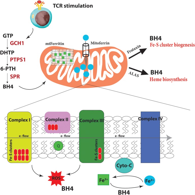
“We indeed observed increases at the protein level of Mitoferrin, the mitochondrial Fe2+ importer; HO-1, which breaks heme into Fe2+; ferritin, the storage regulator of Fe3+, as well as frataxin, responsible for Fe-S biogenesis.”
§ The Role of Iron Regulation in Immunometabolism and Immune-Related Disease > Tetrahydrobiopterin (BH4)—Linking Iron Homeostasis to Mitochondrial Function in Activated T Cells
What the sources say
- Not checked yetcites Cronin et al., 2018 source
plaindirect- Asserted: passes — The authors put it forward as true.
- Checkable: passes — It can be checked against evidence.
- Load-bearing: passes — The paper’s conclusions depend on it.
- Based: passes — The paper gives a reason why it holds.
- It may state two separate claims as one. 75
- Probably not a substantial claim. 18
The paper states this without a source or data behind it here, so there is nothing to check it against. The quote shows it is in the paper.
In the paper
“Under BH4 deficiency, T cells show dysfunctional ETC, enhanced ROS, and reduced ATP production in activated T cells.”
§ The Role of Iron Regulation in Immunometabolism and Immune-Related Disease > Tetrahydrobiopterin (BH4)—Linking Iron Homeostasis to Mitochondrial Function in Activated T Cells
plaindirect- Asserted: passes — The authors put it forward as true.
- Checkable: passes — It can be checked against evidence.
- Load-bearing: passes — The paper’s conclusions depend on it.
- Based: passes — The paper gives a reason why it holds.
It may state two separate claims as one.
In the paper
“Under conditions of BH4 deficiency, as in our Gch1-ablated T cells, cytochrome-C reduction is compromised, resulting in dysfunctional mitochondrial respiration, increased superoxide formation and, ultimately diminished ATP production, all of which we observed (Cronin et al., 2018).”
§ The Role of Iron Regulation in Immunometabolism and Immune-Related Disease > Tetrahydrobiopterin (BH4)—Linking Iron Homeostasis to Mitochondrial Function in Activated T Cells
What the sources say
- Not checked yetcites Cronin et al., 2018 source
plaindirect- Asserted: passes — The authors put it forward as true.
- Checkable: passes — It can be checked against evidence.
- Load-bearing: passes — The paper’s conclusions depend on it.
- Based: passes — The paper gives a reason why it holds.
It may state two separate claims as one.
In the paper
“Using multiple T cell specific ablation strategies, we demonstrated that genetic inactivation of GCH1 or pharmacological inhibition of the terminal enzyme in the biosynthetic pathway of BH4, sepiapterin reductase (SPR), results in severely impaired proliferation of T cells (Cronin et al., 2018).”
§ The Role of Iron Regulation in Immunometabolism and Immune-Related Disease > Tetrahydrobiopterin (BH4)—Linking Iron Homeostasis to Mitochondrial Function in Activated T Cells
What the sources say
- Not checked yetcites Cronin et al., 2018 source
boosteddirect- Asserted: passes — The authors put it forward as true.
- Checkable: passes — It can be checked against evidence.
- Load-bearing: passes — The paper’s conclusions depend on it.
- Based: passes — The paper gives a reason why it holds.
It may state two separate claims as one.
In the paper
“However, interestingly, T cell development or homeostasis as well as B cell and regulatory T cell (Treg) functions are unaffected by inhibition of this pathway.”
§ The Role of Iron Regulation in Immunometabolism and Immune-Related Disease > Tetrahydrobiopterin (BH4)—Linking Iron Homeostasis to Mitochondrial Function in Activated T Cells
What the sources say
- Not checked yetcites Cronin et al., 2018 source
plain- Asserted: passes — The authors put it forward as true.
- Checkable: passes — It can be checked against evidence.
- Load-bearing: unclear — It is unclear whether the paper’s conclusions depend on it.
- Based: passes — The paper gives a reason why it holds.
It may state two separate claims as one.
The paper states this without a source or data behind it here, so there is nothing to check it against. The quote shows it is in the paper.
In the paper
“Moreover, the T cells show dysregulated Mitoferrin, Frataxin, HO-1, Ferritin and overall reduced iron levels in the cell”
§ The Role of Iron Regulation in Immunometabolism and Immune-Related Disease > Tetrahydrobiopterin (BH4)—Linking Iron Homeostasis to Mitochondrial Function in Activated T Cells
plaindirect- Asserted: passes — The authors put it forward as true.
- Checkable: passes — It can be checked against evidence.
- Load-bearing: passes — The paper’s conclusions depend on it.
- Based: passes — The paper gives a reason why it holds.
It may state two separate claims as one.
The paper states this without a source or data behind it here, so there is nothing to check it against. The quote shows it is in the paper.
In the paper
“BH4 can not only act as a superoxide ROS scavenger but also directly reduce Fe3+ to Fe2+ and therefore affect cytochrome C (cyto-C) activity in the ETC.”
§ The Role of Iron Regulation in Immunometabolism and Immune-Related Disease > Tetrahydrobiopterin (BH4)—Linking Iron Homeostasis to Mitochondrial Function in Activated T Cells
plaindirect- Asserted: passes — The authors put it forward as true.
- Checkable: passes — It can be checked against evidence.
- Load-bearing: passes — The paper’s conclusions depend on it.
- Based: passes — The paper gives a reason why it holds.
It may state two separate claims as one.
In the paper
“In genetic inactivation models, we did not detect any differences in iNOS expression or NO production in T cells, nor any detectable signs of activated T cells producing biogenic amine neurotransmitters that require co-factor BH4.”
§ The Role of Iron Regulation in Immunometabolism and Immune-Related Disease > Tetrahydrobiopterin (BH4)—Linking Iron Homeostasis to Mitochondrial Function in Activated T Cells
What the sources say
- Not checked yetcites Cronin et al., 2018 source
plain- Asserted: passes — The authors put it forward as true.
- Checkable: passes — It can be checked against evidence.
- Load-bearing: passes — The paper’s conclusions depend on it.
- Based: passes — The paper gives a reason why it holds.
It may state two separate claims as one.
In the paper
“In orthotopic cancer transfer models, BH4-overproducing T cells have increased anti-cancer immunity, resulting in greatly reduced cancer burden (Cronin et al., 2018).”
§ The Role of Iron Regulation in Immunometabolism and Immune-Related Disease > Tetrahydrobiopterin (BH4)—Linking Iron Homeostasis to Mitochondrial Function in Activated T Cells
What the sources say
- Not checked yetcites Cronin et al., 2018 source
plaindirect- Asserted: passes — The authors put it forward as true.
- Checkable: passes — It can be checked against evidence.
- Load-bearing: passes — The paper’s conclusions depend on it.
- Based: passes — The paper gives a reason why it holds.
It may state two separate claims as one.
In the paper
“To this end, we developed a novel BH4 inhibitor, QM385, which targets SPR, and blocks both mouse and human T cell proliferation at nanomolar potency with good oral bioavailability and no observable side-effects at immune suppressant exposures (Cronin et al., 2018).”
§ The Role of Iron Regulation in Immunometabolism and Immune-Related Disease > Therapeutic Targeting of the BH4 Pathway to Control Autoimmunity and Cancer
What the sources say
- Not checked yetcites Cronin et al., 2018 source
plaindirect- Asserted: passes — The authors put it forward as true.
- Checkable: passes — It can be checked against evidence.
- Load-bearing: passes — The paper’s conclusions depend on it.
- Based: passes — The paper gives a reason why it holds.
It may state two separate claims as one.
In the paper
“In such activated T cells, BH4 regulates both the reduction of Fe3+ and increases the availability of Fe2+ in the cell, which aid activated T cells in coordinating the increased metabolic needs for effector functions.”
§ The Role of Iron Regulation in Immunometabolism and Immune-Related Disease > Conclusions
What the sources say
- Not checked yetcites Cronin et al., 2018 source
plaindirect- Asserted: passes — The authors put it forward as true.
- Checkable: passes — It can be checked against evidence.
- Load-bearing: passes — The paper’s conclusions depend on it.
- Based: passes — The paper gives a reason why it holds.
It may state two separate claims as one.
- Rests on an assumption: Fe2+ is needed for metalloproteins like cytochrome-C to function optimally, for Fe-S cluster biogenesis, which are essential for complex I and II activity of the ETC, and for heme production.
In the paper
“To this end, we developed a novel BH4 inhibitor, QM385, which targets SPR, and blocks both mouse and human T cell proliferation at nanomolar potency with good oral bioavailability and no observable side-effects at immune suppressant exposures (Cronin et al., 2018).”
§ The Role of Iron Regulation in Immunometabolism and Immune-Related Disease > Therapeutic Targeting of the BH4 Pathway to Control Autoimmunity and Cancer
What the sources say
- Not checked yetcites Cronin et al., 2018 source
plain- Asserted: passes — The authors put it forward as true.
- Checkable: passes — It can be checked against evidence.
- Load-bearing: passes — The paper’s conclusions depend on it.
- Based: passes — The paper gives a reason why it holds.
It may state two separate claims as one.
In the paper
“We also showed that by enhancing BH4 genetically, or pharmacologically with BH4 itself, T cells displayed heightened proliferation and, importantly, increased anti-tumor activity (Cronin et al., 2018).”
§ The Role of Iron Regulation in Immunometabolism and Immune-Related Disease > Therapeutic Targeting of the BH4 Pathway to Control Autoimmunity and Cancer
What the sources say
- Not checked yetcites Cronin et al., 2018 source
plaindirect- Asserted: passes — The authors put it forward as true.
- Checkable: passes — It can be checked against evidence.
- Load-bearing: passes — The paper’s conclusions depend on it.
- Based: passes — The paper gives a reason why it holds.
It may state two separate claims as one.
In the paper
“To this end, we developed a novel BH4 inhibitor, QM385, which targets SPR, and blocks both mouse and human T cell proliferation at nanomolar potency with good oral bioavailability and no observable side-effects at immune suppressant exposures (Cronin et al., 2018).”
§ The Role of Iron Regulation in Immunometabolism and Immune-Related Disease > Therapeutic Targeting of the BH4 Pathway to Control Autoimmunity and Cancer
What the sources say
- Not checked yetcites Cronin et al., 2018 source
plaindirect- Asserted: passes — The authors put it forward as true.
- Checkable: passes — It can be checked against evidence.
- Load-bearing: passes — The paper’s conclusions depend on it.
- Based: passes — The paper gives a reason why it holds.
It may state two separate claims as one.
In the paper
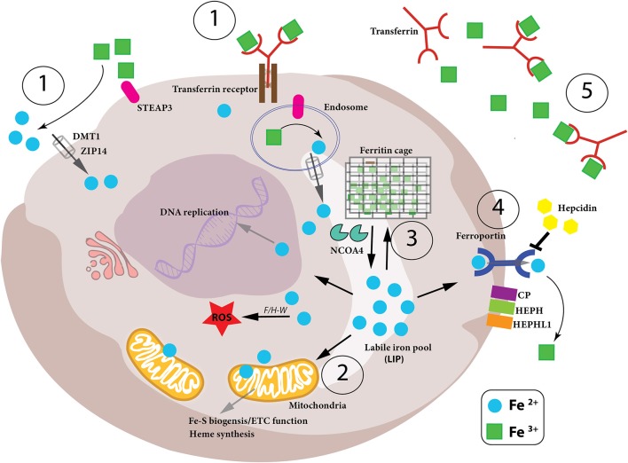
“Transferrin (Tf) is a glycoprotein containing two high affinity binding sites for ferric iron (Fe3+) (Aisen et al., 1978), which captures Fe3+ in the circulation (Figure 1).”
§ The Role of Iron Regulation in Immunometabolism and Immune-Related Disease > Iron Cycle—Cellular Uptake of Iron
What the sources say
- Not checked yetcites Aisen et al., 1978 source
plaindirect- Asserted: passes — The authors put it forward as true.
- Checkable: passes — It can be checked against evidence.
- Load-bearing: unclear — It is unclear whether the paper’s conclusions depend on it.
- Based: passes — The paper gives a reason why it holds.
It may state two separate claims as one.
In the paper
“Iron deficiency is the most common pathology of iron homeostasis, eventually resulting in iron deficiency anemia, the most frequent anemia worldwide (Camaschella, 2015).”
§ The Role of Iron Regulation in Immunometabolism and Immune-Related Disease > Introduction
What the sources say
- Not checked yetcites Camaschella, 2015 source
plaindirect- Asserted: passes — The authors put it forward as true.
- Checkable: passes — It can be checked against evidence.
- Load-bearing: fails — The paper’s conclusions may not depend on it.
- Based: passes — The paper gives a reason why it holds.
Probably not a substantial claim. The paper’s conclusions may not depend on it. It may state two separate claims as one.
In the paper
“Ferric iron in the circulation is then bound by transferrin (Tf) and carried to cells and tissues (Muckenthaler et al., 2017).”
§ The Role of Iron Regulation in Immunometabolism and Immune-Related Disease > Iron Cycle—Dietary Uptake of Iron
What the sources say
- Not checked yetcites Muckenthaler et al., 2017 source
plaindirect- Asserted: passes — The authors put it forward as true.
- Checkable: passes — It can be checked against evidence.
- Load-bearing: fails — The paper’s conclusions may not depend on it.
- Based: passes — The paper gives a reason why it holds.
Probably not a substantial claim. The paper’s conclusions may not depend on it.
In the paper
“This diferric Tf conjugate (Tf-Fe3+) not only prevents free, non-transferrin bound iron (NTBI), from engaging in Fenton chemistry to produce dangerous hydroxyl radicals, but also deprives invading pathogens of free iron to block their expansion and proliferation (Barber and Elde, 2014).”
§ The Role of Iron Regulation in Immunometabolism and Immune-Related Disease > Iron Cycle—Cellular Uptake of Iron
What the sources say
- Not checked yetcites Barber and Elde, 2014 source
plaindirect- Asserted: passes — The authors put it forward as true.
- Checkable: passes — It can be checked against evidence.
- Load-bearing: passes — The paper’s conclusions depend on it.
- Based: passes — The paper gives a reason why it holds.
It may state two separate claims as one.
None of the sources cited for this claim have been checked yet.
In the paper
“Accordingly, iron trafficking is controlled by cytokines and acute phase proteins, whereas the metal itself promotes lymphocyte and macrophage differentiation, anti-microbial immune effector function, and immune cell metabolism, as we will discuss later (Ganz and Nemeth, 2015; Soares and Weiss, 2015).”
§ The Role of Iron Regulation in Immunometabolism and Immune-Related Disease > Introduction
What the sources say
plaindirect- Asserted: passes — The authors put it forward as true.
- Checkable: passes — It can be checked against evidence.
- Load-bearing: passes — The paper’s conclusions depend on it.
- Based: passes — The paper gives a reason why it holds.
It may state two separate claims as one.
None of the sources cited for this claim have been checked yet.
In the paper
“The freed ferric iron is then reduced to its bio-active ferrous form, Fe2+ by the ferrireductase STEAP3 (six-transmembrane epithelial antigen of prostate 3) and shuttled into the cytosol from the endosome by DMT1 (Fleming et al., 1997; Gunshin et al., 1997; Ohgami et al., 2005; Figure 1).”
§ The Role of Iron Regulation in Immunometabolism and Immune-Related Disease > Iron Cycle—Cellular Uptake of Iron
What the sources say
plaindirect- Asserted: passes — The authors put it forward as true.
- Checkable: passes — It can be checked against evidence.
- Load-bearing: unclear — It is unclear whether the paper’s conclusions depend on it.
- Based: passes — The paper gives a reason why it holds.
It may state two separate claims as one.
In the paper
“It has also been demonstrated that heme-conjugated iron is absorbed by enterocytes in the duodenum although this heme importer has not yet been characterized (Gräsbeck et al., 1979).”
§ The Role of Iron Regulation in Immunometabolism and Immune-Related Disease > Iron Cycle—Dietary Uptake of Iron
What the sources say
- Not checked yetcites Gräsbeck et al., 1979 source
boosteddirect- Asserted: passes — The authors put it forward as true.
- Checkable: passes — It can be checked against evidence.
- Load-bearing: fails — The paper’s conclusions may not depend on it.
- Based: passes — The paper gives a reason why it holds.
Probably not a substantial claim. The paper’s conclusions may not depend on it. It may state two separate claims as one.
None of the sources cited for this claim have been checked yet.
In the paper
“This latter step is controlled by the iron exporter, ferroportin (FPN, also called SLC40A1) followed by extracellular oxidation to ferric iron by the copper enzyme hephaestin (HEPH) (Vulpe et al., 1999; Muckenthaler et al., 2017).”
§ The Role of Iron Regulation in Immunometabolism and Immune-Related Disease > Iron Cycle—Dietary Uptake of Iron
What the sources say
plaindirect- Asserted: passes — The authors put it forward as true.
- Checkable: passes — It can be checked against evidence.
- Load-bearing: unclear — It is unclear whether the paper’s conclusions depend on it.
- Based: passes — The paper gives a reason why it holds.
It may state two separate claims as one.
In the paper
“When systemic iron levels in the blood stream are low, expression of DMT1 (divalent metal transporter 1, also called NRAMP2 as well as SLC11A2) and DcytB (duodenal membrane associated cytochrome-b) ferrireductase increases on the surface of intestinal enterocytes on the luminal side enabling the reduction of Fe3+ into its ferrous form (Fe2+) which can then be taken up by DMT1 (McKie et al., 2001).”
§ The Role of Iron Regulation in Immunometabolism and Immune-Related Disease > Iron Cycle—Dietary Uptake of Iron
What the sources say
- Not checked yetcites McKie et al., 2001 source
plaindirect- Asserted: passes — The authors put it forward as true.
- Checkable: passes — It can be checked against evidence.
- Load-bearing: unclear — It is unclear whether the paper’s conclusions depend on it.
- Based: passes — The paper gives a reason why it holds.
It may state two separate claims as one.
In the paper
“Tf-Fe3+ binds to the high-affinity transferrin receptor (TFR1, CD71) on the surface of cells, and this complex is subsequently endocytosed (Harding et al., 1983).”
§ The Role of Iron Regulation in Immunometabolism and Immune-Related Disease > Iron Cycle—Cellular Uptake of Iron
What the sources say
- Not checked yetcites Harding et al., 1983 source
plaindirect- Asserted: passes — The authors put it forward as true.
- Checkable: passes — It can be checked against evidence.
- Load-bearing: unclear — It is unclear whether the paper’s conclusions depend on it.
- Based: passes — The paper gives a reason why it holds.
It may state two separate claims as one.
None of the sources cited for this claim have been checked yet.
In the paper
“Hepcidin overexpression leads to low plasma iron levels (hypoferremia) and anemia (Weinstein et al., 2002; Roy et al., 2007; Theurl et al., 2009; Altamura et al., 2014).”
§ The Role of Iron Regulation in Immunometabolism and Immune-Related Disease > Iron Cycle—Iron Export From the Cell
What the sources say
plaindirect- Asserted: passes — The authors put it forward as true.
- Checkable: passes — It can be checked against evidence.
- Load-bearing: unclear — It is unclear whether the paper’s conclusions depend on it.
- Based: passes — The paper gives a reason why it holds.
It may state two separate claims as one.
In the paper
“Caenorhabditis elegans fed on a mutant strain of Escherichia coli, which exhibits reduced iron uptake, lead to diminished iron uptake by the worms resulting in significantly impaired mitochondrial ETC function, enhanced ROS production and developmental defects (Zhang et al., 2019).”
§ The Role of Iron Regulation in Immunometabolism and Immune-Related Disease > Iron Cycle—Mitochondrial Utilization of Iron
What the sources say
- Not checked yetcites Zhang et al., 2019 source
plaindirect- Asserted: passes — The authors put it forward as true.
- Checkable: passes — It can be checked against evidence.
- Load-bearing: passes — The paper’s conclusions depend on it.
- Based: passes — The paper gives a reason why it holds.
It may state two separate claims as one.
In the paper
“Hepcidin then enters the circulation and binds to the extracellular part of FPN targeting it for degradation, thus preventing iron from being exported into the circulation and instead being sequestered in the cell (Nemeth et al., 2004).”
§ The Role of Iron Regulation in Immunometabolism and Immune-Related Disease > Iron Cycle—Iron Export From the Cell
What the sources say
- Not checked yetcites Nemeth et al., 2004 source
plaindirect- Asserted: passes — The authors put it forward as true.
- Checkable: passes — It can be checked against evidence.
- Load-bearing: passes — The paper’s conclusions depend on it.
- Based: passes — The paper gives a reason why it holds.
It may state two separate claims as one.
A conclusion the paper draws from its own results — it cites no other source for it, so there is none to check. The quote shows it is in the paper.
In the paper
“These reports demonstrate how certain cell types rely more heavily on TFR1-mediated iron uptake while other cell types have adapted other mechanisms to import iron into their cells.”
§ The Role of Iron Regulation in Immunometabolism and Immune-Related Disease > Iron Cycle—Cellular Uptake of Iron
boostedcorrelational- Asserted: passes — The authors put it forward as true.
- Checkable: passes — It can be checked against evidence.
- Load-bearing: unclear — It is unclear whether the paper’s conclusions depend on it.
- Based: passes — The paper gives a reason why it holds.
It may state two separate claims as one.
In the paper
“NCOA4 is a key regulator of ferritinophagy and intracellular iron levels—NCOA4-null mice are unable to undergo ferritinophagy and this increased retention of iron within ferritin complexes results in reduced iron export from cells and ultimately in iron-deficient anemia (Bellelli et al., 2014, 2016).”
§ The Role of Iron Regulation in Immunometabolism and Immune-Related Disease > Iron Cycle—Intracellular Storage of Iron
What the sources say
- Not matchedcites Bellelli et al., 2014, 2016
Couldn’t be matched to a paper in Crossref, so it can’t be checked.
plaindirect- Asserted: passes — The authors put it forward as true.
- Checkable: passes — It can be checked against evidence.
- Load-bearing: unclear — It is unclear whether the paper’s conclusions depend on it.
- Based: passes — The paper gives a reason why it holds.
It may state two separate claims as one.
- Not matched
In the paper
“The Fe source of the cluster is not yet known, although frataxin (FXN) has been identified as an important iron regulator of Fe-S biogenesis (Colin et al., 2013).”
§ The Role of Iron Regulation in Immunometabolism and Immune-Related Disease > Iron Cycle—Mitochondrial Utilization of Iron
What the sources say
- Not checked yetcites Colin et al., 2013 source
plaincorrelational- Asserted: passes — The authors put it forward as true.
- Checkable: passes — It can be checked against evidence.
- Load-bearing: fails — The paper’s conclusions may not depend on it.
- Based: passes — The paper gives a reason why it holds.
Probably not a substantial claim. The paper’s conclusions may not depend on it.
In the paper
“Furthermore, free heme and hemoglobin released during red blood cell (RBC) lysis are bound in the circulation by hemopexin and haptoglobin, respectively, and these iron-containing complexes are then taken up by cells expressing the CD91 and CD163 receptors (Nairz et al., 2017).”
§ The Role of Iron Regulation in Immunometabolism and Immune-Related Disease > Iron Cycle—Cellular Uptake of Iron
What the sources say
- Not checked yetcites Nairz et al., 2017 source
plaindirect- Asserted: passes — The authors put it forward as true.
- Checkable: passes — It can be checked against evidence.
- Load-bearing: unclear — It is unclear whether the paper’s conclusions depend on it.
- Based: passes — The paper gives a reason why it holds.
It may state two separate claims as one.
In the paper
“The key iron exporter is ferroportin (FPN, also called SLC40A1) which exports Fe2+ iron (McKie et al., 2000).”
§ The Role of Iron Regulation in Immunometabolism and Immune-Related Disease > Iron Cycle—Iron Export From the Cell
What the sources say
- Not checked yetcites McKie et al., 2000 source
plain- Asserted: passes — The authors put it forward as true.
- Checkable: passes — It can be checked against evidence.
- Load-bearing: unclear — It is unclear whether the paper’s conclusions depend on it.
- Based: passes — The paper gives a reason why it holds.
It may state two separate claims as one.
None of the sources cited for this claim have been checked yet.
In the paper
“Frataxin has been proposed to provide the iron while GLRX5 acts not only as a scaffolding protein but may also facilitate the transfer of Fe-S clusters to target proteins (Yoon and Cowan, 2003; Ye et al., 2010).”
§ The Role of Iron Regulation in Immunometabolism and Immune-Related Disease > Iron Cycle—Mitochondrial Utilization of Iron
What the sources say
- Not checked yetcites Yoon and Cowan, 2003
- Not checked yetcites Ye et al., 2010
hedgedconditional- Asserted: passes — The authors put it forward as true.
- Checkable: passes — It can be checked against evidence.
- Load-bearing: fails — The paper’s conclusions may not depend on it.
- Based: passes — The paper gives a reason why it holds.
Probably not a substantial claim. The paper’s conclusions may not depend on it. It may state two separate claims as one.
None of the sources cited for this claim have been checked yet.
In the paper
“Hepatic iron deposits can range from 300 mg to 1 g, but can reach up to 25–30 g in patients suffering from genetic iron overload, a condition called hereditary hemochromatosis (HH) (Janssen and Swinkels, 2009; Anderson and Shah, 2013; Procaccini and Kowdley, 2017).”
§ The Role of Iron Regulation in Immunometabolism and Immune-Related Disease > Iron Cycle—Iron Export From the Cell
What the sources say
- Not checked yetcites Janssen and Swinkels, 2009
- Not checked yetcites Anderson and Shah, 2013
- Not checked yetcites Procaccini and Kowdley, 2017
plain- Asserted: passes — The authors put it forward as true.
- Checkable: passes — It can be checked against evidence.
- Load-bearing: fails — The paper’s conclusions may not depend on it.
- Based: passes — The paper gives a reason why it holds.
Probably not a substantial claim. The paper’s conclusions may not depend on it.
None of the sources cited for this claim have been checked yet.
In the paper
“NF-kB, an important integrator transcription factor downstream from multiple inflammatory signals, as well as NRF2 (nuclear factor erythroid 2-related factor-2), the main stress-responsive transcription factor, both increase ferritin transcription (Miller et al., 1991; Pham et al., 2004).”
§ The Role of Iron Regulation in Immunometabolism and Immune-Related Disease > Iron Cycle—Intracellular Storage of Iron
What the sources say
plaindirect- Asserted: passes — The authors put it forward as true.
- Checkable: passes — It can be checked against evidence.
- Load-bearing: unclear — It is unclear whether the paper’s conclusions depend on it.
- Based: passes — The paper gives a reason why it holds.
It may state two separate claims as one.
In the paper
“Notably, as we discuss later, iron not readily used for metabolic purposes is stored by the protein ferritin and ferritin-conjugated iron released from various cells is taken up by Scara5 (Scavenger receptor class A member 5) or TIM-2 (T Cell Immunoglobulin And Mucin Domain Containing 2) receptors (Chen et al., 2005).”
§ The Role of Iron Regulation in Immunometabolism and Immune-Related Disease > Iron Cycle—Cellular Uptake of Iron
What the sources say
- Not checked yetcites Chen et al., 2005 source
plaindirect- Asserted: passes — The authors put it forward as true.
- Checkable: passes — It can be checked against evidence.
- Load-bearing: unclear — It is unclear whether the paper’s conclusions depend on it.
- Based: passes — The paper gives a reason why it holds.
It may state two separate claims as one.
In the paper
“There also exists a mitochondrial form of ferritin (Levi et al., 2001) which may both protect mitochondria against iron-mediated toxicity and provide a quick and efficient source of iron in this organelle rather than relying solely on the cytosolic ferritin.”
§ The Role of Iron Regulation in Immunometabolism and Immune-Related Disease > Iron Cycle—Intracellular Storage of Iron
What the sources say
- Not checked yetcites Levi et al., 2001 source
hedgedconditional- Asserted: passes — The authors put it forward as true.
- Checkable: passes — It can be checked against evidence.
- Load-bearing: unclear — It is unclear whether the paper’s conclusions depend on it.
- Based: passes — The paper gives a reason why it holds.
It may state two separate claims as one.
None of the sources cited for this claim have been checked yet.
In the paper
“Fe-S clusters are ubiquitous, inorganic co-factors that contribute to a wide range of cellular pathways from genome integrity and gene regulation to energy production and immune responses, and are required for numerous biological functions, particularly enzymatic activity (Beinert, 2000; Johnson et al., 2005; Lee et al., 2009; Fuss et al., 2015).”
§ The Role of Iron Regulation in Immunometabolism and Immune-Related Disease > Iron Cycle—Mitochondrial Utilization of Iron
What the sources say
plain- Asserted: passes — The authors put it forward as true.
- Checkable: passes — It can be checked against evidence.
- Load-bearing: fails — The paper’s conclusions may not depend on it.
- Based: passes — The paper gives a reason why it holds.
Probably not a substantial claim. The paper’s conclusions may not depend on it. It may state two separate claims as one.
None of the sources cited for this claim have been checked yet.
In the paper
“Mitochondrial iron is also required for the synthesis of Fe-S clusters and indeed their biogenesis is thought to be one of the most important functions of the mitochondria—even though some Fe-S assembly can occur in the cytosol and nucleus (Tong, 2000; Tong et al., 2003; Tong and Rouault, 2006).”
§ The Role of Iron Regulation in Immunometabolism and Immune-Related Disease > Iron Cycle—Mitochondrial Utilization of Iron
What the sources say
hedgeddirect- Asserted: passes — The authors put it forward as true.
- Checkable: passes — It can be checked against evidence.
- Load-bearing: unclear — It is unclear whether the paper’s conclusions depend on it.
- Based: passes — The paper gives a reason why it holds.
It may state two separate claims as one.
None of the sources cited for this claim have been checked yet.
In the paper
“The importance of these oxidases for successful iron export into the circulation has been demonstrated with gene-ablation murine models as well as identification of patients deficient in CP and HEPH, presenting with iron-deficient anemia (Yoshida et al., 1995; Hahn et al., 2004).”
§ The Role of Iron Regulation in Immunometabolism and Immune-Related Disease > Iron Cycle—Iron Export From the Cell
What the sources say
boosteddirect- Asserted: passes — The authors put it forward as true.
- Checkable: passes — It can be checked against evidence.
- Load-bearing: unclear — It is unclear whether the paper’s conclusions depend on it.
- Based: passes — The paper gives a reason why it holds.
It may state two separate claims as one.
In the paper
“However, pathogens are constantly evolving and adapting to host responses and have created modified siderophores, such as the heavily glycoslyated siderophore, salmochelin, secreted by Salmonella typhimurium, which is not recognized and targeted by lipocalin-2 thus making these bacteria unresponsive to this host defense strategy (Hantke et al., 2003).”
§ The Role of Iron Regulation in Immunometabolism and Immune-Related Disease > Immune Pathologies Associated With Iron Dysregulation
What the sources say
- Not checked yetcites Hantke et al., 2003 source
plaindirect- Asserted: passes — The authors put it forward as true.
- Checkable: passes — It can be checked against evidence.
- Load-bearing: unclear — It is unclear whether the paper’s conclusions depend on it.
- Based: passes — The paper gives a reason why it holds.
It may state two separate claims as one.
None of the sources cited for this claim have been checked yet.
In the paper
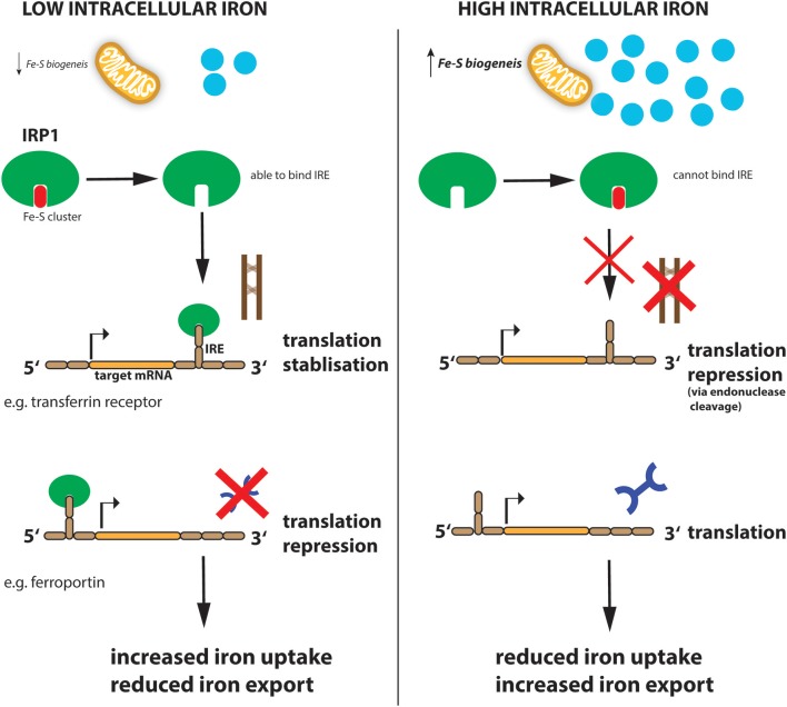
“However, IRP1 binding to the 3′ UTR increases the stability of the respective target mRNAs such as TFR and DMT1, thereby promoting their expression and subsequently enhancing iron uptake into the cell to replenish intracellular iron levels.”
§ The Role of Iron Regulation in Immunometabolism and Immune-Related Disease > Iron Cycle—Iron Export From the Cell
plaindirect- Asserted: passes — The authors put it forward as true.
- Checkable: passes — It can be checked against evidence.
- Load-bearing: unclear — It is unclear whether the paper’s conclusions depend on it.
- Based: passes — The paper gives a reason why it holds.
It may state two separate claims as one.
In the paper
“Other related hemoproteins include salivary peroxidase and lactoperoxidase (LPO) which are present in multiple human exocrine secretions, including tears, milk, saliva, and vaginal fluid, and also serves anti-microbial killing actions similar to MPO (Sarr et al., 2018).”
§ The Role of Iron Regulation in Immunometabolism and Immune-Related Disease > Neutrophils and Iron
What the sources say
- Not checked yetcites Sarr et al., 2018 source
plaindirect- Asserted: passes — The authors put it forward as true.
- Checkable: passes — It can be checked against evidence.
- Load-bearing: fails — The paper’s conclusions may not depend on it.
- Based: passes — The paper gives a reason why it holds.
Probably not a substantial claim. The paper’s conclusions may not depend on it. It may state two separate claims as one.
In the paper
“Erythroferrone (ERFE) is produced by erythropoietin (EPO)-stimulation of erythroid progenitors and blocks hepcidin synthesis in the hepatocytes (Kautz et al., 2014).”
§ The Role of Iron Regulation in Immunometabolism and Immune-Related Disease > Iron Cycle—Iron Export From the Cell
What the sources say
- Not checked yetcites Kautz et al., 2014 source
plaindirect- Asserted: passes — The authors put it forward as true.
- Checkable: passes — It can be checked against evidence.
- Load-bearing: unclear — It is unclear whether the paper’s conclusions depend on it.
- Based: passes — The paper gives a reason why it holds.
It may state two separate claims as one.
In the paper
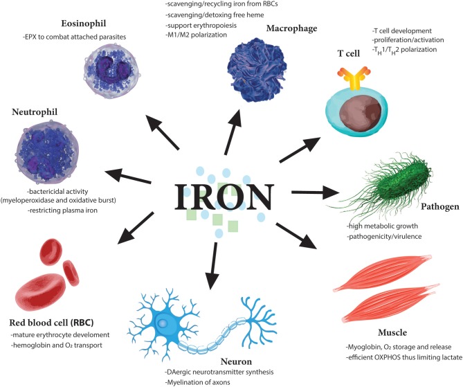
“In addition, neutrophils produce large amounts of calprotectin and lactoferrin, both of which scavenge iron, thereby inhibiting bacterial proliferation (Nakashige et al., 2015).”
§ The Role of Iron Regulation in Immunometabolism and Immune-Related Disease > Neutrophils and Iron
What the sources say
- Not checked yetcites Nakashige et al., 2015 source
plaindirect- Asserted: passes — The authors put it forward as true.
- Checkable: passes — It can be checked against evidence.
- Load-bearing: unclear — It is unclear whether the paper’s conclusions depend on it.
- Based: passes — The paper gives a reason why it holds.
It may state two separate claims as one.
In the paper
“Conversely, individuals with insufficient hepcidin production, suffer from iron-overload in the blood, and hepatitis (Pietrangelo, 2004).”
§ The Role of Iron Regulation in Immunometabolism and Immune-Related Disease > Iron Cycle—Iron Export From the Cell
What the sources say
- Not checked yetcites Pietrangelo, 2004 source
plaindirect- Asserted: passes — The authors put it forward as true.
- Checkable: passes — It can be checked against evidence.
- Load-bearing: unclear — It is unclear whether the paper’s conclusions depend on it.
- Based: passes — The paper gives a reason why it holds.
It may state two separate claims as one.
In the paper
“Indeed, IRP2 deficiency switches cellular metabolic pathways from oxidative phosphorylation (OXPHOS) to aerobic glycolysis (Li et al., 2019) through induction of hypoxia-inducible factors (HIF)-1α and−2α which enhances glycolytic pathway proteins and, at the same time, blocks mitochondrial Fe-S cluster biogenesis and OXPHOS.”
§ The Role of Iron Regulation in Immunometabolism and Immune-Related Disease > Iron Cycle—Iron Export From the Cell
What the sources say
- Not checked yetcites Li et al., 2019
plaindirect- Asserted: passes — The authors put it forward as true.
- Checkable: passes — It can be checked against evidence.
- Load-bearing: unclear — It is unclear whether the paper’s conclusions depend on it.
- Based: passes — The paper gives a reason why it holds.
It may state two separate claims as one.
None of the sources cited for this claim have been checked yet.
In the paper
“When iron concentrations are low, IRP1 loses it Fe-S cluster and, therefore, its catalytic abilities but instead can now bind to IREs in the 5′ UTR of mRNAs such as ferritin-H and -L subunits as well as the rate-limiting enzyme of heme biogenesis, ALAS (aminolevulinic acid synthase) which results in inhibition of their translation and thus reduced storage and utilization of the metal (Figure 2).”
§ The Role of Iron Regulation in Immunometabolism and Immune-Related Disease > Iron Cycle—Iron Export From the Cell
plaindirect- Asserted: passes — The authors put it forward as true.
- Checkable: passes — It can be checked against evidence.
- Load-bearing: unclear — It is unclear whether the paper’s conclusions depend on it.
- Based: passes — The paper gives a reason why it holds.
It may state two separate claims as one.
In the paper
“Siderophores are low molecular weight iron-binding complexes that are secreted by bacteria to compete with transferrin for iron sequestration (Williams, 1988).”
§ The Role of Iron Regulation in Immunometabolism and Immune-Related Disease > Immune Pathologies Associated With Iron Dysregulation
What the sources say
- Not checked yetcites Williams, 1988 source
plaindirect- Asserted: passes — The authors put it forward as true.
- Checkable: passes — It can be checked against evidence.
- Load-bearing: fails — The paper’s conclusions may not depend on it.
- Based: passes — The paper gives a reason why it holds.
Probably not a substantial claim. The paper’s conclusions may not depend on it.
In the paper
“Superoxide radicals can free up Fe2+ iron and this can further react with hydrogen peroxide (H2O2) to give rise to the hydroxyl radicals (OH•) (Koskenkorva-Frank et al., 2013).”
§ The Role of Iron Regulation in Immunometabolism and Immune-Related Disease > Neutrophils and Iron
What the sources say
- Not checked yetcites Koskenkorva-Frank et al., 2013 source
plaindirect- Asserted: passes — The authors put it forward as true.
- Checkable: passes — It can be checked against evidence.
- Load-bearing: unclear — It is unclear whether the paper’s conclusions depend on it.
- Based: passes — The paper gives a reason why it holds.
It may state two separate claims as one.
A conclusion the paper draws from its own results — it cites no other source for it, so there is none to check. The quote shows it is in the paper.
In the paper
“Therefore, the availability and restriction of systemic iron during times of infection and inflammation represents an essential defense mechanism against pathogens but under pathophysiological conditions can lead to anemia and further severe pathologies resulting from reduced RBC development.”
§ The Role of Iron Regulation in Immunometabolism and Immune-Related Disease > Immune Pathologies Associated With Iron Dysregulation
plaindirect- Asserted: passes — The authors put it forward as true.
- Checkable: passes — It can be checked against evidence.
- Load-bearing: passes — The paper’s conclusions depend on it.
- Based: passes — The paper gives a reason why it holds.
It may state two separate claims as one.
In the paper
“However, when iron levels are low, FBXL5 is itself degraded. This is due to a Fe-binding N-terminal hemerythrin-like (Hr) iron-sensing domain in FBXL5. The Hr domain undergoes a conformational change when iron is not present—this structural change leads to FBXL5 polyubiquitination and degradation resulting in IRP2 accumulation (Thompson et al., 2012).”
§ The Role of Iron Regulation in Immunometabolism and Immune-Related Disease > Iron Cycle—Iron Export From the Cell
What the sources say
- Not checked yetcites Thompson et al., 2012 source
plaindirect- Asserted: passes — The authors put it forward as true.
- Checkable: passes — It can be checked against evidence.
- Load-bearing: unclear — It is unclear whether the paper’s conclusions depend on it.
- Based: passes — The paper gives a reason why it holds.
It may state two separate claims as one.
None of the sources cited for this claim have been checked yet.
In the paper
“Imbalanced iron levels in the cell can lead to dysfunctional mitochondria, increased oxidative damage and increased toxicity (Richardson et al., 2010; Volani et al., 2017).”
§ The Role of Iron Regulation in Immunometabolism and Immune-Related Disease > Iron Cycle—Iron Export From the Cell
What the sources say
plaindirect- Asserted: passes — The authors put it forward as true.
- Checkable: passes — It can be checked against evidence.
- Load-bearing: unclear — It is unclear whether the paper’s conclusions depend on it.
- Based: passes — The paper gives a reason why it holds.
It may state two separate claims as one.
None of the sources cited for this claim have been checked yet.
In the paper
“When iron levels normalize again, IRP1 regains its Fe-S cluster and aconitase activity. This results in increased ferritin translation and decreased TFR expression, resulting in reduced Fe uptake (Li et al., 2004; Rouault, 2006; Leipuviene and Theil, 2007; Sanchez et al., 2011; Figure 2).”
§ The Role of Iron Regulation in Immunometabolism and Immune-Related Disease > Iron Cycle—Iron Export From the Cell
What the sources say
- Not checked yetcites Li et al., 2004
- Not checked yetcites Rouault, 2006 source
- Not checked yetcites Leipuviene and Theil, 2007
plaindirect- Asserted: passes — The authors put it forward as true.
- Checkable: passes — It can be checked against evidence.
- Load-bearing: unclear — It is unclear whether the paper’s conclusions depend on it.
- Based: passes — The paper gives a reason why it holds.
It may state two separate claims as one.
None of the sources cited for this claim have been checked yet.
In the paper
“Macrophages and Kupffer cells scavenge heme from damaged or aged RBCs in the bone marrow, liver and spleen, and extract the Fe2+ iron using HO-1 and then either exports it back into the circulation through FPN or stores it in ferritin intracellularly (Knutson et al., 2005; Theurl et al., 2016).”
§ The Role of Iron Regulation in Immunometabolism and Immune-Related Disease > Macrophages and Iron
What the sources say
plaindirect- Asserted: passes — The authors put it forward as true.
- Checkable: passes — It can be checked against evidence.
- Load-bearing: passes — The paper’s conclusions depend on it.
- Based: passes — The paper gives a reason why it holds.
It may state two separate claims as one.
In the paper
“Furthermore, hematopoietic-specific deletion of the ferritin heavy chain (Fth) gene results in reduced numbers of lymphocytes while other cell types such as granulocytes and monocytes are unaffected, again highlighting the essential nature of iron regulation to the adaptive immune system (Vanoaica et al., 2014).”
§ The Role of Iron Regulation in Immunometabolism and Immune-Related Disease > T Cells and Iron
What the sources say
- Not checked yetcites Vanoaica et al., 2014 source
plaindirect- Asserted: passes — The authors put it forward as true.
- Checkable: passes — It can be checked against evidence.
- Load-bearing: passes — The paper’s conclusions depend on it.
- Based: passes — The paper gives a reason why it holds.
It may state two separate claims as one.
In the paper
“In mice, this is done by increased production of nitric oxide (NO) (Nairz et al., 2013), which activates the stress-responsive transcription factor, NRF2 resulting in transcriptional induction of FPN expression leading to subsequent increase of iron export from the cell, thus again restricting iron availability to the intracellular pathogen.”
§ The Role of Iron Regulation in Immunometabolism and Immune-Related Disease > Macrophages and Iron
What the sources say
- Not checked yetcites Nairz et al., 2013 source
plaindirect- Asserted: passes — The authors put it forward as true.
- Checkable: passes — It can be checked against evidence.
- Load-bearing: passes — The paper’s conclusions depend on it.
- Based: passes — The paper gives a reason why it holds.
It may state two separate claims as one.
In the paper
“Ablation of Fth leads to an increase in the labile iron pool (LIP) within the cell contributing to increased oxidative stress and cell death, though when cells are stimulated the increased availability of Fe2+ iron enhances proliferative capacity before ultimately undergoing cell death, possibly due to proliferating cells using up the excess iron (Vanoaica et al., 2014).”
§ The Role of Iron Regulation in Immunometabolism and Immune-Related Disease > T Cells and Iron
What the sources say
- Not checked yetcites Vanoaica et al., 2014 source
hedgeddirect- Asserted: passes — The authors put it forward as true.
- Checkable: passes — It can be checked against evidence.
- Load-bearing: passes — The paper’s conclusions depend on it.
- Based: passes — The paper gives a reason why it holds.
It may state two separate claims as one.
None of the sources cited for this claim have been checked yet.
In the paper
“However, degeneration of cells in the brain also result in the deposition of iron in the extracellular space, which then induces waves of oxidative stress (via Fenton chemistry) in these brain regions (Craelius et al., 1982; Bagnato et al., 2011).”
§ The Role of Iron Regulation in Immunometabolism and Immune-Related Disease > Macrophages and Iron
What the sources say
- Not checked yetcites Craelius et al., 1982
- Not checked yetcites Bagnato et al., 2011 source
plaindirect- Asserted: passes — The authors put it forward as true.
- Checkable: passes — It can be checked against evidence.
- Load-bearing: unclear — It is unclear whether the paper’s conclusions depend on it.
- Based: passes — The paper gives a reason why it holds.
It may state two separate claims as one.
In the paper
“However, unlike neutrophils which release MPO into the phagosome, eosinophils release their EPX-containing granules extracellularly to kill parasites to which they are attached (Acharya and Ackerman, 2014).”
§ The Role of Iron Regulation in Immunometabolism and Immune-Related Disease > Neutrophils and Iron
What the sources say
- Not checked yetcites Acharya and Ackerman, 2014
plaindirect- Asserted: passes — The authors put it forward as true.
- Checkable: passes — It can be checked against evidence.
- Load-bearing: fails — The paper’s conclusions may not depend on it.
- Based: passes — The paper gives a reason why it holds.
Probably not a substantial claim. The paper’s conclusions may not depend on it.
In the paper
“Patients with iron-overload in beta-thalassemia major have decreased CD4+ T cells but increased CD8+ T cell numbers (Porto and De Sousa, 2007), while patients suffering from hereditary hemochromatosis (HH) have apparently normal CD4+ and reduced CD8+ numbers, with the latter displaying a more “effector” phenotype, suggesting again that increased intracellular iron availability may have hyper-prol”
§ The Role of Iron Regulation in Immunometabolism and Immune-Related Disease > T Cells and Iron
What the sources say
- Not checked yetcites Porto and De Sousa, 2007
plaincorrelational- Asserted: passes — The authors put it forward as true.
- Checkable: passes — It can be checked against evidence.
- Load-bearing: unclear — It is unclear whether the paper’s conclusions depend on it.
- Based: passes — The paper gives a reason why it holds.
It may state two separate claims as one.
In the paper
“However, in the context of the tumor microenvironment (TME), the situation is quite complicated as M2 TAMs (tumor-infiltrating macrophages) which are “iron-releasing” will support tumor growth while M1 “iron-retaining” limit tumor progression (Jung et al., 2019).”
§ The Role of Iron Regulation in Immunometabolism and Immune-Related Disease > Macrophages and Iron
What the sources say
- Not checked yetcites Jung et al., 2019 source
plaindirect- Asserted: passes — The authors put it forward as true.
- Checkable: passes — It can be checked against evidence.
- Load-bearing: passes — The paper’s conclusions depend on it.
- Based: passes — The paper gives a reason why it holds.
It may state two separate claims as one.
None of the sources cited for this claim have been checked yet.
In the paper
“NRAMP1 acts as a transporter for divalent metals, including iron, and promotes iron export from the phagolyosome and then from the cytoplasm by induction of FPN expression (Nairz et al., 2007; Lim et al., 2018).”
§ The Role of Iron Regulation in Immunometabolism and Immune-Related Disease > Macrophages and Iron
What the sources say
plaindirect- Asserted: passes — The authors put it forward as true.
- Checkable: passes — It can be checked against evidence.
- Load-bearing: unclear — It is unclear whether the paper’s conclusions depend on it.
- Based: passes — The paper gives a reason why it holds.
It may state two separate claims as one.
In the paper
“This allows the M2 macrophages to promote iron recirculation and aid in tissue healing, a process which requires large amount of iron to rebuild damaged tissue (Recalcati et al., 2010a).”
§ The Role of Iron Regulation in Immunometabolism and Immune-Related Disease > Macrophages and Iron
What the sources say
- Not checked yetcites Recalcati et al., 2010a
plaindirect- Asserted: passes — The authors put it forward as true.
- Checkable: passes — It can be checked against evidence.
- Load-bearing: unclear — It is unclear whether the paper’s conclusions depend on it.
- Based: passes — The paper gives a reason why it holds.
It may state two separate claims as one.
In the paper
“Interestingly, eosinophils have a homolog of MPO called eosinophil peroxidase (EPX) which shares ~70% similarity at the amino acid level with neutrophilic MPO (Ten et al., 1989).”
§ The Role of Iron Regulation in Immunometabolism and Immune-Related Disease > Neutrophils and Iron
What the sources say
- Not checked yetcites Ten et al., 1989
plain- Asserted: passes — The authors put it forward as true.
- Checkable: passes — It can be checked against evidence.
- Load-bearing: fails — The paper’s conclusions may not depend on it.
- Based: passes — The paper gives a reason why it holds.
Probably not a substantial claim. The paper’s conclusions may not depend on it.
None of the sources cited for this claim have been checked yet.
In the paper
“Thus, HO-1 exerts cytoprotective effects by reducing the pro-oxidative activity of heme and by re-programming metabolic traits, thereby limiting tissue damage in the course of inflammatory processes, including sepsis (Gozzelino et al., 2010; Weis et al., 2017).”
§ The Role of Iron Regulation in Immunometabolism and Immune-Related Disease > T Cells and Iron
What the sources say
plaindirect- Asserted: passes — The authors put it forward as true.
- Checkable: passes — It can be checked against evidence.
- Load-bearing: unclear — It is unclear whether the paper’s conclusions depend on it.
- Based: passes — The paper gives a reason why it holds.
It may state two separate claims as one.
In the paper
“Such microglial dystrophy has been observed in Alzheimer's brains as well as aged brains (Lopes et al., 2008).”
§ The Role of Iron Regulation in Immunometabolism and Immune-Related Disease > Macrophages and Iron
What the sources say
- Not checked yetcites Lopes et al., 2008 source
plaincorrelational- Asserted: passes — The authors put it forward as true.
- Checkable: passes — It can be checked against evidence.
- Load-bearing: fails — The paper’s conclusions may not depend on it.
- Based: passes — The paper gives a reason why it holds.
Probably not a substantial claim. The paper’s conclusions may not depend on it.
In the paper
“Indeed within a minute of T cell receptor (TCR) engagement, surface TFR is upregulated from endosomal compartments and is localized to the immunological synapse (IS), the receptor-filled contact area between an antigen-presenting cell and a T cell, necessary for full T cell activation (Batista et al., 2004).”
§ The Role of Iron Regulation in Immunometabolism and Immune-Related Disease > T Cells and Iron
What the sources say
- Not checked yetcites Batista et al., 2004 source
plaindirect- Asserted: passes — The authors put it forward as true.
- Checkable: passes — It can be checked against evidence.
- Load-bearing: unclear — It is unclear whether the paper’s conclusions depend on it.
- Based: passes — The paper gives a reason why it holds.
It may state two separate claims as one.
In the paper
“In lung cancer for example, iron-laden TAMs have been demonstrated to enhance production of ROS and pro-inflammatory cytokines (TNFα and IL-6) and to directly kill tumor cells (da Silva et al., 2017), prompting the exciting possibility of delivering iron to TAMs as a simple adjuvant therapeutic strategy.”
§ The Role of Iron Regulation in Immunometabolism and Immune-Related Disease > Macrophages and Iron
What the sources say
- Not checked yetcites da Silva et al., 2017 source
boosteddirect- Asserted: passes — The authors put it forward as true.
- Checkable: passes — It can be checked against evidence.
- Load-bearing: passes — The paper’s conclusions depend on it.
- Based: passes — The paper gives a reason why it holds.
It may state two separate claims as one.
In the paper
“Heme scavenging proteins such as hemopexin can bind free heme, thus clearing it from the circulation and delivering it to cells (Smith et al., 1988).”
§ The Role of Iron Regulation in Immunometabolism and Immune-Related Disease > T Cells and Iron
What the sources say
- Not checked yetcites Smith et al., 1988 source
plaindirect- Asserted: passes — The authors put it forward as true.
- Checkable: passes — It can be checked against evidence.
- Load-bearing: passes — The paper’s conclusions depend on it.
- Based: passes — The paper gives a reason why it holds.
It may state two separate claims as one.
None of the sources cited for this claim have been checked yet.
In the paper
“In contrast, M1 macrophages enhance iron uptake and storage, and display an attenuated iron-release phenotype favoring intracellular iron sequestration and storage, which has been shown to enhance antimicrobial effector functions such as increased pro-inflammatory TNFα expression while suppressing expression of the anti-inflammatory cytokine, IL-10 (Weiss et al., 1994; Mulero et al., 2002; Fritsch”
§ The Role of Iron Regulation in Immunometabolism and Immune-Related Disease > Macrophages and Iron
What the sources say
boosteddirect- Asserted: passes — The authors put it forward as true.
- Checkable: passes — It can be checked against evidence.
- Load-bearing: passes — The paper’s conclusions depend on it.
- Based: passes — The paper gives a reason why it holds.
It may state two separate claims as one.
In the paper
“GTP cyclohydrolase I (GCH1) is the rate-limiting enzyme for BH4 biosynthesis (Werner et al., 2011; Figure 4).”
§ The Role of Iron Regulation in Immunometabolism and Immune-Related Disease > Tetrahydrobiopterin (BH4)—Linking Iron Homeostasis to Mitochondrial Function in Activated T Cells
What the sources say
- Not checked yetcites Werner et al., 2011 source
plaindirect- Asserted: passes — The authors put it forward as true.
- Checkable: passes — It can be checked against evidence.
- Load-bearing: fails — The paper’s conclusions may not depend on it.
- Based: passes — The paper gives a reason why it holds.
Probably not a substantial claim. The paper’s conclusions may not depend on it.
In the paper
“Levels of intracellular iron were increased significantly in SLE CD4+ T cells compared to healthy controls and the researchers suggest a link between iron homeostasis and global DNA methylation status (Zhao et al., 2018), by which SLE T cells exhibit reduced DNA demethylation resulting in enhanced immune-related gene expression.”
§ The Role of Iron Regulation in Immunometabolism and Immune-Related Disease > T Cells and Iron
What the sources say
- Not checked yetcites Zhao et al., 2018 source
hedgeddirect- Asserted: passes — The authors put it forward as true.
- Checkable: passes — It can be checked against evidence.
- Load-bearing: passes — The paper’s conclusions depend on it.
- Based: passes — The paper gives a reason why it holds.
It may state two separate claims as one.
None of the sources cited for this claim have been checked yet.
In the paper
“GM-CSF is essential for the development and progression of EAE. Mice deficient in GM-CSF are resistant to EAE induction, and blockade of GM-CSF in wild-type mice suppresses ongoing disease (McQualter et al., 2001; Codarri et al., 2011).”
§ The Role of Iron Regulation in Immunometabolism and Immune-Related Disease > T Cells and Iron
What the sources say
plaindirect- Asserted: passes — The authors put it forward as true.
- Checkable: passes — It can be checked against evidence.
- Load-bearing: passes — The paper’s conclusions depend on it.
- Based: passes — The paper gives a reason why it holds.
It may state two separate claims as one.
A conclusion the paper draws from its own results — it cites no other source for it, so there is none to check. The quote shows it is in the paper.
In the paper
“suggesting that BH4 deficiency affects iron metabolism and the cell compensates by trying to increase Fe2+ levels in the mitochondria.”
§ The Role of Iron Regulation in Immunometabolism and Immune-Related Disease > Tetrahydrobiopterin (BH4)—Linking Iron Homeostasis to Mitochondrial Function in Activated T Cells
hedgeddirect- Asserted: passes — The authors put it forward as true.
- Checkable: passes — It can be checked against evidence.
- Load-bearing: passes — The paper’s conclusions depend on it.
- Based: passes — The paper gives a reason why it holds.
It may state two separate claims as one.
In the paper
“Ferroptosis is an iron-dependent oxidative form of cell death associated with increased lipid peroxidation (Xie et al., 2016).”
§ The Role of Iron Regulation in Immunometabolism and Immune-Related Disease > T Cells and Iron
What the sources say
- Not checked yetcites Xie et al., 2016 source
plaincorrelational- Asserted: passes — The authors put it forward as true.
- Checkable: passes — It can be checked against evidence.
- Load-bearing: passes — The paper’s conclusions depend on it.
- Based: passes — The paper gives a reason why it holds.
Probably not a substantial claim.
In the paper
“It was demonstrated that iron protects PCBP1 (Poly(RC)-binding protein 1), an RNA binding protein which stabilizes GMCSF mRNA, from caspase-mediated proteolysis thus leading to enhanced GMCSF production when iron levels are high within the T cell to drive inflammation (Wang et al., 2018).”
§ The Role of Iron Regulation in Immunometabolism and Immune-Related Disease > T Cells and Iron
What the sources say
- Not checked yetcites Wang et al., 2018 source
boosteddirect- Asserted: passes — The authors put it forward as true.
- Checkable: passes — It can be checked against evidence.
- Load-bearing: passes — The paper’s conclusions depend on it.
- Based: passes — The paper gives a reason why it holds.
It may state two separate claims as one.
In the paper
“Through these enzymes, BH4 is required for nitric oxide (NO) production, metabolism of ether lipids, phenylalanine catabolism, and synthesis of the amine neurotransmitters norepinephrine, epinephrine, serotonin, and dopamine.”
§ The Role of Iron Regulation in Immunometabolism and Immune-Related Disease > Tetrahydrobiopterin (BH4)—Linking Iron Homeostasis to Mitochondrial Function in Activated T Cells
What the sources say
- Not checked yetcites Werner et al., 2011 source
plaindirect- Asserted: passes — The authors put it forward as true.
- Checkable: passes — It can be checked against evidence.
- Load-bearing: fails — The paper’s conclusions may not depend on it.
- Based: passes — The paper gives a reason why it holds.
Probably not a substantial claim. The paper’s conclusions may not depend on it. It may state two separate claims as one.
In the paper
“Moreover, ferroptosis can be blocked with iron chelators but not with inhibitors of apoptosis and necroptosis (Xie et al., 2016).”
§ The Role of Iron Regulation in Immunometabolism and Immune-Related Disease > T Cells and Iron
What the sources say
- Not checked yetcites Xie et al., 2016 source
plaindirect- Asserted: passes — The authors put it forward as true.
- Checkable: passes — It can be checked against evidence.
- Load-bearing: passes — The paper’s conclusions depend on it.
- Based: passes — The paper gives a reason why it holds.
It may state two separate claims as one.
In the paper
“A common phenotype of defective Fe-S biogenesis is mitochondrial iron loading, resulting in mitochondrial dysfunction and oxidative stress (Reeve et al., 2012).”
§ The Role of Iron Regulation in Immunometabolism and Immune-Related Disease > Tetrahydrobiopterin (BH4)—Linking Iron Homeostasis to Mitochondrial Function in Activated T Cells
What the sources say
- Not checked yetcites Reeve et al., 2012 source
plaindirect- Asserted: passes — The authors put it forward as true.
- Checkable: passes — It can be checked against evidence.
- Load-bearing: passes — The paper’s conclusions depend on it.
- Based: passes — The paper gives a reason why it holds.
It may state two separate claims as one.
In the paper
“while patients suffering from hereditary hemochromatosis (HH) have apparently normal CD4+ and reduced CD8+ numbers, with the latter displaying a more “effector” phenotype, suggesting again that increased intracellular iron availability may have hyper-proliferative effects in T cells, although this may well-depend on which oxidation form of Fe is available (Costa et al., 2015).”
§ The Role of Iron Regulation in Immunometabolism and Immune-Related Disease > T Cells and Iron
What the sources say
- Not checked yetcites Costa et al., 2015 source
hedgedconditional- Asserted: passes — The authors put it forward as true.
- Checkable: passes — It can be checked against evidence.
- Load-bearing: unclear — It is unclear whether the paper’s conclusions depend on it.
- Based: passes — The paper gives a reason why it holds.
It may state two separate claims as one.
In the paper
“Intriguingly, sulfasalazine, a drug used for decades to treat autoimmune inflammatory bowel disease (IBD) and psoriatic arthritis (Gupta et al., 1990), is now recognized as targeting the BH4 pathway by inhibiting SPR (Haruki et al., 2013), in addition to other targets, such as NFκB (Wahl et al., 1998).”
§ The Role of Iron Regulation in Immunometabolism and Immune-Related Disease > Therapeutic Targeting of the BH4 Pathway to Control Autoimmunity and Cancer
What the sources say
- Not checked yetcites Gupta et al., 1990 source
plain- Asserted: passes — The authors put it forward as true.
- Checkable: passes — It can be checked against evidence.
- Load-bearing: fails — The paper’s conclusions may not depend on it.
- Based: passes — The paper gives a reason why it holds.
Probably not a substantial claim. The paper’s conclusions may not depend on it. It may state two separate claims as one.
None of the sources cited for this claim have been checked yet.
In the paper
“IFNγ has a strong regulatory role in iron homeostasis as it enhances the expression of cellular iron-regulating genes such as HO-1 but also reduces the iron-exporting gene ferroportin (Ludwiczek et al., 2003; Nairz et al., 2015).”
§ The Role of Iron Regulation in Immunometabolism and Immune-Related Disease > Tetrahydrobiopterin (BH4)—Linking Iron Homeostasis to Mitochondrial Function in Activated T Cells
What the sources say
boosteddirect- Asserted: passes — The authors put it forward as true.
- Checkable: passes — It can be checked against evidence.
- Load-bearing: passes — The paper’s conclusions depend on it.
- Based: passes — The paper gives a reason why it holds.
It may state two separate claims as one.
In the paper
“Intriguingly, sulfasalazine, a drug used for decades to treat autoimmune inflammatory bowel disease (IBD) and psoriatic arthritis (Gupta et al., 1990), is now recognized as targeting the BH4 pathway by inhibiting SPR (Haruki et al., 2013), in addition to other targets, such as NFκB (Wahl et al., 1998).”
§ The Role of Iron Regulation in Immunometabolism and Immune-Related Disease > Therapeutic Targeting of the BH4 Pathway to Control Autoimmunity and Cancer
What the sources say
- Not checked yetcites Wahl et al., 1998 source
plaindirect- Asserted: passes — The authors put it forward as true.
- Checkable: passes — It can be checked against evidence.
- Load-bearing: fails — The paper’s conclusions may not depend on it.
- Based: passes — The paper gives a reason why it holds.
Probably not a substantial claim. The paper’s conclusions may not depend on it.
In the paper
“Interestingly, high doses of sulfasalazine are associated with RBC abnormalities (Pounder et al., 1975).”
§ The Role of Iron Regulation in Immunometabolism and Immune-Related Disease > Therapeutic Targeting of the BH4 Pathway to Control Autoimmunity and Cancer
What the sources say
- Not checked yetcites Pounder et al., 1975 source
plaincorrelational- Asserted: passes — The authors put it forward as true.
- Checkable: passes — It can be checked against evidence.
- Load-bearing: fails — The paper’s conclusions may not depend on it.
- Based: passes — The paper gives a reason why it holds.
Probably not a substantial claim. The paper’s conclusions may not depend on it.
In the paper
“Targeting this pathway offers a novel therapeutic and new mode-of-action opportunity to control T cell proliferation, and in this way help to control autoimmunity or enhance anti-cancer immunity.”
§ The Role of Iron Regulation in Immunometabolism and Immune-Related Disease > Conclusions
What the sources say
- Not checked yetcites Cronin et al., 2018 source
plaindirect- Asserted: passes — The authors put it forward as true.
- Checkable: passes — It can be checked against evidence.
- Load-bearing: passes — The paper’s conclusions depend on it.
- Based: passes — The paper gives a reason why it holds.
It may state two separate claims as one.
In the paper
“A general decrease in Fe3+ reduction resulting in a scarcity of Fe2+ and increase of Fe3+ would affect many aspects of cellular metabolism (Volani et al., 2017) which contribute to the reduced T cell proliferation observed with BH4-deficiency.”
§ The Role of Iron Regulation in Immunometabolism and Immune-Related Disease > Tetrahydrobiopterin (BH4)—Linking Iron Homeostasis to Mitochondrial Function in Activated T Cells
What the sources say
- Not checked yetcites Volani et al., 2017 source
hedgeddirect- Asserted: passes — The authors put it forward as true.
- Checkable: passes — It can be checked against evidence.
- Load-bearing: passes — The paper’s conclusions depend on it.
- Based: passes — The paper gives a reason why it holds.
It may state two separate claims as one.
- Rests on an assumption: Fe2+ is needed for metalloproteins like cytochrome-C to function optimally, for Fe-S cluster biogenesis, which are essential for complex I and II activity of the ETC, and for heme production.
The paper relies on this without support.
Iron is essential to almost all organisms.
“Iron is one of the most abundant elements on Earth and essential to almost all organisms.”
The paper relies on this without support.
Normal physiology is a balance between reactive oxygen species and antioxidants, with antioxidants maintaining useful ROS levels while minimizing bystander damage.
“Normal physiology is a balance between the two: antioxidants maintain levels of ROS that permit them to perform useful biological functions, such as neutrophil-mediated killing of phagocytosed bacteria or enhanced T cell proliferation after TCR stimulation, while minimizing by-stander damage.”
The paper relies on this without support.
Iron is essential for many physiological processes including erythropoiesis, immune function, host defense, DNA replication and repair, mitochondrial function including OXPHOS, and enzymatic reactions requiring iron as a cofactor.
“Iron is essential for many physiological processes in the body including erythropoiesis, immune function and host defense, as well as essential cellular activities such as DNA replication and repair, mitochondrial function including OXPHOS and enzymatic reactions which require iron as a cofactor.”
The paper relies on this without support.
There exists no effective excretion mechanism for iron surplus.
“As there exists no effective excretion mechanism for iron surplus, iron is stored in parenchymal organs, mainly the liver.”
The paper relies on this without support.
Intracellular iron levels are tightly controlled.
“Similar to systemic iron levels in the bloodstream, the intracellular iron levels are tightly controlled.”
The paper relies on this without support.
The iron cycle is present in almost all cells of the body.
“The iron cycle is present in almost all cells of the body, and so the regulatory networks described above are also essential to the cells of the immune system.”
The paper relies on this without support.
Both iron deficiency and iron excess can influence both the innate and adaptive arms of the immune system.
“Both iron deficiency and iron excess can influence both the innate and adaptive arms of the immune system.”
The paper relies on this without support.
Iron is essential for cell division.
“Notably, as iron is so essential for cell division, one of the earliest tasks of invading pathogens is to capture host iron in the circulation to aid their own growth and expansion.”
The paper relies on this without support.
Restricting free iron is a first line of defense against invading pathogens.
“Therefore, restricting free iron is a first line of defense against invading pathogens.”
The paper relies on this without support.
Hepcidin promotes intracellular iron retention, thus restricting its availability for pathogens in the circulation.
“Hepcidin is an important gatekeeper which promotes intracellular iron retention thus restricting its availability for pathogens in the circulation.”
The paper relies on this without support.
The increase of hepcidin within hours of an infection or inflammatory insult results in a dramatic reduction of plasma iron levels.
“The increase of hepcidin within hours of an infection or inflammatory insult, results in a dramatic reduction of plasma iron levels.”
The paper relies on this without support.
Iron is essential for ferroptosis because Fe2+ converts hydrogen peroxide produced by mitochondrial respiration into hydroxyl radical through Fenton chemistry, which then takes electrons from lipids resulting in lipid peroxidation and ferroptosis.
“Iron is essential for the process of ferroptosis as free divalent iron (Fe2+) converts the hydrogen peroxide (H2O2) produced by mitochondrial respiration into the toxic free hydroxyl radical (OH•) through Fenton chemistry which then “takes” electrons from lipids resulting in lipid peroxidation and ferroptosis.”
The paper relies on this without support.
Iron homeostasis, its regulation through uptake, use, storage and export, influences the activity of metabolic pathways to couple the activation, growth and survival of T cells.
“Iron homeostasis, its regulation through uptake, use, storage and export, influences the activity of metabolic pathways to couple the activation, growth and survival of T cells.”
The paper relies on this without support.
Fe2+ is needed for metalloproteins like cytochrome-C to function optimally, for Fe-S cluster biogenesis, which are essential for complex I and II activity of the ETC, and for heme production.
“Fe2+ is not only needed for metalloproteins like cytochrome-C to function optimally but also for Fe-S cluster biogenesis, which are essential for complex I and II activity of the ETC, and heme production (Figure 4).”
Claims other papers make about this one
Each is checked against this paper’s own text: does it say what the citing paper says it does?
The cited source says what the claim attributes to it.
In the citing paper
“The interactions of iron homeostasis, infections and immune pathways are active areas of investigations that expand from basic research in immunometabolism to clinical studies in infectious diseases (Cronin et al., 2020).”
From Iron in infection and immunity (2020) · cites this paper as Cronin et al., 2020
In the citing paper
“iNOS produces large amounts of •NO as a defense mechanism and is synthesized by several cell types in response to cytokine release and is an important factor in the body’s response to parasite attack, bacterial infection, and tumor growth [109].”
From The Role of Reactive Species on Innate Immunity (2022) · cites this paper as [109]
Citations
Cites
Ferroptosis: An Iron-Dependent Form of Nonapoptotic Cell Death
Dixon, S. J., Lemberg, K. M., Lamprecht, M. R., Skouta, R., Zaitsev, E. M., Gleason, C. E., Patel, D. N., Bauer, A. J., Cantley, A. M., Yang, W. S., et al. (2012) · Cell · doi:10.1016/j.cell.2012.03.042 · Cited by 18,691
An Integrative Theory of Prefrontal Cortex Function
Miller, E. K., & Cohen, J. D. (2001) · Annual Review of Neuroscience · doi:10.1146/annurev.neuro.24.1.167 · Cited by 12,803
Metal-organic framework materials as catalysts
Lee, J., Farha, O. K., Roberts, J., Scheidt, K. A., Nguyen, S. T., & Hupp, J. T. (2009) · Chemical Society Reviews · doi:10.1039/b807080f · Cited by 7,969
Meijer, L., Nelson, D. J., Riazanski, V., Gabdoulkhakova, A. G., Héry-Arnaud, G., Berre, R. L., Loaëc, N., Oumata, N., Galons, H., Nowak, E., et al. (2016) · Journal of Innate Immunity · doi:10.1159/000444256 · Cited by 5,008
Hepcidin Regulates Cellular Iron Efflux by Binding to Ferroportin and Inducing Its Internalization
Nemeth, E., Tuttle, M. S., Powelson, J., Vaughn, M. B., Donovan, A., Ward, D. M., Ganz, T., & Kaplan, J. (2004) · Science · doi:10.1126/science.1104742 · Cited by 4,803
Ferroptosis: process and function
Xie, Y., Hou, W., Song, X., Yu, Y., Huang, J., Sun, X., Kang, R., & Tang, D. (2016) · Cell Death & Differentiation · doi:10.1038/cdd.2015.158 · Cited by 3,727
Cloning and characterization of a mammalian proton-coupled metal-ion transporter
Gunshin, H., Mackenzie, B., Berger, U. V., Gunshin, Y., Romero, M. F., Boron, W. F., Nussberger, S., Gollan, J. L., & Hediger, M. A. (1997) · Nature · doi:10.1038/41343 · Cited by 3,166
CD8+ T cells regulate tumour ferroptosis during cancer immunotherapy
Wang, W., Green, M., Choi, J. E., Gijón, M., Kennedy, P. D., Johnson, J. K., Liao, P., Lang, X., Kryczek, I., Sell, A., et al. (2019) · Nature · doi:10.1038/s41586-019-1170-y · Cited by 2,880
Friedreich's Ataxia: Autosomal Recessive Disease Caused by an Intronic GAA Triplet Repeat Expansion
Campuzano, V., Montermini, L., Moltò, M. D., Pianese, L., Cossée, M., Cavalcanti, F., Monros, E., Rodius, F., Duclos, F., Monticelli, A., et al. (1996) · Science · doi:10.1126/science.271.5254.1423 · Cited by 2,786
Quantitative proteomics identifies NCOA4 as the cargo receptor mediating ferritinophagy
Mancias, J. D., Wang, X., Gygi, S. P., Harper, J. W., & Kimmelman, A. C. (2014) · Nature · doi:10.1038/nature13148 · Cited by 2,084
Cited by
Sorted by citations
On Iron Metabolism and Its Regulation
Vogt, A. S., Arsiwala, T., Mohsen, M. O., Vogel, M., Manolova, V., & Bachmann, M. F. (2021) · International Journal of Molecular Sciences · doi:10.3390/ijms22094591 · Cited by 505
Iron in infection and immunity
Nairz, M., & Weiss, G. (2020) · Molecular Aspects of Medicine · doi:10.1016/j.mam.2020.100864 · Cited by 359
Self-citationAltered Iron Metabolism and Impact in Cancer Biology, Metastasis, and Immunology
Brown, R. A. M., Richardson, K. L., Kabir, T. D., Trinder, D., Ganß, R., & Leedman, P. J. (2020) · Frontiers in Oncology · doi:10.3389/fonc.2020.00476 · Cited by 284
Fluorescent probes for the detection of disease-associated biomarkers
Dou, W., Han, H., Sedgwick, A. C., Zhu, G.-B., Zang, Y., Yang, X., Yoon, J., James, T. D., Li, J. X., & He, X. (2022) · 中国科学通报:英文版 · doi:10.1016/j.scib.2022.01.014 · Cited by 267
Iron Metabolism and Immune Regulation
Ni, S., Yuan, Y., Kuang, Y., & Li, X. (2022) · Frontiers in Immunology · doi:10.3389/fimmu.2022.816282 · Cited by 282
Targeting iron metabolism in cancer therapy
Morales, M., & Xue, X. (2021) · Theranostics · doi:10.7150/thno.59092 · Cited by 262
Mechanism-based criteria to improve therapeutic outcomes in progressive multiple sclerosis
Yong, H. Y. F., & Yong, V. W. (2021) · Nature Reviews Neurology · doi:10.1038/s41582-021-00581-x · Cited by 170
Iron Deficiency in Heart Failure: Mechanisms and Pathophysiology
Alnuwaysir, R. I. S., Hoes, M. F., van Veldhuisen, D. J., van der Meer, P., & Beverborg, N. G. (2021) · Journal of Clinical Medicine · doi:10.3390/jcm11010125 · Cited by 175
Torti, S. V., & Torti, F. M. (2020) · Molecular Aspects of Medicine · doi:10.1016/j.mam.2020.100860 · Cited by 153
Klebba, P. E., Newton, S. M. C., Six, D. A., Kumar, A., Yang, T., Nairn, B. L., Munger, C., & Chakravorty, S. (2021) · Chemical Reviews · doi:10.1021/acs.chemrev.0c01005 · Cited by 150
The Role of Nutrition in COVID-19 Susceptibility and Severity of Disease: A Systematic Review
James, P. T., Ali, Z., Armitage, A. E., Bonell, A., Cerami, C., Drakesmith, H., Jobe, M., Jones, K. S., Liew, Z., Moore, S. E., et al. (2021) · The Journal of Nutrition · doi:10.1093/jn/nxab059 · Cited by 147
In vivo imaging of nanoparticle-labeled CAR T cells
Kiru, L., Zlitni, A., Tousley, A. M., Dalton, G. N., Wu, W. E., Lafortune, F., Liu, A., Cunanan, K. M., Nejadnik, H., Sulchek, T., et al. (2022) · Proceedings of the National Academy of Sciences · doi:10.1073/pnas.2102363119 · Cited by 152
The Role of Reactive Species on Innate Immunity
Andrés, C., Pérez de la Lastra, J., Juan, C., Plou, F., & Pérez-Lebeña, E. (2022) · Vaccines · doi:10.3390/vaccines10101735 · Cited by 142
Adipose tissue macrophages: Unique polarization and bioenergetics in obesity
Caslin, H. L., Bhanot, M., Bolus, W. R., & Hasty, A. H. (2020) · Immunological Reviews · doi:10.1111/imr.12853 · Cited by 127
Iron: An Essential Element of Cancer Metabolism
Hsu, M. Y., Mina, E., Roetto, A., & Porporato, P. E. (2020) · Cells · doi:10.3390/cells9122591 · Cited by 119
Elevated transferrin receptor impairs T cell metabolism and function in systemic lupus erythematosus
Voss, K., Sewell, A. E., Krystofiak, E., Gibson-Corley, K. N., Young, A., Basham, J. H., Sugiura, A., Arner, E. N., Beavers, W. N., Kunkle, D. E., et al. (2023) · Science Immunology · doi:10.1126/sciimmunol.abq0178 · Cited by 112
Why cells need iron: a compendium of iron utilisation
Teh, M. R., Armitage, A. E., & Drakesmith, H. (2024) · Trends in Endocrinology & Metabolism · doi:10.1016/j.tem.2024.04.015 · Cited by 114
Min, D. K., Kim, Y. E., Kim, M. K., Choi, S. W., Park, N., & Kim, J. (2023) · ACS Nano · doi:10.1021/acsnano.3c11089 · Cited by 120
Vascular calcification: Molecular mechanisms and therapeutic interventions
Pan, W. D., Jie, W., & Huang, H. (2023) · MedComm · doi:10.1002/mco2.200 · Cited by 121
Iron-dependent epigenetic modulation promotes pathogenic T cell differentiation in lupus
Gao, X., Song, Y., Wu, J., Lu, S., Min, X., Liu, L., Hu, L., Zheng, M., Du, P., Yu, Y., et al. (2022) · Journal of Clinical Investigation · doi:10.1172/jci152345 · Cited by 94
Metabolic requirements of NK cells during the acute response against retroviral infection
Littwitz-Salomon, E., Moreira, D., Frost, J. N., Choi, C., Liou, K. T., Ahern, D. K., O’Shaughnessy, S., Wagner, B., Biron, C. A., Drakesmith, H., et al. (2021) · Nature Communications · doi:10.1038/s41467-021-25715-z · Cited by 86
Plasma iron controls neutrophil production and function
Frost, J. N., Wideman, S. K., Preston, A. E., Teh, M. R., Ai, Z., Wang, L., Cross, A. R., White, N., Yazicioglu, Y. F., Bonadonna, M., et al. (2022) · Science Advances · doi:10.1126/sciadv.abq5384 · Cited by 89
Omaveloxolone: a groundbreaking milestone as the first FDA-approved drug for Friedreich ataxia
Pilotto, F., Chellapandi, D. M., & Puccio, H. M. (2024) · Trends in Molecular Medicine · doi:10.1016/j.molmed.2023.12.002 · Cited by 88
Ferroptosis Promotes Cyst Growth in Autosomal Dominant Polycystic Kidney Disease Mouse Models
Zhang, X., Li, L. X., Ding, H., Torres, V. E., Yu, C., & Li, X. (2021) · Journal of the American Society of Nephrology · doi:10.1681/asn.2021040460 · Cited by 86
Cancer Nanovaccines: Nanomaterials and Clinical Perspectives
Desai, N., Chavda, V. P., Singh, T. R. R., Thorat, N. D., & Vora, L. K. (2024) · Small · doi:10.1002/smll.202401631 · Cited by 83
Iron Metabolism in the Tumor Microenvironment: Contributions of Innate Immune Cells
Liang, W. P., & Ferrara, N. (2021) · Frontiers in Immunology · doi:10.3389/fimmu.2020.626812 · Cited by 75
Comprehensive review of lipocalin 2-mediated effects in lung inflammation
Guardado, E. S., Ojeda-Juárez, D., Kaul, M., & Nordgren, T. M. (2021) · American Journal of Physiology-Lung Cellular and Molecular Physiology · doi:10.1152/ajplung.00080.2021 · Cited by 70
COVID-19: Proposing a Ketone-Based Metabolic Therapy as a Treatment to Blunt the Cytokine Storm
Bradshaw, P. C., Seeds, W. A., Miller, A. C., Mahajan, V. R., & Curtis, W. M. (2020) · Oxidative Medicine and Cellular Longevity · doi:10.1155/2020/6401341 · Cited by 73
Key players in the regulation of iron homeostasis at the host-pathogen interface
Ullah, I., & Lang, M. (2023) · Frontiers in Immunology · doi:10.3389/fimmu.2023.1279826 · Cited by 80
Cancer Related Anemia: An Integrated Multitarget Approach and Lifestyle Interventions
Natalucci, V., Virgili, E., Calcagnoli, F., Valli, G., Agostini, D., Zeppa, S. D., Barbieri, E., & Emili, R. (2021) · Nutrients · doi:10.3390/nu13020482 · Cited by 72
Iron Brain Menace: The Involvement of Ferroptosis in Parkinson Disease
Lin, K.-J., Chen, S., Lin, K.-L., Liou, C., Lan, M., Chuang, Y., Wang, P., Lee, J., Wang, F., Lin, H., et al. (2022) · Cells · doi:10.3390/cells11233829 · Cited by 68
Iron imbalance in cancer: Intersection of deficiency and overload
Basak, T., & Kanwar, R. K. (2022) · Cancer Medicine · doi:10.1002/cam4.4761 · Cited by 60
Wang, J., Li, J., Liu, J., Chan, K.-Y., Lee, J. H. S., Lin, K. N., Wang, C. C., & Lau, T. (2024) · Cancers · doi:10.3390/cancers16030512 · Cited by 64
Cell‐type‐specific insights into iron regulatory processes
Mleczko‐Sanecka, K., & Silvestri, L. (2020) · American Journal of Hematology · doi:10.1002/ajh.26001 · Cited by 64
Thyroid hormones and minerals in immunocorrection of disorders in autoimmune thyroid diseases
Kravchenko, V. I., & Zakharchenko, T. F. (2023) · Frontiers in Endocrinology · doi:10.3389/fendo.2023.1225494 · Cited by 55
Iron homeostasis in older adults: balancing nutritional requirements and health risks
Zeidan, R. S., Martenson, M. S., Tamargo, J. A., McLaren, C., Ezzati, A., Lin, Y., Yang, J. J., Yoon, H., McElroy, T., Collins, J. F., et al. (2024) · The journal of nutrition health & aging · doi:10.1016/j.jnha.2024.100212 · Cited by 59
Rodríguez-García, C., Heerman, M. C., Cook, S. C., Evans, J. D., DeGrandi‐Hoffman, G., Banmeke, O., Zhang, Y., Huang, S., Hamilton, M. C., & Chen, Y. P. (2021) · PLoS Pathogens · doi:10.1371/journal.ppat.1009270 · Cited by 54
Iron metabolism in colorectal cancer: a balancing act
Estêvão, D., da Cruz-Ribeiro, M., Cardoso, A. P., Costa, Â. M., Oliveira, M. J., Duarte, T. L., & Cruz, T. (2023) · Cellular Oncology · doi:10.1007/s13402-023-00828-3 · Cited by 56
Liu, Y., Duan, C., Dai, R., & Zeng, Y. (2021) · Frontiers in Cell and Developmental Biology · doi:10.3389/fcell.2021.739392 · Cited by 58
Allergens and Adjuvants in Allergen Immunotherapy for Immune Activation, Tolerance, and Resilience
Jensen‐Jarolim, E., Roth‐Walter, F., Jordakieva, G., & Pali‐Schöll, I. (2021) · The Journal of Allergy and Clinical Immunology In Practice · doi:10.1016/j.jaip.2020.12.008 · Cited by 51
Understanding Metal Dynamics Between Cancer Cells and Macrophages: Competition or Synergism?
Serra, M., Columbano, A., Ammarah, U., Mazzone, M., & Menga, A. (2020) · Frontiers in Oncology · doi:10.3389/fonc.2020.00646 · Cited by 49
Hu, Z., Chen, L., Ma, R., Wei, F., Wen, Y., Zeng, X., Sun, W., & Wen, W. (2021) · Aging · doi:10.18632/aging.202841 · Cited by 52
Karami, H., Derakhshani, A., GhasemiGol, M., Fereidouni, M., Miri‐Moghaddam, E., Baradaran, B., Tabrizi, N. J., Najafi, S., Solimando, A. G., Marsh, L. M., et al. (2021) · Journal of Clinical Medicine · doi:10.3390/jcm10163567 · Cited by 48
Wincup, C., Sawford, N., & Rahman, A. (2021) · Expert Review of Clinical Immunology · doi:10.1080/1744666x.2021.1953981 · Cited by 48
Nascimento, C. S., Alves, É. A. R., de Melo, C. P., Corrêa‐Oliveira, R., & Calzavara-Silva, C. E. (2021) · Nanomedicine · doi:10.2217/nnm-2021-0255 · Cited by 48
Yu, W., Hu, Y., Liu, Z., Guo, K., Ma, D., Peng, M., Wang, Y., Zhang, J., Zhang, X., Wang, P., et al. (2023) · Acta Pharmaceutica Sinica B · doi:10.1016/j.apsb.2023.08.016 · Cited by 51
The role of iron metabolism in the pathogenesis and treatment of multiple sclerosis
Duarte-Silva, E., Meuth, S. G., & Peixoto, C. A. (2023) · Frontiers in Immunology · doi:10.3389/fimmu.2023.1137635 · Cited by 46
Sexual dimorphism in immunometabolism and autoimmunity: Impact on personalized medicine
Manuel, R. S. J., & Liang, Y. (2021) · Autoimmunity Reviews · doi:10.1016/j.autrev.2021.102775 · Cited by 44
Impaired iron recycling from erythrocytes is an early hallmark of aging
Slusarczyk, P., Mandal, P. K., Zurawska, G., Niklewicz, M., Chouhan, K., Mahadeva, R., Jończy, A., Macias, M., Szybińska, A., Cybulska, M., et al. (2023) · eLife · doi:10.7554/elife.79196 · Cited by 40
bu, X., & Wang, L. (2024) · International Journal of Molecular Medicine · doi:10.3892/ijmm.2024.5480 · Cited by 54
Fruits and Vegetables in the Management of Underlying Conditions for COVID-19 High-Risk Groups
Moreb, N. A., Albandary, A., Jaiswal, S., & Jaiswal, A. K. (2021) · Foods · doi:10.3390/foods10020389 · Cited by 42
Iron deficiency, pregnancy, and neonatal development
Ataíde, R., Fielding, K., Pasricha, S., & Bennett, C. (2023) · International Journal of Gynecology & Obstetrics · doi:10.1002/ijgo.14944 · Cited by 39
Vu, L. T., Ahmed, F., Zhu, H., Iu, D. S., Fogarty, E. A., Kwak, Y., Chen, W., Franconi, C. J., Munn, P. R., Tate, A., et al. (2024) · Cell Reports Medicine · doi:10.1016/j.xcrm.2023.101373 · Cited by 45
Injecting Immunosuppressive M2 Macrophages Alleviates the Symptoms of Periodontitis in Mice
Miao, Y., He, L., Qi, X., & Lin, X. (2020) · Frontiers in Molecular Biosciences · doi:10.3389/fmolb.2020.603817 · Cited by 38
Trace metal elements: a bridge between host and intestinal microorganisms
Ma, Y., Fei, Y., Ding, S., Jiang, H., Fang, J. Q., & Liu, G. (2023) · Science China Life Sciences · doi:10.1007/s11427-022-2359-4 · Cited by 39
Dysregulated Iron Homeostasis as Common Disease Etiology and Promising Therapeutic Target
Holbein, B. E., & Lehmann, C. (2023) · Antioxidants · doi:10.3390/antiox12030671 · Cited by 37
Epigenomic regulation by labile iron
Camarena, V., Huff, T. C., & Wang, G. (2021) · Free Radical Biology and Medicine · doi:10.1016/j.freeradbiomed.2021.01.026 · Cited by 35
Complementary Feeding and Iron Status: “The Unbearable Lightness of Being” Infants
Miniello, V. L., Verga, M. C., Miniello, A., Mauro, C. D., Diaferio, L., & Francavilla, R. (2021) · Nutrients · doi:10.3390/nu13124201 · Cited by 47
Iron as a therapeutic target in chronic liver disease
Kouroumalis, E., Tsomidis, I., & Voumvouraki, A. (2023) · World Journal of Gastroenterology · doi:10.3748/wjg.v29.i4.616 · Cited by 42
Mechanism and role of regulated cell death in tumor immunity and immunotherapy
Hu, J., Li, Y., Lian, B., Mao, Y., & Zhao, L. (2025) · 癌症:英文版 · doi:10.1002/cac2.70064 · Cited by 40
Carter, A., Racey, S. N., & Veuger, S. J. (2022) · Applied Sciences · doi:10.3390/app121910161 · Cited by 34
Imaging of innate immunity activation in vivo with a redox-tuned PET reporter
Pisaneschi, F., Gammon, S. T., Paolillo, V., Qureshy, S. A., & Piwnica-Worms, D. R. (2022) · Nature Biotechnology · doi:10.1038/s41587-021-01169-y · Cited by 34
Enhanced postoperative cancer therapy by iron-based hydrogels
Zhang, H., Zhang, M., Zhang, X., Gao, Y., Ma, Y., Chen, H., Wan, J., Li, C., Wang, F., & Sun, X. J. (2022) · Biomaterials Research · doi:10.1186/s40824-022-00268-4 · Cited by 36
Iron Deficiency, Anemia, and the Immune System
Mitterstiller, A., von Raffay, L., & Nairz, M. (2022) · Nutrition and health · doi:10.1007/978-3-031-14521-6_18 · Cited by 35
Rapid increase in transferrin receptor recycling promotes adhesion during T cell activation
Rossatti, P., Redpath, G., Ziegler, L., Samson, G. P. B., Clamagirand, C. D., Legler, D. F., & Rossy, J. (2022) · BMC Biology · doi:10.1186/s12915-022-01386-0 · Cited by 29
The versatile biochemistry of iron in macrophage effector functions
Behmoaras, J. V. (2020) · FEBS Journal · doi:10.1111/febs.15682 · Cited by 30
Iron in multiple sclerosis – Neuropathology, immunology, and real-world considerations
Zierfuss, B., Wang, Z., Jackson, A. N., Moezzi, D., & Yong, V. W. (2023) · Multiple Sclerosis and Related Disorders · doi:10.1016/j.msard.2023.104934 · Cited by 34
Nutrition impact on ILC3 maintenance and function centers on a cell-intrinsic CD71–iron axis
Xiong, L., Helm, E. Y., Dean, J. W., Sun, N., Jimenez, F. R., & Zhou, L. (2023) · Nature Immunology · doi:10.1038/s41590-023-01612-z · Cited by 35
Signaling Integration of Hydrogen Sulfide and Iron on Cellular Functions
Arif, H. M., Qian, Z., & Wang, R. (2021) · Antioxidants and Redox Signaling · doi:10.1089/ars.2021.0203 · Cited by 33
Lucas, S. E., & Garg, M. (2023) · Internal Medicine Journal · doi:10.1111/imj.16184 · Cited by 27
Mohammadi, S., & Ghaderi, S. (2024) · Heliyon · doi:10.1016/j.heliyon.2024.e27950 · Cited by 30
Drosophila melanogaster Mitochondrial Carriers: Similarities and Differences with the Human Carriers
Curcio, R., Lunetti, P., Zara, V., Ferramosca, A., Marra, F., Fiermonte, G., Cappello, A. R., Leonardis, F. D., Capobianco, L., & Dolce, V. (2020) · International Journal of Molecular Sciences · doi:10.3390/ijms21176052 · Cited by 28
Comprehensive Analysis of the Components of Walnut Kernel (Juglans regia L.) in China
Geng, S., Ning, D., Ma, T., Chen, H., Zhang, Y., & Sun, X. (2021) · Journal of Food Quality · doi:10.1155/2021/9302181 · Cited by 31
Iron deprivation restrains the differentiation and pathogenicity of T helper 17 cell
Li, L., Xia, Y., Yuan, S., Li, F., Xie, X., Luo, Y. M., Yang, X., & He, R. (2021) · Journal of Leukocyte Biology · doi:10.1002/jlb.3ma0821-015r · Cited by 28
Iron metabolism disorder and multiple sclerosis: a comprehensive analysis
Tang, C. K., Yang, J., Zhu, C., Ding, Y., Yang, S., Xu, B., & He, D. (2024) · Frontiers in Immunology · doi:10.3389/fimmu.2024.1376838 · Cited by 31
Integrating microbial siderophores into concepts of plant iron nutrition
Gu, S., Wang, N., Zheng, Y., Wang, T., Shen, Q., Zhang, F. S., Kümmerli, R., Wei, Z., & Zuo, Y. (2025) · Nature Plants · doi:10.1038/s41477-025-02171-x · Cited by 39
Association of iron-related biomarkers with severity and mortality in COVID-19 patients
Yadav, D., Kumar, P. K., Tomo, S., Sankanagoudar, S., Charan, J., Purohit, A. H. L., Nag, V., Bhatia, P. K., Singh, K., Dutt, N., et al. (2022) · Journal of Trace Elements in Medicine and Biology · doi:10.1016/j.jtemb.2022.127075 · Cited by 28
Nascimento, C. S., Castro, F., Domingues, M., Lage, A. C. P., Alves, É. A. R., Junior, R. D. J. O., de Melo, C. P., Calzavara-Silva, C. E., & Sarmento, B. F. C. C. (2023) · International Journal of Pharmaceutics · doi:10.1016/j.ijpharm.2023.122866 · Cited by 29
Holbein, B. E., Ang, M. T. C., Allan, D. S., Chen, W., & Lehmann, C. (2021) · Environmental Chemistry Letters · doi:10.1007/s10311-021-01242-7 · Cited by 27
Gupta, S., Krishnakumar, V., Soni, N., Rao, E. P., Banerjee, A. K., & Mohanty, S. (2022) · Experimental Cell Research · doi:10.1016/j.yexcr.2022.113354 · Cited by 26
Lycopene Prevents Phthalate-Induced Cognitive Impairment via Modulating Ferroptosis
Wang, H., Li, M.-Z., Cui, J.-G., Zhang, H., Zhao, Y., & Li, J. (2023) · Journal of Agricultural and Food Chemistry · doi:10.1021/acs.jafc.3c04801 · Cited by 28
Redox Pathogenesis in Rheumatic Diseases
Laniak, O. T., Winans, T., Patel, A., Park, J. S., & Perl, A. (2024) · ACR Open Rheumatology · doi:10.1002/acr2.11668 · Cited by 28
Nabavi-Rad, A., Azizi, M., Jamshidizadeh, S., Sadeghi, A., Aghdaei, H. A., Yadegar, A., & Zali, M. R. (2022) · Journal of Immunology Research · doi:10.1155/2022/4713684 · Cited by 34
Ye, J., Qin, Y., Liu, H., Xiong, H., Zhang, H., Shen, H., Zeng, F., Shi, C., & Zhou, Z. (2025) · ACS Nano · doi:10.1021/acsnano.4c18555 · Cited by 26
Iron acquisition strategies in pathogenic fungi
Takemura, K., Kolasinski, V., Poeta, M. D., de Sa, N. F. V., Garg, A., Ojima, I., Poeta, M. D., & de Sá, N. P. (2025) · mBio · doi:10.1128/mbio.01211-25 · Cited by 32
Iron deficiency after kidney transplantation
Vinke, J. S. J., Francke, M. I., Eisenga, M. F., Hesselink, D. A., & de Borst, M. H. (2020) · Nephrology Dialysis Transplantation · doi:10.1093/ndt/gfaa123 · Cited by 28
Advances in Ferroptosis-Inducing Agents by Targeted Delivery System in Cancer Therapy
Xiang, D., Zhou, L., Yang, R., et al. (2024) · International Journal of Nanomedicine · doi:10.2147/ijn.s448715 · Cited by 26
Fernández-García, V., González-Ramos, S., Ortiz, J. A., Martín‐Sanz, P., Delgado, C., Castrillo, A., & Boscá, L. (2022) · Biomedicine & Pharmacotherapy · doi:10.1016/j.biopha.2022.112769 · Cited by 26
Obeagu, G. U., Altraide, B. O., & Obeagu, E. I. (2025) · Annals of Medicine and Surgery · doi:10.1097/ms9.0000000000003224 · Cited by 46
A double-edged sword with a therapeutic target: iron and ferroptosis in immune regulation
Ni, S., Yuan, Y., Song, S., & Li, X. (2022) · Nutrition Reviews · doi:10.1093/nutrit/nuac071 · Cited by 24
An Overview of Heavy Chain Ferritin in Cancer
Shi, X., Zhang, A., Lu, J., Wang, X., Yi, C., & Yang, F. (2023) · Frontiers in Bioscience-Landmark · doi:10.31083/j.fbl2808182 · Cited by 28
Morel, L. M., & Scindia, Y. M. (2024) · Clinical Immunology · doi:10.1016/j.clim.2024.110181 · Cited by 21
Bonilla, D. A., Moreno, Y., Petro, J. L., Forero, D. A., Vargas-Molina, S., Odriozola-Martínez, A., Orozco, C. A., Stout, J. R., Rawson, E. S., & Kreider, R. B. (2022) · Biomedicines · doi:10.3390/biomedicines10030724 · Cited by 26
Detecting anomalous anatomic regions in spatial transcriptomics with STANDS
Xu, K., Lu, Y., Hou, S., Liu, K., Du, Y., Huang, M., Feng, H., Wu, H., & Sun, X. (2024) · Nature Communications · doi:10.1038/s41467-024-52445-9 · Cited by 20
Iron Status of Vegans, Vegetarians and Pescatarians in Norway
Henjum, S., Groufh-Jacobsen, S., Stea, T. H., Tonheim, L. E., & Almendingen, K. (2021) · Biomolecules · doi:10.3390/biom11030454 · Cited by 22
Tetrahydrobiopterin modulates the behavioral neuroinflammatory response to an LPS challenge in mice
Vancassel, S., Fanet, H., Castanon, N., de Oliveira, C. M., Cussotto, S., & Capuron, L. (2022) · Brain Behavior and Immunity · doi:10.1016/j.bbi.2022.06.016 · Cited by 21
Zhao, L., Wei, Y., Liu, Q., Cai, J., Mo, X., Tang, X. A., Wang, X., Qin, L., Liang, Y., Cao, J., et al. (2024) · BMC Public Health · doi:10.1186/s12889-024-18638-z · Cited by 22
Lee, Y.-B., Shin, H.-W., Shrestha, S., Kim, J.-K., Jung, S.-J., Shin, M., & Hong, C.-W. (2025) · Journal of Leukocyte Biology · doi:10.1093/jleuko/qiaf039 · Cited by 24
Endophilin A2 regulates B-cell endocytosis and is required for germinal center and humoral responses
Malinova, D., Wasim, L., Newman, R., Riaño, A. M., Engels, N., & Tolar, P. (2021) · EMBO Reports · doi:10.15252/embr.202051328 · Cited by 20
An Iron Refractory Phenotype in Obese Adipose Tissue Macrophages Leads to Adipocyte Iron Overload
Ameka, M., Beavers, W. N., Shaver, C. M., Ware, L. B., Kerchberger, V. E., Schoenfelt, K. Q., Sun, L., Koyama, T., Skaar, E. P., Becker, L., et al. (2022) · International Journal of Molecular Sciences · doi:10.3390/ijms23137417 · Cited by 20
Metadata
- Authors
- Cronin, S. J. F., Woolf, C. J., Weiss, G., & Penninger, J. M.
- Affiliations
- Institute of Molecular Biotechnology (AT)
- Boston Children's Hospital (US)
- Corresponding author
- Penninger, Josef M.
- Venue
- Frontiers in Molecular Biosciences
- Publisher
- Frontiers Media SA
- Published
- 2019-11-22
- Volume
- 6
- Pages
- 116-116
- ISSN
- 2296-889X (ISSN-L)
- PMC
- PMC6883604
- OpenAlex
- W2991203943
- Cited by
- 299
- References
- 214
- Citations by year
- 2026: 28 · 2025: 50 · 2024: 49 · 2023: 45 · 2022: 56 · 2021: 47 · 2020: 24
- FWCI
- 5.06
- Type
- journal article
- Language
- en
- Open access
- gold
- doi.org
- Licence
- Locations
- Frontiers in Molecular Biosciences (publishedVersion, cc-by)
- PubMed Central (publishedVersion, cc-by)
- PubMed (publishedVersion)
- DOAJ (DOAJ: Directory of Open Access Journals) (submittedVersion, cc-by-sa)
- Topics
- Health Sciences › Medicine › Hematology › Iron Metabolism and Disorders
- Life Sciences › Biochemistry, Genetics and Molecular Biology › Cancer Research › Ferroptosis and cancer prognosis
- Life Sciences › Immunology and Microbiology › Immunology › Immune cells in cancer
- Keywords
- iron metabolism, immunometabolism, immune cell metabolism, iron bioavailability, immune cell homeostasis, inflammation, autoimmunity, tumor immunology, metabolic reprogramming, immune-related diseases
About these checks
Registry, retraction, Cites and statistics results are computed, not judged, with one exception: a printed reference that no index lists is read once by a model before it counts as a citation error. Claims are extracted by a model and kept only when their quote appears verbatim in the text; each cited source is then checked, with a model’s help, for whether it says what the claim relies on. Those verdicts can be wrong — read the claim’s quote and the cited source before relying on one.
A language model drew the claims from the paper on 2026-10-07, under version 3 of the claim definition (model deepseek-v4.1-flash, prompt extract-v3).
A second model labelled each claim on 2026-10-07 (model jev-1.13-20260917, definition revision gold-guideline-v0.2).
Statistics and arithmetic are recomputed, not judged.
Confidence bands are shown only for fields calibrated on our annotated test set. None is calibrated yet, so no band is shown.
What counts as a claim ›How we measure confidence ›How we check this ›
Are you an author, or do you see a problem with this page? Request a review
