

The DOI matches its Crossref record; 142 of the 142 references it cites were found in a registry or an index.
Brain and Retinal Pericytes: Origin, Function and Role
Open accessAbstract
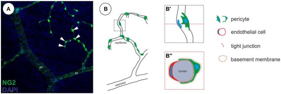
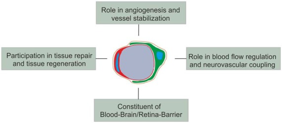
Pericytes are specialized mural cells located at the abluminal surface of capillary blood vessels, embedded within the basement membrane. In the vascular network these multifunctional cells fulfil diverse functions, which are indispensable for proper homoeostasis. They serve as microvascular stabilizers, are potential regulators of microvascular blood flow and have a central role in angiogenesis, as they for example regulate endothelial cell proliferation. Furthermore, pericytes, as part of the neurovascular unit, are a major component of the blood-retina/brain barrier. CNS pericytes are a heterogenic cell population derived from mesodermal and neuro-ectodermal germ layers acting as modulators of stromal and niche environmental properties. In addition, they display multipotent differentiation potential making them an intriguing target for regenerative therapies. Pericyte-deficiencies can be cause or consequence of many kinds of diseases. In diabetes, for instance, pericyte-loss is a severe pathological process in diabetic retinopathy (DR) with detrimental consequences for eye sight in millions of patients. In this review, we provide an overview of our current understanding of CNS pericyte origin and function, with a special focus on the retina in the healthy and diseased. Finally, we highlight the role of pericytes in de- and regenerative processes.
Overview
The claims and statistics of this paper haven’t been checked yet.
Run the check — free, no account needed. It reads the open-access text and checks the sources it cites.
Integrity signals
Registry
Cites
Statistics
Figures
Verification
Claims the paper makes
The claims and statistics of this paper haven’t been checked yet.
Run the check — free, no account needed. It reads the open-access text and checks the sources it cites.
Claims other papers make about this one
Each is checked against this paper’s own text: does it say what the citing paper says it does?
In the citing paper
“PDGFRβ expression in pericytes is noticeably higher than in SMCs18,25,26.”
From Blood–brain barrier breakdown is an early biomarker of human cognitive dysfunction (2019) · cites this paper as [26]
Self-check
Percentages, sums and reported differences re-computed from the paper’s own numbers.
The self-check needs the paper’s full text.
Citations
Cites
2 retracted
RETRACTED ARTICLE: Pericyte loss influences Alzheimer-like neurodegeneration in mice
Sagare, A. P., Bell, R. D., Zhao, Z., Ma, Q., Winkler, E. A., Ramanathan, A., & Zlokovic, B. V. (2013) · Nature Communications · doi:10.1038/ncomms3932 · Cited by 589
RetractedRetracted Article: Pericyte-mediated vasoconstriction underlies TBI-induced hypoperfusion
Dore‐Duffy, P., Wang, S. S., Mehedi, A., Katyshev, V., Cleary, K., Tapper, A., Reynolds, C. A., Ding, Y., Zhan, P., Rafols, J. A., et al. (2011) · Neurological Research · doi:10.1179/016164111x12881719352372 · Cited by 69
RetractedLang, G. E., Vaiser, A., Snyder, W. B., & Lynn, J. R. (2007) · Developments in ophthalmology · doi:10.1159/isbn.978-3-318-01439-6 · Cited by 5,607
A perivascular origin for mesenchymal stem cells in multiple human organs
Crisan, M., Yap, S., Casteilla, L., Chen, C.-W., Corselli, M., Park, T. S., Andriolo, G., Sun, B., Zheng, B., Zhang, L., et al. (2008) · Cell Stem Cell · doi:10.1016/j.stem.2008.07.003 · Cited by 3,991 · cited 2×
“As these cells, next to pericyte markers (NG2+, CD146+, aSMA+), also expressed markers typical for MSCs, the authors suggested that MSCs may have developed from perivascular cells/pericytes (Caplan, 2008; Crisan et al., 2008).”
BackgroundThe Blood-Brain Barrier in Health and Chronic Neurodegenerative Disorders
Zlokovic, B. V. (2008) · Neuron · doi:10.1016/j.neuron.2008.01.003 · Cited by 3,280
Pericytes regulate the blood–brain barrier
Armulik, A., Genové, G., Mäe, M., Nisancioglu, M. H., Wallgard, E., Niaudet, C., He, L., Norlin, J., Lindblom, P., Strittmatter, K., et al. (2010) · Nature · doi:10.1038/nature09522 · Cited by 2,843
Pericytes: Developmental, Physiological, and Pathological Perspectives, Problems, and Promises
Armulik, A., Genové, G., & Betsholtz, C. (2011) · Developmental Cell · doi:10.1016/j.devcel.2011.07.001 · Cited by 2,653
Pericyte Loss and Microaneurysm Formation in PDGF-B-Deficient Mice
Lindahl, P., Johansson, B. R., Levéen, P., & Betsholtz, C. (1997) · Science · doi:10.1126/science.277.5323.242 · Cited by 2,226
Pericytes are required for blood–brain barrier integrity during embryogenesis
Daneman, R., Zhou, L., Kebede, A. A., & Barres, B. A. (2010) · Nature · doi:10.1038/nature09513 · Cited by 2,128
Endothelial/Pericyte Interactions
Armulik, A., Abramsson, A., & Betsholtz, C. (2005) · Circulation Research · doi:10.1161/01.res.0000182903.16652.d7 · Cited by 2,048
Research works printed but not linked: 2
- Rouget, 1879, Sur la contractilite des capillaries sanguins, CR Acad. Sci., 88, 916
- Tidhar, 2001, A novel transgenic marker for migrating limb muscle precursors and for vascular smooth muscle cells, Dev. Dyn., 220, 60, 10.1002/1097-0177(2000)9999:9999::aid-dvdy10893.3.co;2-o · DOI
Cited by
Sorted by citations
Molecular diversity and specializations among the cells of the adult mouse brain
Saunders, A., Macosko, E. Z., Wysoker, A., Goldman, M., Krienen, F. M., de Rivera, H., Bien, E., Baum, M., Bortolin, L., Wang, S., et al. (2018) · Cell · doi:10.1016/j.cell.2018.07.028 · Cited by 1,531
“Mural cells are intrinsic to the endothelium and control vascular development, stability, and homeostasis (Sweeney et al., 2016; Trost et al., 2016).”
BackgroundBlood–brain barrier breakdown is an early biomarker of human cognitive dysfunction
Nation, D. A., Sweeney, M. D., Montagne, A., Sagare, A. P., D’Orazio, L. M., Pachicano, M., Sepehrband, F., Nelson, A. R., Buennagel, D. P., Harrington, M. G., et al. (2019) · Nature Medicine · doi:10.1038/s41591-018-0297-y · Cited by 1,728
Mechanisms of macular edema: Beyond the surface
Daruich, A., Matet, A., Moulin, A., Kowalczuk, L., Nicolas, M., Sellam, A., Rothschild, P.-R., Omri, S., Gélizé, E., Jonet, L., et al. (2017) · Progress in Retinal and Eye Research · doi:10.1016/j.preteyeres.2017.10.006 · Cited by 744
Neurodegeneration in diabetic retinopathy: does it really matter?
Simó, R., Stitt, A. W., & Gardner, T. W. (2018) · Diabetologia · doi:10.1007/s00125-018-4692-1 · Cited by 617
Retinal amyloid pathology and proof-of-concept imaging trial in Alzheimer’s disease
Koronyo, Y., Biggs, D., Barrón, E., Boyer, D. S., Pearlman, J. A., Au, W. J., Kile, S. J., Blanco, A., Fuchs, D.-T. S., Ashfaq, A., et al. (2017) · JCI Insight · doi:10.1172/jci.insight.93621 · Cited by 521
Pericyte loss leads to circulatory failure and pleiotrophin depletion causing neuron loss
Nikolakopoulou, A. M., Montagne, A., Kisler, K., Dai, Z., Wang, Y., Huuskonen, M. T., Sagare, A. P., Lazić, D., Sweeney, M. D., Kong, P., et al. (2019) · Nature Neuroscience · doi:10.1038/s41593-019-0434-z · Cited by 386
Spectral-Domain OCT Measurements in Alzheimer’s Disease
Chan, V. T. T., Sun, Z., Tang, S., Chen, L. J., Wong, A., Tham, C. C. Y., Wong, T. Y., Chen, C., Ikram, M. K., Whitson, H. E., et al. (2018) · Ophthalmology · doi:10.1016/j.ophtha.2018.08.009 · Cited by 368
The blood–retina barrier in health and disease
O’Leary, F., & Campbell, M. (2021) · FEBS Journal · doi:10.1111/febs.16330 · Cited by 372
Bridging barriers: a comparative look at the blood–brain barrier across organismsPreprint
O’Brown, N. M., Pfau, S. J., & Gu, C. (2018) · Genes & Development · doi:10.1101/gad.309823.117 · Cited by 268
Ocular indicators of Alzheimer’s: exploring disease in the retina
Hart, N. J., Koronyo, Y., Black, K. L., & Koronyo‐Hamaoui, M. (2016) · Acta Neuropathologica · doi:10.1007/s00401-016-1613-6 · Cited by 264
Advances in the development of new biomarkers for Alzheimer’s disease
Klyucherev, T. O., Olszewski, P., Shalimova, A. A., Chubarev, V. N., Tarasov, V. V., Attwood, M. M., Syvänen, S., & Schiöth, H. B. (2022) · Translational Neurodegeneration · doi:10.1186/s40035-022-00296-z · Cited by 241
Development and Function of the Blood-Brain Barrier in the Context of Metabolic Control
Haddad-Tóvolli, R., Dragano, N. R. V., Ramalho, A. F. S., & Velloso, L. A. (2017) · Frontiers in Neuroscience · doi:10.3389/fnins.2017.00224 · Cited by 226
Revolution of Alzheimer Precision Neurology. Passageway of Systems Biology and Neurophysiology
Hampel, H. J., (APMI), F. T. A. P. M. I., Toschi, N., Babiloni, C., Baldacci, F., Black, K. L., Bokde, A. L. W., Bun, R. S., Cacciola, F., Cavedo, E., et al. (2018) · Journal of Alzheimer s Disease · doi:10.3233/jad-179932 · Cited by 184
Optical Coherence Tomography in Alzheimer’s Disease and Other Neurodegenerative Diseases
Doustar, J., Torbati, T., Black, K. L., Koronyo, Y., & Koronyo‐Hamaoui, M. (2017) · Frontiers in Neurology · doi:10.3389/fneur.2017.00701 · Cited by 180
Tissue specific origin, development, and pathological perspectives of pericytes
Yamazaki, T., & Mukouyama, Y.-S. (2018) · Frontiers in Cardiovascular Medicine · doi:10.3389/fcvm.2018.00078 · Cited by 175
Immune Regulation by Pericytes: Modulating Innate and Adaptive Immunity
Navarro, R., Compte, M., Álvarez‐Vallina, L., & Sanz, L. (2016) · Frontiers in Immunology · doi:10.3389/fimmu.2016.00480 · Cited by 186
Autoimmunity, Autoinflammation, and Infection in Uveitis
Forrester, J. V., Kuffová, L., & Dick, A. D. (2018) · American Journal of Ophthalmology · doi:10.1016/j.ajo.2018.02.019 · Cited by 177
Past, present and future role of retinal imaging in neurodegenerative disease
Kashani, A. H., Asanad, S., Chan, J. W., Singer, M., Zhang, J., Sharifi, M., Khansari, M. M., Abdolahi, F., Shi, Y., Biffi, A., et al. (2021) · Progress in Retinal and Eye Research · doi:10.1016/j.preteyeres.2020.100938 · Cited by 169
Singh, A. V., Chandrasekar, V., Janapareddy, P., Mathews, D. E., Laux, P., Luch, A., Yang, Y., Canibaño, B., Balakrishnan, S., Abinahed, J. A., et al. (2021) · ACS Chemical Neuroscience · doi:10.1021/acschemneuro.1c00087 · Cited by 160
The Tumor Microenvironment in Tumorigenesis and Therapy Resistance Revisited
Dzobo, K., Senthebane, D. A., & Dandara, C. (2023) · Cancers · doi:10.3390/cancers15020376 · Cited by 146
Mechanisms Mediating Functional Hyperemia in the Brain
Nippert, A. R., Biesecker, K. R., & Newman, E. A. (2017) · The Neuroscientist · doi:10.1177/1073858417703033 · Cited by 145
Pericytes in Microvessels: From “Mural” Function to Brain and Retina Regeneration
Caporarello, N., D’Angeli, F., Cambria, M. T., Candido, S., Giallongo, C., Salmeri, M., Lombardo, C., Longo, A., Giurdanella, G., Anfuso, C. D., et al. (2019) · International Journal of Molecular Sciences · doi:10.3390/ijms20246351 · Cited by 138
The Neurovascular Unit in Glaucomatous Neurodegeneration
Wareham, L. K., & Calkins, D. J. (2020) · Frontiers in Cell and Developmental Biology · doi:10.3389/fcell.2020.00452 · Cited by 134
Alzheimer’s Retinopathy: Seeing Disease in the Eyes
Mirzaei, N., Shi, H., Oviatt, M., Doustar, J., Rentsendorj, A., Fuchs, D.-T. S., Sheyn, J., Black, K. L., Koronyo, Y., & Koronyo‐Hamaoui, M. (2020) · Frontiers in Neuroscience · doi:10.3389/fnins.2020.00921 · Cited by 136
Retinal capillary basement membrane thickening: Role in the pathogenesis of diabetic retinopathy
Roy, S., & Kim, D. (2020) · Progress in Retinal and Eye Research · doi:10.1016/j.preteyeres.2020.100903 · Cited by 127
Diverse Functions and Mechanisms of Pericytes in Ischemic Stroke
Yang, S., Jin, H., Zhu, Y., Wan, Y., Opoku, E. N., Zhu, L., & Hu, B. (2017) · Current Neuropharmacology · doi:10.2174/1570159x15666170112170226 · Cited by 125
Blood–brain barrier pericytes as a target for HIV-1 infection
Bertrand, L., Cho, H. J., & Toborek, M. J. (2018) · Brain · doi:10.1093/brain/awy339 · Cited by 119
Pericytes, inflammation, and diabetic retinopathy
Spencer, B. G., Estevez, J. J., Liu, E., Craig, J. E., & Finnie, J. W. (2019) · Inflammopharmacology · doi:10.1007/s10787-019-00647-9 · Cited by 116
Alzheimer's disease pathophysiology in the Retina
Gaire, B. P., Koronyo, Y., Fuchs, D.-T. S., Shi, H., Rentsendorj, A., Danziger, R., Vit, J., Mirzaei, N., Doustar, J., Sheyn, J., et al. (2024) · Progress in Retinal and Eye Research · doi:10.1016/j.preteyeres.2024.101273 · Cited by 107
Contribution of pericyte paracrine regulation of the endothelium to angiogenesis
Caporali, A., Martello, A., Miscianinov, V., Maselli, D., Vono, R., & Spinetti, G. (2016) · Pharmacology & Therapeutics · doi:10.1016/j.pharmthera.2016.10.001 · Cited by 96
Blood–Brain Barrier and Neurodegenerative Diseases—Modeling with iPSC-Derived Brain Cells
Wu, Y., Sonninen, T.-M., Peltonen, S., Koıstınaho, J., & Lehtonen, Š. (2021) · International Journal of Molecular Sciences · doi:10.3390/ijms22147710 · Cited by 91
The regulatory mechanisms of NG2/CSPG4 expression
Ampofo, E., Schmitt, B., Menger, M. D., & Laschke, M. W. (2017) · Cellular & Molecular Biology Letters · doi:10.1186/s11658-017-0035-3 · Cited by 89
Neurovascular dysfunction in glaucoma
Alarcón-Martínez, L., Shiga, Y., Villafranca‐Baughman, D., Vargas, J. L. C., Vidal-Paredes, I. A., Quintero, H., Fortune, B. A., Danesh-Meyer, H. V., & Polo, A. D. (2023) · Progress in Retinal and Eye Research · doi:10.1016/j.preteyeres.2023.101217 · Cited by 89
The Retina in Alzheimer's Disease: Histomorphometric Analysis of an Ophthalmologic Biomarker
Asanad, S., Ross‐Cisneros, F. N., Nassisi, M., Barrón, E., Karanjia, R., & Sadun, A. A. (2019) · Investigative Ophthalmology & Visual Science · doi:10.1167/iovs.18-25966 · Cited by 86
Apelin/APJ system as a therapeutic target in diabetes and its complications
Hu, H., He, L., Li, L., & Chen, L. (2016) · Molecular Genetics and Metabolism · doi:10.1016/j.ymgme.2016.07.012 · Cited by 85
Darden, J. A., Payne, L. B., Zhao, H., & Chappell, J. C. (2018) · Angiogenesis · doi:10.1007/s10456-018-9648-z · Cited by 82
Role of oxidative stress in diabetes-induced complications and their management with antioxidants
Singh, H. B., Singh, R., Singh, A., Singh, H., Singh, G., Kaur, S., & Singh, B. (2023) · Archives of Physiology and Biochemistry · doi:10.1080/13813455.2023.2243651 · Cited by 86
Jung, B., Arnold, T. D., Raschperger, E., Gaengel, K., & Betsholtz, C. (2017) · Journal of Cerebral Blood Flow & Metabolism · doi:10.1177/0271678x17697720 · Cited by 80
Methods to label, image, and analyze the complex structural architectures of microvascular networks
Corliss, B. A., Mathews, C., Doty, R. W., Rohde, G. K., & Peirce, S. M. (2018) · Microcirculation · doi:10.1111/micc.12520 · Cited by 77
Established and Emerging Strategies for Drug Delivery Across the Blood-Brain Barrier in Brain Cancer
Parodi, A., Rudzińska, M., Deviatkin, A. A., Soond, S. M., Baldin, A. V., & Zamyatnin, A. A. (2019) · Pharmaceutics · doi:10.3390/pharmaceutics11050245 · Cited by 81
Deligne, C., Hachani, J., Duban‐Deweer, S., Meignan, S., Leblond, P., Carcaboso, A. M., Sano, Y., Shimizu, F., Kanda, T., Gosselet, F., et al. (2020) · Fluids and Barriers of the CNS · doi:10.1186/s12987-020-00198-0 · Cited by 77
Chaulagain, B., Gothwal, A., Lamptey, R. N. L., Trivedi, R., Mahanta, A. K., Layek, B., & Singh, J. (2023) · International Journal of Molecular Sciences · doi:10.3390/ijms24032710 · Cited by 89
Gagnon, L., Smith, A. F., Boas, D. A., Devor, A., Secomb, T. W., & Sakadžić, S. (2016) · Frontiers in Computational Neuroscience · doi:10.3389/fncom.2016.00082 · Cited by 76
Pericyte morphology and function.
Alarcón-Martínez, L., Yemişçi, M., & Dalkara, T. (2021) · PubMed · doi:10.14670/hh-18-314 · Cited by 85
Pericytes in the Premetastatic Niche
Paiva, A. E., Lousado, L., Guerra, D. A. P., de Azevedo, P. O., Sena, I. F. G., Andreotti, J. P., Santos, G. S. P., Gonçalves, R., Mintz, A., & Birbrair, A. (2018) · Cancer Research · doi:10.1158/0008-5472.can-17-3883 · Cited by 73
Selective drug delivery to the retinal cells: Biological barriers and avenues
Ramsay, E., Lajunen, T., Bhattacharya, M., Reinisalo, M., Rilla, K. J., Kidron, H., Terasaki, T., & Urtti, A. O. (2023) · Journal of Controlled Release · doi:10.1016/j.jconrel.2023.07.028 · Cited by 75
Xue, X., Wu, X., Fan, Y., Han, S., Zhang, H., Sun, Y., Yin, Y., Yin, M., Chen, B., Sun, Z., et al. (2024) · Nature Communications · doi:10.1038/s41467-024-50564-x · Cited by 78
Pyroptosis in the Retinal Neurovascular Unit: New Insights Into Diabetic Retinopathy
Meng, C., Gu, C., He, S., Su, T., Lhamo, T., Draga, D., & Qiu, Q. (2021) · Frontiers in Immunology · doi:10.3389/fimmu.2021.763092 · Cited by 72
The pericyte microenvironment during vascular development
Payne, L. B., Zhao, H., James, C. C., Darden, J. A., McGuire, D. R., Taylor, S., Smyth, J. W., & Chappell, J. C. (2019) · Microcirculation · doi:10.1111/micc.12554 · Cited by 69
foxc1 is required for embryonic head vascular smooth muscle differentiation in zebrafish
Whitesell, T. R., Chrystal, P. W., Ryu, J., Munsie, N. M., Grosse, A. S., French, C. R., Workentine, M. L., Li, R., Zhu, L. J., Waskiewicz, A. J., et al. (2019) · Developmental Biology · doi:10.1016/j.ydbio.2019.06.005 · Cited by 65
Drug delivery to retinal photoreceptors
Himawan, E., Ekström, P. A. R., Buzgo, M., Gaillard, P. J., Stefánsson, E., Marigo, V., Loftsson, Þ., & Paquet‐Durand, F. (2019) · Drug Discovery Today · doi:10.1016/j.drudis.2019.03.004 · Cited by 65
The Ion Channel and GPCR Toolkit of Brain Capillary Pericytes
Hariharan, A., Weir, N., Robertson, C., He, L., Betsholtz, C., & Longden, T. A. (2020) · Frontiers in Cellular Neuroscience · doi:10.3389/fncel.2020.601324 · Cited by 64
Garnett, C. N., Watson, J. B., Higgins, E. K., Quan, N., & Bachstetter, A. D. (2021) · Frontiers in Immunology · doi:10.3389/fimmu.2021.688254 · Cited by 62
Basic Components of Vascular Connective Tissue and Extracellular Matrix
Halper, J. T. (2017) · Advances in pharmacology · doi:10.1016/bs.apha.2017.08.012 · Cited by 60
Uwamori, H., Ono, Y., Yamashita, T., Arai, K., & Sudo, R. (2018) · Microvascular Research · doi:10.1016/j.mvr.2018.11.007 · Cited by 61
Gonzales‐Aloy, E., Ahmed-Cox, A., Tsoli, M., Ziegler, D. S., & Kavallaris, M. (2023) · Advanced Drug Delivery Reviews · doi:10.1016/j.addr.2023.114777 · Cited by 60
Noble Metals and Soft Bio-Inspired Nanoparticles in Retinal Diseases Treatment: A Perspective
Matteis, V. D., & Rizzello, L. (2020) · Cells · doi:10.3390/cells9030679 · Cited by 57
Zheng, Y., Liu, Y., Wang, L., Xu, H., Lu, Z.-Q., Xuan, Y., Meng, W., Ye, L., Fang, D., Zhou, Y., et al. (2020) · International Journal of Molecular Medicine · doi:10.3892/ijmm.2020.4775 · Cited by 57
The Pathogenesis and Therapeutic Approaches of Diabetic Neuropathy in the Retina
Oshitari, T. (2021) · International Journal of Molecular Sciences · doi:10.3390/ijms22169050 · Cited by 58
Yong, Z., Cui, Z., Liu, J., Chen, J., & Tang, S. (2020) · Frontiers in Physiology · doi:10.3389/fphys.2019.01621 · Cited by 52
Location Matters: Navigating Regional Heterogeneity of the Neurovascular Unit
Bernier, L., Brunner, C., Cottarelli, A., & Balbi, M. (2021) · Frontiers in Cellular Neuroscience · doi:10.3389/fncel.2021.696540 · Cited by 52
Pericyte Plasticity in the Brain
Santos, G. S. P., Magno, L. A. V., Romano‐Silva, M. A., Mintz, A., & Birbrair, A. (2018) · Neuroscience Bulletin · doi:10.1007/s12264-018-0296-5 · Cited by 53
Retinal nerve fiber layer thickness predicts CSF amyloid/tau before cognitive decline
Asanad, S., Fantini, M., Sultan, W., Nassisi, M., Felix, C. M., Wu, J., Karanjia, R., Ross‐Cisneros, F. N., Sagare, A. P., Zloković, B. V., et al. (2020) · PLoS ONE · doi:10.1371/journal.pone.0232785 · Cited by 55
Pericytes Across the Lifetime in the Central Nervous System
Bennett, H. C., & Kim, Y. (2021) · Frontiers in Cellular Neuroscience · doi:10.3389/fncel.2021.627291 · Cited by 49
The Role of Pericytes in Ischemic Stroke: Fom Cellular Functions to Therapeutic Targets
Zhou, S., Guo, Z., Zhang, D., Qu, Y., & Jin, H. (2022) · Frontiers in Molecular Neuroscience · doi:10.3389/fnmol.2022.866700 · Cited by 52
Zéboulon, P., Lévêque, P.-M., Brasnu, E., Aragno, V., Hamard, P., Baudouin, C., & Labbé, A. (2017) · Journal of Glaucoma · doi:10.1097/ijg.0000000000000652 · Cited by 46
Sheng, X., Zhang, C., Zhao, J., Xu, J., Zhang, P., Ding, Q., & Zhang, J. (2024) · Cell & Bioscience · doi:10.1186/s13578-024-01269-7 · Cited by 53
Macrophages Generate Pericytes in the Developing Brain
Prazeres, P. H. D. M., de Almeida, V. M., Lousado, L., Andreotti, J. P., Paiva, A. E., Santos, G. S. P., de Azevedo, P. O., Souto, L., Almeida, G. G., Filev, R., et al. (2017) · Cellular and Molecular Neurobiology · doi:10.1007/s10571-017-0549-2 · Cited by 45
Yeh, T.-C., Kuo, C.-T., & Chou, Y.-B. (2022) · Frontiers in Aging Neuroscience · doi:10.3389/fnagi.2022.860759 · Cited by 43
Jackson, S., ElAli, A., Virgintino, D., & Gilbert, M. R. (2017) · Neuro-Oncology · doi:10.1093/neuonc/nox058 · Cited by 44
Microvascular bioengineering: a focus on pericytes
Zhao, H., & Chappell, J. C. (2019) · Journal of Biological Engineering · doi:10.1186/s13036-019-0158-3 · Cited by 44
Organ-On-A-Chip Technologies for Advanced Blood–Retinal Barrier Models
Ragelle, H., Gonçalves, A., Kustermann, S., Antonetti, D. A., & Jayagopal, A. (2019) · Journal of Ocular Pharmacology and Therapeutics · doi:10.1089/jop.2019.0017 · Cited by 46
Neurovascular Impairment and Therapeutic Strategies in Diabetic Retinopathy
Oshitari, T. (2021) · International Journal of Environmental Research and Public Health · doi:10.3390/ijerph19010439 · Cited by 45
López‐de‐Eguileta, A., López‐García, S., Lage, C., Pozueta, A., García‐Martínez, M., Kazimierczak, M., Bravo, M., Irure, J., López‐Hoyos, M., Muñoz, P., et al. (2022) · Alzheimer s Research & Therapy · doi:10.1186/s13195-022-00998-6 · Cited by 41
Caporarello, N., Olivieri, M., Cristaldi, M., Scalia, M., Toscano, M. A., Genovese, C., Addamo, A. P., Salmeri, M., Lupo, G., & Anfuso, C. D. (2017) · Molecular Neurobiology · doi:10.1007/s12035-017-0769-y · Cited by 41
Anatomy and physiology of blood-brain barrier
Gupta, S. N., Dhanda, S., & Sandhir, R. (2018) · Elsevier eBooks · doi:10.1016/b978-0-12-814001-7.00002-0 · Cited by 42
Anatomy of the Human Optic Nerve: Structure and Function
Salazar, J. J., Ramirez, A. I., de Hoz, R., Salobrar‐García, E., Rojas, P., Fernández‐Albarral, J. A., López‐Cuenca, I., Rojas, B., Triviño, A., & Ramirez, J. M. (2019) · IntechOpen eBooks · doi:10.5772/intechopen.79827 · Cited by 42
Pericytes Act as Key Players in Spinal Cord Injury
Picoli, C. C., Coimbra-Campos, L. M. C., Guerra, D. A. P., da Silva, W. N., Prazeres, P. H. D. M., Costa, A. C., Magno, L. A. V., Romano‐Silva, M. A., Mintz, A., & Birbrair, A. (2019) · American Journal Of Pathology · doi:10.1016/j.ajpath.2019.03.008 · Cited by 40
Mechanisms of pulmonary fibrosis induced by core fucosylation in pericytes
Sun, W., Tang, H., Gao, L., Sun, X., Liu, J., Wang, W., Wu, T., & Lin, H. (2017) · The International Journal of Biochemistry & Cell Biology · doi:10.1016/j.biocel.2017.05.010 · Cited by 38
Mah, J. L., & Dunn, C. W. (2024) · Nature Ecology & Evolution · doi:10.1038/s41559-023-02281-9 · Cited by 38
Liu, W.-J., Yin, Y., Sun, J., Feng, S., Ma, J., Fu, X., Hou, Y., Yang, M., Sun, B., & Fan, C. (2018) · OncoTargets and Therapy · doi:10.2147/ott.s174498 · Cited by 37
The Role of Pericytes in Regulation of Innate and Adaptive Immunity
Dabravolski, S. A., Andreeva, E. R., Eremin, I. I., Markin, A. M., Nadelyaeva, I. I., Orekhov, A. N., & Melnichenko, A. А. (2023) · Biomedicines · doi:10.3390/biomedicines11020600 · Cited by 36
Injury and stress responses of adult neural crest-derived cells
Parfejevs, V., Antunes, A. T., & Sommer, L. (2018) · Developmental Biology · doi:10.1016/j.ydbio.2018.05.011 · Cited by 35
The role of pigment epithelium-derived factor in protecting against cellular stress
Brook, N., Brook, E., Dharmarajan, A., Chan, A., & Dass, C. R. (2019) · Free Radical Research · doi:10.1080/10715762.2019.1697809 · Cited by 36
Pericyte-like differentiation of human adipose-derived mesenchymal stem cells: An in vitro study
Mannino, G., Gennuso, F., Giurdanella, G., Conti, F., Drago, F., Salomone, S., Furno, D. L., Bucolo, C., & Giuffrida, R. (2020) · World Journal of Stem Cells · doi:10.4252/wjsc.v12.i10.1152 · Cited by 36
Retinal Dysfunction in Alzheimer’s Disease and Implications for Biomarkers
Liao, C., Xu, J., Chen, Y., & Ip, N. Y. (2021) · Biomolecules · doi:10.3390/biom11081215 · Cited by 37
Pericytes and their potential in regenerative medicine across species
Esteves, C. L., & Donadeu, F. X. (2017) · Cytometry Part A · doi:10.1002/cyto.a.23243 · Cited by 35
The Pericytic Phenotype of Adipose Tissue-Derived Stromal Cells Is Promoted by NOTCH2
Terlizzi, V., Kolibabka, M., Burgess, J. K., Hammes, H. P., & Harmsen, M. C. (2017) · Stem Cells · doi:10.1002/stem.2726 · Cited by 35
Tang, D., Zhang, S., Shi, X., Wu, J., Yin, G., Tan, X., Liu, F., Wu, X., & Du, X. (2019) · Integrative Cancer Therapies · doi:10.1177/1534735418824408 · Cited by 37
The secreted neuronal signal Spock1 promotes blood-brain barrier development
O’Brown, N. M., Patel, N. B., Hartmann, U., Klein, A. M., Gu, C., & Megason, S. G. (2023) · Developmental Cell · doi:10.1016/j.devcel.2023.06.005 · Cited by 33
Central Nervous System Pericytes Contribute to Health and Disease
Girolamo, F., Errede, M., Bizzoca, A., Virgintino, D., & Ribatti, D. (2022) · Cells · doi:10.3390/cells11101707 · Cited by 32
Leptin Receptor Expression in Mouse Intracranial Perivascular Cells
Yuan, X., Caron, A., Wu, H., & Gautron, L. (2018) · Frontiers in Neuroanatomy · doi:10.3389/fnana.2018.00004 · Cited by 33
Vitronectin from brain pericytes promotes adult forebrain neurogenesis by stimulating CNTF
Jia, C., Keasey, M. P., Malone, H. M., Lovins, C., Sante, R. R., Razskazovskiy, V., & Hagg, T. (2018) · Experimental Neurology · doi:10.1016/j.expneurol.2018.11.002 · Cited by 32
Alpha-Smooth Muscle Actin-Positive Perivascular Cells in Diabetic Retina and Choroid
Kim, S., Kim, S. A., Choi, Y. A., Park, D. Y., & Lee, J. (2020) · International Journal of Molecular Sciences · doi:10.3390/ijms21062158 · Cited by 31
Tu, Y., Li, Q., Zhou, Y.-C., Ye, Z., Wu, C., Xie, E., Li, Y., Li, P., Wu, Y., Guo, Z., et al. (2022) · Frontiers in Cardiovascular Medicine · doi:10.3389/fcvm.2022.995216 · Cited by 32
Vision-Threatening Behcet’s Disease: Severity of Ocular Involvement Predictors
Hussein, M. A., Eissa, I. M., & Dahab, A. A. (2018) · Journal of Ophthalmology · doi:10.1155/2018/9518065 · Cited by 28
Increased Optic Nerve Head Capillary Blood Flow in Early Primary Open-Angle Glaucoma
Gardiner, S. K., Cull, G. A., Fortune, B. A., & Wang, L. (2019) · Investigative Ophthalmology & Visual Science · doi:10.1167/iovs.19-27389 · Cited by 28
Caire, R., Roche, B., Picot, T., Aanei, C.-M., He, Z., Campos, L., Thomas, M., Malaval, L., Vico, L., & Lafage-Proust, M.-H. (2019) · Journal of Bone and Mineral Research · doi:10.1002/jbmr.3728 · Cited by 27
Pericytes in Alzheimer’s Disease: Novel Clues to Cerebral Amyloid Angiopathy Pathogenesis
Salmina, A. B., Komleva, Y. K., Lopatina, O. L., & Birbrair, A. (2019) · Advances in experimental medicine and biology · doi:10.1007/978-3-030-16908-4_7 · Cited by 29
Fu, J., Liang, H., Yuan, P., Wei, Z., & Zhong, P. (2023) · Frontiers in Cellular Neuroscience · doi:10.3389/fncel.2023.1267785 · Cited by 28
Metadata
- Authors
- Trost, A., Lange, S., Schroedl, F., Bruckner, D., Motloch, K. A., Bogner, B., Kaser-Eichberger, A., Strohmaier, C., Runge, C., Aigner, L., Rivera, F. J., & Reitsamer, H. A.
- Affiliations
- Paracelsus Medical University (AT)
- Corresponding author
- Reitsamer, Herbert A.
- ORCID
- Venue
- Frontiers in Cellular Neuroscience
- Publisher
- Frontiers Media SA
- Published
- 2016-02-04
- Volume
- 10
- Pages
- 20-20
- ISSN
- 1662-5102 (ISSN-L)
- PMC
- PMC4740376
- OpenAlex
- W2312261107
- Cited by
- 244
- References
- 141
- Citations by year
- 2026: 9 · 2025: 23 · 2024: 20 · 2023: 23 · 2022: 25 · 2021: 27 · 2020: 24 · 2019: 33 · 2018: 26 · 2017: 26 · 2016: 8
- FWCI
- 2.72
- Type
- journal article
- Language
- en
- Open access
- gold
- doi.org
- Licence
- cc-by
- Locations
- Frontiers in Cellular Neuroscience (publishedVersion, cc-by)
- PubMed Central (publishedVersion, cc-by)
- PubMed (publishedVersion)
- DOAJ (DOAJ: Directory of Open Access Journals) (submittedVersion, cc-by-sa)
- Funders
- Glaucoma Research Foundation
- Topics
- Life Sciences › Neuroscience › Neurology › Barrier Structure and Function Studies
- Health Sciences › Medicine › Ophthalmology › Retinal Diseases and Treatments
- Life Sciences › Biochemistry, Genetics and Molecular Biology › Molecular Biology › Angiogenesis and VEGF in Cancer
- Keywords
- pericytes, retinal pericytes, brain pericytes, neurovascular unit, blood-retinal barrier, blood-brain barrier, angiogenesis, pericyte loss, diabetic retinopathy, pericyte heterogeneity, multipotent differentiation, regenerative therapy
About these checks
Registry, retraction, Cites and statistics results are computed, not judged, with one exception: a printed reference that no index lists is read once by a model before it counts as a citation error. Claims are extracted by a model and kept only when their quote appears verbatim in the text; each cited source is then checked, with a model’s help, for whether it says what the claim relies on. Those verdicts can be wrong — read the claim’s quote and the cited source before relying on one.
Statistics and arithmetic are recomputed, not judged.
Confidence bands are shown only for fields calibrated on our annotated test set. None is calibrated yet, so no band is shown.
What counts as a claim ›How we measure confidence ›How we check this ›
Are you an author, or do you see a problem with this page? Request a review
