

The DOI matches its Crossref record; 159 of the 159 references it cites were found in a registry or an index.
The Temperature Dependence of Sleep
Open accessAbstract
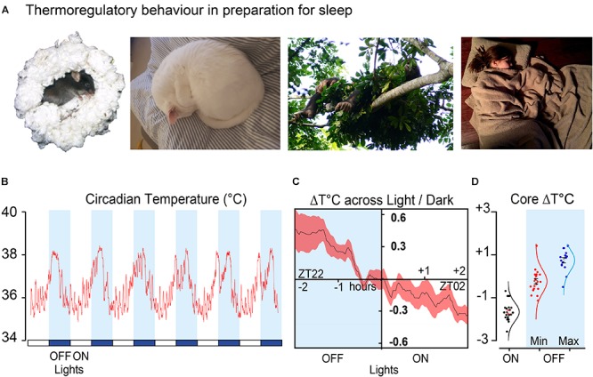
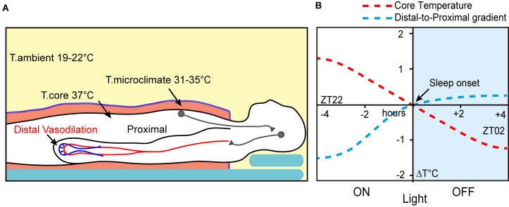
Mammals have evolved a range of behavioural and neurological mechanisms that coordinate cycles of thermoregulation and sleep. Whether diurnal or nocturnal, sleep onset and a reduction in core temperature occur together. Non-rapid eye movement (NREM) sleep episodes are also accompanied by core and brain cooling. Thermoregulatory behaviours, like nest building and curling up, accompany this circadian temperature decline in preparation for sleeping. This could be a matter of simply comfort as animals seek warmth to compensate for lower temperatures. However, in both humans and other mammals, direct skin warming can shorten sleep-latency and promote NREM sleep. We discuss the evidence that body cooling and sleep are more fundamentally connected and that thermoregulatory behaviours, prior to sleep, form warm microclimates that accelerate NREM directly through neuronal circuits. Paradoxically, this warmth might also induce vasodilation and body cooling. In this way, warmth seeking and nesting behaviour might enhance the circadian cycle by activating specific circuits that link NREM initiation to body cooling. We suggest that these circuits explain why NREM onset is most likely when core temperature is at its steepest rate of decline and why transitions to NREM are accompanied by a decrease in brain temperature. This connection may have implications for energy homeostasis and the function of sleep.
Overview
The claims and statistics of this paper haven’t been checked yet.
Run the check — free, no account needed. It reads the open-access text and checks the sources it cites.
Integrity signals
Registry
Cites
Statistics
Figures
Verification
Claims the paper makes
The claims and statistics of this paper haven’t been checked yet.
Run the check — free, no account needed. It reads the open-access text and checks the sources it cites.
Claims other papers make about this one
Each is checked against this paper’s own text: does it say what the citing paper says it does?
The check wasn’t confident either way, so it gives no verdict — never disputed.
In the citing paper
“Similarly, features of sleep-wake time series can directly capture insomnia-like patterns in sleep related to DM6,34,43,44.”
From Sleep and temperature data from wearable devices support noninvasive detection of diabetes mellitus in a large-scale, retrospective analysis (2026) · cites this paper as [34]
Experimental — accuracy not yet measuredThe check wasn’t confident either way, so it gives no verdict — never disputed.
In the citing paper
“Within a night, the complexity of the distal body temperature time series has been related to the autonomic nervous system’s (ANS) health during sleep6,34.”
From Sleep and temperature data from wearable devices support noninvasive detection of diabetes mellitus in a large-scale, retrospective analysis (2026) · cites this paper as [34]
Experimental — accuracy not yet measuredThe check wasn’t confident either way, so it gives no verdict — never disputed.
In the citing paper
“Studies suggest associations between DMN connectivity and subsequent sleep quality as measured by polysomnography, with greater DMNcortical connectivity associated with be er sleep efficiency and total sleep time [37].”
From Pre-Sleep Anxiety: Mechanisms and Therapeutic Approaches in Clinical Practice. (2026) · cites this paper as [37]
Experimental — accuracy not yet measuredIn the citing paper
“As a practical example, a hot bath (inducing vasodilation) prior to, but not immediately before the sleep period, is known to help with its initiation [9,52].”
From Human thermal perception and time of day: A review (2021) · cites this paper as [9]
Self-check
Percentages, sums and reported differences re-computed from the paper’s own numbers.
The self-check needs the paper’s full text.
Citations
Cites
Brown Adipose Tissue: Function and Physiological Significance
CANNON, B., & NEDERGAARD, J. (2004) · Physiological Reviews · doi:10.1152/physrev.00015.2003 · Cited by 6,433 · cited 1×
“It decouples the electron transport chain from the ATP-synthase, facilitating heat production through proton gradient dissipation, without ATP production, and compensatory metabolism (Cannon and Nedergaard, 2004).”
Two-Process Model of Sleep Regulation
Binder, M. D., Hirokawa, N., & Windhorst, U. (2008) · Springer eBooks · doi:10.1007/978-3-540-29678-2_6166 · Cited by 3,900 · cited 3×
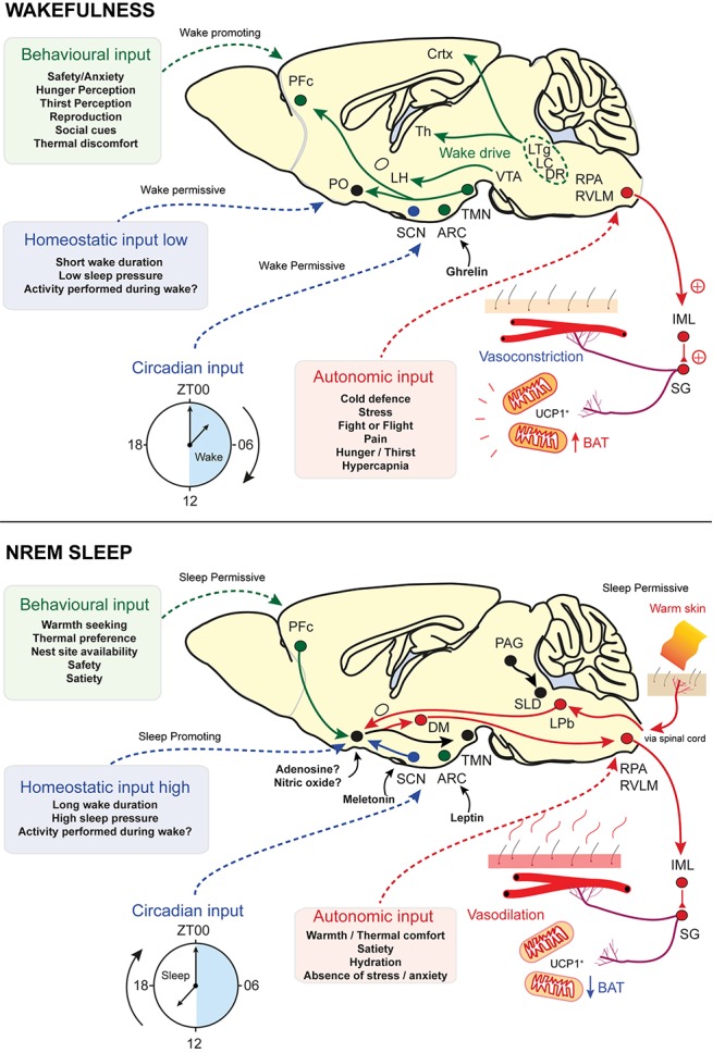
“According to current thinking, these are known as Process C and Process S, the circadian and the homeostatic input, respectively, and are part of the two-process model (Borbély, 1982).”
BackgroundLeptin activates anorexigenic POMC neurons through a neural network in the arcuate nucleus
Cowley, M. A., Smart, J. L., Rubinstein, M., Cerdán, M. G., Diano, S., Horvath, T. L., Cone, R. D., & Low, M. J. (2001) · Nature · doi:10.1038/35078085 · Cited by 2,356 · cited 1×
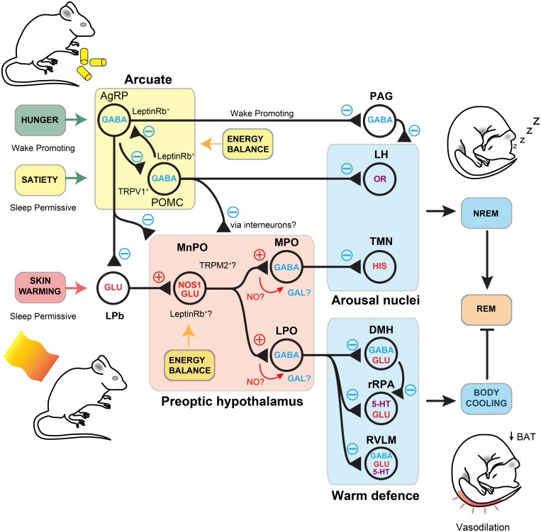
“POMC oppose the action of AgRP neurons and are activated by leptin (Cowley et al., 2001).”
BackgroundThe two‐process model of sleep regulation: a reappraisal
Borbély, A. A., Daan, S., Wirz‐Justice, A., & Deboer, T. (2016) · Journal of Sleep Research · doi:10.1111/jsr.12371 · Cited by 1,577 · cited 1×
“However, as we have seen, sleep onset is also gated by other inputs: ambient temperature, as well as levels of satiety, mating opportunities and the need to escape predators all determine the appropriate moment for NREM onset (Borbély, 1982; Borbély et al., 2016; EbanRothschild et al., 2017; Logan and McClung, 2019).”
BackgroundMolecular, spatial, and functional single-cell profiling of the hypothalamic preoptic region
Moffitt, J. R., Bambah-Mukku, D., Eichhorn, S. W., Vaughn, E., Shekhar, K., Perez, J. D., Rubinstein, N. D., Hao, J., Regev, A., Dulac, C., et al. (2018) · Science · doi:10.1126/science.aau5324 · Cited by 1,365
Mammalian Hibernation: Cellular and Molecular Responses to Depressed Metabolism and Low Temperature
Carey, H. V., Andrews, M. T., & Martin, S. L. (2003) · Physiological Reviews · doi:10.1152/physrev.00008.2003 · Cited by 1,145 · cited 4×
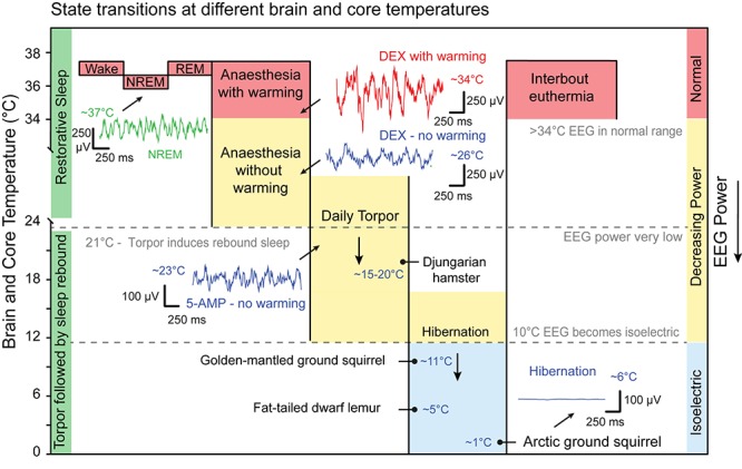
“These are periods of interbout euthermia (Boyer and Barnes, 1999; Carey et al., 2003).”
BackgroundInfluentialAssessing nest building in mice
Deacon, R. M. J. (2006) · Nature Protocols · doi:10.1038/nprot.2006.170 · Cited by 1,115 · cited 1×
“0 universal and the nesting mouse is adapted from Deacon (2006).”
BackgroundLeptin Differentially Regulates NPY and POMC Neurons Projecting to the Lateral Hypothalamic Area
Elias, C. F., Aschkenasi, C., Lee, C., Kelly, J., Ahima, R. S., Bjorbæk, C., Flier, J. S., Saper, C. B., & Elmquist, J. K. (1999) · Neuron · doi:10.1016/s0896-6273(01)80035-0 · Cited by 924 · cited 2×
“In rats, chronic total-sleep deprivation and selective REM deprivation, using the disk-overwater method for many days, leads to profound physiological effects and eventually death (Everson et al., 1989).”
BackgroundMethodDaily torpor and hibernation in birds and mammals
Ruf, T., & Geiser, F. (2014) · Biological reviews/Biological reviews of the Cambridge Philosophical Society · doi:10.1111/brv.12137 · Cited by 839 · cited 5×
“A range of mammals from ground squirrels to brown bears also use annual hibernation strategies for winter survival and reproduction (Carey et al., 2003; Ruf and Geiser, 2015).”
BackgroundInfluentialRegulation of Body Temperature by the Nervous System
Tan, C. L., & Knight, Z. A. (2018) · Neuron · doi:10.1016/j.neuron.2018.02.022 · Cited by 834 · cited 1×
“Neurons that receive afferent temperature information, but are not direct temperature ‘sensors’, have been distinguished by the term ‘warm-activated’ neurons (Tan and Knight, 2018).”
Background
Research works printed but not linked: 1
- Ma, 2019, Galanin neurons in the hypothalamus link sleep homeostasis, body temperature and actions of the alpha2 adrenergic agonist dexmedetomidine., bioRxiv, 10.1101/565747 · DOI
Cited by
Sorted by citations
Rising temperatures erode human sleep globally
Minor, K., Bjerre-Nielsen, A., Jonasdottir, S. S., Lehmann, S., & Obradovich, N. (2022) · One Earth · doi:10.1016/j.oneear.2022.04.008 · Cited by 261
Sleep Deprivation and Neurological Disorders
Bishir, M., Bhat, A., Essa, M. M., Ekpo, O. E., Ihunwo, A. O., Veeraraghavan, V. P., Surapaneni, K. M., Mahalakshmi, A. M., Ray, B., Tuladhar, S., et al. (2020) · BioMed Research International · doi:10.1155/2020/5764017 · Cited by 267
“NREM sleep is characterized by a low metabolic rate and an increase in brain temperature which helps to overcome the damages that are introduced during the wake cycle [7].”
The Promise of Sleep: A Multi-Sensor Approach for Accurate Sleep Stage Detection Using the Oura Ring
Altini, M., & Kinnunen, H. O. (2021) · Sensors · doi:10.3390/s21134302 · Cited by 208
“The reason is that core body temperature reductions are mechanistically accomplished through vasodilation of peripheral surface blood vessels of the skin in the extremities, particularly the hands and feet [48].”
BackgroundA systematic review of ambient heat and sleep in a warming climate
Chevance, G., Minor, K., Vielma, C., Campi, E., O’Callaghan-Gordo, C., Basagaña, X., Ballester, J., & Bernard, P. (2024) · Sleep Medicine Reviews · doi:10.1016/j.smrv.2024.101915 · Cited by 150
Galanin Neurons Unite Sleep Homeostasis and α2-Adrenergic Sedation
Ma, Y., Miracca, G., Yu, X., Harding, E. C., Miao, A., Yustos, R., Vyssotski, A. L., Franks, N. P., & Wisden, W. (2019) · Current Biology · doi:10.1016/j.cub.2019.07.087 · Cited by 130
“Given that sleep induction occurs when the rate of core body temperature decline is maximal [40, 41], this could explain why LPO-DGal mice are actually more awake compared with LPO-Gal-GFP mice at the start of lights on (Figure 1A).”
BackgroundSelf-citationMattingly, S. M., Grover, T., Martinez, G. J., Aledavood, T., Robles-Granda, P., Nies, K., Striegel, A., & Mark, G. J. (2021) · npj Digital Medicine · doi:10.1038/s41746-021-00435-2 · Cited by 123
“…circadian responses to optimize to the environment 46 – 53 , e.g., in humans to promote sleep when it is dark and to be awake when it is light, to be asleep at the lowest point of core body temperature 43,54 – 56 , and/or to avoid extremes in ambient temperature that can impair sleep 57 – 61 .”
BackgroundLim, C. L. (2020) · International Journal of Environmental Research and Public Health · doi:10.3390/ijerph17217795 · Cited by 137
Harding, E. C., Franks, N. P., & Wisden, W. (2019) · Current Opinion in Physiology · doi:10.1016/j.cophys.2019.11.008 · Cited by 127
Self-citationAutomating sleep stage classification using wireless, wearable sensors
Boe, A. J., McGee Koch, L. L., O’Brien, M. K., Shawen, N., Rogers, J. A., Lieber, R. L., Reid, K. J., Zee, P. C., & Jayaraman, A. (2019) · npj Digital Medicine · doi:10.1038/s41746-019-0210-1 · Cited by 98
Tossell, K., Yu, X., Giannos, P., Soto, B. A., Nollet, M., Yustos, R., Miracca, G., Vicente, M., Miao, A., Hsieh, B., et al. (2023) · Nature Neuroscience · doi:10.1038/s41593-023-01430-4 · Cited by 86
“Nesting provides thermal microclimates, warming the skin; this in turn, via Nos1 -, Vglut2 -expressing cells in the MPO hypothalamus, promotes NREM sleep and concomitant body cooling when sleep starts 3,9,50 .”
BackgroundSelf-citationCommon Sleep Disorders Affecting Older Adults
Jaqua, E. E., Hanna, M., Labib, W., Moore, C., & Matossian, V. (2022) · The Permanente Journal · doi:10.7812/tpp/22.114 · Cited by 74
Dream engineering: Simulating worlds through sensory stimulation
Carr, M., Horowitz, A. H., Amores, J., Lopes, P., Bernal, G., Gálvez, T. A. V., Rosello, O., Jain, A., & Maes, P. (2020) · Consciousness and Cognition · doi:10.1016/j.concog.2020.102955 · Cited by 74
Irwin, M. R. (2022) · Temperature · doi:10.1080/23328940.2022.2109932 · Cited by 80
“contributes further to sleep-related decrease in core body temperature [3].”
BackgroundThe Sleep-Promoting Ventrolateral Preoptic Nucleus: What Have We Learned over the Past 25 Years?
Arrigoni, E., & Fuller, P. M. (2022) · International Journal of Molecular Sciences · doi:10.3390/ijms23062905 · Cited by 77
“, heat loss [100], and this process is linked with the inhibition of thermoregulatory neurons in the dorsomedial hypothalamic nucleus (DMH) and the raphe pallidus (RPa) [101,102].”
BackgroundThe contribution of sleep to the neuroendocrine regulation of rhythms in human leukocyte traffic
Lange, T., Luebber, F., Graßhoff, H., & Besedovsky, L. (2022) · Seminars in Immunopathology · doi:10.1007/s00281-021-00904-6 · Cited by 54
Lavender essential oil on postmenopausal women with insomnia: Double-blind randomized trial
Lucena, L. D. R., Santos-Junior, J. G., Tufik, S., & Hachul, H. (2021) · Complementary Therapies in Medicine · doi:10.1016/j.ctim.2021.102726 · Cited by 54
Richardson, R. B., & Mailloux, R. J. (2023) · Antioxidants · doi:10.3390/antiox12030674 · Cited by 60
“Nevertheless, it is unlikely that systemic energy conservation, or indeed cerebral free radical flux control, are the primary purposes of sleep [200].”
BackgroundTargeting Sleep Physiology to Modulate Glymphatic Brain Clearance
van Hattem, T., Verkaar, L., Krugliakova, E., Adelhöfer, N., Zeising, M., Drinkenburg, W. H. I. M., Claassen, J. A. H. R., Bódizs, R., Dresler, M., & Rosenblum, Y. (2024) · Physiology · doi:10.1152/physiol.00019.2024 · Cited by 57
Interoception relates to sleep and sleep disorders
Wei, Y., & Someren, E. J. W. V. (2019) · Current Opinion in Behavioral Sciences · doi:10.1016/j.cobeha.2019.11.008 · Cited by 54
“Based on these findings, an extension of the traditional two-process model of sleep has been proposed [14].”
Lateral hypothalamic neuronal ensembles regulate pre-sleep nest-building behavior
Sotelo, M. I., Tyan, J., Markunas, C., Sulaman, B. A., Horwitz, L., Lee, H. K., Morrow, J. G., Rothschild, G., Duan, B., & Eban-Rothschild, A. (2022) · Current Biology · doi:10.1016/j.cub.2021.12.053 · Cited by 49
Shriane, A. E., Rigney, G., Ferguson, S. A., Bin, Y. S., & Vincent, G. E. (2023) · SLEEP · doi:10.1093/sleep/zsad182 · Cited by 53
“…routine [63–66], transitioning to days off work [67–72], napping for fatigue management [73–80], maintaining a comfortable sleep environment [81–86], managing exposure to light [87–92], managing caffeine consumption [18, 93, 94], limiting alcohol and nicotine [95–98], considering medication…”
BackgroundHuman thermal perception and time of day: A review
Vellei, M., Chinazzo, G., Zitting, K.-M., & Hubbard, J. (2021) · Temperature · doi:10.1080/23328940.2021.1976004 · Cited by 45
“This is corroborated through observations in other mammals [9].”
BackgroundResultSleep disorder in patients with chronic liver disease: a narrative review
Shah, N. M., Malhotra, A. M., & Kaltsakas, G. (2020) · Journal of Thoracic Disease · doi:10.21037/jtd-cus-2020-012 · Cited by 44
“Sleep onset relies on core body temperature cooling through thermoregulatory mechanisms (19).”
BackgroundThe Molecular Clock and Neurodegenerative Disease: A Stressful Time
Carter, B., Justin, H. S., Gulick, D., & Gamsby, J. J. (2021) · Frontiers in Molecular Biosciences · doi:10.3389/fmolb.2021.644747 · Cited by 42
“This could be related to the lowering of core body temperature that occurs in healthy mammalian sleep cycles (Harding et al., 2019).”
BackgroundBe cool to be far: Exploiting hibernation for space exploration
Cerri, M., Hitrec, T., Luppi, M., & Amici, R. (2021) · Neuroscience & Biobehavioral Reviews · doi:10.1016/j.neubiorev.2021.03.037 · Cited by 38
Pho, H., Berger, S., Freire, C., Kim, L. J., Shin, M., Streeter, S. R., Hosamane, N. S., Cabassa, M. E., Anokye‐Danso, F., Dergacheva, O., et al. (2021) · SLEEP · doi:10.1093/sleep/zsab046 · Cited by 39
Vera, L. M., de Alba, G., Santos, S., Szewczyk, T. M., MacKenzie, S. A., Sánchez‐Vázquez, F. J., & Rey, S. (2023) · Journal of Thermal Biology · doi:10.1016/j.jtherbio.2023.103544 · Cited by 37
“Likewise, similar results have been found in terrestrial animals and humans regarding the effect of temperature on the sleep pattern (Murphy and Campbell, 1997; Harding et al., 2019).”
ResultThe relationship between fasting-induced torpor, sleep, and wakefulness in laboratory mice
Huang, Y., Flaherty, S. J., Pothecary, C. A., Foster, F., Peirson, S. N., & Vyazovskiy, V. V. (2021) · SLEEP · doi:10.1093/sleep/zsab093 · Cited by 42
“This drop in T b is facilitated by a decline in the set-point temperature of the thermoregulatory system, which is likely to decline initially upon the wake–NREM transition and decline even further as torpor progresses [81, 93].”
BackgroundAhmadzadeh, S., Luo, J., & Wiffen, R. (2020) · IEEE Reviews in Biomedical Engineering · doi:10.1109/rbme.2020.3033930 · Cited by 39
“Skin temperature increases during sleep and decreases when we wakeup [136].”
BackgroundHung, S.-T., Cheng, Y.-C., Wu, C.-C., & Su, C.-H. (2023) · Journal of Multidisciplinary Healthcare · doi:10.2147/jmdh.s419306 · Cited by 35
“The bedroom should be quiet, dark, and cool, and comfortable bedding should be used.(94) Additionally, it is important to avoid stimulants before bedtime.”
BackgroundSleep in Myopic and Non-Myopic Children
Ostrin, L. A., Read, S. A., Vincent, S. J., & Collins, M. J. (2020) · Translational Vision Science & Technology · doi:10.1167/tvst.9.9.22 · Cited by 35
Elevated body temperature is associated with depressive symptoms: results from the TemPredict Study
Mason, A. E., Kasl, P., Soltani, S., Green, A., Hartogensis, W., Dilchert, S., Chowdhary, A., Pandya, L. S., Siwik, C. J., Foster, S. L., et al. (2024) · Scientific Reports · doi:10.1038/s41598-024-51567-w · Cited by 31
“This has been observed in the form of elevated body temperature, particularly at night 13–16 , when thermoregulatory cooling responses are critical for sleep onset and quality 17,18 .”
Tai, Y., Obayashi, K., Yamagami, Y., Yoshimoto, K., Kurumatani, N., Nishio, K., & Saeki, K. (2021) · Journal of Clinical Sleep Medicine · doi:10.5664/jcsm.9180 · Cited by 32
Li, S., Hu, S., Liu, R., Liang, S., He, M., & Sun, J. (2022) · Journal of Building Engineering · doi:10.1016/j.jobe.2022.104375 · Cited by 29
Enhanced conductive body heat loss during sleep increases slow-wave sleep and calms the heart
Herberger, S., Penzel, T., Fietze, I., Glos, M., Cicolin, A., Fattori, E., Grimaldi, D., Reid, K. J., Zee, P. C., Mason, M., et al. (2024) · Scientific Reports · doi:10.1038/s41598-024-53839-x · Cited by 28
“Mammals, including humans, exhibit typical sleep preparation behaviors such as curling up, nest building and use of bedding 38 .”
BackgroundTurn it off and on again: characteristics and control of torpor
Ambler, M. T., Hitrec, T., & Pickering, A. E. (2021) · Wellcome Open Research · doi:10.12688/wellcomeopenres.17379.1 · Cited by 24
Many faces of sleep regulation: beyond the time of day and prior wake time
Duhart, J. M., Inami, S., & Koh, K. (2021) · FEBS Journal · doi:10.1111/febs.16320 · Cited by 22
How Temperature Influences Sleep
Fan, Y., Wang, Y., Gu, P., Han, J., & Tian, Y. (2022) · International Journal of Molecular Sciences · doi:10.3390/ijms232012191 · Cited by 25
Core body temperature changes before sleep are associated with nocturnal heart rate variability
Bigalke, J. A., Cleveland, E. L., Barkstrom, E., Gonzalez, J. E., & Carter, J. R. (2023) · Journal of Applied Physiology · doi:10.1152/japplphysiol.00020.2023 · Cited by 22
Planetary sleep medicine: Studying sleep at the individual, population, and planetary level
Bragazzi, N. L., Garbarino, S., Puce, L., Trompetto, C., Marinelli, L., Currà, A., Jahrami, H., Trabelsi, K., Garcia, B. R. M., Asgary, A., et al. (2022) · Frontiers in Public Health · doi:10.3389/fpubh.2022.1005100 · Cited by 21
“Endogenous temperature represents, indeed, an important Zeitgeber (a biological timekeeping cue), which regulates human sleep (82).”
BackgroundPotential effects of shift work on skin autoimmune diseases
Stenger, S., Graßhoff, H., Hundt, J. E., & Lange, T. (2023) · Frontiers in Immunology · doi:10.3389/fimmu.2022.1000951 · Cited by 22
“These changes would induce an increase in skin temperature, a decrease in core body temperature and in this way her body would get prepared for the sleep period (34, 44, 45).”
BackgroundLiu, W., Wang, Y., Chang, L.-T., Wu, C., Wu, D., Tsai, C.-Y., Lo, C.-C., Lo, K., Chung, K. F., Chang, T.-Y., et al. (2022) · Environmental Science and Pollution Research · doi:10.1007/s11356-022-18922-8 · Cited by 20
“Temperature-related alterations in sleep stages may result from a general disruption of sleep processes rather than being specifically related to the status of the thermoregulatory system during REM sleep (Harding et al. 2019).”
BackgroundRe-examining extreme sleep duration in bats: implications for sleep phylogeny, ecology, and function
Harding, C. D., Yovel, Y., Peirson, S. N., Hackett, T. D., & Vyazovskiy, V. V. (2022) · SLEEP · doi:10.1093/sleep/zsac064 · Cited by 19
“Factors such as photoperiod (Borb´ely, 1978; Hubbard et al., 2013), light intensity (Borb´ely, 1978), ambient temperature (Harding et al., 2019), diet (St-Onge et al., 2016), social environment (Lendrem, 1984) and predation risk (Lima et al., 2005) have all been identified as capable of influencing…”
BackgroundPattnaik, H., Mir, M., Boike, S., Kashyap, R., Khan, S. A., & Surani, S. (2022) · Cureus · doi:10.7759/cureus.32803 · Cited by 21
“Discussion around sleep hygiene highlights this role, as proper temperature [8], darkness [9,10], silence [11], and comfort, along with sleep-conducive behaviors before bedtime, are factors that influence the quality as well as the quantity of sleep [12].”
BackgroundTai, Y., Obayashi, K., Yamagami, Y., & Saeki, K. (2023) · Journal of Clinical Sleep Medicine · doi:10.5664/jcsm.10590 · Cited by 18
Beyond day and night: The importance of ultradian rhythms in mouse physiology
Škop, V., Liu, N., Xiao, C., Stinson, E. J., Chen, K. Y., Hall, K. D., Piaggi, P., Gavrilova, O., & Reitman, M. L. (2024) · Molecular Metabolism · doi:10.1016/j.molmet.2024.101946 · Cited by 20
“One possibility is that it does augment core functions of sleep [75].”
Landen, J. G., Vandendoren, M., Killmer, S., Bedford, N. L., & Nelson, A. C. (2024) · Communications Biology · doi:10.1038/s42003-024-06781-7 · Cited by 20
“3F, G,lightgreenpoints),con fi rming previousreports that sleep is associated with lower Tb 51 – 53 .”
Temperature effects on the neuronal dynamics and Hamilton energy
Xie, Y., Ye, Z., Wang, X., Jia, Y., Hu, X., & Li, X. (2025) · Chaos Solitons & Fractals · doi:10.1016/j.chaos.2025.116325 · Cited by 16
Schork, I. G., Manzo, I. A., de Oliveira, M. R. B., da Costa, F. V., Palme, R., Young, R. J., & de Azevedo, C. S. (2022) · Behavioural Processes · doi:10.1016/j.beproc.2022.104662 · Cited by 19
Enhancing circadian rhythms—the circadian MEGA bundle as novel approach to treat critical illness
Prin, M., Bertazzo, J., Walker, L. A., Scott, B. K., & Eckle, T. D. L. G. (2023) · Annals of Translational Medicine · doi:10.21037/atm-22-5127 · Cited by 18
“The timing of daily temperature variation is integral to sleep and reveals the phase shift of circadian rhythms (118).”
Sleep and global warming: How will we sleep when the Earth is hotter?
Buguet, A., Reis, J. J., & Radomski, M. W. (2023) · Journal of the Neurological Sciences · doi:10.1016/j.jns.2023.120859 · Cited by 16
The new science of sleep: From cells to large-scale societies
Sharon, O., Simon, E. B., Shah, V. D., Desel, T., & Walker, M. P. (2024) · PLoS Biology · doi:10.1371/journal.pbio.3002684 · Cited by 18
“Temperature, therefore, offers 1 novel and newly harnessed pathway for enhancing human sleep [74,75].”
BackgroundThe sociality of sleep in animal groups
Chakravarty, P., Ashbury, A. M., Strandburg‐Peshkin, A., Iffelsberger, J., Goldshtein, A., Schuppli, C., Snell, K. R. S., Charpentier, M. J. E., Núñez, C. L., Gaggioni, G., et al. (2024) · Trends in Ecology & Evolution · doi:10.1016/j.tree.2024.07.011 · Cited by 17
Mortlock, E., Silovský, V., Güldenpfennig, J., Faltusová, M., Olejarz, A., Börger, L., Ježek, M., Jennings, D. J., & Capellini, I. (2024) · Proceedings of the Royal Society B Biological Sciences · doi:10.1098/rspb.2023.2115 · Cited by 16
“Ambient temperature and light affect sleep quantity, fragmentation and quality in the laboratory [27,62]; thus, they are considered important mediators of sleep behaviour.”
BackgroundTossell, K., Yu, X., Soto, B. A., Vicente, M., Miracca, G., Giannos, P., Miao, A., Hsieh, B., Ma, Y., Yustos, R., et al. (2020) · bioRxiv (Cold Spring Harbor Laboratory) · doi:10.1101/2020.07.01.179671 · Cited by 15
Self-citationHaghayegh, S., Smolensky, M. H., Khoshnevis, S., Hermida, R. C., Castriotta, R. J., & Diller, K. R. (2021) · Comprehensive physiology · doi:10.1002/cphy.c210008 · Cited by 16
“The importance of the decline and rise phases of the CBT circadian rhythm in affecting cognitive performance and the gating of sleep onset and offset is well recognized (50, 53, 70, 73, 76, 119, 128, 138).”
Ninham, B. W., Bunkin, N. F., & Battye, M. (2025) · Advances in Colloid and Interface Science · doi:10.1016/j.cis.2025.103401 · Cited by 15
Raymond, J. S., Everett, N. A., Gururajan, A., & Bowen, M. T. (2023) · SLEEP · doi:10.1093/sleep/zsad112 · Cited by 19
“During the sleep initiation period, for both humans and rats, sleep onset is most likely to occur during the steepest drop in body temperature [62, 63], suggesting that body cooling and NREM sleep are linked.”
BackgroundBiological sex influences sleep phenotype in mice experiencing spontaneous opioid withdrawal
Tisdale, R. K., Sun, Y., Park, S., Ma, S., Haire, M., Allocca, G., Bruchas, M. R., Morairty, S. R., & Kilduff, T. S. (2023) · Journal of Sleep Research · doi:10.1111/jsr.14037 · Cited by 14
“Behavioural immobility and reduced body temperature are characteristic of sleep (Harding et al., 2019).”
BackgroundEffects of quilts on comfortable indoor temperatures and human thermal responses during sleep
Zheng, Q., Yan, F., Wang, H., & Ke, Y. (2022) · Indoor Air · doi:10.1111/ina.13122 · Cited by 14
Rivas, M., Serantes, D., Peña, F., González, J., Ferreira, A., Torterolo, P., & Benedetto, L. (2021) · Neuroscience · doi:10.1016/j.neuroscience.2021.08.034 · Cited by 13
Linking Hypothermia and Altered Metabolism with TrkB Activation
Alitalo, O., González-Hernández, G., Rosenholm, M., Kohtala, P., Matsui, N., Müller, H. K., Theilmann, W., Klein, A. B., Kärkkäinen, O. K., Rozov, S. V., et al. (2023) · ACS Chemical Neuroscience · doi:10.1021/acschemneuro.3c00350 · Cited by 11
Rapid Eye Movement Sleep Engages Melanin-Concentrating Hormone Neurons to Reduce Cocaine Seeking
Guo, R., Wang, Y., Yan, R., Chen, B., Ding, W., Gorczyca, M. T., Ozsoy, S., Cai, L., Hines, R. L., Tseng, G. C., et al. (2022) · Biological Psychiatry · doi:10.1016/j.biopsych.2022.06.006 · Cited by 13
Thermoregulation and Sleep: Functional Interaction and Central Nervous Control
Cerri, M., & Amici, R. (2021) · Comprehensive physiology · doi:10.1002/cphy.c140012 · Cited by 21
“The intrinsic complexity of the pre-optic region is beyond the scope of this review; we, therefore, refer the reader to more specialized publications (17, 54, 89, 121, 140, 143, 159, 195, 274, 275).”
InfluentialWang, J., Huffman, D., Ajwad, A. A., McLouth, C. J., Bachstetter, A. D., Kohler, K., Murphy, M. P., O’Hara, B. F., Duncan, M. J., & Sunderam, S. (2024) · SLEEP · doi:10.1093/sleep/zsae078 · Cited by 14
Disrupting flight increases sleep and identifies a novel sleep-promoting pathway in Drosophila
Melnattur, K. V., Zhang, B., & Shaw, P. J. (2020) · Science Advances · doi:10.1126/sciadv.aaz2166 · Cited by 12
Mortlock, E., English, H. M., Fitzsimmons, J., Börger, L., Jennings, D. J., & Capellini, I. (2024) · Animal Behaviour · doi:10.1016/j.anbehav.2024.03.006 · Cited by 11
Neuropeptide Y – Its role in human performance and extreme environments
Kienast, C., Gunga, H., & Steinach, M. (2019) · REACH · doi:10.1016/j.reach.2019.100032 · Cited by 12
Some evidence of a time-varying thermal perception
Vellei, M., O’Brien, W., Martinez, S., & Le Dréau, J. (2022) · Indoor and Built Environment · doi:10.1177/1420326x211034563 · Cited by 11
“From a biological perspective, the lower MET and core body temperature occurring during sleep minimize energy expenditure and help to deal with the recovering of the cell damage done during the metabolically active periods of the day.(21) In the literature, few studies have investigated diurnal variations in thermal p…”
BackgroundInfluentialMammalian NREM and REM sleep: Why, when and how
Rial, R. V., Akaârir, M., Cañellas, F., Barceló, P., Díaz, J. Á. R., Martin-Reina, A., Gamundí, A., & Nicolau, M. C. (2023) · Neuroscience & Biobehavioral Reviews · doi:10.1016/j.neubiorev.2023.105041 · Cited by 12
Horgan, B. G., Tee, N., West, N. P., Drinkwater, E. J., Halson, S. L., Colomer, C. M. E., Fonda, C. J., Tatham, J., Chapman, D. W., & Haff, G. G. (2023) · The Journal of Strength and Conditioning Research · doi:10.1519/jsc.0000000000004428 · Cited by 12
“, heat loss by peripheral vasodilation (26), and thereby inducing sleep onset by core body temperature cooling (20,43), or by a placebo response to a postexercise recovery strategy.”
BackgroundMoyen, N. E., Ediger, T. R., Taylor, K. M., Hancock, E. G., Holden, L. D., Tracy, E. E., Kay, P. M., Kotzen, K., Irick, C. R., & He, D. D. (2024) · Bioengineering · doi:10.3390/bioengineering11040352 · Cited by 11
“Body temperature is one of the main factors that affects sleep quality and, therefore, must be tightly regulated for optimal sleep [8–10].”
BackgroundAmbient chemical and physical approaches for the modulation of sleep and wakefulness
Wang, Y., Ma, W., Kong, L.-X., Zhang, H., Yuan, P., Qu, W., Liu, C., & Huang, Z. (2024) · Sleep Medicine Reviews · doi:10.1016/j.smrv.2024.102015 · Cited by 13
Wearing, O. H., & Scott, G. R. (2022) · American Journal of Physiology-Regulatory, Integrative and Comparative Physiology · doi:10.1152/ajpregu.00075.2022 · Cited by 11
Top 10 evidence-based countermeasures for night shift workers
Wallace, P., & Haber, J. J. (2020) · Emergency Medicine Journal · doi:10.1136/emermed-2019-209134 · Cited by 11
“28 29 Some studies have shown sleeping with a warm water bottle (or other modality of warming the feet) induces this peripheral vasodilation leading to sleep induction.”
Stein, W., Torres, G., Giménez, L., Espinosa-Novo, N., Geißel, J. P., Vidal-Gadea, A., & Harzsch, S. (2023) · Frontiers in Cellular Neuroscience · doi:10.3389/fncel.2023.1263591 · Cited by 10
“The nervous system is particularly affected by temperature changes since these can significantly disrupt cellular and circuit activity (Robertson and Money, 2012; Harding et al., 2019; Marder and Rue, 2021; Robertson et al., 2023) with far-reaching consequences for animal survival and performance.”
The effects of temperature on sleep experience: evidence from China
Wang, C., Liu, K., & Wang, H. (2022) · Applied Economics · doi:10.1080/00036846.2022.2130870 · Cited by 10
“According to the National Sleep Foundation poll in the U.S. and Harding, Franks, and Wisden (2019), the ideal temperature for sleeping is around 15.5℃ to 19℃.2 When the temperature is higher than the ideal temperature, it interrupts normal sleep physiology by affecting circadian thermoregulation…”
MethodInfluentialFabbri, R., Spennato, D., Conte, G., Konstantoulaki, A., Lazzarini, C., Saracino, E., Nicchia, G. P., Frigeri, A., Zamboni, R., Spray, D. C., et al. (2022) · Pharmacology & Therapeutics · doi:10.1016/j.pharmthera.2023.108403 · Cited by 10
Kennedy, K. E. R., Wills, C. C. A., Holt, C., & Grandner, M. A. (2023) · Journal of Clinical Sleep Medicine · doi:10.5664/jcsm.10648 · Cited by 10
Tóth, A., & Dobolyi, Á. (2025) · Neuroscience & Biobehavioral Reviews · doi:10.1016/j.neubiorev.2024.106000 · Cited by 13
Murphy, C. J., Hartescu, I., Roberts, I. E., Leicht, C. A., & Goosey-Tolfrey, V. L. (2021) · Frontiers in Sports and Active Living · doi:10.3389/fspor.2021.643233 · Cited by 10
“For example, appropriate bed time temperatures (Harding et al., 2019), cognitive behavioral therapy (Haynes et al.”
BackgroundStevens, T. R., Crane, N. B., & Mulford, R. B. (2023) · Energies · doi:10.3390/en16196978 · Cited by 9
Eat Well, Sleep Well: Exploring the Association Between Eating Behavior and Sleep Quality
Bazzani, A., & Faraguna, U. (2025) · Nutrients · doi:10.3390/nu17172908 · Cited by 9
“This reduction in energy use is partly mediated by central thermoregulatory downshifts, which conserve energy and mitigate environmental stress by lowering the body’s core temperature set point [25,26,28].”
Prevention of suicides associated with global warming: perspectives from early career psychiatrists
Shoib, S., Hussaini, S. S., Armiya’u, A. Y., Saeed, F., Őri, D., Roza, T. H., Gürcan, A., Agrawal, A., Solerdelcoll, M., Lucero‐Prisno, D. E., et al. (2023) · Frontiers in Psychiatry · doi:10.3389/fpsyt.2023.1251630 · Cited by 9
“Sleep is fundamentally tied to body cooling (14).”
BackgroundEarly life thermal stress: impacts on future temperature preference in weaned pigs (3 to 15 kg).
Robbins, L. A., Green-Miller, A. R., Johnson, J. S., & Gaskill, B. N. (2020) · PubMed Central · doi:10.1093/jas/skaa327 · Cited by 9
Wu, J., Liu, D.-Q., Li, J., Sun, J., Huang, Y., Zhang, S., Gao, S., & Mei, W. (2021) · Current Neuropharmacology · doi:10.2174/1570159x19666210225152728 · Cited by 8
“The effect of the circadian input signal of the SCN onto the CNCs-Tcore has been outlined previously [2].”
BackgroundInfluentialEffects of mattress support on sleeping position and low-back pain
Bolton, R., Hulshof, H., Daanen, H. A. M., & van Dieën, J. H. (2022) · Sleep Science and Practice · doi:10.1186/s41606-022-00073-x · Cited by 9
Lee, M., Ho, C.-S., Hsu, Y.-J., Kan, N., Fei, C.-Y., Yang, H.-J., & Huang, C. (2024) · Clocks & Sleep · doi:10.3390/clockssleep6030033 · Cited by 8
“This, in turn, aids in the release of melatonin, increasing the duration of both deep sleep and REM sleep [33].”
BackgroundSleep circuits and physiology in non-mammalian systems
Lyons, D. G., & Rihel, J. (2020) · Current Opinion in Physiology · doi:10.1016/j.cophys.2020.03.006 · Cited by 8
“Although some aspects of mammalian sleep physiology, such as the 35 interaction between thermoregulation and sleep [6], are unlikely to be broadly 36 conserved, those elements of sleep physiology that constitute and regulate the core 37”
BackgroundHemostasis and Cognitive Brain: 5P-Medicine and Chronotherapy of Arterial Hypertension
Pyatin, V. F., Maslova, O., Romanchuk, N., Bulgakova, S. V., & Volobuev, A. N. (2021) · Bulletin of Science and Practice · doi:10.33619/2414-2948/66/16 · Cited by 8
Molecular and Neural Mechanisms of Temperature Preference Rhythm in Drosophila melanogaster
Goda, T., Umezaki, Y., & Hamada, F. N. (2023) · Journal of Biological Rhythms · doi:10.1177/07487304231171624 · Cited by 9
“Therefore, there is an obvious correlation between sleep induction and body cooling (Harding et al., 2019; Harding et al., 2020).”
BackgroundEaston, D. F., Gupta, C. C., Vincent, G. E., Vandelanotte, C., Duncan, M. J., Tucker, P., Milia, L. V. D., & Ferguson, S. A. (2025) · Chronobiology International · doi:10.1080/07420528.2025.2503866 · Cited by 8
“In line with previous research recommendations, temperature and humidity were maintained at 21°C + 2°C and 40–60%, respectively (Cao et al. 2021; Harding et al. 2019).”
Analyzing potential correlations between light exposure and skin temperature in a climate chamber
Tamura, C. A., Krüger, E. L., Wagner, A., & Strauhs, F. D. R. (2021) · Building and Environment · doi:10.1016/j.buildenv.2021.108242 · Cited by 8
Moraes, M. M., Marques, A. L., Borges, L., Hatanaka, E., Heller, D., Núñez-Espinosa, C. A., Gonçalves, D. A. P., Soares, D. D., Wanner, S. P., Mendes, T. T., et al. (2023) · Scientific Reports · doi:10.1038/s41598-023-42910-8 · Cited by 8
“Individuals preserved their Tsk oscillating from 31 to 35 °C 54 ; however, along the nights, the temperatures in the thigh and arm, distal body sites, were lower than in the chest, a proximal body site.”
BackgroundDittmar, M., Stark, T., & Wedell, S. (2024) · Geriatrics · doi:10.3390/geriatrics9040102 · Cited by 7
“Changes in human temperature rhythms with aging are of special interest because the thermoregulatory system is coupled with sleep initiation [17,18].”
BackgroundBerciu, A.-G., Micu, D. D., & Dulf, E. H. (2024) · International Conference on Electrical, Computer and Energy Technologies (ICECET) · doi:10.1109/icecet61485.2024.10698066 · Cited by 7
“Furthermore, the skin temperature should ideally fall within the range of 31-35 °C [19], while the perceived indoor noise level should be carefully regulated between 40 and 55 decibels (dB) [20] to mitigate disturbances during sleep.”
BackgroundPasquier, F., Chauvineau, M., Castellini, G., Gianola, S., Bargeri, S., Vitale, J. A., & Nédélec, M. (2024) · Journal of Thermal Biology · doi:10.1016/j.jtherbio.2024.104030 · Cited by 7
RNA velocity prediction via neural ordinary differential equation
Xie, C., Yang, Y., Yu, H., He, Q., Yuan, M., Dong, B., Zhang, L., & Yang, M. (2024) · iScience · doi:10.1016/j.isci.2024.109635 · Cited by 7
“Wefurtheranalyzespecificdifferential genesofvelocity,aspresentedinFigureS4C,includingGart,Ret,andOlfm2,whichmediateneuralstemcellandprogenitorcellproliferationand expansion, 14,15 nucleosome acetylation, 16 and maturation of OLs, 17 respectively.”
Pantelic, J., Aristizabal, S., Liu, Q., Senerat, A. M., Son, Y. J., Byun, K., Li, L., Mullan, A. F., Zhang, R., Johnson, B. D., et al. (2023) · Frontiers in Built Environment · doi:10.3389/fbuil.2023.1117992 · Cited by 9
“Although environmental air quality factors such as noise, temperature, and humidity have been shown to impact the sleep (Okamoto-Mizuno and Mizuno, 2012; Harding et al., 2019; Akiyama et al., 2021), there is limited research on the effect of ambient and indoor air pollution exposures and sleep health measures, includi…”
BackgroundZhang, Y., Xiang, S., Cheng, Z., Lu, Y., & Xu, J. (2024) · Journal of Building Engineering · doi:10.1016/j.jobe.2024.111236 · Cited by 7
Metadata
- Authors
- Harding, E. C., Franks, N. P., & Wisden, W.
- Affiliations
- Imperial College London (GB)
- UK Dementia Research Institute (GB)
- Corresponding author
- Franks, Nicholas P.
- Wisden, William
- Venue
- Frontiers in Neuroscience
- Publisher
- Frontiers Media SA
- Published
- 2019-04-24
- Volume
- 13
- Pages
- 336-336
- ISSN
- 1662-453X (ISSN-L)
- PMC
- PMC6491889
- OpenAlex
- W2940903564
- Cited by
- 261
- References
- 159
- Citations by year
- 2026: 53 · 2025: 48 · 2024: 40 · 2023: 38 · 2022: 30 · 2021: 28 · 2020: 17 · 2019: 7
- FWCI
- 2.60
- Type
- journal article
- Language
- en
- Open access
- gold
- doi.org
- Licence
- Locations
- Frontiers in Neuroscience (publishedVersion, cc-by)
- Europe PMC (PubMed Central) (publishedVersion, cc-by)
- PubMed Central (publishedVersion, cc-by)
- PubMed (publishedVersion)
- DOAJ (DOAJ: Directory of Open Access Journals) (submittedVersion, cc-by-sa)
- Spiral (Imperial College London) (submittedVersion, other-oa)
- Funders
- Wellcome Trust
- Topics
- Life Sciences › Neuroscience › Cognitive Neuroscience › Sleep and Wakefulness Research
- Life Sciences › Neuroscience › Endocrine and Autonomic Systems › Circadian rhythm and melatonin
- Social Sciences › Psychology › Experimental and Cognitive Psychology › Sleep and related disorders
- Keywords
- sleep, thermoregulation, core body temperature, brain cooling, circadian rhythm, behavioral thermoregulation, nest building, skin warming, sleep latency, vasodilation, energy homeostasis, sleep function
About these checks
Registry, retraction, Cites and statistics results are computed, not judged, with one exception: a printed reference that no index lists is read once by a model before it counts as a citation error. Claims are extracted by a model and kept only when their quote appears verbatim in the text; each cited source is then checked, with a model’s help, for whether it says what the claim relies on. Those verdicts can be wrong — read the claim’s quote and the cited source before relying on one.
Statistics and arithmetic are recomputed, not judged.
Confidence bands are shown only for fields calibrated on our annotated test set. None is calibrated yet, so no band is shown.
What counts as a claim ›How we measure confidence ›How we check this ›
Are you an author, or do you see a problem with this page? Request a review
