

The DOI matches its Crossref record; 12 of the 12 references it cites were found in a registry or an index.
miR-212 and miR-132 Are Downregulated in Neurally Derived Plasma Exosomes of Alzheimer’s Patients
Overview
The claims and statistics of this paper haven’t been checked yet.
Run the check — free, no account needed. It reads the open-access text and checks the sources it cites.
Integrity signals
Registry
Cites
Statistics
Figures
Verification
Claims the paper makes
The claims and statistics of this paper haven’t been checked yet.
Run the check — free, no account needed. It reads the open-access text and checks the sources it cites.
Claims other papers make about this one
Each is checked against this paper’s own text: does it say what the citing paper says it does?
In the citing paper
“Many miRNAs of EVs isolated from plasma are dysregulated in AD patients [79,85,86,87,88]”
From MicroRNAs in Extracellular Vesicles of Alzheimer’s Disease (2023) · cites this paper as [79]
In the citing paper
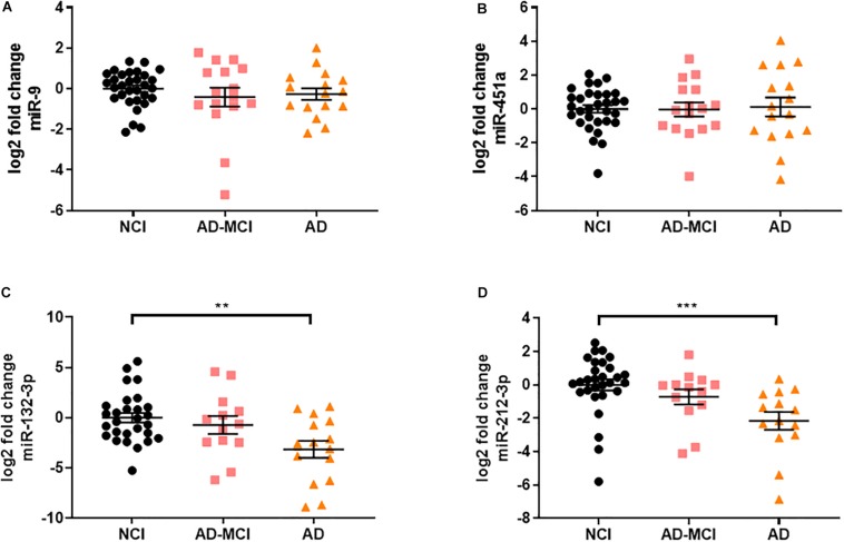
“miR-132-3p and miR-212-3p were downregulated in neural exosomes derived from the plasma of AD patients [79]”
From MicroRNAs in Extracellular Vesicles of Alzheimer’s Disease (2023) · cites this paper as [79]
In the citing paper
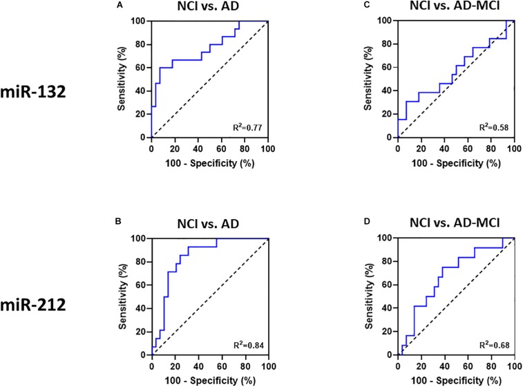
“miR-132-3p (AUC = 0.77) and miR-212-3p (AUC = 0.84) from exosomes in plasma were used as biomarkers to distinguish AD from HC, respectively [79]”
From MicroRNAs in Extracellular Vesicles of Alzheimer’s Disease (2023) · cites this paper as [79]
In the citing paper
“miR-132-3p was downregulated in AD brains [56,84] and EVs from plasma [79] and CSF [49].”
From MicroRNAs in Extracellular Vesicles of Alzheimer’s Disease (2023) · cites this paper as [79]
In the citing paper
“Moreover, miR-212-5p was found decreased in several other neurodegenerative diseases such as AD, Huntington’s disease, and PD [62, 66, 69–75], indicating a key role in neurodegenerative diseases.”
From A role for astrocytic miR-129-5p in frontotemporal dementia (2025) · cites this paper as [74]
Self-check
Percentages, sums and reported differences re-computed from the paper’s own numbers.
The self-check needs the paper’s full text.
Citations
Cites
NIA‐AA Research Framework: Toward a biological definition of Alzheimer's disease
Jack, C. R., Bennett, D. A., Blennow, K., Carrillo, M. C., Dunn, B., Haeberlein, S. B., Holtzman, D. M., Jagust, W., Jessen, F., Karlawish, J., et al. (2018) · Alzheimer's & Dementia · doi:10.1016/j.jalz.2018.02.018 · Cited by 10,537
Self-citationExtracellular vesicles: Exosomes, microvesicles, and friends
Raposo, G., & Stoorvogel, W. (2013) · Journal of Cell Biology · doi:10.1083/jcb.201211138 · Cited by 8,099
Overview of MicroRNA Biogenesis, Mechanisms of Actions, and Circulation
O'Brien, J., Hayder, H., Zayed, Y., & Peng, C. (2018) · Frontiers in Endocrinology · doi:10.3389/fendo.2018.00402 · Cited by 5,287
Johnstone, R. M., Adam, M., Hammond, J. R., Orr, L., & Turbide, C. (1987) · Journal of Biological Chemistry · doi:10.1016/s0021-9258(18)48095-7 · Cited by 2,931
Two Phase 3 Trials of Bapineuzumab in Mild-to-Moderate Alzheimer's Disease
Salloway, S., Sperling, R., Fox, N. C., Blennow, K., Klunk, W., Raskind, M., Sabbagh, M., Honig, L. S., Porsteinsson, A. P., Ferris, S., et al. (2014) · New England Journal of Medicine · doi:10.1056/nejmoa1304839 · Cited by 1,960
Neuropathology of older persons without cognitive impairment from two community-based studies
Bennett, D. A., Schneider, J. A., Arvanitakis, Z., Kelly, J. F., Aggarwal, N. T., Shah, R. C., & Wilson, R. S. (2006) · Neurology · doi:10.1212/01.wnl.0000219668.47116.e6 · Cited by 1,336
Self-citationReligious Orders Study and Rush Memory and Aging Project
Bennett, D. A., Buchman, A. S., Boyle, P. A., Barnes, L. L., Wilson, R. S., & Schneider, J. A. (2018) · Journal of Alzheimer's Disease · doi:10.3233/jad-179939 · Cited by 1,260
Self-citationFiandaca, M. S., Kapogiannis, D., Mapstone, M., Boxer, A., Eitan, E., Schwartz, J. B., Abner, E. L., Petersen, R. C., Federoff, H. J., Miller, B. L., et al. (2015) · Alzheimer's & Dementia · doi:10.1016/j.jalz.2014.06.008 · Cited by 959
Self-citationPlasma β-amyloid in Alzheimer's disease and vascular disease
Janelidze, S., Stomrud, E., Palmqvist, S., Zetterberg, H., van Westen, D., Jeromin, A., Song, L., Hanlon, D., Tan Hehir, C. A., Baker, D., et al. (2016) · Scientific Reports · doi:10.1038/srep26801 · Cited by 626
Emerging roles of exosomes in neuron-glia communication
Frühbeis, C., Fröhlich, D., & Krämer-Albers, E.-M. (2012) · Frontiers in Physiology · doi:10.3389/fphys.2012.00119 · Cited by 275
Fluid Biomarkers in Alzheimer Disease
Blennow, K., Zetterberg, H., & Fagan, A. M. (2012) · Cold Spring Harbor Perspectives in Medicine · doi:10.1101/cshperspect.a006221 · Cited by 208
Monsell, S. E., Kukull, W. A., Roher, A. E., Maarouf, C. L., Serrano, G., Beach, T. G., Caselli, R. J., Montine, T. J., & Reiman, E. M. (2015) · JAMA Neurology · doi:10.1001/jamaneurol.2015.1721 · Cited by 82
Research works printed but not linked: 36
- Abner, 2016, Plasma neuronal exosomal levels of Alzheimer’s disease biomarkers in normal aging., Ann. Clin. Transl. Neurol., 3, 399, 10.1002/acn3.309 · DOI
- Banzhaf-Strathmann, 2014, MicroRNA-125b induces tau hyperphosphorylation and cognitive deficits in Alzheimer’s disease., EMBO J., 33, 1667, 10.15252/embj.201387576 · DOI · Paper page
- Bao, 2017, PET Imaging for Early Detection of Alzheimer’s Disease: from pathologic to physiologic biomarkers., PET Clin., 12, 329, 10.1016/j.cpet.2017.03.001 · DOI
- Bateman, 2019, Plasma biomarkers of AD emerging as essential tools for drug development: an EU/US CTAD task force report., J. Prev. Alzheimers Dis., 6, 169, 10.14283/jpad.2019.21 · DOI
- Bhatnagar, 2014, Increased microRNA-34c abundance in Alzheimer’s disease circulating blood plasma., Front. Mol. Neurosci., 7, 10.3389/fnmol.2014.00002 · DOI
- Chevillet, 2014, Quantitative and stoichiometric analysis of the microRNA content of exosomes., Proc. Natl. Acad. Sci. U.SA., 111, 14888, 10.1073/pnas.1408301111 · DOI
- Coolen, 2013, miR-9: a versatile regulator of neurogenesis., Front. Cell Neurosci., 7, 10.3389/fncel.2013.00220 · DOI · Paper page
- Denk, 2018, Specific serum and CSF microRNA profiles distinguish sporadic behavioural variant of frontotemporal dementia compared with Alzheimer patients and cognitively healthy controls., PLoS One, 13, 10.1371/journal.pone.0197329 · DOI
- El Fatimy, 2018, MicroRNA-132 provides neuroprotection for tauopathies via multiple signaling pathways., Acta Neuropathol., 136, 537, 10.1007/s00401-018-1880-5 · DOI · Paper page
- Galasko, 2015, Expanding the repertoire of biomarkers for alzheimer’s disease: targeted and non-targeted approaches., Front. Neurol., 6, 10.3389/fneur.2015.00256 · DOI
- Goetzl, Altered lysosomal proteins in neural-derived plasma exosomes in preclinical Alzheimer disease., Neurology, 85, 40, 10.1212/WNL.0000000000001702 · DOI · Paper page
- Goetzl, Low neural exosomal levels of cellular survival factors in Alzheimer’s disease., Ann. Clin. Transl. Neurol., 2, 769, 10.1002/acn3.211 · DOI
- Goetzl, 2019, Neuron-derived exosome proteins may contribute to progression from repetitive mild traumatic brain injuries to chronic traumatic encephalopathy., Front. Neurosci., 13, 10.3389/fnins.2019.00452 · DOI
- Guix, 2018, Detection of aggregation-competent tau in neuron-derived extracellular vesicles., Int. J. Mol. Sci., 19, 10.3390/ijms19030663 · DOI
- Hebert, 2008, Loss of microRNA cluster miR-29a/b-1 in sporadic Alzheimer’s disease correlates with increased BACE1/beta-secretase expression., Proc. Natl. Acad. Sci. U.S.A., 105, 6415, 10.1073/pnas.0710263105 · DOI · Paper page
- Heinzelman, 2016, Nanoscale extracellular vesicle analysis in Alzheimer’s disease diagnosis and therapy., Int. J. Alzheimers Dis., 2016, 10.1155/2016/8053139 · DOI
- Hernandez-Rapp, 2016, microRNA-132/212 deficiency enhances Abeta production and senile plaque deposition in Alzheimer’s disease triple transgenic mice., Sci. Rep., 6, 10.1038/srep30953 · DOI
- Herrera-Espejo, 2019, A Systematic Review of MicroRNA Expression as Biomarker of Late-Onset Alzheimer’s Disease., Mol. Neurobiol, 10.1007/s12035-019-01676-9 · DOI · Paper page
- Jack, 2013, Tracking pathophysiological processes in Alzheimer’s disease: an updated hypothetical model of dynamic biomarkers., Lancet Neurol., 12, 207, 10.1016/S1474-4422(12)70291-0 · DOI · Paper page
- Kapogiannis, 2015, Dysfunctionally phosphorylated type 1 insulin receptor substrate in neural-derived blood exosomes of preclinical Alzheimer’s disease., FASEB J., 29, 589, 10.1096/fj.14-262048 · DOI
- Kosik, 2006, The neuronal microRNA system., Nat. Rev. Neurosci., 7, 911, 10.1038/nrn2037 · DOI
- Lau, 2013, Alteration of the microRNA network during the progression of Alzheimer’s disease., EMBO Mol. Med., 5, 1613, 10.1002/emmm.201201974 · DOI · Paper page
- Okamoto, 2018, MicroRNA-451a in extracellular, blood-resident vesicles attenuates macrophage and dendritic cell responses to influenza whole-virus vaccine., J. Biol. Chem., 293, 18585, 10.1074/jbc.RA118.003862 · DOI
- Pan, 1985, Electron microscopic evidence for externalization of the transferrin receptor in vesicular form in sheep reticulocytes., J. Cell Biol., 101, 942, 10.1083/jcb.101.3.942 · DOI
- Paolicelli, 2019, Cell-to-cell communication by extracellular vesicles: focus on microglia., Neuroscience, 405, 148, 10.1016/j.neuroscience.2018.04.003 · DOI · Paper page
- Patrick, 2017, Dissecting the role of non-coding RNAs in the accumulation of amyloid and tau neuropathologies in Alzheimer’s disease., Mol. Neurodegener., 12, 10.1186/s13024-017-0191-y · DOI · Paper page
- Pichler, 2017, The miRNome of Alzheimer’s disease: consistent downregulation of the miR-132/212 cluster., Neurobiol. Aging, 50, 10.1016/j.neurobiolaging.2016.09.019 · DOI · Paper page
- Pucella, 2015, miR-182 is largely dispensable for adaptive immunity: lack of correlation between expression and function., J. Immunol., 194, 2635, 10.4049/jimmunol.1402261 · DOI
- Salta, 2016, miR-132 loss de-represses ITPKB and aggravates amyloid and TAU pathology in Alzheimer’s brain., EMBO Mol. Med., 8, 1005, 10.15252/emmm.201606520 · DOI · Paper page
- Sambandan, 2017, Activity-dependent spatially localized miRNA maturation in neuronal dendrites., Science, 355, 634, 10.1126/science.aaf8995 · DOI
- Swarbrick, 2019, Systematic review of miRNA as biomarkers in alzheimer’s disease., Mol. Neurobiol., 56, 6156, 10.1007/s12035-019-1500-y · DOI
- Vella, 2016, Focus on extracellular vesicles: exosomes and their role in protein trafficking and biomarker potential in Alzheimer’s and Parkinson’s disease., Int. J. Mol. Sci., 17, 10.3390/ijms17020173 · DOI
- Wanet, 2012, miR-212/132 expression and functions: within and beyond the neuronal compartment., Nucleic Acids Res., 40, 4742, 10.1093/nar/gks151 · DOI
- Weinberg, 2015, Evidence for a neuroprotective microRNA pathway in amnestic mild cognitive impairment., Front. Neurosci., 9, 10.3389/fnins.2015.00430 · DOI
- Winston, 2016, Prediction of conversion from mild cognitive impairment to dementia with neuronally derived blood exosome protein profile., Alzheimers Dement., 3, 63, 10.1016/j.dadm.2016.04.001 · DOI
- Wong, 2013, De-repression of FOXO3a death axis by microRNA-132 and -212 causes neuronal apoptosis in Alzheimer’s disease., Hum. Mol. Genet., 22, 3077, 10.1093/hmg/ddt164 · DOI · Paper page
Cited by
Sorted by citations
Exosome Processing and Characterization Approaches for Research and Technology Development
Lai, J. J., Chau, Z. L., Chen, S., Hill, J. J., Korpany, K. V., Liang, N., Lin, L., Lin, Y., Liu, J. K., Liu, Y., et al. (2022) · Advanced Science · doi:10.1002/advs.202103222 · Cited by 623
Biomarkers used in Alzheimer’s disease diagnosis, treatment, and prevention
Mahaman, Y. A. R., Embaye, K. S., Huang, F., Li, L., Zhu, F., Wang, J.-Z., Liu, R., Feng, J., & Wang, X. (2022) · Ageing Research Reviews · doi:10.1016/j.arr.2021.101544 · Cited by 149
Xu, X., Iqbal, Z., Xu, L., Wen, C., Duan, L., Xia, J., Yang, N., Zhang, Y., & Liang, Y. (2024) · Psychiatry and Clinical Neurosciences · doi:10.1111/pcn.13610 · Cited by 68
Emerging prospects of extracellular vesicles for brain disease theranostics
Wang, R., Wang, X., Zhang, Y., Zhao, H., Cui, J., Li, J., & Di, L. (2022) · Journal of Controlled Release · doi:10.1016/j.jconrel.2021.12.024 · Cited by 48
MicroRNAs in Extracellular Vesicles of Alzheimer’s Disease
Li, W., & Zheng, Y. (2023) · Cells · doi:10.3390/cells12101378 · Cited by 43
Metadata
- Authors
- Cha, D. J., Mengel, D., Mustapic, M., Liu, W., Selkoe, D. J., Kapogiannis, D., Galasko, D., Rissman, R. A., Bennett, D. A., & Walsh, D. M.
- Venue
- Frontiers in Neuroscience
- Publisher
- Frontiers Media SA
- Published
- 2019-11-26
- Volume
- 13
- ISSN
- 1662-453X
- PMC
- PMC6902042
- Cited by
- 204
- References
- 12
- Funders
- Alzheimer's Drug Discovery Foundation
- Alzheimer's Association
- Deutsche Forschungsgemeinschaft
About these checks
Registry, retraction, Cites and statistics results are computed, not judged, with one exception: a printed reference that no index lists is read once by a model before it counts as a citation error. Claims are extracted by a model and kept only when their quote appears verbatim in the text; each cited source is then checked, with a model’s help, for whether it says what the claim relies on. Those verdicts can be wrong — read the claim’s quote and the cited source before relying on one.
Statistics and arithmetic are recomputed, not judged.
Confidence bands are shown only for fields calibrated on our annotated test set. None is calibrated yet, so no band is shown.
What counts as a claim ›How we measure confidence ›How we check this ›
Are you an author, or do you see a problem with this page? Request a review
