

The DOI matches its Crossref record; 190 of the 192 references it cites were found in a registry or an index.
Integrin-Mediated TGFβ Activation Modulates the Tumour Microenvironment
Open accessAbstract
TGFβ (transforming growth factor-beta) is a pleotropic cytokine with contrasting effects in cancer. In normal tissue and early tumours, TGFβ acts as a tumour suppressor, limiting proliferation and inducing apoptosis. However, these effects are eventually abrogated by the loss or inactivation of downstream signalling within the TGFβ pathway, and in established tumours, TGFβ then acts as a tumour promotor through multiple mechanisms including inducing epithelial-to-mesenchymal transition (EMT), promoting formation of cancer-associated fibroblasts (CAFs) and increasing angiogenesis. TGFβ is secrereted as a large latent complex and is embedded in the extracellular matrix or held on the surface of cells and must be activated before mediating its multiple functions. Thus, whilst TGFβ is abundant in the tumour microenvironment (TME), its functionality is regulated by local activation. The αv-integrins are major activators of latent-TGFβ. The potential benefits of manipulating the immune TME have been highlighted by the clinical success of immune-checkpoint inhibitors in a number of solid tumour types. TGFβ is a potent suppressor of T-cell-mediated immune surveillance and a key cause of resistance to checkpoint inhibitors. Therefore, as certain integrins locally activate TGFβ, they are likely to have a role in the immunosuppressive TME, although this remains to be confirmed. In this review, we discussed the role of TGFβ in cancer, the role of integrins in activating TGFβ in the TME, and the potential benefits of targeting integrins to augment immunotherapies.
Overview
The claims and statistics of this paper haven’t been checked yet.
Run the check — free, no account needed. It reads the open-access text and checks the sources it cites.
Integrity signals
Registry
Cites
Statistics
Figures
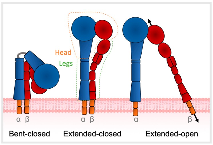
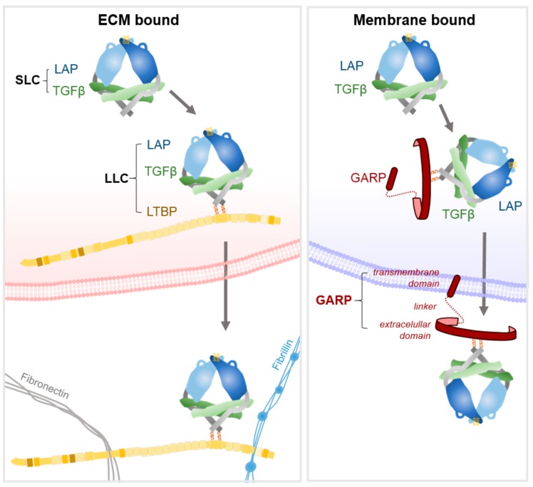
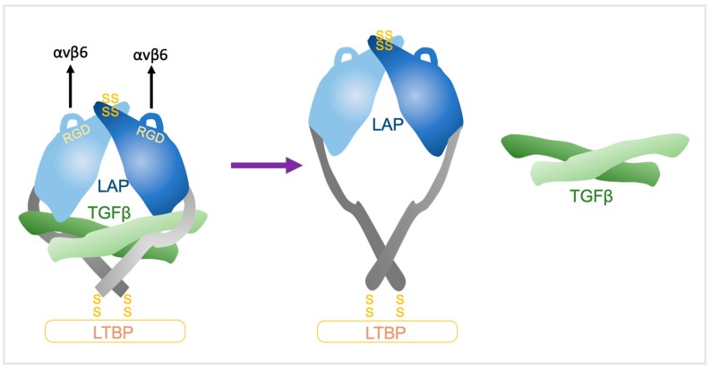
Verification
Claims the paper makes
The claims and statistics of this paper haven’t been checked yet.
Run the check — free, no account needed. It reads the open-access text and checks the sources it cites.
Claims other papers make about this one
Each is checked against this paper’s own text: does it say what the citing paper says it does?
The source backs part of the claim, not all of it — or backs it for a different population, condition or comparison.
In the citing paper
“A recent report further demonstrated that Atf4 expression is enriched in hepatic Tregs from Western diet-induced obese mice, where it contributes to liver fibrosis by promoting TGF-β activation through integrin αvβ8.”
From The double-edged sword of regulatory T cells in MASLD: from inflammation control to fibrosis promotion (2026) · cites this paper as Brown and Marshall 2019
Experimental — accuracy not yet measuredIn the citing paper
“Since αvβ1, αvβ6 and αvβ8 mediate TGF-β1 activation81”
From TGFβ biology in cancer progression and immunotherapy (2021) · cites this paper as [81]
Self-check
Percentages, sums and reported differences re-computed from the paper’s own numbers.
The self-check needs the paper’s full text.
Citations
Cites
PD-1 and PD-L1 Immune Checkpoint Blockade to Treat Breast Cancer
Hartkopf, A. D., Taran, F., Wallwiener, M., Walter, C. B., Krämer, B., Grischke, E., & Brucker, S. Y. (2016) · Breast Care · doi:10.1159/000453569 · Cited by 30,989
Epithelial-Mesenchymal Transitions in Development and Disease
Thiery, J. P., Acloque, H., Huang, R. Y. J., & Nieto, M. A. (2009) · Cell · doi:10.1016/j.cell.2009.11.007 · Cited by 9,931
Hynes (2002) · doi:10.1016/s0092-8674(02)00971-6 · Cited by 8,606
Mechanisms of TGF-β Signaling from Cell Membrane to the Nucleus
Shi, Y., & Massagué, J. (2003) · Cell · doi:10.1016/s0092-8674(03)00432-x · Cited by 5,860
Smad-dependent and Smad-independent pathways in TGF-β family signalling
Derynck, R., & Zhang, Y. E. (2003) · Nature · doi:10.1038/nature02006 · Cited by 5,498
TGFβ attenuates tumour response to PD-L1 blockade by contributing to exclusion of T cells
Mariathasan, S., Turley, S. J., Nickles, D., Castiglioni, A., Yuen, K., Wang, Y., Kadel III, E. E., Koeppen, H., Astarita, J. L., Cubas, R., et al. (2018) · Nature · doi:10.1038/nature25501 · Cited by 5,400
Predictive correlates of response to the anti-PD-L1 antibody MPDL3280A in cancer patients
Herbst, R. S., Soria, J.-C., Kowanetz, M., Fine, G. D., Hamid, O., Gordon, M. S., Sosman, J. A., McDermott, D. F., Powderly, J. D., Gettinger, S. N., et al. (2014) · Nature · doi:10.1038/nature14011 · Cited by 5,231
Massagué, J. (2008) · Cell · doi:10.1016/j.cell.2008.07.001 · Cited by 3,907
Rosenberg, J. E., Hoffman-Censits, J., Powles, T., van der Heijden, M. S., Balar, A. V., Necchi, A., Dawson, N., O'Donnell, P. H., Balmanoukian, A., Loriot, Y., et al. (2016) · The Lancet · doi:10.1016/s0140-6736(16)00561-4 · Cited by 3,669
Polarization of Tumor-Associated Neutrophil Phenotype by TGF-β: “N1” versus “N2” TAN
Fridlender, Z. G., Sun, J., Kim, S., Kapoor, V., Cheng, G., Ling, L. E., Worthen, G. S., & Albelda, S. Μ. (2009) · Cancer Cell · doi:10.1016/j.ccr.2009.06.017 · Cited by 3,470
Pembrolizumab as Second-Line Therapy for Advanced Urothelial Carcinoma
Bellmunt, J., de Wit, R., Vaughn, D. J., Fradet, Y., Lee, J.-L., Fong, L., Vogelzang, N. J., Climent, M. A., Petrylak, D. P., Choueiri, T. K., et al. (2017) · New England Journal of Medicine · doi:10.1056/nejmoa1613683 · Cited by 3,395
Transitions between epithelial and mesenchymal states: acquisition of malignant and stem cell traits
Polyak, K., & Weinberg, R. A. (2009) · Nature Reviews Cancer · doi:10.1038/nrc2620 · Cited by 3,302
The Transforming Growth Factor-Beta Family
Massagué, J. (1990) · Annual Review of Cell Biology · doi:10.1146/annurev.cb.06.110190.003121 · Cited by 3,041
Shull, M. M., Ormsby, I., Kier, A. B., Pawlowski, S., Diebold, R. J., Yin, M., Allen, R., Sidman, C., Proetzel, G., Calvin, D., et al. (1992) · Nature · doi:10.1038/359693a0 · Cited by 2,985
Özdemir, B. C., Pentcheva-Hoang, T., Carstens, J. L., Zheng, X., Wu, C.-C., Simpson, T. R., Laklai, H., Sugimoto, H., Kahlert, C., Novitskiy, S. V., et al. (2014) · Cancer Cell · doi:10.1016/j.ccr.2014.04.005 · Cited by 2,681
Distinct populations of inflammatory fibroblasts and myofibroblasts in pancreatic cancer
Öhlund, D., Handly-Santana, A., Biffi, G., Elyada, E., Almeida, A. S., Ponz-Sarvise, M., Corbo, V., Oni, T. E., Hearn, S. A., Lee, E. J., et al. (2017) · Journal of Experimental Medicine · doi:10.1084/jem.20162024 · Cited by 2,590
Role of Transforming Growth Factor β in Human Disease
Blobe, G. C., Schiemann, W. P., & Lodish, H. F. (2000) · New England Journal of Medicine · doi:10.1056/nejm200005043421807 · Cited by 2,453
Transforming Growth Factor-β Signaling in Immunity and Cancer
Batlle, E., & Massagué, J. (2019) · Immunity · doi:10.1016/j.immuni.2019.03.024 · Cited by 2,366
Mechanism of activation of the TGF-β receptor
Wrana, J. L., Attisano, L., Wieser, R., Ventura, F., & Massagué, J. (1994) · Nature · doi:10.1038/370341a0 · Cited by 2,340
Balar, A. V., Galsky, M. D., Rosenberg, J. E., Powles, T., Petrylak, D. P., Bellmunt, J., Loriot, Y., Necchi, A., Hoffman-Censits, J., Perez-Gracia, J. L., et al. (2017) · The Lancet · doi:10.1016/s0140-6736(16)32455-2 · Cited by 2,199
Immune-related adverse events with immune checkpoint blockade: a comprehensive review
Michot, J. M., Bigenwald, C., Champiat, S., Collins, M., Carbonnel, F., Postel-Vinay, S., Berdelou, A., Varga, A., Bahleda, R., Hollebecque, A., et al. (2016) · European Journal of Cancer · doi:10.1016/j.ejca.2015.11.016 · Cited by 2,127
Stromal Elements Act to Restrain, Rather Than Support, Pancreatic Ductal Adenocarcinoma
Rhim, A. D., Oberstein, P. E., Thomas, D. H., Mirek, E. T., Palermo, C. F., Sastra, S. A., Dekleva, E. N., Saunders, T., Becerra, C. P., Tattersall, I. W., et al. (2014) · Cancer Cell · doi:10.1016/j.ccr.2014.04.021 · Cited by 2,085
Munger, J. S., Huang, X., Kawakatsu, H., Griffiths, M. J. D., Dalton, S. L., Wu, J., Pittet, J.-F., Kaminski, N., Garat, C., Matthay, M. A., et al. (1999) · Cell · doi:10.1016/s0092-8674(00)80545-0 · Cited by 2,076
Kulkarni, A. B., Huh, C. G., Becker, D., Geiser, A., Lyght, M., Flanders, K. C., Roberts, A. B., Sporn, M. B., Ward, J. M., & Karlsson, S. (1993) · Proceedings of the National Academy of Sciences · doi:10.1073/pnas.90.2.770 · Cited by 1,967
Brabletz, T., Kalluri, R., Nieto, M. A., & Weinberg, R. A. (2018) · Nature Reviews Cancer · doi:10.1038/nrc.2017.118 · Cited by 1,961
Yu, Q., & Stamenkovic, I. (2000) · PubMed · Cited by 1,952
Massagué, J. (2000) · Nature Reviews Molecular Cell Biology · doi:10.1038/35043051 · Cited by 1,925
TGFβ drives immune evasion in genetically reconstituted colon cancer metastasis
Tauriello, D. V. F., Palomo-Ponce, S., Stork, D., Berenguer-Llergo, A., Badia-Ramentol, J., Iglesias, M., Sevillano, M., Ibiza, S., Cañellas, A., Hernando-Momblona, X., et al. (2018) · Nature · doi:10.1038/nature25492 · Cited by 1,851
Molecular requirements for epithelial–mesenchymal transition during tumor progression
Huber, M. A., Kraut, N., & Beug, H. (2005) · Current Opinion in Cell Biology · doi:10.1016/j.ceb.2005.08.001 · Cited by 1,850
Yu, Q., & Stamenkovic, I. (2000) · Genes & Development · doi:10.1101/gad.14.2.163 · Cited by 1,754
Humphries, J. D., Byron, A., & Humphries, M. J. (2006) · Journal of Cell Science · doi:10.1242/jcs.03098 · Cited by 1,711
Structural Basis of Integrin Regulation and Signaling
Luo, B., Carman, C. V., & Springer, T. A. (2007) · Annual Review of Immunology · doi:10.1146/annurev.immunol.25.022106.141618 · Cited by 1,662
Nakamura, K., Kitani, A., & Strober, W. (2001) · The Journal of Experimental Medicine · doi:10.1084/jem.194.5.629 · Cited by 1,596
Brooks, P. C., Strömblad, S., Sanders, L. C., Schalscha, T. V., Aimes, R. T., Stetler‐Stevenson, W. G., Quigley, J. P., & Cheresh, D. A. (1996) · Cell · doi:10.1016/s0092-8674(00)81235-0 · Cited by 1,566
Nishida, N., Yano, H., Nishida, T., Kamura, T., & Kojiro, M. (2006) · Vascular Health and Risk Management · doi:10.2147/vhrm.2006.2.3.213 · Cited by 1,509
Every step of the way: integrins in cancer progression and metastasis
Hamidi, H., & Ivaska, J. (2018) · Nature Reviews Cancer · doi:10.1038/s41568-018-0038-z · Cited by 1,493
Myofibroblast contraction activates latent TGF-β1 from the extracellular matrix
Wipff, P.-J., Rifkin, D. B., Meister, J.-J., & Hinz, B. (2007) · The Journal of Cell Biology · doi:10.1083/jcb.200704042 · Cited by 1,346
TGF-β and the TGF-β family: context-dependent roles in cell and tissue physiology
Morikawa, M., Derynck, R., & Miyazono, K. (2016) · Cold Spring Harbor Perspectives in Biology · doi:10.1101/cshperspect.a021873 · Cited by 1,321
Hargadon, K. M., Johnson, C. E., & Williams, C. J. (2018) · International Immunopharmacology · doi:10.1016/j.intimp.2018.06.001 · Cited by 1,211
Integrin Structure, Activation, and InteractionsPreprint
Campbell, I. D., & Humphries, M. J. (2011) · Cold Spring Harbor Perspectives in Biology · doi:10.1101/cshperspect.a004994 · Cited by 1,192
Takagi, J., Petre, B. M., Walz, T., & Springer, T. A. (2002) · Cell · doi:10.1016/s0092-8674(02)00935-2 · Cited by 1,168
Stromal gene expression defines poor-prognosis subtypes in colorectal cancer
Calon, A., Lonardo, E., Berenguer-Llergo, A., Espinet, E., Hernando-Momblona, X., Iglesias, M., Sevillano, M., Palomo-Ponce, S., Tauriello, D. V. F., Byrom, D., et al. (2015) · Nature Genetics · doi:10.1038/ng.3225 · Cited by 1,070
Stupp, R., Hegi, M. E., Gorlia, T., Erridge, S. C., Perry, J., Hong, Y.-K., Aldape, K. D., Lhermitte, B., Pietsch, T., Grujicic, D., et al. (2014) · The Lancet Oncology · doi:10.1016/s1470-2045(14)70379-1 · Cited by 1,052
Defective haematopoiesis and vasculogenesis in transforming growth factor-β1 knock out mice
Dickson, M. C., Martin, J. S., Cousins, F. M., Kulkarni, A. B., Karlsson, S., & Akhurst, R. J. (1995) · Development · doi:10.1242/dev.121.6.1845 · Cited by 1,029
TGF-β and immune cells: an important regulatory axis in the tumor microenvironment and progression
Yang, L., Pang, Y., & Moses, H. L. (2010) · Trends in Immunology · doi:10.1016/j.it.2010.04.002 · Cited by 1,023
Latent TGF-β structure and activation
Shi, M., Zhu, J., Wang, R., Chen, X., Mi, L., Walz, T., & Springer, T. A. (2011) · Nature · doi:10.1038/nature10152 · Cited by 1,013
Ghiringhelli, F., Menard, C., Terme, M., Flament, C., Taı̈eb, J., Chaput, N., Puig, P. E., Novault, S., Escudier, B. J., Vivier, É., et al. (2005) · The Journal of Experimental Medicine · doi:10.1084/jem.20051511 · Cited by 1,004
Hegde, P. S., Karanikas, V., & Evers, S. (2016) · Clinical Cancer Research · doi:10.1158/1078-0432.ccr-15-1507 · Cited by 1,000
Yin, J. J., Selander, K. S., Chirgwin, J. M., Dallas, M., Grubbs, B. G., Wieser, R., Massagué, J., Mundy, G. R., & Guise, T. A. (1999) · Journal of Clinical Investigation · doi:10.1172/jci3523 · Cited by 988
Shang, B., Liu, Y., Jiang, S.-J., & Liu, Y. (2015) · Scientific Reports · doi:10.1038/srep15179 · Cited by 985
Moustakas, A., & Heldin, C.-H. (2005) · Journal of Cell Science · doi:10.1242/jcs.02554 · Cited by 958
The polarization of immune cells in the tumour environment by TGFβ
Flavell, R. A., Sanjabi, S., Wrzesinski, S. H., & Licona-Limón, P. (2010) · Nature Reviews Immunology · doi:10.1038/nri2808 · Cited by 940
Miettinen, P. J., Ebner, R., Lopez, A. R., & Derynck, R. (1994) · The Journal of Cell Biology · doi:10.1083/jcb.127.6.2021 · Cited by 939
The roles of TGFβ in the tumour microenvironment
Pickup, M. W., Novitskiy, S. V., & Moses, H. L. (2013) · Nature reviews. Cancer · doi:10.1038/nrc3603 · Cited by 936
TGF-β: A Master of All T Cell Trades
Li, M. O., & Flavell, R. A. (2008) · Cell · doi:10.1016/j.cell.2008.07.025 · Cited by 916
Henderson, N. C., Arnold, T. D., Katamura, Y., Giacomini, M. M., Rodriguez, J. D., McCarty, J. H., Pellicoro, A., Raschperger, E., Betsholtz, C., Ruminski, P. G., et al. (2013) · Nature Medicine · doi:10.1038/nm.3282 · Cited by 866
TGF-β Activation and Function in Immunity
Travis, M. A., & Sheppard, D. (2014) · Annual Review of Immunology · doi:10.1146/annurev-immunol-032713-120257 · Cited by 856
Phenotypic Diversity and Plasticity in Circulating Neutrophil Subpopulations in Cancer
Sagiv, J. Y., Michaeli, J., Assi, S., Mishalian, I., Kisos, H., Levy, L., Damti, P., Lumbroso, D., Polyansky, L., Sionov, R. V., et al. (2015) · Cell Reports · doi:10.1016/j.celrep.2014.12.039 · Cited by 842
Smad3 and NFAT cooperate to induce Foxp3 expression through its enhancer
Tone, Y., Furuuchi, K., Kojima, Y., Tykocinski, M. L., Greene, M. I., & Tone, M. (2008) · Nature Immunology · doi:10.1038/ni1549 · Cited by 807
Gorelik, L., & Flavell, R. A. (2001) · Nature Medicine · doi:10.1038/nm1001-1118 · Cited by 770
Li, M. O., Wan, Y. Y., & Flavell, R. A. (2007) · Immunity · doi:10.1016/j.immuni.2007.03.014 · Cited by 769
TGF-β Superfamily Signaling in Embryonic Development and Homeostasis
Wu, M., & Hill, C. S. (2009) · Developmental Cell · doi:10.1016/j.devcel.2009.02.012 · Cited by 743
The integrin αvβ8 mediates epithelial homeostasis through MT1-MMP–dependent activation of TGF-β1
Mu, D., Cambier, S., Fjellbirkeland, L., Baron, J. L., Munger, J. S., Kawakatsu, H., Sheppard, D., Broaddus, V. C., & Nishimura, S. L. (2002) · The Journal of Cell Biology · doi:10.1083/jcb.200109100 · Cited by 729
Bertrand, A., Kostine, M., Barnetche, T., Truchetet, M.-E., & Schaeverbeke, T. (2015) · BMC Medicine · doi:10.1186/s12916-015-0455-8 · Cited by 704
Integrin–TGF‐β crosstalk in fibrosis, cancer and wound healing
Margadant, C., & Sonnenberg, A. (2010) · The EMBO Reports · doi:10.1038/embor.2009.276 · Cited by 653
Targeting the TGFβ pathway for cancer therapy
Neuzillet, C., Tijeras‐Raballand, A., Cohen, R., Cros, J. F., Faivre, S. J., Raymond, É., & Gramont, A. D. E. (2014) · Pharmacology & Therapeutics · doi:10.1016/j.pharmthera.2014.11.001 · Cited by 631
Enhanced pathological angiogenesis in mice lacking β3 integrin or β3 and β5 integrins
Reynolds, L. E., Wyder, L., Lively, J. C., Taverna, D., Robinson, S. D., Huang, X., Sheppard, D., Hynes, R. O., & Hodivala‐Dilke, K. (2002) · Nature Medicine · doi:10.1038/nm0102-27 · Cited by 627
Giampieri, S., Manning, C. S., Hooper, S., Jones, J. L., Hill, C. S., & Sahai, E. A. (2009) · Nature Cell Biology · doi:10.1038/ncb1973 · Cited by 602
Lahn, M. M. F., Herbertz, S., Sawyer, J. S., Stauber, A. J., Gueorguieva, I., Driscoll, K. E., Estrem, S. T., Cleverly, A., Desaiah, D., Guba, S. C., et al. (2015) · Drug Design Development and Therapy · doi:10.2147/dddt.s86621 · Cited by 571
LOX-Mediated Collagen Crosslinking Is Responsible for Fibrosis-Enhanced Metastasis
Cox, T. R., Bird, D., Baker, A.-M., Barker, H. E., Ho, M. W.-Y., Lang, G., & Erler, J. T. (2013) · Cancer Research · doi:10.1158/0008-5472.can-12-2233 · Cited by 571
Yang, P., Li, Q.-J., Feng, Y., Zhang, Y., Markowitz, G. J., Ning, S., Deng, Y., Zhao, J., Jiang, S., Yuan, Y., et al. (2012) · Cancer Cell · doi:10.1016/j.ccr.2012.07.023 · Cited by 545
Alterations in components of the TGF-β superfamily signaling pathways in human cancer
LEVY, L., & HILL, C. (2006) · Cytokine & Growth Factor Reviews · doi:10.1016/j.cytogfr.2005.09.009 · Cited by 539
Integrins and the activation of latent transforming growth factor β1 – An intimate relationship
Wipff, P., & Hinz, B. (2008) · European Journal of Cell Biology · doi:10.1016/j.ejcb.2008.01.012 · Cited by 532
Zhang, M., He, Y., Sun, X., Li, Q. Y., Wang, W., Zhao, A., & Di, W. J. (2014) · Journal of Ovarian Research · doi:10.1186/1757-2215-7-19 · Cited by 530
T cells that cannot respond to TGF-β escape control by CD4+CD25+ regulatory T cells
Fahlén, L., Read, S., Gorelik, L., Hurst, S. D., Coffman, R. L., Flavell, R. A., & Powrie, F. M. (2005) · The Journal of Experimental Medicine · doi:10.1084/jem.20040685 · Cited by 516
Loss of integrin αvβ8 on dendritic cells causes autoimmunity and colitis in mice
Travis, M. A., Reizis, B., Melton, A. C., Masteller, E., Tang, Q., Proctor, J. M., Wang, Y., Bernstein, X., Huang, X., Reichardt, L. F., et al. (2007) · Nature · doi:10.1038/nature06110 · Cited by 503
Integrin αVβ6-mediated activation of latent TGF-β requires the latent TGF-β binding protein-1
Annes, J. P., Chen, Y., Munger, J. S., & Rifkin, D. B. (2004) · The Journal of Cell Biology · doi:10.1083/jcb.200312172 · Cited by 500
Marie, J. C., Liggitt, D., & Rudensky, A. Y. (2006) · Immunity · doi:10.1016/j.immuni.2006.07.012 · Cited by 480
Tran, D. Q., Andersson, J., Wang, R., Ramsey, H. E., Unutmaz, D., & Shevach, E. M. (2009) · Proceedings of the National Academy of Sciences · doi:10.1073/pnas.0901944106 · Cited by 473
Calderwood, D. (2004) · Journal of Cell Science · doi:10.1242/jcs.01014 · Cited by 449
TGFβ signaling plays a critical role in promoting alternative macrophage activation
Gong, D., Shi, W., Yi, S.-J., Chen, H., Groffen, J., & Heisterkamp, N. (2012) · BMC Immunology · doi:10.1186/1471-2172-13-31 · Cited by 440
Latent Transforming Growth Factor-β (TGF-β) Binding Proteins: Orchestrators of TGF-β Availability
Rifkin, D. B. (2004) · Journal of Biological Chemistry · doi:10.1074/jbc.r400029200 · Cited by 422
Differentiation plasticity regulated by TGF-β family proteins in development and disease
Derynck, R., & Akhurst, R. J. (2007) · Nature Cell Biology · doi:10.1038/ncb434 · Cited by 421
Nucleocytoplasmic Shuttling of Smads 2, 3, and 4 Permits Sensing of TGF-β Receptor Activity
Inman, G. J., Nicolás, F. J., & Hill, C. S. (2002) · Molecular Cell · doi:10.1016/s1097-2765(02)00585-3 · Cited by 417
Kaminski, N., Allard, J. D., Pittet, J., Zuo, F., Griffiths, M., Morris, D. G., Huang, X., Sheppard, D., & Heller, R. A. (2000) · Proceedings of the National Academy of Sciences · doi:10.1073/pnas.97.4.1778 · Cited by 416
Regulation of the Bioavailability of TGF-β and TGF-β-Related Proteins
Robertson, I. B., & Rifkin, D. B. (2016) · Cold Spring Harbor Perspectives in Biology · doi:10.1101/cshperspect.a021907 · Cited by 403
Grady, W. M., Myeroff, L. L., Swinler, S. E., Rajput, A., Thiagalingam, S., Lutterbaugh, J., Neumann, A., Brattain, M. G., Chang, J., Kim, S., et al. (1999) · PubMed · Cited by 391
Bhowmick, N. A., Zent, R., Ghiassi, M., McDonnell, M. A., & Moses, H. L. (2001) · Journal of Biological Chemistry · doi:10.1074/jbc.m106176200 · Cited by 388
Structural Biology and Evolution of the TGF-β FamilyPreprint
Hinck, A. P., Mueller, T. D., & Springer, T. A. (2016) · Cold Spring Harbor Perspectives in Biology · doi:10.1101/cshperspect.a022103 · Cited by 381
Bates, R. C., Bellovin, D. I., Brown, C. L., Maynard, E., Wu, B., Kawakatsu, H., Sheppard, D., Oettgen, P., & Mercurio, A. M. (2005) · Journal of Clinical Investigation · doi:10.1172/jci23183 · Cited by 380
The role of proteases in transforming growth factor-β activation
Jenkins, G. R. (2008) · The International Journal of Biochemistry & Cell Biology · doi:10.1016/j.biocel.2007.11.026 · Cited by 374
Complexities of TGF-β Targeted Cancer Therapy
Connolly, E. C., Freimuth, J., & Akhurst, R. J. (2012) · International Journal of Biological Sciences · doi:10.7150/ijbs.4564 · Cited by 354
Latent TGF-β Binding Proteins: Extracellular Matrix Association and Roles in TGF-β Activation
Hyytiäinen, M. M., Penttinen, C., & Keski‐Oja, J. (2004) · Critical Reviews in Clinical Laboratory Sciences · doi:10.1080/10408360490460933 · Cited by 351
Platelets subvert T cell immunity against cancer via GARP-TGFβ axis
Rachidi, S., Metelli, A., Riesenberg, B., Wu, B. X., Nelson, M. H., Wallace, C., Paulos, C. M., Rubinstein, M. P., Garrett-Mayer, E. S., Hennig, M., et al. (2017) · Science Immunology · doi:10.1126/sciimmunol.aai7911 · Cited by 347
Rolli, M., Fransvea, E., Pilch, J., Saven, A., & Felding‐Habermann, B. (2003) · Proceedings of the National Academy of Sciences · doi:10.1073/pnas.1633689100 · Cited by 333
Absence of integrin-mediated TGFβ1 activation in vivo recapitulates the phenotype of TGFβ1-null mice
Yang, Z., Mu, Z., Dabovic, B., Jurukovski, V., Yu, D., Sung, J. J., Xiong, X., & Munger, J. S. (2007) · The Journal of Cell Biology · doi:10.1083/jcb.200611044 · Cited by 330
Smad4 signalling in T cells is required for suppression of gastrointestinal cancer
Kim, B., Li, C., Qiao, W., Mamura, M., Kasperczak, B., Anver, M. R., Wolfraim, L. A., Hong, S., Mushinski, E. B., Potter, M., et al. (2006) · Nature · doi:10.1038/nature04846 · Cited by 325
Wakefield, L. M., Winokur, T. S., Hollands, R. S., Christopherson, K. S., Levinson, A. D., & Sporn, M. B. (1990) · Journal of Clinical Investigation · doi:10.1172/jci114932 · Cited by 320
Sun, L., Wu, G., Willson, J. K. V., Zborowska, E., Yang, J., Rajkarunanayake, I., Wang, J., Gentry, L. E., Wang, X. F., & Brattain, M. G. (1994) · Journal of Biological Chemistry · doi:10.1016/s0021-9258(18)47215-8 · Cited by 319
Huang, X., Wu, J. F., Cass, D. L., Erle, D. J., Corry, D. B., Young, S. G., Farese, R. V., & Sheppard, D. (1996) · The Journal of Cell Biology · doi:10.1083/jcb.133.4.921 · Cited by 316
v Integrins in Angiogenesis and CancerPreprint
Weis, S. M., & Cheresh, D. A. (2011) · Cold Spring Harbor Perspectives in Medicine · doi:10.1101/cshperspect.a006478 · Cited by 311
Neuzillet, C., Tijeras‐Raballand, A., Ragulan, C., Cros, J. F., Patil, Y., Martinet, M., Erkan, M., Kleeff, J. H., Wilson, J. S., Apte, M. V., et al. (2018) · The Journal of Pathology · doi:10.1002/path.5224 · Cited by 310
Reszka, A. A., Hayashi, Y., & Horwitz, A. (1992) · The Journal of Cell Biology · doi:10.1083/jcb.117.6.1321 · Cited by 281
β8 integrins are required for vascular morphogenesis in mouse embryos
Zhu, J., Motejlek, K., Wang, D., Zang, K., Schmidt, A., & Reichardt, L. F. (2002) · Development · doi:10.1242/dev.129.12.2891 · Cited by 274
Force interacts with macromolecular structure in activation of TGF-β
Dong, X., Zhao, B., Iacob, R. E., Zhu, J., Koksal, A. C., Lu, C., Engen, J. R., & Springer, T. A. (2017) · Nature · doi:10.1038/nature21035 · Cited by 273
Guasch, G., Schöber, M., Pasolli, H. A., Conn, E. B., Polak, L., & Fuchs, E. V. (2007) · Cancer Cell · doi:10.1016/j.ccr.2007.08.020 · Cited by 271
The Single-Molecule Mechanics of the Latent TGF-β1 Complex
Buscemi, L., Ramonet, D., Klingberg, F., Formey, A., Smith-Clerc, J., Meister, J.-J., & Hinz, B. (2011) · Current Biology · doi:10.1016/j.cub.2011.11.037 · Cited by 266
Silva, R., D’Amico, G., Hodivala‐Dilke, K., & Reynolds, L. E. (2008) · Arteriosclerosis Thrombosis and Vascular Biology · doi:10.1161/atvbaha.108.172015 · Cited by 263
Nabors, B., Fink, K., Mikkelsen, T., Grujičić, D., Tarnawski, R., Nam, D. H., Mazurkiewicz, M., Salacz, M. E., Ashby, L. S., Zagonel, V., et al. (2015) · Neuro-Oncology · doi:10.1093/neuonc/nou356 · Cited by 242
Munger, J. S., Harpel, J. G., Giancotti, F. G., & Rifkin, D. B. (1998) · Molecular Biology of the Cell · doi:10.1091/mbc.9.9.2627 · Cited by 239
Aluwihare, P., Mu, Z., Zhao, Z., Yu, D., Weinreb, P. H., Horan, G. S. B., Violette, S. M., & Munger, J. S. (2008) · Journal of Cell Science · doi:10.1242/jcs.035246 · Cited by 229
Differentiation plasticity regulated by TGF-|[beta]| family proteins in development and disease
Derynck, R., & Akhurst, R. J. (2007) · Nature · doi:10.1038/ncb0907-1000 · Cited by 227
TGFβ: a sleeping giant awoken by integrins
Worthington, J. J., Klementowicz, J. E., & Travis, M. A. (2010) · Trends in Biochemical Sciences · doi:10.1016/j.tibs.2010.08.002 · Cited by 226
Complete integrin headpiece opening in eight steps
Zhu, J., Zhu, J., & Springer, T. A. (2013) · The Journal of Cell Biology · doi:10.1083/jcb.201212037 · Cited by 215
Worthington, J. J., Kelly, A., Smedley, C., Bauché, D., Campbell, S. S., Marie, J. C., & Travis, M. A. (2015) · Immunity · doi:10.1016/j.immuni.2015.04.012 · Cited by 215
Targeting TGF-β signaling in cancer
Katz, L. H., Li, Y., Chen, J.-S., Muñoz, N. M., Majumdar, A., Chen, J., & Mishra, L. (2013) · Expert Opinion on Therapeutic Targets · doi:10.1517/14728222.2013.782287 · Cited by 212
The TGF-β Signaling Regulator PMEPA1 Suppresses Prostate Cancer Metastases to Bone
Fournier, P. G. J., Juárez, P., Jiang, G., Clines, G. A., Niewolna, M., Kim, H. S., Walton, H. W., Peng, X., Liu, Y., Mohammad, K. S., et al. (2015) · Cancer Cell · doi:10.1016/j.ccell.2015.04.009 · Cited by 212
Taipale, J., Saharinen, J., & Keski‐Oja, J. (1998) · Advances in cancer research · doi:10.1016/s0065-230x(08)60740-x · Cited by 209
Switching TGFβ from a tumor suppressor to a tumor promoter
Inman, G. J. (2011) · Current Opinion in Genetics & Development · doi:10.1016/j.gde.2010.12.004 · Cited by 206
Disruption of the antiproliferative TGF-β signaling pathways in human pancreatic cancer cells
Villanueva, A., García, C., Paules, A. B., Vicente, M., Megías, M., Reyes, G., Villalonga, P., Agell, N., Lluı́s, F., Bachs, O., et al. (1998) · Oncogene · doi:10.1038/sj.onc.1202118 · Cited by 194
Hersey, P., Sosman, J. A., O'Day, S. J., Richards, J. M., Bedikian, A. Y., Gonzalez, R. C., Sharfman, W. H., Weber, R. W., Logan, T. F., Buzoianu, M., et al. (2010) · Cancer · doi:10.1002/cncr.24821 · Cited by 190
Posey, J. A., Khazaeli, M. B., Delgrosso, A., Saleh, M. N., Lin, C., Huse, W. D., & LoBuglio, A. F. (2001) · Cancer Biotherapy and Radiopharmaceuticals · doi:10.1089/108497801300189218 · Cited by 189
Elayadi, A. N., Samli, K. N., Prudkin, L., Liu, Y.-H., Bian, A., Xie, X., Wistuba, I. I., Roth, J. A., McGuire, M. J., & Brown, K. C. (2007) · Cancer Research · doi:10.1158/0008-5472.can-07-0245 · Cited by 189
GARP regulates the bioavailability and activation of TGFβ
Wang, R., Zhu, J., Dong, X., Shi, M., Lu, C., & Springer, T. A. (2012) · Molecular Biology of the Cell · doi:10.1091/mbc.e11-12-1018 · Cited by 188
Moustakas, A., Lin, H. Y.-F., Henis, Y. I., Plamondon, J., O'Connor‐McCourt, M. D., & Lodish, H. F. (1993) · Journal of Biological Chemistry · doi:10.1016/s0021-9258(18)41509-8 · Cited by 181
Fontana, L., Chen, Y., Prijatelj, P., Sakai, T., Fässler, R., Sakai, L. Y., & Rifkin, D. B. (2005) · The FASEB Journal · doi:10.1096/fj.05-4134com · Cited by 181
Hazelbag, S., Kenter, G. G., Gorter, A., Dreef, E., Koopman, L., Violette, S., Weinreb, P. H., & Fleuren, G. J. (2007) · The Journal of Pathology · doi:10.1002/path.2168 · Cited by 178
Cloning and expression of a divergent integrin subunit beta 8.
Moyle, M., Napier, M. A., & McLean, J. W. (1991) · Journal of Biological Chemistry · doi:10.1016/s0021-9258(18)55042-0 · Cited by 177
Asano, Y., Ihn, H., Yamane, K., Jinnin, M., & Tamaki, K. (2006) · The American Journal of Pathology · doi:10.2353/ajpath.2006.041306 · Cited by 175
The integrin αVβ6 binds and activates latent TGFβ3
Annes, J. P., Rifkin, D. B., & Munger, J. S. (2001) · FEBS Letters · doi:10.1016/s0014-5793(01)03280-x · Cited by 173
The αvβ6 Integrin Is Transferred Intercellularly via Exosomes
Fedele, C., Singh, A., Zerlanko, B. J., Iozzo, R. V., & Languino, L. R. (2015) · Journal of Biological Chemistry · doi:10.1074/jbc.c114.617662 · Cited by 167
Structural basis of latent TGF-β1 presentation and activation by GARP on human regulatory T cells
Liénart, S., Merceron, R., Vanderaa, C., Lambert, F., Colau, D., Stockis, J., van der Woning, B., de Haard, H., Saunders, M. J., Coulie, P. G., et al. (2018) · Science · doi:10.1126/science.aau2909 · Cited by 160
Signal transduction by members of the transforming growth factor-β superfamily
Attisano, L., & Wrana, J. L. (1996) · Cytokine & Growth Factor Reviews · doi:10.1016/s1359-6101(96)00042-1 · Cited by 159
Dumont, N., Bakin, A. V., & Arteaga, C. L. (2003) · Journal of Biological Chemistry · doi:10.1074/jbc.m204623200 · Cited by 159
Singh, A., Fedele, C., Lu, H., Nevalainen, M. T., Keen, J. H., & Languino, L. R. (2016) · Molecular Cancer Research · doi:10.1158/1541-7786.mcr-16-0058 · Cited by 159
Cuende, J., Liénart, S., Dedobbeleer, O., van der Woning, B., De Boeck, G., Stockis, J., Huygens, C., Colau, D., Somja, J., Delvenne, P., et al. (2015) · Science Translational Medicine · doi:10.1126/scitranslmed.aaa1983 · Cited by 155
Integrin αvβ5-Mediated TGF-β Activation by Airway Smooth Muscle Cells in Asthma
Tatler, A. L., John, A. E., Jolly, L., Habgood, A., Porte, J., Brightling, C. E., Knox, A. J., Pang, L., Sheppard, D., Huang, X., et al. (2011) · The Journal of Immunology · doi:10.4049/jimmunol.1003507 · Cited by 153
The L3 loop and C-terminal phosphorylation jointly define Smad protein trimerization.
Chacko, B. M., Qin, B. Y., Correia, J. J., Lam, S. S., de Caestecker, M. P., & Lin, K. F. (2001) · Nature Structural Biology · doi:10.1038/84995 · Cited by 151
Budhu, S., Schaer, D., Li, Y., Toledo‐Crow, R., Panageas, K. S., Yang, X., Zhong, H., Houghton, A. N., Silverstein, S. C., Merghoub, T., et al. (2017) · Science Signaling · doi:10.1126/scisignal.aak9702 · Cited by 148
Koopman Van Aarsen, L. A., Leone, D. R., Ho, S., Dolinski, B. M., McCoon, P. E., LePage, D. J., Kelly, R., Heaney, G., Rayhorn, P., Reid, C., et al. (2008) · Cancer Research · doi:10.1158/0008-5472.can-07-2307 · Cited by 146
Therapeutic Targeting of Integrin αvβ6 in Breast Cancer
Moore, K. M., Thomas, G. J., Duffy, S. W., Warwick, J., Gabe, R., Chou, P., Ellis, I. O., Green, A. R., Haider, S. A., Brouilette, K., et al. (2014) · JNCI Journal of the National Cancer Institute · doi:10.1093/jnci/dju169 · Cited by 144
Self-citationStructural determinants of integrin β-subunit specificity for latent TGF-β
Dong, X., Hudson, N. E., Lu, C., & Springer, T. A. (2014) · Nature Structural & Molecular Biology · doi:10.1038/nsmb.2905 · Cited by 141
Naber, H. P. H., Drabsch, Y., Snaar-Jagalska, E. B., ten Dijke, P., & van Laar, T. (2013) · Biochemical and Biophysical Research Communications · doi:10.1016/j.bbrc.2013.04.037 · Cited by 135
Marcoe, J. P., Lim, J., Schaubert, K. L., Fodil-Cornu, N., Matka, M., McCubbrey, A. L., Farr, A. R., Vidal, S. M., & Laouar, Y. (2012) · Nature Immunology · doi:10.1038/ni.2388 · Cited by 134
Ludbrook, S. B., Barry, S. T., Delves, C. J., & Horgan, C. M. T. (2003) · Biochemical Journal · doi:10.1042/bj20020809 · Cited by 126
Melton, A. C., Bailey-Bucktrout, S. L., Travis, M. A., Fife, B. T., Bluestone, J. A., & Sheppard, D. (2010) · Journal of Clinical Investigation · doi:10.1172/jci43786 · Cited by 126
Élez, E., Kocáková, I., Höhler, T., Martens, U. M., Bokemeyer, C., Cutsem, E. V., Melichar, B., Šmakal, M., Csőszi, T., Топузов, Э. Э., et al. (2014) · Annals of Oncology · doi:10.1093/annonc/mdu474 · Cited by 124
Sipos, B., Hahn, D., Carceller, A., Piulats, J., Hedderich, J., Kalthoff, H., Goodman, S. L., Kosmahl, M., & Klöppel, G. (2004) · Histopathology · doi:10.1111/j.1365-2559.2004.01919.x · Cited by 124
Human monocytes and macrophages regulate immune tolerance via integrin αvβ8–mediated TGFβ activation
Kelly, A., Gunaltay, S., McEntee, C. P., Shuttleworth, E. E., Smedley, C., Houston, S. A., Fenton, T. M., Levison, S., Mann, E. R., & Travis, M. A. (2018) · Journal of Experimental Medicine · doi:10.1084/jem.20171491 · Cited by 124
Biological Significance of Local TGF-β Activation in Liver Diseases
Hayashi, H., & Sakai, T. (2012) · Frontiers in Physiology · doi:10.3389/fphys.2012.00012 · Cited by 121
Bacman, D., Merkel, S., Croner, R. S., Papadopoulos, T., Brueckl, W. M., & Dimmler, A. W. (2007) · BMC Cancer · doi:10.1186/1471-2407-7-156 · Cited by 118
Glick, A. B., Weinberg, W. C., Wu, I. H., Quan, W., & Yuspa, S. H. (1996) · PubMed · Cited by 117
Reader, C. S., Vallath, S., Steele, C. W., Haider, S., Brentnall, A. R., Desai, A., Moore, K. M., Jamieson, N. B., Chang, D. K.-F., Bailey, P. J., et al. (2019) · The Journal of Pathology · doi:10.1002/path.5320 · Cited by 116
Self-citationTruong, H. H., Xiong, J., Ghotra, V. P. S., Nirmala, E., Haazen, L. C. J. M., Dévédec, S. E. L., Balcioglu, H. E., He, S., Snaar-Jagalska, E. B., Vreugdenhil, E., et al. (2014) · Science Signaling · doi:10.1126/scisignal.2004751 · Cited by 115
Blocking immunosuppression by human Tregs in vivo with antibodies targeting integrin αVβ8
Stockis, J., Liénart, S., Colau, D., Collignon, A., Nishimura, S. L., Sheppard, D., Coulie, P. G., & Lucas, S. (2017) · Proceedings of the National Academy of Sciences · doi:10.1073/pnas.1710680114 · Cited by 111
Edwards, J. P., Thornton, A. M., & Shevach, E. M. (2014) · The Journal of Immunology · doi:10.4049/jimmunol.1401102 · Cited by 106
TGF-β and αvβ6 Integrin Act in a Common Pathway to Suppress Pancreatic Cancer Progression
Hezel, A. F., Deshpande, V., Zimmerman, S. M., Contino, G., Alagesan, B., O’Dell, M. R., Rivera, L. B., Harper, J. W., Lonning, S. M., Brekken, R. A., et al. (2012) · Cancer Research · doi:10.1158/0008-5472.can-12-0634 · Cited by 99
Crescenzo, G. D., Hinck, C. S., Shu, Z., Zúñiga, J. E., Yang, J., Tang, Y., Baardsnes, J., Mendoza, V., Sun, L., López‐Casillas, F., et al. (2005) · Journal of Molecular Biology · doi:10.1016/j.jmb.2005.10.022 · Cited by 90
Allen, M. D., Thomas, G. J., Clark, S. J., Dawoud, M. M., Vallath, S., Payne, S. J., Gomm, J. J., Dreger, S. A., Dickinson, S., Edwards, D. R., et al. (2013) · Clinical Cancer Research · doi:10.1158/1078-0432.ccr-13-1504 · Cited by 90
Self-citationLiu, Z., Kobayashi, K., van Dinther, M., van Heiningen, S. H., Valdimarsdóttir, G., van Laar, T., Scharpfenecker, M., Löwik, C. W. G. M., Goumans, M., ten Dijke, P., et al. (2009) · Journal of Cell Science · doi:10.1242/jcs.048942 · Cited by 89
A role for the integrin alphavbeta8 in the negative regulation of epithelial cell growth.
Cambier, S., Mu, D., O'Connell, D., Boylen, K., Travis, W. D., Liu, W. H., Broaddus, V. C., & Nishimura, S. L. (2000) · PubMed · Cited by 89
Cilengitide in newly diagnosed glioblastoma: biomarker expression and outcome
Weller, M., Nabors, B., Gorlia, T., Leske, H., Rushing, E. J., Bady, P., Hicking, C., Perry, J., Hong, Y.-K., Roth, P., et al. (2016) · Oncotarget · doi:10.18632/oncotarget.7588 · Cited by 89
Eberlein, C. A., Kendrew, J., McDaid, K., Alfred, A., Kang, J., Jacobs, V. N., Ross, S. J., Rooney, C., Smith, N. R., Rinkenberger, J. L., et al. (2012) · Oncogene · doi:10.1038/onc.2012.460 · Cited by 87
Self-citationOwens, D. M., Romero, M. R., Gardner, C., & Watt, F. M. (2003) · Journal of Cell Science · doi:10.1242/jcs.00725 · Cited by 87
Suppression of TGFβand Angiogenesis by Type VII Collagen in Cutaneous SCC
Martins, V., Caley, M. P., Moore, K. M., Szentpétery, Z., Marsh, S., Murrell, D. F., Kim, M. H., Avari, M., McGrath, J. A., Cerio, R., et al. (2015) · JNCI Journal of the National Cancer Institute · doi:10.1093/jnci/djv293 · Cited by 87
Self-citationChen, X., Song, C., Feng, B.-S., Li, T., Li, P., Zheng, P.-Y., Chen, X., Xing, Z., & Yang, P.-C. (2011) · Journal of Leukocyte Biology · doi:10.1189/jlb.1210696 · Cited by 86
Sheppard, D. (2015) · Annals of the American Thoracic Society · doi:10.1513/annalsats.201406-245mg · Cited by 82
αvβ6 Integrin Promotes the Invasion of Morphoeic Basal Cell Carcinoma through Stromal Modulation
Marsh, D. J., Dickinson, S., Neill, G. W., Marshall, J. F., Hart, I. R., & Thomas, G. J. (2008) · Cancer Research · doi:10.1158/0008-5472.can-08-0174 · Cited by 81
Self-citationConnexins and Integrins in Exosomes
Shimaoka, M., Kawamoto, E., Gaowa, A., Okamoto, T., & Park, E. J. (2019) · Cancers · doi:10.3390/cancers11010106 · Cited by 81
The dual adverse effects of TGF-β secretion on tumor progression
Trapani, J. A. (2005) · Cancer Cell · doi:10.1016/j.ccr.2005.10.018 · Cited by 80
Activation of the Mitogen-Activated Protein Kinase Pathway by Transforming Growth Factor-β
Yue, J., & Mulder, K. M. (2003) · Humana Press eBooks · doi:10.1385/1-59259-053-5:125 · Cited by 77
Role of TGF-β in breast cancer bone metastases
Chiechi, A., Waning, D. L., Stayrook, K. R., Buijs, J. T., Guise, T. A., & Mohammad, K. S. (2013) · Advances in Bioscience and Biotechnology · doi:10.4236/abb.2013.410a4003 · Cited by 76
Languino, L. R., Singh, A., di Prisco, M., Inman, G. J., Luginbuhl, A., Curry, J., & South, A. P. (2016) · PubMed Central · Cited by 74
Exosomal αvβ6 integrin is required for monocyte M2 polarization in prostate cancer
Lu, H., Bowler, N., Harshyne, L. A., Hooper, D. C., Krishn, S. R., Kurtoglu, S., Fedele, C., Liu, Q., Tang, H., Kossenkov, A. V., et al. (2018) · Matrix Biology · doi:10.1016/j.matbio.2018.03.009 · Cited by 73
Hayashida, T., Wu, M.-H., Pierce, A. A., Poncelet, A.-C., Varga, J., & Schnaper, H. W. (2007) · Journal of Cell Science · doi:10.1242/jcs.03492 · Cited by 70
Exosomal regulation of lymphocyte homing to the gut
Park, E. J., Prajuabjinda, O., Soe, Z. Y., Darkwah, S. O., Appiah, M. G., Kawamoto, E., Momose, F., Shiku, H., & Shimaoka, M. (2018) · Blood Advances · doi:10.1182/bloodadvances.2018024877 · Cited by 69
Integrin αvβ6 Promotes an Osteolytic Program in Cancer Cells by Upregulating MMP2
Dutta, A., Li, J., Lu, H., Akech, J., Pratap, J., Wang, T., Zerlanko, B. J., FitzGerald, T. J., Jiang, Z., Birbe, R., et al. (2014) · Cancer Research · doi:10.1158/0008-5472.can-13-1796 · Cited by 68
Yang, L., & Kaartinen, V. (2007) · Developmental Biology · doi:10.1016/j.ydbio.2007.09.034 · Cited by 67
Rahmouni, S., Lindner, A., Rechenmacher, F., Neubauer, S., Sobahi, T. R., Kessler, H., Cavalcanti‐Adam, E. A., & Spatz, J. P. (2013) · Advanced Materials · doi:10.1002/adma.201301338 · Cited by 66
Qian, S. W., Burmester, J. K., Tsang, M., Weatherbee, J. A., Hinck, A. P., Ohlsen, D. J., Sporn, M. B., & Roberts, A. B. (1996) · Journal of Biological Chemistry · doi:10.1074/jbc.271.48.30656 · Cited by 63
Schniewind, B., Groth, S., Müerköster, S. S., Sipos, B., Schäfer, H., Kalthoff, H., Fändrich, F., & Ungefroren, H. (2007) · Oncogene · doi:10.1038/sj.onc.1210272 · Cited by 62
Role of GARP in the activation of latent TGF-β1
Stockis, J., Dedobbeleer, O., & Lucas, S. (2017) · Molecular BioSystems · doi:10.1039/c7mb00251c · Cited by 62
Eberlein, C. A., Rooney, C., Ross, S. J., Farren, M. R., Weir, H. M., & Barry, S. T. (2014) · Oncogene · doi:10.1038/onc.2013.600 · Cited by 60
Characterization of integrin ?6 and thrombospondin-1 double-null mice
Ludlow, A., Yee, K. O., Lipman, R. D., Bronson, R. T., Weinreb, P. H., Huang, X., Sheppard, D., & Lawler, J. (2005) · Journal of Cellular and Molecular Medicine · doi:10.1111/j.1582-4934.2005.tb00367.x · Cited by 59
Integrin α3β1 Potentiates TGFβ-Mediated Induction of MMP-9 in Immortalized Keratinocytes
Lamar, J. M., Iyer, V., & DiPersio, C. M. (2007) · Journal of Investigative Dermatology · doi:10.1038/sj.jid.5701042 · Cited by 49
Feldkoren, B., Hutchinson, R., Rapoport, Y., Mahajan, A., & Margulis, V. (2017) · Experimental Cell Research · doi:10.1016/j.yexcr.2017.03.051 · Cited by 46
Tchaicha, J. H., Mobley, A. K., Hossain, M. G., Aldape, K., & McCarty, J. H. (2010) · Oncogene · doi:10.1038/onc.2010.199 · Cited by 40
Activation of the mitogen-activated protein kinase pathway by transforming growth factor-beta.
Yue, J., & Mulder, K. (2000) · The HKU Scholars Hub (University of Hong Kong) · Cited by 29
αvβ6 integrin is required for TGFβ1-mediated matrix metalloproteinase2 expression
Dutta, A., Li, J., Fedele, C., Sayeed, A., Singh, A., Violette, S. M., Manes, T. D., & Languino, L. R. (2015) · Biochemical Journal · doi:10.1042/bj20140698 · Cited by 28
Wu, B. X., Li, A., Lei, L., Kaneko, S., Wallace, C. H., Li, X., & Li, Z. (2017) · Journal of Biological Chemistry · doi:10.1074/jbc.m117.797613 · Cited by 24
Cited by
Sorted by citations
TGFβ biology in cancer progression and immunotherapy
Derynck, R., Turley, S. J., & Akhurst, R. J. (2021) · Nature Reviews Clinical Oncology · doi:10.1038/s41571-020-0403-1 · Cited by 944
Emerging therapeutic opportunities for integrin inhibitors
Slack, R. J., Macdonald, S. J. F., Roper, J. A., Jenkins, G. R., & Hatley, R. J. D. (2021) · Nature Reviews Drug Discovery · doi:10.1038/s41573-021-00284-4 · Cited by 558
TGF-β1 – A truly transforming growth factor in fibrosis and immunity
Lodyga, M., & Hinz, B. (2019) · Seminars in Cell and Developmental Biology · doi:10.1016/j.semcdb.2019.12.010 · Cited by 467
Targeting TGFβ signal transduction for cancer therapy
Liu, S., Ren, J., & ten Dijke, P. (2021) · Signal Transduction and Targeted Therapy · doi:10.1038/s41392-020-00436-9 · Cited by 413
“Previous studies showed that genetically and pharmacologically inhibition of αvβ integrin can inhibit TGFβ signaling and suppress metastasis.(201,203) For example, the cyclic RGD pentapeptide cilengitide (EMD121974) is a potent and selective inhibitor of integrin αvβ3/5 that showed a reduction of TGFβ1 and TGFβ2 messe…”
BackgroundOvercoming TGFβ-mediated immune evasion in cancer
Tauriello, D. V. F., Sancho, E., & Batlle, E. (2022) · Nature Reviews Cancer · doi:10.1038/s41568-021-00413-6 · Cited by 377
Metadata
- Authors
- Brown, N. F., & Marshall, J. F.
- Affiliations
- Queen Mary University of London (GB)
- Corresponding author
- Marshall, John F.
- Venue
- Cancers
- Publisher
- MDPI AG
- Published
- 2019-08-21
- Volume
- 11
- Issue
- 9
- Pages
- 1221
- ISSN
- 2072-6694 (ISSN-L)
- PMC
- PMC6769837
- OpenAlex
- W2969253527
- Cited by
- 116
- References
- 192
- Citations by year
- 2026: 13 · 2025: 17 · 2024: 18 · 2023: 11 · 2022: 16 · 2021: 23 · 2020: 16 · 2019: 2
- FWCI
- 1.26
- Type
- journal article
- Language
- en
- Open access
- gold
- dx.doi.org
- Licence
- Locations
- Cancers (publishedVersion, cc-by)
- Europe PMC (PubMed Central) (publishedVersion, cc-by)
- PubMed Central (publishedVersion, cc-by)
- PubMed (publishedVersion)
- DOAJ (DOAJ: Directory of Open Access Journals) (submittedVersion, cc-by-sa)
- Queen Mary Research Online (Queen Mary University of London) (submittedVersion, cc-by)
- Funders
- Cancer Research UK (S_3565)
- Topics
- Life Sciences › Biochemistry, Genetics and Molecular Biology › Molecular Biology › TGF-β signaling in diseases
- Health Sciences › Medicine › Immunology and Allergy › Cell Adhesion Molecules Research
- Health Sciences › Medicine › Oncology › Cancer Immunotherapy and Biomarkers
- Keywords
- integrins, ITGAV, tumor microenvironment, TGF-beta signaling, immunosuppression, immune checkpoint inhibitors, cancer-associated fibroblasts, epithelial-mesenchymal transition, angiogenesis
About these checks
Registry, retraction, Cites and statistics results are computed, not judged, with one exception: a printed reference that no index lists is read once by a model before it counts as a citation error. Claims are extracted by a model and kept only when their quote appears verbatim in the text; each cited source is then checked, with a model’s help, for whether it says what the claim relies on. Those verdicts can be wrong — read the claim’s quote and the cited source before relying on one.
Statistics and arithmetic are recomputed, not judged.
Confidence bands are shown only for fields calibrated on our annotated test set. None is calibrated yet, so no band is shown.
What counts as a claim ›How we measure confidence ›How we check this ›
Are you an author, or do you see a problem with this page? Request a review
